1 NNT : 2017SACLS286 Thèse de doctorat
de
L’Université Paris-Saclay
préparée à
l’Université Paris-Sud
Ecole Doctorale n° 582CBMS Cancérologie : biologie - médecine - santé
Spécialité de doctorat :
Aspects moléculaires et cellulaires de la biologie ParMr. Maximilien GRANDCLAUDON
Multivariate study of human CD4 T cell cytokine diversity: generation and association with breast cancer subtypes
Thèse présentée et soutenue à Paris, le 27/10/2017 Composition du Jury :
Mme BENDRISS-VERMARE Nathalie – Rapporteur Mr ROGGE Lars – Rapporteur
Mr LATOUCHE Aurelien – Examinateur
Mme CHAPUT-GRAS Nathalie – Examinateur et Présidente du Jury SOUMELIS Vassili – Directeur de Thèse
2 INDEX
1 INTRODUCTION ... 5
1.1 Complexity of signal integration in systems biology ... 5
1.1.1 What is a complex system? ... 5
1.1.2 Complex systems in biology ... 7
1.1.3 Reductionism and Holism: how to study complex biological systems? ... 8
1.1.4 Communication and signal integration in complex biological systems ... 9
1.1.4.1 Requirements for information exchanges ... 9
1.1.4.2 Complex features of the response to an information signal ... 10
1.1.4.3 Communication in cell biology ... 11
1.1.5 Signal integration in complex systems at the cell level ... 12
1.1.5.1 Signal integration in cell: levels of complexity ... 12
1.1.5.2 Signal integration and context dependency ... 13
1.1.5.3 Signal integration complexity: interaction between signals ... 15
1.2 Acquisition and diversity of T helper phenotypes in health and disease: a complex system of communication ... 18
1.2.1 The immune system: basic concepts ... 18
1.2.2 Dendritic cell derived The T helper differentiation process: the three signals theory . 19 1.2.3 The DC control of Th differentiation: a complex system of signal integration beyond the three signals theory ... 21
1.2.4 T helper Cytokines profiles defines an increasing number of Th subsets ... 26
1.2.5 Diversity of T helper cytokines and their association to disease states ... 28
1.2.5.1 Th1 and Th2 diseases: a historical dichotomy ... 28
1.2.5.2 Multiple Th subsets associated to the same disease? ... 29
1.2.5.3 Cancer and Th states ... 31
1.2.5.4 The different breast cancer subtypes ... 33
1.3 Mathematical modeling: an important tool to study complex systems ... 35
1.3.1 What is mathematical modeling and how does it work? ... 36
1.3.2 The different steps of mathematical modeling ... 37
1.3.3 Different types of model currently used in biology ... 38
1.3.3.1 Modeling kinetics through ordinary differential equation (ODE) ... 38
1.3.3.2 Boolean models ... 39
1.3.3.3 Statistical modeling ... 40
3 2.1 First objective: the study of the integration of numerous signals to specify Th phenotypes
during CD4 T cells differentiation process. ... 42
2.2 Second objective: the study of the association of Th cytokine diversity with the different Breast Cancer subtypes ... 43
3 RESULTS ... 44
3.1 Article 1: Combinatorial flexibility of cytokine function during human T helper cell differentiation ... 44
3.2 Article 2: Multivariate modeling of human T helper cell differentiation reveals a context specific induction of Th17 by IL-12p70 ... 45
3.3 Article 3: A Th17 multivariate signature for prognostic stratification in triple negative breast cancer 46 4 DISCUSSION AND PERSPECTIVES ... 47
4.1 Drive of the discussion ... 47
4.1.1 Study T helper as a complex system: importance of mathematical and global unbiased approach 48 4.1.2 Advantages and originality of our statistical modeling strategies ... 50
4.1.3 The limits or missing parameters of our T helper differentiation model ... 52
4.2 Perspectives on clinical applications ... 56
4.2.1 Multivariate model of Th differentiation: immune checkpoint therapy ... 56
4.2.2 Th cytokine diversity in tumor microenvironment for personalized medicine ... 57
5 APPENDIX ... 59
5.1 Collaboration on Tfh differentiation induced by TSLP treated dendritic cells ... 59
5.2 Collaboration with the group of Denis Thieffry on the development of a Boolean model of Th differentiation coupled to the use of model checking tools ... 60
5.3 Collaboration on a new methodology to study and reconstruct inter-cellular communication network from large scale expression data ... 61
Unpublished manuscript ... 61
5.4 Collaboration on the role of TSLP in the differentiation of Langerhans –like cells from BDCA-1 blood dendritic cell ... 62
6 REFERENCES ... 63
7 ANALYSES MULTIVARIEES DE LA GENERATION DE LA DIVERSITE DES CYTOKINES DES CELLULES T CD4 ET ASSOCIATION DE CETTE DIVERSITE AUX DIFFERENTS SOUS-TYPES DE CANCER DU SEIN ... 69
4 LIST OF ABBREVIATIONS
DC Dendritic Cells
CBA Cytometric Bead Array
GM-CSF Granulocyte-Macrophage Colony-Stimulating Factor
IFN Interferon
IL Interleukin
LPS Lipopolysaccharide
MHC Major Histocompatibility Complex
PBMCs Peripheral Blood Mononuclear Cells
BC Breast Cancer
TNBC Triple Negative Breast Cancer
BC Breast Cancer
EAE Experimental Allergic Encephalomyelitis
MS Multiple sclerosis
AD Atopic Dermatitis
RA Rheumatoid Arthritis
SLE Systemic Lupus Erythematosus
5 1 INTRODUCTION
1.1 Complexity of signal integration in systems biology 1.1.1 What is a complex system?
The term complexity emerged from the Latin word “Complexus”, which means “bound together”. This notion is present in current definitions of complexity in different scientific fields, such as mathematics, computer science, physics and biology. The concept of complexity can be directly applied to the notion of system, which is defined by a set of different elements acting together to perform a function.
In complexity theory, a complex system always refers to the interactions of high numbers of components possessing emergent functions or properties of a system as a whole that would not be seen at lower scales, or in subsystems. A complex system is made of several levels of hierarchies of very heterogeneous substructures that can evolve in time with different dynamic rates through information exchanges in order to adapt to different conditions notably through self-organization(1, 2).
6 Figure 1- Overview of the characteristics of complex systems figure adapted from: Julia Slingo et al. Phil. Trans. R. Soc. A (2009) 367, 815–831
The different subparts of a complex system function with rules that can be either shared, or completely independent. These rules and the elements they act on can differ in natures and types. Sometimes different rules can even appear to be contradictory within different subparts of the system. The main consequence is that the study of the individual elements interacting at a lower scale does not allow a full comprehension of the global system at a higher scale. Therefore, complex systems are “non-fragmentable”, which means that reducing the global system to one of its subpart automatically implies losing major characteristics of the system. This goes with the fact that all the properties of a complex system cannot be explained by a single formalism, but rather by different models specific to different modules and scales of a complex system(3). In addition, another feature of complex systems is that small changes in the conditions of a complex system may have dramatic and unpredictable effects locally, or on the whole system, which is also known as the butterfly effect(4).
Scientists from different fields needed to evaluate how much complex a system was and tried to find impartial criteria to compare complex systems with each other. These elements are important to complete the definition of complexity. In mathematics complexity was quantified by the number of operations needed to solve a problem. In computer science, the time required to run a program or the minimal space required to store all data were proposed to quantify complexity.
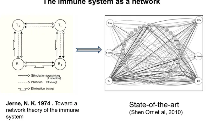
In biology, the main criteria usually used to quantify complexity are the number of components or nodes, and the number of links/edges among the components of a network (5). However, these numbers are highly dependent on the state of our knowledge, which usually underestimates complexity, to the differences of “exact” sciences analyzing systems with a clearly defined composition and structure. For example the Nobel Prize laureate Niels Jerne in the 70’s, proposed a network view of immune system’s complexity included 4 nodes focused mainly on B and T cells. The current networks are usually comprised of a representation of a cytokine network
7
between different players of the immune system containing a hundreds of nodes in the current view (Figure 2).
Figure 2- Two representative views of the immune system as a network.
1.1.2 Complex systems in biology
Following this definitions of complexity, all living organisms can be seen as complex systems. Indeed, living creatures are all self-organized through multi-level and hierarchical architectures of highly interactive components, which define emergent and unpredictable properties.
In biology, each level of study can be seen as a complex system integrated within other scales interacting together or not, but allowing the emergence of functions that ultimately participate into making a live organism. For instance, a cell can be seen as a complex system composed of different sub-compartments all containing different sets of proteins, carbohydrates and lipids having different dynamics, life time and interaction partners, that will collectively achieve functions such as production of energy, DNA synthesis and repair, transcription and translation to obtain new proteins, which collectively allow the cell to perform its specific functions. At the
8
macroscopic levels the different organs are components of the human organism, and different human organisms define groups of people and large populations (3). In conclusion, complexity is therefore one of the main features of biology (4).
1.1.3 Reductionism and Holism: how to study complex biological systems?
The ultimate goal of biology is to understand how life functions. We have seen that all living systems are intrinsically complex systems. In order to reach a comprehension of living organism in science, a main and predominant approach was Reductionism. Reductionism was first defined by Sir Isaac Newton (1643–1727): “Truth is ever to be found in the simplicity and not in the multiplicity and confusion of things“. This philosophy impacted deeply science and biologists in the way they answered scientific questions, using simplified models in controlled environments with mainly loss or gain of function experiments as a way to study the role of a given element of the system, such as a protein or a gene. These approaches were intensively used in molecular biology and were successful in making important discovery on basic cell mechanisms, which led to derived applications such as drug design for medicine.
However, the reductionist approach is unable to take into account many of the key aspects of complexity, such as interactions between components, emergent properties, multiple scales and hierarchical levels. Therefore, scientists developed a theory of complexity which tries to understand complex systems globally, without using reduced or simple models, therefore belonging to the holism philosophy (6). The holistic movement sees the truth has reachable only by the knowledge derived from the whole system. From this movement, and in opposition to the reductionist one, a specific field of biology emerged called systems biology.
Systems biology aims at the description of complex biological systems at the large scale using mathematical and computational tools in order to study the elements of a system altogether and not taken one by one. Recently, systems biology took an even more important meaning in biology. In 2003, the successful end of the human genomic project definitely launched biology into the “OMICS” era (7, 8). OMICS
9
studies regrouped notably genomics, proteomics, transcriptomics, metabolomics and lipidomics. All these OMICS studies are based on technologies allowing the study of a system by measuring all its components at once. For instance, the human genomic project allowed the full characterization of more than 20 000 genes of a human individual (9).
With the emergence of these new OMICS technologies more and more biologists started to ask systems level questions using tools from Systems Biology rather than the classical reductionist approaches. Systems Biology is dedicated to the study of biological systems at the large scale, and is therefore associated to the study of complexity.
1.1.4 Communication and signal integration in complex biological systems
1.1.4.1 Requirements for information exchanges
As defined before, biological systems are complex systems involving many components able to dynamically evolve and to define several modules with self-organization. These key features of complex system are supported by exchange of information or communication through emission and integration of information signals among the different components of the system.
In the field of communication, an information signal or stimulus is a physical entity that encodes or conveys a message. By definition, a signal is produced by a transmitter and will then circulate in a given communication channel that can be of different nature and composed of different noises. A signal must be able to be decoded or perceived by observers that possess the required machinery to receive and process it in order to retrieve the original information sent by the source signal (FIGURE 2). Exchanges of such signals define what is commonly called communication.
10 Figure 2: The basic features of a communication system
1.1.4.2 Complex features of the response to an information signal
If information exchanges are an important part of complex systems, the response of information signal itself contains several levels of complexity. Indeed, in communication an important notion is not only the transmission of information but also how the receiver will respond to the message. The response to a single signal possesses different features. One of the main feature is that the response to a signal can be unique or multiple. In a simple dialogue between two entities a signal triggers the production of a response signal that will be received by the original transmitter of the first emitted signal. In complex situations, a single signal can trigger multiple responses from the same receiver. The other key features of the response to a single signal are the dynamical range of the response, the different scales in which the response can be produced, but also the different nature of the multiple responses produced and the strength of the response(s).
The communication channel in which the signal is transmitted also determines the nature and the number of receivers which can produce different responses that will ultimately generate one response at the population levels. This global population level response can change based on the different entities that constitute the considered population.
11 1.1.4.3 Communication in cell biology
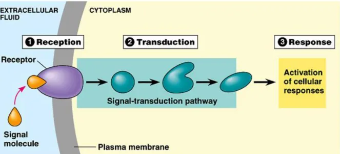
As described before, in communication theory, the signal physically encodes a message that is transmitted to a receiver that possesses the specific machinery to decode this signal. Among the different biological system, we will focus here on the different ways cells can communicate.
Cells can communicate by direct contact, either through cell to cell junctions, which imply direct cytoplasm exchange between the two cells, or through membrane contacts. Cells can also communicate through short distances in absence of direct cell-cell contacts through paracrine communication. Paracrine communication remains local within a given tissue and neighboring cells constitute the targets. Paracrine communication is performed via the secretion of communication molecules outside the cell. Cells can also reach long distances through endocrine communication. It involves the secretion of molecules such as hormones within the whole body through the blood stream, in order to reach distant target cells. All these different communication types, through short or long distances, by direct contact or through secretion of communication molecules, involve the recognition by a receptor expressed by the receiver cell of a specific ligand produced by the transmitter cells. This is the first step of signal integration.
FIGURE 3: The three different steps of signal integration by a cell: reception, transduction and response. This scheme was taken from the Chapter 11 of Cell Communication
12
PowerPoint Lectures for Biology, Seventh Edition Neil Campbell and Jane Reece Lectures by Chris Romero 2005 Pearson Education, Inc. publishing as Benjamin Cummings.
The signal is encoded physically by ligands that can be expressed at the cell surface or secreted outside the cell. After the recognition of the ligand by the receptor, the signal is usually transduced through the cytoplasm by relay molecules in specific signal transduction pathways that end into the nucleus of the cells where specific transcription factors are targeted and act on the regulation of the transcription of genes that can be activated or repressed in order to control the cellular response to the signal.
1.1.5 Signal integration in complex systems at the cell level 1.1.5.1 Signal integration in cell: levels of complexity
All the cells of the human body possess different sensors that allow them to respond to signals from their microenvironments. Signal integration is the fact for a cell to sense, transduce and respond to different signals. This signal integration can be studied itself as a complex system.
In cells integrating signals, three main levels can be seen as direct characteristics of complex systems: 1) the cell itself. For instance, a human cell is composed of more than 20 000 genes. In average, each gene can give rise to three different proteins and each protein can then be differentially regulated or interact with other proteins and/or be addressed with different dynamics to specific cellular compartments where they will perform different functions(10). As for all proteins, the proteins involved in the signaling pathways downstream of a receptor can be also multiple, and can be found in different states, with multiple binding sites for distinct partners. This defines an enormous combinatorial complexity of protein states, which can influence the way a cell, respond to a signal (11, 12). 2) The multiplicity of signals that can act on a given cell. Indeed, one fundamental aspect of cells is that the information they receive is multivariate: the cell microenvironment can contain hundreds of thousands of concurrent molecular signals that can be sensed by a cell at a given time and influence the cell state(13).3) The multiplicity of responses that a cell can produce
13
to a given signal(14). Indeed, as described before in this manuscript, the response to a signal can be multiple. Since the cell itself, and more precisely the group of proteins that defines the signaling pathway, are complex systems based on its high number of components and the high combinatorial of possible protein states, the way this system respond to a signal is in consequence also of a complex nature.
Signal integration in the context of systems biology and complex systems raised important questions that still remain largely unanswered. How the cell states influence the integration of a single signal? How the multiplicity of signals impacts the cell states and cell responses to the stimulation? How information is conserved within these different levels of complexity? Several efforts have been made in the field of signal integration to answer such questions, and some concepts are essential to be presented here to fully understand this field of research.
1.1.5.2 Signal integration and context dependency
In literature or text analysis, context dependency refers to a differential interpretation of a given sentence based on the other sentences from the same paragraph, chapter or book. In this example, a context could also be even more global, such as the author of the sentence or the time in which he wrote it. In cell biology and signal integration, context dependency refers to the exact same concept. Mechanistically the sensing, signaling and response of a cell to a signal can be different based on different contexts.
In a cell-centered signal integration system, a context can be a signal or group of signals sensed by the cell. Indeed, one specific signal that a cell has to interpret can be integrated together with different groups of other signals. The context can also be the cell itself; one signal can act on different cell types or on different cell states of a given cell type. All variations in type, nature and intensities of these different contexts could potentially influence the response of a cell to a single signal (FIGURE 4).
As an example of context dependent mechanisms we can cite the paper of Janes K.A et al, where they showed that Jun N-terminal kinase (JNK) activation can be
14
either anti- or pro-apoptotic depending on the state of the molecular network of cells that received growth factor cues(15).
FIGURE 4: Context dependency in signal integration by cells
More globally any condition or dimension of different types and scales, such as genetic variations, disease states, time or aging, organisms or tissue location can define theoretically a context for the cells. However, whether all putative contexts lead to relevant and differential signal integration by the cell is still largely unknown. The specificity and definitions of cell-intrinsic versus context-dependent responses of cells to a given signal are key challenges emerging in the field of signal integration in systems biology. It opens direct fundamental questions for which no answer can be formulated yet. Notably, in cell biology, if the message encoded by a single signal is defined by the response of a given cell to this signal, is this response highly context dependent? If yes, how essential information is conserved in complex systems? If no, which information is context-independent?
15 1.1.5.3 Signal integration complexity: interaction between signals
Several levels of complexity emerge from the integration at the cell level of a multiplicity of signals.
Considering the integration of two signals on a single output response, this response can be either additive or defines interactions (FIGURE 5). Additivity defines a situation where interactions between the two considered signals are completely absent. In this case, the response of one signal perfectly sums up to the response of the other signal.
FIGURE5: Mathematical definition of signal interaction by cells
This figure was taken from Cappuccio A. et al, Nature Communications 2015
In opposition, interactions correspond to situations where the output response of the two signals combined is different from the sum of each signal taken individually.
In these cases, theoretical combinatorial analysis reveals 82 possible interaction profiles, which were biologically and mathematically grouped into five positive and five negative interaction modes (16) (FIGURE 6).
16 FIGURE 6: Systematic mathematical description of all interaction modes defined by the integration of two distinct signals X and Y and driving a single output response. This figure was taken from Cappuccio A et al, Nature
Communications 2015
All these interaction profiles describe theoretically the behavior of one given cell response, for example a given gene, when two signals interact.
17
The number of theoretical response profiles increase exponentially with the number of signals considered, which leads to an important complexity.
In cell biology it is likely that two different signals can be sensed at the same time by a cell. The complexity of the integration of two signals is increased if we consider all the different output responses triggered by the two signals. In their article, Cappuccio et al showed that up to 9 of the 10 defined modes coexisted in context-dependent proportions. Each interaction mode was preferentially used in specific biological pathways, suggesting a functional role in the adaptation to multiple signals in two different cellular systems (16).
Other studies addressed the question of combinatorial complexity in signal integration. This is the case notably to answer questions regarding interactions in drug combinations (17, 18). But also to study at the large scale level the response of cells to different signals in terms of transcriptional activity or signaling network (19, 20).
Whether or not the number of different integration modes increases with the number of signals considered is still an open question in the field. It will be very complicated to answer this question because of the high number of theoretical profiles of cell response that can be described. However, intrinsic biological limitations in the way cells respond to multiple signals may exist. For instance, mechanisms like general dominance of a signal over the others or redundancy of the effect of different signals could drastically decrease the observed number of integration modes of a cell compared to its theoretical number. Such limitations would decrease the enormous number of putative profiles obtained theoretically and allow data-driven quantification of integration modes. These modes may then be categorized in biologically relevant categories such as synergy or inhibition.
In the first part of this manuscript, we detailed how signal integration was a key element of communication in cell biology. We also showed that cells, as any biological system, can be seen as a complex system as defined by the complexity theory. We notably detailed how the signal integration process or the response to one or multiple signals can be by themselves very complex. Among the different
18
biological systems of an individual, the immune system is of particular interest because it is composed by numerous cells that act together to fight infections. Indeed, communication is central in immunity to coordinate the immune response. The entire immune response to a threat can be seen as a dynamical network of cells exchanging information to achieve a specific function, which is to eliminate a pathogen. In the second part of this manuscript, we will focus our attention on the transfer of information between two immune cell types: the dendritic cell (DC) and the CD4 T cell. We will see why, among this large network of communication, this specific transfer of information is crucial to the global immune response. Specifically, we will detail why CD4 T cell differentiation can be seen on its own as a complex system. To achieve this goal, we will focus first on the integration of multiple signals coming from DC by the CD4 T cells. Then we will see how this information leads to multiple responses of the CD4 T cells leading to the definition of different subsets. Finally, we will see how these multiple subsets can play a role in complex diseases such as allergy, auto-immune disorders or cancer.
1.2 Acquisition and diversity of T helper phenotypes in health and disease: a complex system of communication
1.2.1 The immune system: basic concepts
The immune system involves many different cell types that communicate with each other, and act together to eliminate non-self-threats encountered by an organism. The human immune system is usually segregated in two different sub-systems: the innate and the adaptive immunity. Among different mechanisms, innate immune system includes cells such as macrophages that are resident into peripheral tissues and are able to sense and directly fight different classes of microbes. The principal categories of microbes are fungi, bacteria, virus and parasites. Innate immune cells are able to recognize these different classes of microbes and to reduce the microbial burden at infection sites; in that sense it functions as a first barrier against infections. However, this first line of defense is often not sufficient to completely clear the presence of pathogens. In such cases adaptive immunity comes into action. Adaptive immunity can both select cells specific for the general categories of microbe but also specific to the particular variants within each different species of microbes. For
19
instance, CD8 T cells which are specialized in killing infected cells will be recruited to the infection sites, and will recognize specifically the virus variant involved. For an extracellular threat, such as gram negative bacteria, an antibody response mediated by B cells will be generated. These different cells of the adaptive immune systems are selected for their direct receptor affinity against specific peptides of a given individual of microbial specie (21).
1.2.2 Dendritic cell derived The T helper differentiation process: the three signals theory
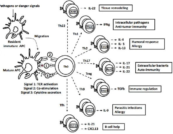
Among the different cells of the adaptive immune system, CD4 T helper (Th) cells are key players that organize and orchestrate the global immune responses (22). To perform this task, Th cells are specialized in communication with other cells through the production of specific communication molecules of the immune system called cytokines. The cytokine or sets of cytokines produced by Th cells will alert, activate and recruit other immune cells specific to the type of danger or pathogen encountered (FIGURE 7). At the initial state of an infection, CD4 T cells are present as naïve and resting in secondary lymphoid organs such as lymph nodes or the spleen and do not produce any cytokine. In order to become functional, naïve CD4 T cells need to enter into the process of cell differentiation to become effector T helper (Th) cells. This process ends up with the generation of different types of Th subsets characterized by the sets of cytokines they are able to secrete. In other words, different Th subsets will secrete different sets of cytokines that act on different types of immune cells in order to specifically coordinate innate and adaptive immune responses against a specific pathogen (22).
The initiation of the differentiation process of naïve CD4 T cells requires an important step of interaction with antigen presenting cells such as Dendritic Cells (DC) (23). DCs are innate immune cells specialized in the activation and the communication with the adaptive immune cells.
After the encounter of pathogens or danger signals at the periphery, DCs become activated and migrate to the secondary lymphoid organs to initiate Th cell
20
differentiation. Three types of signals are involved in this process called signal 1, 2 and 3(24) (FIGURE 7).
FIGURE 7: Dendritic cell derived T helper differentiation: the three signals theory
Signal 1 corresponds to the specific engagement of a T cell Receptor (TCR) by the CMH-II – peptide complex expressed at the surface of DCs. This step is essential to select TCR highly specific to the foreign antigen. Signal 2 constitute the signal of co-stimulation; it is performed through cell-cell contact between DCs and the naïve T cells. Activated DCs express co-stimulatory molecules such as CD80 or CD86, which will signal through the CD28 receptor expressed by the CD4 T cells. These co-stimulation signals together with the TCR signaling allow the CD4 T cells to get activated and to enter into several cycles of clonal division. The Signal 3, is composed of cytokines secreted by DCs which are responsible for the polarization of the naive T cells towards distinct lineages, among which the canonical Th subsets Th1, Th2, Th17, Treg, Tfh, Th9 and Th22 (FIGURE 7) (22). Here the term
21
polarization defines the fact for a Th effector cells to acquire the ability to secrete specific sets of cytokines defining different subsets. Therefore, polarization is only a subpart of the global concept Th differentiation which also defines other features such as T cell proliferation and generation of memory. However, differentiation is often used in papers or reviews to specifically describe polarization.
The different Th subsets are characterized by a set of cytokines they express under the control of a ’master regulator’ transcriptional factor. Each master regulator is critically involved in driving of the differentiation of a specific Th lineage. These subsets are associated to specific physio-pathological functions. For instance, Th1 cells express IFN-γ as a hallmark cytokine under the control of the master regulator T-bet, which directs Th1 lineage commitment, and is involved in the clearance of intracellular pathogens (FIGURE 7).
1.2.3 The DC control of Th differentiation: a complex system of signal integration beyond the three signals theory
The three signals theory emerged from the basic characterization of the T helper differentiation process and constitutes one of the first useful models to understand and ask questions about Th polarization. It is still largely cited in scientific conferences and textbooks and general reviews (24-26), since it gives a rapid understanding of the Th differentiation process.
However, its view of three main categories of signals corresponding to three different functions acting together to specify Th differentiation is partially wrong. Indeed, many different research teams independently studied different aspects of Th differentiation and showed results that contradict many views or derived concepts of the three signals theory. In fact, all these different results taken together describe a complex system of signal integration.
The most important point that was demonstrated in various studies is that factors involved in Th polarization cannot be limited to cytokines, the original signal 3. Indeed, it has been shown that elements belonging to signal 1 or 2 were also
22
important in specifying Th phenotypes. Notably, in different mouse models strength of the TCR signaling, affinity of the TCR for the foreign antigen peptide or the dose of antigen, which are parameters related to signal 1, have been shown to play a role in the differential regulation of Th1 versus Th2 during differentiation (27, 28). Also, co-stimulatory molecules, originally constitutive of signal 2, such as CD80 and CD86 have been shown to preferentially induce Th2 phenotypes (27). In opposition, CD40 another costimulatory molecule has been shown to induce preferentially a Th1 response (28, 29). Further studies have shown that many costimulatory molecules have a role in specifying Th phenotype. In addition, parameters originally absent from the description of the three signals can also play a role in Th differentiation. For instance, the ratio between the number of DCs and naïve T cells (30) can influence Th polarization. At a low ratio (1 DC for 300 T cells) mature DCs induced naive T cells to become Th2, while a high ratio (1 DC for 4 T cells) allows the emergence of a Th1 response (30). The presence of chemokines, another parameter absent from the 3 signals theory, also showed its importance in Th polarization (31). Collectively these studies demonstrate that cytokines are not the only players in Th polarization (32).It also demonstrates that Th polarization is controlled by a large number of different parameters delivered by the DCs to the naïve CD4 T cells during the differentiation process, which is another point ignored from the three signals theory and numerous reviews on Th differentiation.
These numerous signals and their relationships to Th phenotypes were usually identified in different studies through classical deletion or addition types of experiments, such as gene knock-out or supplementation using recombinant proteins, focused through reductionist approaches on one main parameter.
Since the historical discovery of the first signal inducing a Th1 phenotype through the induction of IFN-γ by IL-12 by antigen presenting cells (23, 33), at least 64 different DC parameters have been shown to be involved in Th polarization. These DC parameters, their nature, their receptors on T cells, but also important scientific articles demonstrating their role in Th polarization are detailed in TABLE 1.
All these discoveries in terms of Th phenotype control, recapitulated in TABLE1, lead to a new conceptual view of the Th differentiation as a complex system of signal
23
integrations driven by dendritic cells as previously defined. First, because many of these parameters can be expressed simultaneously by DCs in various combinations and at different levels and define altogether a signal for naïve T cells. This can be indirectly interpreted from several studies performed on DCs (34), however specific studies on the number of distinct matured DC states and their precise molecular characteristic are still lacking in the field. In addition, the co-expression of a large number of parameters acting on T helper cells induce the possibility of signal interactions or context dependent mechanisms which are two important features of signal integration in complex systems as previously detailed in section 1.1.3.
Context dependent control of T helper differentiation and specific signal interactions have already been characterized in the field in different studies, underlying the importance of studying the complexity in the control of Th polarization. In the field of human Th17 differentiation it has been shown that IL-17A was induced through an emergent positive synergy interaction mode involving the co-signaling of IL-1b, IL-23, IL-6 and TGF-b (35, 36). A context dependent action has been shown for OX40L, which induces a Th1 profile in the presence of IL-12 but induces a Th2 response in the absence of IL-12 (37). More recently, it has been shown that TGF-b could be a major driver of Tfh differentiation when combined with IL-12 or IL-23 (38).These different studies constitute a proof of concept that two or more factors can collectively define an emergent phenotype in Th cells, that would be induced by the same signaling element independently of the others.
24
DC
Molecule Other names Localization Category Receptor Articles PMID
CD86 B7.2 Membrane B7 CTLA4 + CD28 Arlene H. Sharpe et al. JI (1999) / Ranger AM, MP et al. IntImmunol (1996)
10453003 / 8921434
CD80 B7.1 Membrane B7 CTLA4 + CD28 Freeman GJ et al. Immunity, (1995) / Tao X et al. JI (1997)
7538442 / 9550393 B7H4 B7X / VTCN1 Membrane B7 unknown I-Fang Lee (Cellular Immunology 2013) 23623902 ICOSL B7-H2 / CD275 Membrane B7 ICOS Nurieva RI (PNAS 2003) / Ito T (J exp Med 2007) 14615582 / 17200410 B7-H3 B7RP-2 /CD276 Membrane B7 unknown Nagashima O et al. JI (2008) / Suh WK et al. Nat
Immunol (2003)
18768862 / 12925852 VISTA B7-H5 Membrane B7 unknown J. Louise Lines et al. Cancer Research (2014) 24691993 PDL1 B7-H1 /CD274 Membrane B7 PD1 Loise M. Francisco et al (J Exp Med 2009) 20008522 PDL2 B7-DC Membrane B7 PD1 By Su-Yi Tseng (2001 J exp Med) / Tahiro Shin et al
(J exp Med 2005)
11283156 / 15897272
HHLA2 - Membrane B7 unknown Zhao R et al (PNAS 2013) 23716685
CD30L CD153 /
TNFSF8 Membrane TNF CD30 Xun Sun et al (JI 2010) 20639486
CD70 CD27L /
TNFSF7 Membrane TNF CD27
Coquet JM et al (Immunity 2013) / Libregts et al (ImmunolLetters 2011)
23159439 / 21277898 4-1BBL CD137 /
TNFSF9 Membrane TNF 4-1BB Kim YH et al (JI 2011) 21715692
CD40 TNFRSF5 Membrane TNF CD40L GiandomenicaIezzi (PNAS 2009) 19136631
OX40-L CD252 Membrane TNF OX40 Ito T et al (JI 2004)/ Ito T et al (J exp Med 2005) 15034038 / 16275760
HVEM TNFRSF14 Membrane TNF LIGHT Tamada K et al (JI 2000) 10754304
LIGHT Membrane TNF HVEM Tamada K et al (JI 2000) 10754304
SLAM CD150 Membrane SLAM SLAM (SLAMF1) Cannons et al (Immunity 2004) 15539155
SLAMF3 Ly9 Membrane SLAM SLAMF3 Graham et al (J Immunol 2006) 16365421
SLAMF5 CD84 Membrane SLAM SLAMF5 Cannons et al (AnnuRevImmunol 2011) / Cannons et al (Immunity 2010)
21219180 / 20153220
NTBA SLAMF6 Membrane SLAM SLAMF6 Howie et al (J Immunol 2005) 15879084
CD48 SLAMF2 Membrane SLAM CD2 R de Jong et al Immunology. 1991 October; 74(2): 175–182
PMC13845 90 ICAM-1 CD54 Membrane Integrin LFA1 Christiane Ruedl (Eur J Immunol 2000) / Smits HH
(J immunol 2002)
10940895 / 11823501
LFA1 CD18 + CD11a Membrane Integrin ICAM1 Singh K et al (JI 2013) 23418628
αv Integrin Membrane Integrin MriduAcharya et al (JCI 2010) 21099114
VLA-4 CD29 + CD49D Membrane Integrin VCAM1 Mittelbrunn M (PNAS 2004) 15263094
ICAM-2 CD102 Membrane Integrin LFA1 Bleijis DA et al (EJI 1999) 10427988
ICAM-3 CD50 Membrane Integrin LFA1 Bleijis DA et al (EJI 1999) 10427988
LFA3 CD58 Membrane Integrin CD2 Gollob JA (J exp Med 1995) / Semnani RT et al (J exp Med 1994)
7544396 / 7525848 Jagged 2 SER2 Membrane Notch NOTCH1 Elyaman et al (Immunity 2012); Sauma D et al
(Scand J immunol)
22503540/ 21352254 Jagged 1 CD339 Membrane Notch NOTCH1 DerkAmsen et al (Cell 2004) / Asano et al (J
Immunol 2008)
15137944 /18292500 Delta 1 DLL1 Membrane Notch NOTCH1, 2 and 3 Keerthivasan S et al (2011) JI 21685328 Delta 4 DLL4 Membrane Notch NOTCH1, 2 and 3 DerkAmsen et al (Cell 2004) / Mukherjee et al (J
Immunology 2009)
15137944 / 19494260 Galectin 9 LGALS9 Secreted /
Membrane Galectins TIM3 Seki M (ClinImmunol 2008) 18282810
Galectin 3 LGALS3 Membrane Galectins Gal 3 Breuilh et al et Oliveira et al (2007) Galectin 1 LGALS1 Membrane Galectins CD69 Martin P et al (Mol Cell Biol 2010) / Martin P et al
(Sci Signal 2011)
20696842 / 21427408
25 Table 1: 64 dendritic cell-derived parameters known to act on T helper polarization.
Literature review of all signals that can be express by dendritic cells and that can act on the T helper differentiation process to specify Th phenotypes.
TIM4 TIMD4 Membrane TIM Tao Liu et al (Mol Immunol 2007) 17439824
PVR CD155 Membrane Nectin CD226 +TIGIT+
CRTAM Lozano E et al ( JI 2013) / Seth S et al (EJI 2009)
23980210 / 19688744
Nectin 2 CD112 Membrane Nectin CD226 Chan et al (CurOpinImmunol 2012) 22285893
Nectin 3 CD113 Membrane Nectin TIGIT Chan et al (CurOpinImmunol 2012) 22285893
CD39 ENTPD1 Membrane Ivan D Mascanfroni (Nat Immunol et al) 23995234
SEMA4A SEMAB Membrane Semaphorin PLXNB Kumanogoh A et al (2005 immunity) 15780988 IL-1b - Secreted Interleukin IL-1RA, IL-1RB Volpe E (Nat Immunol 2008) 18454150 IL-1a - Secreted Interleukin IL-1RA, IL-1RB Madera RF. (Plos One 2011) 22206014 IL-6 - Secreted Interleukin IL-6RA + gp130 Roza I. Nurieva ( Immunity 2008) 18599325 TGF-b - Secreted Interleukin TGFbR1, TGFbR2,
TGFbR3 Volpe E (Nat Immunol 2008) 18454150
IL-18 IL-1F4 Secreted Interleukin-1 IL18R1 + IL18RAP Lim HX (Cytokine 2013) 23697689 IL-10 CSIF Secreted Interleukin IL10RI IL10RII McGuirk (J exp Med 2002) 11805149
TNF-a - Secreted Interleukin TNFRI TNFRII Miller PG et al. Ji 2015 26268655
IFN-alpha IFN-α Secreted Interleukin IFNAR Moschen AR 18926293
IFN-beta IFN-β Secreted Interleukin IFNAR McRae BL et al (Eur JI 1994) 9368622 IL-28A IFN-lambda 2 Secreted Interleukin IL28RA + IL-10Rb Javad ArastehIran et al J Allergy Asthma Immunol
2015 25780882
IL-28B IFN-lambda 3 Secreted Interleukin IL28RA + IL-10Rb I Matthew P. Morrow et al (Blood 2009) 19304955 IL-29 IFN-lambda 1 Secreted Interleukin L28RA + IL-10Rb Dai J et al (Blood 2009) 19346497 IL-27 (p28 + EBI3) Secreted Interleukin IL27R (IL27Ralpha +
gp130) Awasthi, A. et al Nat Immunol (2007) 23995234 IL23 (p19 + p40) Secreted Interleukin IL23R (IL12beta1
+IL23R) McGeachy MJ ( Nat immunol 2009) 19182808 IL-12p70 p35+p40 Secreted Interleukin IL12R (IL12β1 +
IL12β2) Chyi-Song Hsieh (Science 1993) 8097338
CXCL9 MIG Secreted Chemockine CXCR3 Joanna R Groom (immunity 2012) 23123063
CXCL10 IP-10 Secreted Chemockine CXCR3 Joanna R Groom (immunity 2012) 23123063
RANTES CCL5 Secreted Chemockine CCR1, CCR3, CCR5 Gerdes N (ThrombHaemost 2011) 21655676
PF4 CXCL4 Secreted Chemockine CXCR3B Gerdes N (ThrombHaemost 2011) 21655676
CXCL11 I-TAC Secreted Chemockine CXCR3 Liu Z (Clinical and Experimental Immunology 2011) 21438871 Ratio of
DC/T - other Tanaka H (J exp Med 2000) 10934228
Antigen
26 1.2.4 T helper Cytokines profiles defines an increasing number of Th
subsets
The initial description of Th subsets involved the associations of the production of IFN-γ and IL-4 to two different T cell clones, that were respectively named Th1 and Th2(39). Further studies characterized these different Th subsets and showed that subset specific cytokines played major role in the functional specialization of these cells(40).Therefore, discovery and definition of new Th subsets were largely associated to discovery of new cytokines.
In 2003, ten years after the original identification of Interleukin 17A (IL-17 or IL-17A) from a rodent T-cell hybridoma by Rouvier et al (41), a new Th subset producing IL-17 was described (42). This discovery put an end to the concept of the general Th1 and Th2 dichotomy in Th phenotypes. In the following years were reported the discovery of Th22 and Th9 cells expressing respectively IL-22 but no IL-17, or IL-9 but no IL-4(43, 44). In fact, with the increasing number of Th cytokine described, another level of definition of Th subset has emerged: the potential combination or the co-expression pattern of diverse cytokines by the same Th cells.
Tfh cells are a specific Th subset providing B cell help by producing IL-21. Together with IL-21, the co-expression of IFN-g or IL-4 or IL-17 defined three new subsets of Tfh cells, respectively Tfh1, Tfh2 and Tfh17 (45). In an equivalent way distinct populations of Treg were described: Treg1, Treg2, Treg17 and Treg22 described by their suppressive function and expression of distinct cytokines profiles generally associated to Th1, Th2, Th17 and Th22 respectively (46). Another example of this is the differential expression by the same type of Th cells of TNF-α, IFN-γ or IL-10 defining pro-or anti-inflammatory Th subtypes. Notably, It has been shown that based on the original microbial stimulation, Th17 cells could secrete IL-10 or IFN-γ together with IL-17A (47). Expression of TNF-α together with Th2 cytokines was also described in allergy settings, defining pro-inflammatory Th2 cells (37).
Recently, different research groups identified distinct Th2 cell subsets in the memory CD4 T cell compartment. These Th2 subsets produce large amounts of IL-5, IL-17, or
27
IFN-γ in addition to IL-4 and IL-13 (48, 49). Other studies showed that IL-9, usually associated to Th2 or Th9 cells, can also be expressed by Th17 cells, characterized by the production of IL-17A, IL-17F, IL-21 and IL-22 cytokines (50).
As described above, several studies showed that the classical view of well definite and separated subsets of Th cells expressing specific combinations of effector molecules is in fact limited and probably partially untrue. Even if these concepts are still very useful to understand this field of research it seems that the reality of Th phenotype diversity is much more complex. These last years many studies brought knowledge about new subsets of Th cells expressing various combinations of cytokines. Taken altogether these findings describing different subset of Th subsets put an end to the Th1/Th2 paradigm and shed light on new important questions, still largely open in the field.
How many relevant Th profiles exist? Do all putative combinations of output Th cytokines define independent Th subsets occurring in vivo? In this case, the Th phenotypes would be characterized by a continuum of phenotypes. Based on this hypothesis, all subsets described so far based on few output cytokine combinations would appear trivial. However, it triggers other questions regarding the combinations of Th output cytokines that can be co-expressed. Notably, are mutual exclusions of cytokine expression profiles main parameters regulating Th diversity? Are the limitations in Th phenotype diversity Th-intrinsic or regulated by the input received by naïve CD4 T cells?
An interesting study tried to answer such questions. It systematically studied the expression 5 cytokines (IFN-g, IL-4, IL-10, IL-17A, and IL-22) on CD4+ T cell across tissues, identified 12 of the 32 possible combinations (51). These results suggest that all the combinations of Th cytokines are not retrieved in the CD4 memory compartment. So far, more than 20 cytokines have been shown to be secreted by Th cells each of them defining specific functions. Thus, further studies on the production of cytokines by T cells at the single cell level are required to gain knowledge about the real diversity of Th subsets based on the combination of these 20 cytokines.
28
To answer these fundamental questions, it will be required to study the Th cytokines as a whole and therefore to use systems biology approaches to decipher real Th secretion profiles. Large scale data using multi-protein measurement (such as luminex or large intracellular cell staining antibody panel) coupled to single cell RNA-seq studies.
1.2.5 Diversity of T helper cytokines and their association to disease states
1.2.5.1 Th1 and Th2 diseases: a historical dichotomy
5 years after the original discovery of Th1 and Th2 cells, the group of Modlin RG showed for the first time that these two types of Th cells were associated to two different disease states in human Leprosy (52). They showed that Th2 cytokines, IL-4, IL-5 and IL-10, were associated to the multibacillary form of Leprosy, while the Th1 cytokines, IL-2 and IFN-g were predominant in lesions of the resistant form of the disease.
Encouraged by this seminal work, many groups tried to associate specific disease states to either Th1 or Th2 cytokines. Upon time, Th2 was largely associated to allergic disorders. Notably, the impact of the Th2 cytokines, IL-4, IL-5, and IL-13 has been revealed in human asthma, as well as in murine models of allergic inflammation(53, 54).In addition, the pathogenicity of Th2 cytokines has been further proven by the successful use of therapeutic monoclonal antibodies directed against IL-5 (mepolizumab), IL-13 (lebrikizumab) (55) and IL4Ralpha (dupilumab) (56).
On the other side, IFN-γ and Th1 cells were more associated to auto-immune disorders such as multiple sclerosis. In different experimental work, the suppression of IFN-γ, a specific Th1 cytokine, in mouse model of experimental allergic encephalomyelitis (EAE), reduces the disease severity (57-59). The concept of Th1/Th2 balance emerged since it was observed in EAE that the diminution of IFN-γ was associated with the increase of IL-4, and itself associated with improved disease course (60).
29
In human, an approach to determine the Th subset associated to Multiple sclerosis (MS) was to study the cytokines present in the cerebral fluid of patients versus healthy donors. IFN-γ was significantly higher in MS patients compared to controls (61).
These two examples of disease associated either to Th1 or Th2 profiles show how the original dichotomy found in Th clones by Mossman and Coffman in 1986, gave rise to a binary classification of diseases according to the nature of the Th pathogenic responses. However, with the discovery of other cytokines and Th subsets, such as Th17, Tfh, Th22 or Th9, new questions emerged: would a disease be associated to several types of Th subsets? Or will we see specific associations with only one Th subset being pathogenic? Which will be the consequence or the meaning of the infiltration of multiple Th subsets in disease lesions? How could the characterization of the multiplicity of Th phenotype occurring in a single disease help the understanding and cure of this peculiar pathology?
1.2.5.2 Multiple Th subsets associated to the same disease?
In 2003 and following years multiple papers described the discovery of the Th17 subset and the related regulatory mechanisms involved in their differentiation (62).As for Th1 and Th2 subsets, studies also found that Th17 could be associated to bad prognosis or disease severity in several pathologies. Notably, IL17 was shown to be pathogenic in auto-immune disorders such as MS, Psoriasis, Crohn’s diseases or type I diabetes where Th1 were already described to be pathogenic (62-64). In addition, in some studies Th17 cytokines were associated to allergy, notably in asthma where Th cells able to co-produce IL-4 and IL-17 were found (65, 66), but also in atopic dermatitis (AD) (67). Therefore, the discovery of Th17 cells also disrupted the original dichotomy of disease originally classified in two classes either related to Th1 or to Th2.
Th22 are characterized by the secretion of the cytokine IL-22, but also by the lack of IL-4, IL-17 and IFN-γ production. Since their discovery, Th22 and IL-22 were also associated to different pathogenic disorders. Evidences showed that this subset
30
could play a role in allergic disorders, notably AD (67), but also in inflammatory autoimmune disorders such as rheumatoid arthritis (RA) (68), type 1 and 2 diabetes (69, 70), psoriasis (71) and systemic lupus erythematosus (SLE) (72).
Regulatory T cells (Treg), are suppressive Th cells that usually express the transcription factor FOXP3, the cytokines IL-10 and/or TGF-beta and high level of CD25, which is the specific receptor for IL-2. Treg dysfunction was also shown to be associated with various autoimmune pathologies, including multiple sclerosis, type I diabetes, psoriasis (73-75).
In mouse model of allergy, it was demonstrated that important IL-9 production and Th9 differentiation were present (76). In addition, a pathogenic role of Th9 cells, through IL-9 production, has also been shown in inflammatory bowel disease (IBD) (77). Link between IL-9 and psoriasis was also proposed (78).
T follicular helper cells (Tfh) are characterized by the co-expression of several markers that taken independently can belong to different Th subsets. Tfh cells secrete IL-21 and CXCL13; express the transcription factors Bcl-6 and Ascl2 in their nucleus and high level of ICOS, programmed cell death 1 (PD1) and CXCR5 at their cell surface. Tfh cells were found to have detrimental role in SLE, RA, MS and AD (79-81). In addition, we were able to find that Tfh2 cells were induced by the TSLP pathway, well known for its pathogenicity in various allergic disorders and that AD patients had higher percentage of Tfh2 cells, see Annex 1 for details. These findings are corroborated by other studies that associate Tfh with allergic disorders (82, 83).
The global description of these various Th subsets or their derived cytokines provided important new insights into the understanding of the molecular mechanisms involved in the development of complex diseases such as allergic disorders and autoimmune diseases and thus led to revision of the classic Th1/Th2 paradigm and its association to disease.
As a consequence, the classical paradigm evolved from a bipolar view to a complex multipolar one composed of several Th subsets involved in different ways in the pathogenesis of several autoimmune or allergic disorders. This view, of not one but
31
multiple Th subsets present at the same time, in the microenvironment of complex diseases brings another level of difficulty in the analysis of Th-related pathogenic diseases and asks important questions.
Does distinct disease states that can be specific of each human individual, are related to the presence of absence of distinct Th subset in the microenvironment, or by the ratio or combinations of subsets? Can the combination of Th subsets be used in clinics to define distinct classes of patients? How can it be useful in the way patients are treated?
1.2.5.3 Cancer and Th states
In the last decade, emerged the idea that a cancer was more than just cancer cells. Indeed, more and more studies showed that the microenvironment of cancer cells is an important part of the cancer biology itself (84). The microenvironment can be composed of different cells, such as fibroblast and epithelial cells or immune cells such as macrophages, DC and T cells. However, even if the concept of immunosurveillance of cancer implies a positive role of the immune system in preventing the emergence of the disease by suppressing cells at very early steps of carcinogenesis, the role played by the immune system, as part of the tumor microenvironment, once a tumor has developed at later stages of these chronic diseases is much less clear (85).
In some cancer types the presence of an inflammation is correlated to bad prognosis of cancer or to higher susceptibility to cancer (85). In other cases, immune infiltrates have been associated to good prognosis (86).
32 FIGURE 8: Scheme representing the different actors of the tumor microenvironment. Adapted from Fridman WH et al. Nature Reviews Cancer
(2012).
As described before, the immune system is complex and can be polarized towards distinct responses involving distinct cellular actors and distinct sets of communications molecules. The major contributors to this immune polarization are Th cells that are specialized in communicating with other cells to shape the immune responses. Therefore, the study of Th phenotypes in cancer inflammation was largely performed to understand if these distinct associations with good or bad prognosis could be due to distinct inflammation.
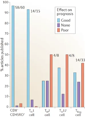
Across all cancer types it has been found that Th1 associated to the activation of the CD8 T cell response was of good prognosis. However, regarding other type of Th subsets, such as Th2, Th17 and Treg, the association to prognosis was largely dependent on the cancer type. For instance, Treg were found to be of poor prognosis in breast cancer (BC) and melanoma (87-90), while of good prognosis in head and neck cancer and colorectal cancer (91-93). These were perfectly illustrated and
33
recapitulated by Fridman W.H. et al in a review from which is extracted Figure 9 and 9 (94).
FIGURE 9: Histograms representing the percentage of article published associating a given Th subsets to their effect on prognosis. Taken from Fridman
WH et al. Nature Reviews Cancer (2012).
The fact to know if different Th subsets have different roles in one cancer type may reflect diversity of cancer diversity that could rely on tumor parameters not yet well understood or taken into account or on the combination of tumor-associated parameters with parameters from the microenvironment. Therefore, this observation raises questions about the diversity of cancers included in each class of cancers primarily defined by its location and tissue origin. One very good example of this complexity of subcategories of cancer within one given cancer type is BC.
1.2.5.4 The different breast cancer subtypes
Based on the expression of specific markers on the tumor cell, scientists and clinicians were able to subdivide BCs in three categories. These three receptors are:
34
the Estrogen Receptor (ER), the Progesterone receptor (PR) and the epidermal growth factor Receptor 2 (HER2). The expression of these three receptors defines three main categories of Breast cancers that have different clinical features and prognosis. First, the Luminal (LUM) Breast cancers are defined by the positivity for ER and/or PR. Then, the second category of breast cancer is defined by the positivity for HER2. And finally a third category is defined by the absence of expression of either ER/PR or HER2 and is called Triple Negative Breast Cancers (TNBC)(95).
Studies have shown that HER2 BC were the most aggressive type of BC, prognosis of this group of patient has been really improved with the use of trastuzumab an IgG1 humanized monoclonal antibody that targets HER2 and blocks the link with its natural ligand, the epidermal growth factor (EGF), and therefore diminishes cancer cell proliferation. Luminal tumors possess the best prognosis compared to other BC subtypes. LUM BC also have specific therapies that aim at blocking the effect of hormones signaling on the cancer cell. This can be done either through aromatase inhibitors that block the aromatase enzymes responsible for the production of estrogen or by blocking the effect of estrogens using estrogen receptor modulators such as tamoxifen that will bind the ER and block the binding of the natural hormone. These treatments greatly improved the overall survival of patients having LUM or HER2 BC. However, patients with TNBC have not any dedicated therapy and harbor today the worst prognosis among the different breast cancer subtypes. Transcriptional profiling of TNBC revealed that this category of BC was highly heterogeneous and could identify 6 subgroups within TNBC, with independent good or bad prognosis (96). Breast cancers illustrate the high diversity of disease types within one general category of cancer.
Whether or not and how different Th subsets or Th derived cytokines are associated to each category of breast cancers is still largely unknown. Some efforts in the field suggest that it could be the case. For example, studies by the group of Dr Palucka showed that Th2 could have pro-tumoral functions in a mouse model of breast cancer (97). Other efforts showed that Tfh could be of good prognosis specifically in HER2 BC (98). In addition the group of Rudensky recently showed that Treg were associated to poor prognosis in BC (99).
35
After describing complex system in general in the first part of the thesis, the second part was dedicated to a very specific point of the immune response, which is Th differentiation. In this second part, we showed why Th differentiation can be seen as a complex system as it was described in the first part of this thesis.
Indeed, 30 years after the characterization of the Th1/Th2 phenotypes, numerous reductionist approaches brought independent pieces of knowledge, conceptual and mechanistic, on T helper differentiation. This revealed at least three levels of complexity; 1) High number of signals putatively integrated by naïve CD4 T cells; 2) High number of cytokines that can be expressed by T cells in multiple combinations defining subsets; 3) The associations of these different subsets to the pathogenicity of different complex diseases and notably in cancer. We also described how the complexity theory challenges the current concept in place in the field of Th differentiation. It notably questions the concept of Th subset but also the three signals theory and the classical association of diseases to Th subset. To investigate these questions, we propose in the third part of this manuscript to see how mathematical modeling can help studying complexity of T helper differentiation process and the associations of different Th subsets to distinct disease states. After describing mathematical modeling in general, we will focus on different types of mathematical models, and illustrate how such models were used by others to study complex biological systems and in some cases to understand T helper differentiation.
1.3 Mathematical modeling: an important tool to study complex systems
Within the tools of systems biology, mathematical models are increasingly used to analyze high-throughput OMICs biomedical or experimental data that are now commonly generated in research laboratories. Notably, the use of dedicated computer interface, allow the simulation of complex biological processes, such as biological networks or multi-factorial diseases, in silico and to test hypotheses that will help and guide either the choice of new experiments or different clinical or therapeutic strategies. However, despite great advances in the field of systems biology and computational biology, having an accurate model of either a whole cell or