HAL Id: dumas-02384411
https://dumas.ccsd.cnrs.fr/dumas-02384411
Submitted on 28 Nov 2019
HAL is a multi-disciplinary open access archive for the deposit and dissemination of sci-entific research documents, whether they are pub-lished or not. The documents may come from teaching and research institutions in France or abroad, or from public or private research centers.
L’archive ouverte pluridisciplinaire HAL, est destinée au dépôt et à la diffusion de documents scientifiques de niveau recherche, publiés ou non, émanant des établissements d’enseignement et de recherche français ou étrangers, des laboratoires publics ou privés.
Caractéristiques cliniques et microbiologiques de
l’infection de canule chez les patients assistés par
membrane d’oxygénation extra-corporelle périphérique
Hugo Lo Pinto
To cite this version:
Hugo Lo Pinto. Caractéristiques cliniques et microbiologiques de l’infection de canule chez les patients assistés par membrane d’oxygénation extra-corporelle périphérique. Sciences du Vivant [q-bio]. 2018. �dumas-02384411�
Université de Bordeaux
U.F.R. DES SCIENCES MÉDICALES
Année 2018 Thèse N° 3008
Thèse pour l’obtention du
DIPLÔME D’ÉTAT DE DOCTEUR EN MÉDECINE
Spécialité Anesthésie Réanimation
Présentée et soutenue publiquement
Le 08 février 2018
Par Hugo LO PINTO
Né le 20 mars 1989 à Romans sur Isère (26)
CARACTÉRISTIQUES CLINIQUES ET MICROBIOLOGIQUES DE
L’INFECTION DE CANULE CHEZ LES PATIENTS ASSISTÉS PAR
MEMBRANE D’OXYGÉNATION EXTRA-CORPORELLE
PÉRIPHÉRIQUE
Directeur de thèse
Monsieur le Docteur Nicolas ALLOU
Membres du Jury
Monsieur le Professeur Alexandre OUATTARA
Président du jury
Monsieur le Professeur Arnaud WINER
Juge
Monsieur le Professeur Éric BRAUNBERGER
Juge
Monsieur le Docteur Jérôme ALLYN
Juge
Remerciements
Au Professeur Alexandre OUATTARA,
Merci d’avoir accepté de présider ce jury, j’ai été particulièrement touché par l’intérêt que vous avez manifesté pour ce travail dès notre première rencontre. Recevez ici le témoignage de mon profond respect.
Au Docteur Nicolas ALLOU,
Merci de m’avoir proposé ce travail, et de m’avoir si bien accompagné dans sa réalisation, depuis mon premier semestre jusqu’à aujourd’hui. Un immense merci également d’avoir permis l’aboutissement de cette thèse : sa publication.
Au Professeur Arnaud WINER,
Merci d’avoir accepté de juger ce travail, mais aussi d’avoir tant participé à la formation de qualité que nous offre l’internat d’anesthésie et réanimation en océan indien.
Au Professeur Éric BRAUNBERGER et au Docteur Jérôme ALLYN,
Merci d’avoir accepté si promptement de faire partie du jury et de juger ce travail. Au Professeur Silvia IACOBELLI,
Merci d’avoir relu avec autant de soins, d’attention et d’intérêt ce travail, et de m’avoir fait partager vos réflexions. Je suis touché par votre investissement.
A mes parents,
Pour votre amour, votre confiance et votre accompagnement plein d’attention et de respect tout au long de ces études exigeantes, malgré la distance. Ma réussite vous revient en grande partie. A Justine,
Merci à toi pour la relecture du manuscrit et toutes les corrections que tu as judicieusement proposées avec beaucoup de compétences.
A Benjamin et Vincent,
Pour m’avoir conseillé et incité à débuter ma thèse au plus tôt pour éviter le « rush » du dernier semestre.
A Myriem,
Pour m’avoir aiguillé dans les méandres administratifs… A toute ma famille, amis sans qui la vie ne serait pas aussi belle. A mes co-internes avec qui j’ai partagé joies et galères d’internat.
A tous les soignants, ceux que j’ai croisés et côtoyés pendant mes études, ceux qui ont pris le temps de me former, ceux qui m’ont marqués par leur humanité et leurs compétences mises au service du patient.
Table des matières
Introduction
4
Objectif du travail de thèse
10
Article original en cours de soumission
11
Résumé de l’article
13
Introduction
14
Méthode
15
Résultats
18
Discussion
21
Conclusion
25
Références de l’article
27
Figure
29
Tableaux
30
Discussion des résultats
35
Conclusion
43
Références
44
Introduction
Membrane d’Oxygénation Extracorporelle
Le monde médical a vu ses pratiques se modifier grâce aux progrès biotechnologiques de ces dernières années. La réanimation n’a pas échappé à cette avancée et de nouveaux outils thérapeutiques ont ainsi pu voir le jour. L’ECMO (Extracorporeal membrane oxygenation) fait partie de ces innovations. Cette
technique de suppléance d’organe, aussi appelée ECLS (Extracorporeal life support), permet une assistance cardiaque et respiratoire. Le sang désoxygéné du patient, provenant d’une canule insérée dans une veine
de gros calibre, est aspiré grâce à une pompe intégrée au circuit ; il traverse ensuite une membrane qui permet l’extraction du dioxyde de carbone et
l’oxygénation. Le sang ainsi oxygéné est réinjecté dans la circulation systémique du patient,
via une deuxième canule. Si le sang oxygéné est réinjecté dans la circulation artérielle, le système est appelé ECMO veino-arterielle (ECMO VA) et apporte un soutien cardio-respiratoire. Par contre si le sang est réinjecté dans la circulation veineuse, le système est appelé ECMO veino-veineuse (ECMO VV) et assure uniquement un soutien respiratoire (1). Concernant l’introduction des canules, il existe deux techniques de mise en place. La première, dont il sera exclusivement traité dans ce travail, est la technique de canulation
périphérique (en percutanée selon la méthode de Seldinger, ou avec un abord chirurgical) au niveau des vaisseaux fémoraux (le plus fréquemment), ou au niveau des vaisseaux supra-cave (artère axillaire, veine jugulaire interne). La deuxième est la technique de canulation centrale, où les canules sont directement placées chirurgicalement, au niveau de l’oreillette droite et de l’aorte ascendante.
Bien que les techniques de circulations extracorporelles soient décrites en chirurgie cardiaque depuis les années cinquante, il faudra attendre 1971 pour voir apparaitre une membrane à oxygénation sur les circuits, marquant les débuts de l’ECMO. Cette technique, s’est initialement développée en réanimation néo-natale. Ainsi en 1989, O’Rourke et al., ont montré que la survie globale des nourrissons traités par ECMO était de 97% (28/29) contre 60% (6/10) dans le groupe traité conventionnellement, chez les patients en détresse respiratoire avec hypertension pulmonaire sévère (2). En réanimation adulte, les débuts de l’ECMO sont moins séduisants, avec des taux de mortalité dans les années quatre-vingt, allant jusqu’à 90% dans les syndromes de détresse respiratoire aigüe (SDRA) (3). Au fil des années,
avec les progrès de biocompatibilités et de matériels, la mortalité des patients sous ECMO a bien diminué, ce qui permit un véritable essor de ce mode d’assistance temporaire. Ainsi, en 2006, Peek et al. dans l’étude randomisée CESAR, ont dévoilé que chez les adultes souffrant de SDRA sévère, la mortalité ou les séquelles graves à 6 mois étaient de 36% (33/90) chez les patients traités par ECMO, contre 51% (46/90) chez les patients ayant bénéficié d’un traitement médical conventionnel (RR 0.69) (4). Le succès de l’ECMO VV en 2009 pour traiter l’épidémie de grippe A H1N1 a fini de populariser son utilisation (5) avec une diminution de 50% de la mortalité en comparaison au traitement habituel (6). Quant à l’ECMO VA, elle permet actuellement, d’après le registre de l’ELSO (Extracorporeal Life
Support Organization) une survie intra-hospitalière de 38.6% des patients en choc cardiogénique réfractaire et de 28.2% des patients en arrêt cardio-respiratoire (ACR) (7).
L'ECMO présente le double avantage de la rapidité de mise en place et de l’efficacité, mais elle est associée à un fort potentiel iatrogène. De ce fait les indications en sont ciblées et restreintes. Les indications et contre-indications de l’ECMO sont résumées dans le tableau ci-dessous, d’après les recommandations sur les indications de l’assistance circulatoire de 2009 (8) ainsi que les travaux de Esper et al. (7) et Richard et al. (9). A l’heure actuelle, les indications de l’ECMO VA sont limitées au choc cardiogénique réfractaire malgré des
thérapeutiques médicales
optimales et peut s’envisager dans le cadre de trois perspectives thérapeutiques. Premièrement dans l’attente de récupération, « bridge-to-recovery » (7), dans le
choc cardiogénique
post-cardiotomie ou
post-transplantation cardiaque ; dans la dysfonction ventriculaire droite secondaire à une hypertension
artérielle pulmonaire
décompensée ou à une embolie pulmonaire massive ; dans la cardiopathie septique ; dans le choc cardiogénique par intoxication aux cardiotropes stabilisateurs de membrane ; dans la myocardite fulminante et la cardiomyopathie du post-partum; dans le choc cardiogénique post-infarctus et enfin dans l’ACR réfractaire avec étiologie identifiée et réanimation initiale efficace (7). Deuxièmement,
dans l’attente d’un autre système d’assistance circulatoire, « bridge-to-bridge », lors d’un choc réfractaire avec défaillances d’organes pour contrôler rapidement l’hémodynamique et
espérer une récupération fonctionnelle des organes périphériques en attendant une transplantation ou une implantation d’une assistance de long cours. Et troisièmement dans l’attente de décision, « bridge-to-décision », pour contrôler rapidement le choc cardiogénique
et prendre le temps d’évaluer le patient et s’orienter secondairement vers une assistance circulatoire de long court ou bien vers un arrêt thérapeutique.
L’ECMO VV est réservée aux SDRA sévères réfractaires au traitement conventionnel (dont le
décubitus ventral) avec un rapport PaO2/ FiO21 inférieur à 50mmHg pendant plus de trois
heures, ou inférieur à 80mmHg pendant plus de six heures. Elle est indiquée également dans l’insuffisance respiratoire hypercapnique et/ou obstructive (10), où l’ECMO VV permet une
ventilation ultraprotectrice (4ml/kg associé à une fréquence basse) et ainsi une diminution des durées de ventilation (11,12).
Complications
Les complications liées à la présence d’une assistance cardio-respiratoire par ECMO sont nombreuses, parfois graves, voire fatales. En effet la méta-analyse de Vaquer et al. en 2017 sur les complications de l’ECMO VV (13), a révélé un taux de complication allant jusqu’à 51% : 40% de complications médicales qui étaient largement dominées par les complications hémorragiques, et 11% de complications mécaniques. La mortalité due aux complications s’élevait à près de 7% (taux de mortalité globale de 37.7%). Les complications infectieuses se
déclarent également fréquemment chez ces patients vulnérables, assistés par ECMO, avec des taux rapportés allant de 8% à 64% (14–19). En 2011, l’ELSO rapportait une incidence des infections sous ECMO de 11.7%, pour un taux de 30.6 par 1000 jours d’assistance pour la population adulte (20). Ces infections peuvent être également associées à des complications graves, jusqu’au choc septique pouvant augmenter considérablement la mortalité hospitalière.
En effet, Grasselli et al. ont publié en octobre 2017, dans une étude rétrospective, que la mortalité était deux fois plus élevée chez les patients présentant une infection nosocomiale au cours d’un traitement par ECMO (40% versus 20%, HR 2.40) (19). Les infections nosocomiales les plus fréquemment signalées chez les patients soutenus par ECMO sont la pneumopathie acquise sous ventilation mécanique et la septicémie, avec des incidences jusqu’à 55% et 18% respectivement (14,17). A propos des germes en causes dans les
infections nosocomiales des patients sous ECMO les études diffèrent : Haneke et al. en 2017, montrent que chez 88 patients traités par ECMO (86% d’ECMO VV) 52% ont présenté une infection nosocomiale et que les pathogènes responsables ne différaient pas de l’écologie générale des services de réanimation (21). Grasselli et al. la même année, trouvaient une très forte part de bactéries multi résistantes ; dans leur étude de 82 patients sous ECMO (87% ECMO VV), 56% ont présenté une infection nosocomiale (19) : 48% de ces infections étaient causées par des bacilles Gram négatifs (BGN) dont 60% étaient multi résistants ; 35% étaient causées par des cocci Gram positifs (CGP) dont 50% étaient multirésistants ; et 17% des infections étaient fongiques.
Infection de canule
Les patients assistés par ECMO périphérique présentent une porte d’entrée infectieuse spécifique, en plus de celles habituellement rapportées en réanimation. En effet, les canules d’admission et de réinjection sont des dispositifs intravasculaires et sont donc propices à la
colonisation bactérienne, pouvant être responsables d’infections systémiques graves chez ces patients précaires. Il semble que l'infection de canule (IDC) soit fréquente : en effet les études rapportent des taux de 4.2 à 21.5% selon les méthodes diagnostiques (17,18,22) avec un taux moyen de 9.9% d’après la récente méta-analyse de 2017 de Vaquer et al. (13). Mais très peu d'études ont rapporté cette complication et aucune grande étude n'a évalué les facteurs de risque et le pronostic de l'IDC chez les patients adultes soutenus par ECMO. A notre
connaissance seules deux études sont actuellement publiées sur le sujet : premièrement le travail préliminaire de Hahne et al., en 2015, sur 94 patients sous ECMO VV et VA (22); et deuxièmement la récente étude rétrospective de Thomas et al. publiée en novembre 2017, sur 99 patients sous ECMO VV (23). Dans cette dernière étude le taux d’infection de canule était de 9.7% et le taux de colonisation des canules sans infection était de 32%. Aucune différence n’a été observée sur la mortalité ou la durée de ventilation ou de séjour de réanimation entre
les patients présentant une infection de canule et ceux n’en présentant pas. À l'heure actuelle, il n'existe pas de recommandation nationale (24) ou même internationale pour le diagnostic et la gestion de l'IDC (25). Schmidt et al. définissaient en 2012 l’IDC comme étant une culture microbiologique positive de l’extrémité de la canule associée à des signes locaux d’infection avec une culture positive de l’aspiration sous-cutanée à l’aiguille fine (17).
Thomas et al. en 2017 ont quant à eux défini l’IDC comme une culture microbiologique positive de l’extrémité de la canule associée à des signes systémiques d’infection sans autre point d’appel (23).
Service de réanimation
La réanimation polyvalente du centre hospitalo-universitaire Felix Guyon à Saint-Denis de la Réunion comporte vingt-trois lits. L’ECMO est utilisée dans ce service depuis 2009, les équipes sont donc entrainées à cette thérapeutique. Le service possède plusieurs machines, avec deux modèles différents : Sorin® ou Maquet®. Cette réanimation, à proximité du service de chirurgie cardiaque, draine les patients d’une grande partie de la Réunion mais aussi de tout l’océan indien. Près de mille patients y sont hospitalisés chaque année, avec plusieurs dizaines d’implantation d’ECMO par an. Ce centre est donc propice à l’élaboration d’une grosse cohorte de patients pour étudier cette complication fréquente mais encore mal connue qu’est l’infection de canule.
Objectif du travail de thèse
Une meilleure connaissance des caractéristiques cliniques et microbiologiques de l’IDC chez les patients assistés par ECMO périphérique pourrait aider à réduire les facteurs de risque associés à cette infection nosocomiale, à adapter des mesures de prévention et pourrait améliorer la prise en charge des patients atteints.
L'objectif de cette étude était d’évaluer l’incidence, les facteurs de risques, les caractéristiques microbiologiques et le pronostic des patients sous membrane d’oxygénation extracorporelle périphérique ayant une infection de canule.
Cannula-related infection in patients supported by peripheral ECMO:
clinical and microbiological characteristics
1Nicolas Allou, MD, 1Hugo Lo Pinto, MD, 1Romain Persichini, MD, 1Bruno Bouchet, MD, 2Eric Braunberger,
MD, 3Nathalie Lugagne, MD, 4Olivier Belmonte, MD, 1Olivier Martinet, MD, Benjamin Delmas, MD,
1Laurence Dangers, MD, PhD, 1Jérôme Allyn, MD
Running title: Cannula-related infection and ECMO Word count: 2879
Abstract Word count: 218
1Réanimation polyvalente, Centre Hospitalier Universitaire Félix Guyon, Saint Denis, Allée
des Topazes 97405, France
2Chirurgie cardiaque, Centre Hospitalier Universitaire Félix Guyon, Saint Denis, Allée des
Topazes 97405, France
3Unité d’Hygiène et de lutte Contre les Infections Hospitalières, Centre Hospitalier
Universitaire Felix Guyon, Allée des Topazes 97405 Saint Denis, France
4Bacteriologie, Centre Hospitalier Universitaire Felix Guyon Allée des Topazes 97405
Saint Denis, France
Corresponding Author: Nicolas Allou, MD
Hôpital Félix Guyon, Réanimation polyvalente, Bellepierre 97405 Saint-Denis, France E-mail: nicolas.allou@chu-reunion.fr
Key words: extracorporeal membrane oxygenation; cannula; mortality; morbidity; bacterial infections; antibiotics.
This work was internally funded. Conflict of interest: none declared.
Abstract
Little is known about cannula-related infection (CRI) in patients supported by extracorporeal membrane oxygenation (ECMO). The aim of this study was to assess the incidence, the risk factors, prognosis, and microbiological characteristics of CRI in patients supported by ECMO. This retrospective cohort study was conducted in one intensive care unit. Among 220 consecutive patients with peripheral ECMO, 39 (17.7%) developed CRI. The incidence of CRI was 17.2 per 1000 ECMO-days. The main isolated microorganisms were Enterobacteriaceae (38%), Staphylococcus spp. (28.2%) (8.5% were methicillin-sensitive
Staphylococcus aureus and 19.7% were coagulase-negative staphylococci) and Pseudomonas aeruginosa (18.3%). Bacteraemia was present in 23 cases (59.7%). In multivariate analysis,
the risk factors for CRI were longer ECMO duration (P=0.006) and higher Simplified Acute Physiology score 2 (P=0.004). Forty-one percent of patients with CRI needed surgical management of the infected site. Cannula-related infection was not associated with higher in-hospital mortality (P = 0.73), but it was associated with a longer stay in ICU (P < 0.0001) and a longer stay in hospital (P = 0.002). In conclusion, CRI is frequent in patients with ECMO and associated with a longer stay in hospital. Risk factors for CRI were longer ECMO duration and higher Simplified Acute Physiology score 2. Concomitant bacteraemia was frequent (59.7%) and CRI should be strongly investigated in cases of positive blood culture.
Background
Extracorporeal membrane oxygenation (ECMO) is a support system that is increasingly being used to aid patients with refractory cardiogenic shock or refractory acute respiratory distress syndrome. Nosocomial infection frequently occurs in patients supported by ECMO, with reported rates ranging between 8% and 64%.1-5 This complication can be associated with worse outcome, including in-hospital mortality due to septic shock .4 The most frequently reported nosocomial infections in patients supported by ECMO are ventilator-associated pneumonia and bloodstream infection .1,4 While it appears that cannula-related infection (CRI) is also frequent, 4,6 few studies have reported this complication and no large study has
assessed the risk factors and prognosis of CRI in patients supported by ECMO.6 At present there are no guidelines for the diagnosis and management of CRI. A better knowledge of the characteristics of CRI in patients supported by ECMO could help reduce the risk factors associated with this infection and could improve the management of affected patients. The aim of this study was to assess the incidence, risk factors, microbiological characteristics, and prognosis of CRI in patients supported by ECMO.
Material and methods
This observational study was approved by the Institutional Review Board of the Committee of the French Intensive Care Society and was declared to the Commission nationale de l’informatique et des libertés (CNIL MR-003, N° 2000694). The need for informed consent
was waived because of the observational and retrospective nature of the study.
Selection of the study sample
This retrospective cohort study was conducted between January 2010 and December 2016 in a 23-bed mixed medical/surgical ICU at a French university hospital.
All patients hospitalized in ICU who underwent ECMO were consecutively evaluated. The exclusion criteria were: age < 18 years old, ECMO duration < 2 days, and application of central ECMO.
Cannulation was either percutaneous by the Seldinger technique or open under direct vision. In case of venoarterial ECMO with femoro-femoral cannulation, both cannulas were usually implanted via vessels of the same groin and a distal perfusion catheter was used to maintain perfusion distal to the arterial cannula.
Cannula-related infection diagnosis
The cannula were removed under sterile conditions at the end of ECMO therapy. The tips were sent for microbiological examination when CRI was suspected.
In the absence of a definition in the literature and guidelines, we chose to define microbiologically-proven CRI as follows: (1) local signs of infection at the site of cannula insertion; (2) a positive microbiological sample taken from: (a) the site of infection via subcutaneous fine-needle aspiration or (b) through surgical exploration, or (c) from the cannula tip; (3) infection occurring within 30 days after ECMO insertion, as per the criteria of
the Centers for Disease Control and Prevention and the National Nosocomial Infections Surveillance System. 7 Only the first episode of CRI was studied.
Blood cultures were performed only when an infection was suspected.
The site of cannula was cleaned with povidone iodine-alcohol and covered with an occlusive, transparent dressing each 2 days
Systemic antibiotic prophylaxis was not used, except in the case of patients who underwent cardiac surgery with cardiopulmonary bypass and who received cefamandole every two hours during surgery.
No protocol changes underwent during the study period
Susceptibility testing and empiric antimicrobial therapy
Empiric antimicrobial therapy based on local guidelines was initiated when CRI was suspected.
Susceptibility testing was determined by the disk-diffusion method, following the criteria of the European Committee on Antimicrobial Susceptibility Testing.8
Data collection and factors associated with cannula-related infection management
The variables associated with the development of CRI were assessed by comparing the characteristics of patients with CRI with those of patients without CRI.
The following characteristics were collected: age; gender; Simplified Acute Physiology Score (SAPS) 2; body mass index; previous coronary artery disease; hypertension; chronic renal failure with dialysis; chronic obstructive pulmonary disease; hyperlipidemia; diabetes mellitus; history of congestive heart failure; immunosuppression; liver cirrhosis, cancer; peripheral vascular disease; smoking (current or former); hazardous alcohol use and biochemistry data and organ failures at ECMO cannulation.
We also recorded the antibiotics administered for any reason within 24 hours prior to ECMO initiation, including the ranking of beta-lactams.9 We also recorded the reason for ECMO support, the lengths of stay in ICU and hospital before ECMO initiation, the location of peripheral ECMO support (operation room or not), the site of ECMO cannulation, and the presence or not of an intra-aortic balloon pump.
Outcome
Patient outcome was documented, including in-hospital mortality and lengths of stay in ICU, and hospital.
Statistical analysis
Results were expressed as total number (percentage) for categorical variables and as median [25th-75th percentiles] for continuous variables, as appropriate. Continuous variables were compared using the Mann-Whitney test, and categorical variables were compared using the Chi-square test or the Fisher’s exact test, as appropriate. In bivariate analysis, the risk factors for CRI with P < 0.1 were entered into a multivariate logistic regression analysis using backward selection with P < 0.05. Collinearity between independent risk factors was tested; when collinearity was identified between two risk factors, the most clinically relevant factor was selected to construct the multivariate model. A P value < 0.05 was considered significant. Analyses were performed using SAS statistical software (8.2, Cary, NC, USA).
Results
Study Population
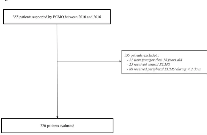
Over the study period, 355 patients were supported by ECMO. Among these, 135 patients were excluded (21 were < 18 years old, 25 received central ECMO, and 89 received peripheral ECMO during < 2 days). The remaining 220 patients formed the cohort of this study (Figure 1).
Patient characteristics at ICU admission and ECMO cannulation are shown in Tables 1 and 2.
The median age was 52 [43-64] and the median SAPS 2 on admission was 56 [42-72].
One hundred and fifty-one patients (68.6%) received venoarterial ECMO. The main indications for ECMO were acute respiratory distress syndrome in 63 cases (28.6%), cardiogenic shock postcardiotomy in 56 cases (25.5%), and acute myocardial infarction in 47 cases (21.4%) (Table 2).
Diagnostic and distribution of microorganisms
Thirty-nine cases of CRI (17.7%) were diagnosed 9 [7-14] days after ECMO cannulation. The incidence of CRI was 17.2 per 1000 ECMO-days: 9.2 per 1000 ECMO days in the venovenous ECMO group and 23.6 per ECMO days in the venoarterial ECMO group (P=0.11). In the femoro-jugular ECMO group 8 of the 61 patients (13.1%) developed CRI, the cannula involved was the jugular one in 3 cases and the femoral one in the remaining 5 cases. Thirty-one of 155 the patients of the femoro-femoral group developed CRI (20%) and 0 of the 4 patients of the femoro-axillar group developed CRI. Leading to a total of 35 femoral cannulae infections.
Diagnosis of CRI was performed during ECMO support in 31 cases (79.5%) and after ECMO was removed in 8 cases (20.5%).
A total of 71 microorganisms were cultured (Table 3), and CRI was found to be polymicrobial in 13 cases (33.3%).
The most frequently isolated bacteria were Enterobacteriaceae (38%), Staphylococcus spp (28.2%) and Pseudomonas aeruginosa (18.3%). Among the 20 Staphylococcus spp strains: 6 were Staphylococcus aureus strains (all were sensitive to methicillin) and 14 were coagulase-negative staphylococci strains (9 were resistant to methicillin) (Table 3). Bacteremia was present in 23 cases (59.7%).
Empiric antibiotic therapy, susceptibility of microorganisms, and surgical management
In the study cohort, 26.2% of Gram-negative bacilli strains were susceptible to amoxicillin/clavulanate, 69% to piperacillin/tazobactam, 73.8% to ceftazidime, and 92.9% to imipenem (Table 4).
Twenty-two patients received a combination therapy with broad-spectrum antibiotic agents targeting non-fermenting Gram-negative bacilli and methicillin-resistant Staphylococcus spp. (56.4%). The most frequently prescribed empiric antibiotic regimen was a combination of piperacillin/tazobactam and vancomycin (or linezolid) (23.1%). The rate of appropriate empiric antimicrobial therapy was 66.7%.
Among the 39 patients with CRI, 23 (59%) were treated only with antibiotics and 16 (41%) received antibiotics and surgical exploration of the infected site with debridement of infected tissue.
Cannulas were removed in 18 cases because of CRI (46.2%). The cannulation site was changed in three cases because of CRI (7.7%).
Risk factors associated with cannula-related infection
In bivariate analysis, the factors (P < 0.1) associated with CRI were: higher SAPS 2 (P = 0.02), male sex (P = 0.04), longer ECMO duration (P = 0.015), shorter prothrombin time (P = 0.065), greater number of units of red blood cells transfused during ECMO support (P = 0.02), lower median daily nadir haemoglobin (P = 0.055), and renal replacement therapy (P = 0.022).
Antibiotic therapy at the time of ECMO cannulation was not associated with reduced CRI (P = 0.57), regardless of levels of beta-lactams (Table 2).
After multivariate analysis, factors independently associated with CRI were longer ECMO duration (per day increment) (OR: 1.065, 95%CI: 1.018-1.114, P=0.006) and higher SAPS 2 (per point increment) (OR: 1.028, 95%CI: 1.009-1.047, P=0.004) (Table 5).
Prognosis
In-ICU mortality was 47.3%. Cannula-related infection was not associated with increased mortality (19 patients died in the group with CRI (48.7%) vs. 85 (47%) in the group without CRI, P = 0.73).
The median duration of stay in ICU was 23 [17-40] days in the CRI group vs. 15 [9-23] days in the non-CRI group (P < 0.0001). The median duration of stay in hospital was 40 [21-59] days in the CRI group vs. 24 [13-40] days in the non-CRI group (P = 0.002).
Discussion
To our knowledge, this is the largest study evaluating the incidence, microbiological characteristics, and impact of CRI in patients supported by ECMO. The main results of this study were as follows: (1) the incidence of CRI in patients supported by ECMO was high; (2) the factors significantly associated with CRI after multivariate analysis were longer ECMO duration and higher SAPS 2.
The rate of CRI observed in our center (17.7%) was high compared to previously reported rates, as these range between 3.5% and 15%.3,4,6 This high rate may be due to the fact that CRI
was the specific focus of our study. Hahne et al. also focused specifically on this complication, and the rate they found (15%) was similar to ours.6 By contrast, Schmidt et al. observed a lower incidence of CRI (10%), likely because they investigated this complication 24 hours after ECMO initiation and within 48 hours after ECMO discontinuation.4 In our study, 20.5% of CRI diagnoses were performed after ECMO removal. Indeed, it seems unlikely that an infection (for instance, of the Scarpa) observed a few days after ECMO discontinuation is unrelated to the cannulation. Thus, the Centers for Disease Control and Prevention defines a surgical site of infection as one in which an infection occurs within 30 days after surgery.7
In the present study, the most frequently isolated microorganisms were Enterobacteriaceae (38%), Staphylococcus spp. (28.2%), and Pseudomonas aeruginosa (18.3%). Studies reporting microorganisms isolated in patients with CRI and supported by ECMO yielded variable results, with rates of isolated Enterobacteriaceae ranging between 7% and 56%, of
Staphylococcus spp. between 17% and 40%, and of non-fermenting Gram-negative bacilli
between 7% and 33%. 2,4,6 In studies evaluating CRI, we (2.6%) and others (0%) have reported very low incidence of fungal infection (Schmidt et al. Hahne et al.).4,6 Nevertheless,
lasting the study period the additional routine use of fungal bottles was not performed that could lead to underestimate the real incidence of fungal infections in patients supported by ECMO.
There is no data in the literature on the susceptibility to antibiotics of pathogens cultured from CRIs. In our study, the rate of appropriate empiric antimicrobial therapy was low (66.7%). This may be explained by the fact that CRI was polymicrobial in 1/3 of the cases, with a high proportion of resistant Gram-negative bacilli strains (31% and 26.2% were resistant to piperacillin/tazobactam and ceftazidime, respectively) and of methicillin-resistant
Staphylococcus spp. This high proportion of resistant microorganisms is related to the
characteristics of the study population: Selection pressure was high (as 65.5% of the patients received antibiotic therapy on the day of ECMO insertion), and the median delay of CRI diagnosis after ECMO insertion was 9 days.10
The only identified risk factors for CRI in our cohort of 220 patients were longer ECMO duration and higher SAPS 2. One previous study specifically investigated the risk factors associated with CRI in 94 adult patients with ECMO, but the only risk factor it identified was positive blood culture.6 ECMO duration 1-3,5 and higher severity score on admission 1,4 are frequently reported in the literature as risk factors for nosocomial infection in patients supported by ECMO. Others factors associated with nosocomial infection in patients with ECMO are presence of an autoimmune disease, and insertion of venovenous as opposed to venoarterial ECMO.3
Many centers administer antibiotic prophylaxis to patients supported by ECMO. Thus, a survey showed that 74% of centers resort to antibiotic prophylaxis, with great variability in the choice of antibiotics and the duration of prophylaxis.11 However, antibiotic prophylaxis
by ECMO.12 In our study, the use of antibiotic prophylaxis was not associated with lower
rates of CRI.
We found concomitant bacteraemia to be frequent among patients with CRI (59.7%). These results are confirmed by those of Hahne et al., who found that positive blood culture constitutes a risk factor for CRI.6 In view of this, they recommend that blood cultures be taken frequently from patients with prolonged duration of ECMO, and that CRI be strongly investigated in cases of positive blood culture. This may be easy to accomplish, as many centers already perform daily blood cultures as part of routine surveillance in patients with ECMO.11,13
While bundle care has been shown to successfully reduce central line-associated bloodstream infections, 14 it is difficult to apply in the case of patients with ECMO. Overall, measures to
prevent CRI are lacking, and a better understanding of this complication is needed. Future studies should investigate other approaches to reducing CRI, including bathing with chlorhexidine gluconate, 15,16 antimicrobial impregnation, coating on cannula tip of ECMO as has been performed with central venous catheters.17
Management of patients with CRI remains a challenge. Early administration of antimicrobial agents in patients with CRI should be systematic. Cannulas, like other medical devices, can serve as a substrate for microorganism’s biofilm infection, necessitating biofilm extirpation. Removal or early change of the infected cannula should always be discussed (ICU physician’s, cardiologist, surgeon) especially in case of septic shock. Bacterial biofilm
formation on the ECMO cannulas may play a key role in infection. Bacterial biofilms on device surfaces are difficult to eradicate and CRI are not easily treated as many ECMO cannulas are not easily replaced. It has been showed that combination of putative anti-biofilm agents and antibiotics might be useful in treatment of biofilm infections.18
The mortality rate reported here (47.3%) is similar to that observed in previous studies of nosocomial infection in patients with ECMO (between 34.2% and 68.3%).1,3,4 We found that patients with CRI did not have higher mortality rates compared with patients without CRI; however, they had longer stays in ICU and hospital. Aubron et al. and Schmidt et al. found similar outcomes in patients with nosocomial infection.1,4
Our study has some limitations. The retrospective nature of the analysis is obviously a weakness. The diagnosis of CRI remains a challenge, as there are no guidelines available for clinicians. The data presented here correspond to usual medical management, which can also be viewed as a limitation. This single-center study does not provide any definitive conclusions for other institutions, especially as regards the choice of antibiotics. Single-center studies are useful to reveal local ecological patterns, while larger studies uncover regional and even global trends not apparent in smaller studies. Our study population may be considered small; yet, to our knowledge, it is the largest cohort addressing the issue of CRI in patients with ECMO. Previous publications analysed a limited number of patients with nosocomial infection: 45 patients in Sun et al.,3 94 in Hahne et al.,6 114 in Hsu et al.,2 and 220 in Schmidt
et al. 4 Only documented cases of CRI were taken into account in our study, which may lead to underestimating their incidence. The cannula tips were sent for microbiological examination and blood cultures were performed only when CRI was suspected this could lead to underestimate the real incidence of CRI. In our study many patients underwent surgery and probably we have an aggressive policy regarding the management of CRI that is debatable.
In addition, this study suffers from a possible lack of power because of the low number of patients and events evaluated. In the multivariate analysis, there were 8 variables per predictor event, and the rule that logistic regression should be used with a minimum of 10 events per predictor variable is based only on few studies. 19,20 It has also been shown that type 1 errors
were not severe, with 5 to 9 events per predictor variable, and were comparable with 10 to 16 events per predictor variable in many circumstances.21
Conclusion
Cannula-related infection is frequent in patients supported by ECMO (17.2 CRI per 1000 ECMO-days), and it is associated with a longer stay in hospital. Risk factors for CRI were longer ECMO duration and a higher risk score. Concomitant bacteraemia was frequent among patients with CRI (59.7%) and CRI should be strongly investigated in cases of positive blood culture. Medical management of this complication could require frequent surgical exploration of the surgical site. Future studies should investigate other approaches to reducing CRI.
Declarations
List of abbreviations
CRI: cannula-related infection
ECMO: extracorporeal membrane oxygenation ICU: Intensive Care Unit
SAPS 2: Simplified Acute Physiology score 2 Ethics approval and consent to participate
This observational study was approved by the Institutional Review Board of the Committee of the French Intensive Care Society (CE SRLF17-21) and was declared to the Commission nationale de l’informatique et des liberté (CNIL MR-003, N° 2000694).
The need for informed consent was waived because of the observational and retrospective nature of the study.
Availability of data and material
The dataset used and/or analyzed during the current study are available from the corresponding author on reasonable request.
Competing interest none declared. Funding
This work was internally funded. Authors Contributions
AN and LPH had full access to all of the data in the study, and they take responsibility for the integrity of the data and the accuracy of the data analysis.
Study concept and design: AN, AJ, LPH and BB Acquisition of data: LPH, AN, BB, NL, OB, BE
Analysis and interpretation of data: AN, AJ, LPH, MO, DL, PR, DB, BE. Drafting of the manuscript: AN, AJ
Critical revision of the manuscript for important intellectual content: AN, AJ, LPH, MO, PR,
DB, DL.
Statistical analysis: AJ, AN, DL.
Obtained funding: Support was provided solely from institutional and/or departmental
sources.
Administrative, technical, or material support: AN, AJ, MO, DL, PR, DB, BE. Study supervision: AN, AJ, MO, DL, PR, DB, BE.
References
1. Aubron C, Cheng AC, Pilcher D, et al. Infections acquired by adults who receive extracorporeal membrane oxygenation: risk factors and outcome. Infect Control Hosp
Epidemiol 34:24-30, 2013.
2. Hsu MS, Chiu KM, Huang YT, Kao KL, Chu SH, Liao CH. Risk factors for nosocomial infection during extracorporeal membrane oxygenation. J Hosp Infect 73:210-6, 2009.
3. Sun HY, Ko WJ, Tsai PR, Sun CC, et al. Infections occurring during extracorporeal membrane oxygenation use in adult patients. J Thorac Cardiovasc Surg 140:1125-32 e2, 2010.
4. Schmidt M, Brechot N, Hariri S, et al. Nosocomial infections in adult cardiogenic shock patients supported by venoarterial extracorporeal membrane oxygenation. Clin Infect Dis 55:1633-41, 2012.
5. O'Neill JM, Schutze GE, Heulitt MJ, Simpson PM, Taylor BJ. Nosocomial infections during extracorporeal membrane oxygenation. Intensive Care Med 27:1247-53, 2001.
6. Hahne K, Horstmann C, Fischer D, Köck R, Peters G, Lebiedz P. Cannula-related infection in adult medical intensive care unit patients undergoing extracorporeal life support and extracorporeal membrane oxygenation. J Hosp Infect 91:372-4, 2015.
7. Procedure-associated Module. Surgical Site Infection Event. Center for disease Control and Prevention (CDC) 2017. https://www.cdc.gov/nhsn/pdfs/pscmanual/9pscssicurrent.pdf, 2017. 8. EUCAST. European Committee on Antimicrobial Susceptibility Testing 2016. www.eucast.org/.
9. Weiss E, Zahar JR, Lesprit P, et al. Elaboration of a consensual definition of de-escalation allowing a ranking of beta-lactams. Clin Microbiol Infect 21:649 e1-10, 2015.
10. Trouillet JL, Chastre J, Vuagnat A, Joly-Guillou ML, Combaux D, Dombret MC. Ventilator-associated pneumonia caused by potentially drug-resistant bacteria. Am J Respir
Crit Care Med 157:531-9, 1998.
11. Kao LS, Fleming GM, Escamilla RJ, Lew DF, Lally KP. Antimicrobial prophylaxis and infection surveillance in extracorporeal membrane oxygenation patients: a multi-institutional survey of practice patterns. ASAIO J 57:231-8, 2011.
12. O'Horo JC, Cawcutt KA, De Moraes AG, Sampathkumar P, Schears GJ. The Evidence Base for Prophylactic Antibiotics in Patients Receiving Extracorporeal Membrane Oxygenation. ASAIO J 62:6-10, 2016.
13. Glater-Welt LB, Schneider JB, Zinger MM, Rosen L, Sweberg TM . Nosocomial Bloodstream Infections in Patients Receiving Extracorporeal Life Support: Variability in Prevention Practices: A Survey of the Extracorporeal Life Support Organization Members. J
Intensive Care Med Feb 10. pii: 0885066615571540, 2015.
14. Blot K, Bergs J, Vogelaers D, Blot S, Vandijck D. Prevention of central line-associated bloodstream infections through quality improvement interventions: a systematic review and meta-analysis. Clin Infect Dis 59:96-105, 2014.
15. Afonso E, Blot K and Blot S. Prevention of hospital-acquired bloodstream infections through chlorhexidine gluconate-impregnated washcloth bathing in intensive care units: a
systematic review and meta-analysis of randomised crossover trials. Euro Surveill 21 Nov 17;21(46). pii: 30400, 2016.
16. Swan JT, Ashton CM, Bui LN, et al. Effect of Chlorhexidine Bathing Every Other Day on Prevention of Hospital-Acquired Infections in the Surgical ICU: A Single-Center, Randomized Controlled Trial. Crit Care Med 44:1822-32, 2016.
17. Lai NM, Chaiyakunapruk N, Lai NA, O'Riordan E, Pau WS, Saint S. Catheter impregnation, coating or bonding for reducing central venous catheter-related infections in adults. Cochrane Database Syst Rev ;3:CD007878, 2016.
18. Belfield K, Bayston R, Hajduk N, Levell G, Birchall JP, Daniel M. Evaluation of combinations of putative anti-biofilm agents and antibiotics to eradicate biofilms of
Staphylococcus aureus and Pseudomonas aeruginosa. J Antimicrob Chemother;72:2531-8,
2017.
19. Concato J, Peduzzi P, Holford TR, Concato J, Peduzzi P, Holford TR. Importance of events per independent variable in proportional hazards analysis. I. Background, goals, and general strategy. J Clin Epidemiol 48 :1495-501, 1995.
20. Peduzzi P, Concato J, Kemper E, Peduzzi P, Concato J, Kemper E. A simulation study of the number of events per variable in logistic regression analysis. J Clin Epidemiol 49:1373-9, 1996.
21. Vittinghoff E, McCulloch CE. Relaxing the rule of ten events per variable in logistic and Cox regression. Am J Epidemiol 165:710-8, 2007.
Table 1 - Clinical characteristics at ICU admission
Variable Total Cannula-related infection P
(n=220) Yes (n=39) No (n=181)
Simplified Acute Physiology Score 2, median [25th-75th percentiles]
56 [42-72] 64 [50-81] 55 [40-71] 0.02
Male, n (%) 149 (67.7) 21 (53.8) 128 (70.2) 0.04
Age (years), median [25th-75th percentiles]
52 [43-64] 56 [42-65] 52 [43-63] 0.79
Body mass index (kg/m²) 25.4 [21.7-30] 26.1 [22.4-29.3] 25 [21.5-30.3] 0.65
Previous coronary artery disease, n (%) 47 (21.3) 6 (15.4) 41 (22.7) 0.32
Hypertension, n (%) 83 (37.7) 16 (41) 67 (37) 0.64
Chronic renal failure with dialysis, n (%) 17 (7.7) 2 (5.1) 15 (8.3) 0.5
Chronic obstructive pulmonary disease, n (%) 11 (5) 2 (5.1) 9 (5) 0.97
Hyperlipidaemia, n (%) 32 (14.5) 3 (7.7) 29 (16) 0.18
Diabetes mellitus, n (%) 64 (29.1) 11 (28.2) 53 (29.3) 0.89
History of congestive heart failure, n (%) 37 (16.8) 9 (23.1) 28 (15.5) 0.25
Immunosuppression, n (%) 12 (5.5) 3 (7.7) 9 (5) 0.5
Liver cirrhosis, n (%) 5 (2.3) 1 (2.6) 4 (2.2) 0.89
Cancer, n (%) 5 (2.3) 0 5 (2.8) 0.29
Peripheral vascular disease, n (%) 20 (9.1) 2 (5.1) 18 (9.9) 0.34
Smoking (current or former), n (%) 73 (33.2) 10 (25.6) 63 (34.8) 0.27
Hazardous alcohol use, n (%) 49 (22.3) 7 (17.9) 42 (23.2) 0.47
Antibiotic therapy in last 3 months, n (%) 43 (19.5) 8 (20.5) 35 (19.3) 0.87
Table 2 - Clinical characteristics at ECMO cannulation
Variable Total Cannula-related infection P
(n=220) Yes (n=39) No (n=181)
Length of stay in hospital before ECMO (day), median [25th-75th
percentiles] 2 [0-8] 1 [0-10] 2 [0-8] 0.37
Length of stay in ICU before ECMO (day), median [25th-75th percentiles] 0 [0-1] 0 [0-1] 0 [0-0] 0.14
Antiplatelet therapy, n (%) 57 (25.9) 7 (17.9) 50 (27.6) 0.21
Glasgow Coma Scale score, median [25th-75th percentiles] 15 [14-15] 15 [13-15] 15 [15-15] 0.19
Catecholamines, n (%) 199 (90.5) 35 (89.7) 164 (90.1) 0.87
Mechanical ventilation, n (%) 201 (91.4) 35 (89.7) 166 (91.7) 0.69
Renal replacement therapy, n (%) 83 (37.7) 21 (53.8) 62 (34.3) 0.022
Lactate level (mmol/L), median [25th-75th percentiles] 4.9 [2.6-10.1] 5.7 [3.6-11.2] 4.8 [2.5-9.6] 0.14
pH, median [25th-75th percentiles] 7.27 [7.16-7.4] 7.26 [7.16-7.4] 7.27 [7.17-7.4] 0.51
Total bilirubin level (µmol/L) , median [25th-75th percentiles] 18 [9-32] 22 [8-36] 9 [16-31] 0.35
Platelet count (G/L), median [25th-75th percentiles] 151 [73-219] 139 [76-192] 151 [73-222] 0.62
Prothrombin time (%), median [25th-75th percentiles] 49 [35-69] 44 [31-60] 50 [36-70] 0.065
Hemoglobin level (g/dL) , median [25th-75th percentiles] 9.3 [8-11] 10.2 [8.2-11.3] 9.1 [7.9-10.9] 0.25
Creatinine level (µmol/L) , median [25th-75th percentiles] 123 [87-181] 136 [100-194] 120 [85-177] 0.12
Lactatemia (mmol/L), median [25th-75th percentiles] 4.9 [2.6-10.1] 5.7 [3.6-11.2] 4.8 [2.5-9.6] 0.34
Operating room for ECMO cannulation, n (%) 104 (47.3) 16 (41) 88 (48.6) 0.39
Percutaneous implantation of ECMO, n (%) 132 (60) 26 (66.7) 106 (58.6) 0.35
Reason for ECMO
Cardiac arrest, n (%) 16 (7.3) 5 (12.8) 11 (6.1) 0.14
Dilated cardiomyopathy, n (%) 10 (4.5) 1 (2.6) 9 (4.8) 0.51
Myocarditis, n (%) 7 (3.2) 2 (5.1) 5 (2.6) 0.45
Acute myocardial infarction, n (%) 47 (21.4) 12 (30.8) 35 (18.5) 0.11
Postcardiotomy, n (%) 56 (25.5) 6 (15.4) 50 (27.6) 0.11
Acute respiratory distress syndrome, n (%) 63 (28.6) 9 (23.1) 54 (28.6) 0.4
Any antibiotic at the time of ECMO cannulation 144 (65.5) 24 (61.5) 120 (66.3) 0.57
Ranking of beta-lactams ≥ 3 a, n (%) 87 (39.5) 14 (35.9) 73 (40.3) 0.61
Ranking of beta-lactams ≥ 4 a, n (%) 38 (17.3) 6 (15.4) 32 (17.7) 0.73
Ranking of beta-lactams of 6 a, n (%) 14 (6.4) 3 (7.7) 11 (6.1) 0.71
Macrolide, n (%) 38 (17.3) 6 (15.4) 32 (17.7) 0.73
Site of ECMO cannulation, n (%)
Femoro-jugular, n (%) 61 (27.7) 8 (20.5) 53 (29.3) 0.27
Femoro-femoral, n (%) Femoro-axillar, n (%) Limb ischemia, n (%)
Major bleeding from cannula, n (%)
155 (70.5) 4 (1.8) 10 (4.5%) 34 (15.5%) 31 (79.5) 0 2 (5.2%) 7 (17.9%) 124 (68.5) 4 (2.2) 8 (4.4%) 27 (14.9%) 0.17 0.78 0.85 0.63
Intra-aortic balloon pump, n (%) 43 (19.5) 10 (25.6) 33 (18.2) 0.93
ECMO duration (days), median [25th-75th percentiles] 7 [5-12] 9 [6-16] 7 [5-12] 0.015
Venoarterial ECMO, n (%) 151 (68.6) 30 (76.9) 121 (66.9) 0.21
Total RBC during ECMO support (units), median [25th-75th percentiles] 6 [2-12] 4 [8-18] 6 [2-10] 0.02
Median daily nadir Hb during ECMO support (g/dL), median [25th-75th
percentiles] 7.9 [7.4-8.5] 7.7 [7.7-8.1] 7.9 [7.4-8.6] 0.055
Results are expressed as median [25th-75th percentiles] or number (%)
ECMO: extracorporeal membrane oxygenation; Hb: hemoglobin; ICU: intensive care unit; RBC: red blood cells
a Ranking of beta-lactams: 1: amoxicillin; 2: amoxicillin/clavulanic acid; 3: 3rd generation cephalosporins with limited broad spectrum;
Table 3 - Seventy-one microorganisms isolated from 39 episodes of cannula-related infection
Microorganisms Number (%)
Non-fermenting gram-negative bacilli, n (%) 15 (21.1)
Pseudomonas aeruginosa, n (%) 13 (18.3) Stenotrophomonas maltophilia, n (%) 2 (2.8) Enterobacteriaceae, n (%) 27 (38) Escherichia coli, n (%) 6 (8.5) Enterobacter spp., n (%) 7 (9.6) Klebsiella spp, n (%) 7 (9.6) Citrobacter spp, n (%). 2 (2.8) Proteus spp, n (%). 1 (1.4) Morganella morganii, n (%) 2 (2.8) Serratia marcescens, n (%) 1 (1.4) Providencia spp. , n (%) 1 (1.4) Bacteroides fragilis, n (%) 1 (1.4) Staphylococcus spp, n (%) 20 (28.2)
Methicillin-sensitive Staphylococcus aureus, n (%) 6 (8.5)
Methicillin-sensitive coagulase-negative staphylococci, n (%) 5 (7)
Methicillin-resistant coagulase-negative staphylococci, n (%) 9 (12.7)
Enterococcus faecalis, n (%) 6 (8.5)
Streptococcus spp., n (%) 1 (1.4)
Candida tropicalis, n (%) 1 (1.4)
Results are expressed as n (%)
Infections were polymicrobial in 13 cases (33.3%) Bacteremia was present in 23 cases (59.7%)
Table 4 - Antimicrobial susceptibilities of the 42 isolated strains of Gram-negative bacilli
Microorganisms (n=42) AMC TIC PTZ CTX CAZ IPM GM AN CIP TMP
Pseudomonas aeruginosa 13 x 85 85 x 85 92 100 100 100 x Stenotrophomonas maltophilia 2 x x x x 50 x x x 50 100 Enterobacter spp. 7 x 57 57 57 57 100 71 86 71 86 Klebsiella spp. 7 71 x 71 100 100 100 100 100 71 71 Escherichia coli 6 67 67 100 100 100 100 100 100 100 86 Citrobacter spp. 2 50 0 50 50 50 100 100 100 100 100 Proteus spp. 1 100 100 100 100 100 100 100 100 100 100 Morganella morganii 2 0 0 0 0 0 100 100 100 100 100 Serratia marcescens 1 0 100 100 100 100 100 100 100 100 100 Providencia spp. 1 0 100 100 100 100 100 100 100 100 100
AMX: amoxicillin; AMC: amoxicillin + clavulanate; TIC: ticarcillin; PIP : piperacillin; TZP: piperacillin + tazobactam; CTX: cefotaxime; CAZ: ceftazidime; IPM: imipenem; GM: gentamicin; AN; amikacin; CIP: ciprofloxacin; TMP: trimethoprim + sulfamethoxazole
Table 5 - Multivariate analysis of factors associated with cannula-related infection
Variables Adjusted Odds Ratio (CI 95%) P-value
Duration of ECMO (per day increment) 1.065 (1.018-1.114) 0.006
Total units of red blood cells transfused - 0.83
Median daily nadir haemoglobin - 0.14
Simplified Acute Physiology score 2 (per point increment) 1.028 (1.009-1.047) 0.004
Prothrombin time - 0.18
Male sex - 0.11
Renal replacement therapy - 0.14
The Hosmer-Lemeshow goodness-of-fit test P-value was 0.0.041. The Nagelkerke and Cox/Snell R squares were 0.118 and 0.0.071, respectively.
Discussion des résultats
A notre connaissance il s’agit de la plus grande étude évaluant l’incidence, les caractéristiques microbiologiques et l’impact de l’infection de canule (IDC) chez les patients assistés par une ECMO (Extracorporeal membrane oxygenation). Les principaux résultats de cette étude étaient les suivants : premièrement, l’infection de canule est très fréquente chez les patients sous ECMO périphérique ; deuxièmement, les facteurs de risque de cette complication étaient une augmentation de la durée de l’assistance par ECMO et un score de gravité IGS2 plus élevé ; troisièmement les germes qui étaient le plus souvent responsable de l’infection étaient des bacilles à Gram négatif multirésistants et des souches de staphylocoques blancs résistants à la méticilline ; enfin, l’infection de canule n’était pas associée à une augmentation de la mortalité mais à une augmentation de la durée de séjour en réanimation et de la durée de séjour hospitalier.
Population
Il est intéressant de constater que notre population d’étude était similaire à celle des autres études sur le sujet. En effet la médiane d’âge de 52 ans dans notre étude était très proche de celle des études de Thomas et al. et de Schmidt et al. (49 et 51 ans respectivement) (17,23). Pareillement pour l’IGS2 médian qui était de 56, contre 46 et 61. La durée médiane d’assistance était de 9 jours dans notre étude contre 11 jours dans le travail de Thomas et al
(23).
Incidence
L’incidence de l’infection de canules (IDC) retrouvée dans notre centre était de 17,7%, avec
17,2 infections de canule pour 1000 jours d’ECMO. Ce taux relativement élevé par rapport à ceux décrits dans la littérature qui varient entre 3,6% et 15% (16,17,22,23), pourrait être dû au fait que l’IDC était l’objet principal de notre étude. En effet Sun et al. (16) ainsi que Schmidt
et al. (17) ont étudié l’ensemble des complications infectieuses et n’ont retrouvé
respectivement que 3,6% et 9,1% d’IDC. En revanche Hahne et al. (22) qui ont, eux aussi, centré leur étude sur l’IDC, ont trouvé une incidence de 15%, qui est plus proche de la nôtre. Cette différence pourrait être également expliquée par le fait que Schmidt et al. qui ont trouvé une incidence plus faible (9,1% avec 7.1 infections de canules pour 1000 jours d’ECMO) n’ont recherché cette complication que sur une période courte allant de 24h après la mise en
place des canules jusqu’à seulement 48h après l’explantation de celles-ci (17). Thomas et al. (23) ont également choisi cette période pour le diagnostic d’IDC, en se basant sur les recommandations de diagnostic des cathéters veineux centraux (26). Dans notre étude cette période était beaucoup plus longue, puisque que l’infection était recherchée jusqu’à trente jours après l’explantation, à l’image des recommandations établies par le « Centers for Diseases Control and Prevention » qui définissent une infection du site opératoire comme une infection se produisant jusqu’à 30 jours après la chirurgie (27). En effet l’abord vasculaire lors de la canulation se fait très fréquemment de manière chirurgicale même si une technique de Seldinger est utilisée pour tunnéliser le vaisseau ; nous donc avons choisi de considérer les infections de canules comme une véritable infection de site opératoire plus qu’une simple infection de cathéter. Ainsi dans notre étude, 20,5% des diagnostics d’IDC ont été effectué à distance de l’explantation de l’ECMO ; il semble en effet peu probable qu’une infection de Scarpa survenant quelques jours après l’ablation de la canule de l’ECMO ne soit pas liée à la canule. L’incidence plus faible de l’IDC dans le travail récent de Thomas et al. (9,7%) qui avait évalué 103 patients ayant une ECMO-VV pourrait être expliquée par le fait que leur définition propre de l’IDC était différente. En effet ils n’ont considéré les IDC que chez les patients qui avaient une culture microbiologiquement positive de l’extrémité de canule associée à une hémoculture positive (au même germe) et/ou des signes systémiques d’infection (à la manière des infections de cathéter). Or nous avons aussi considéré comme
IDC la présence de signes locaux d’infection associée à la culture positive de l’extrémité de la canule. Il est donc nécessaire que des recommandations soient établies sur le sujet, pour pouvoir uniformiser nos prises en charge et nos recherches cliniques.
Facteurs de risque
Les seuls facteurs de risque identifiés pour l’IDC dans notre étude étaient une durée d’assistance par ECMO prolongée avec un Odds Ratio de 1,06 par jour d’assistance
supplémentaire, ainsi qu’un score IGS2 élevé avec un Odds Ratio de 1,03 par point supplémentaire. Hahne et al. n’avaient identifié comme seul facteur de risque la présence d’une hémoculture positive (22). La durée de l’assistance par ECMO (14,16–18) et le score de
gravité à l’admission (14,17) sont fréquemment rapportés dans la littérature comme facteurs de risque d’infection nosocomiale chez les patients sous ECMO, mais cela n’avait pas été décrit spécifiquement pour l’IDC en elle-même. D’autre facteurs de risque d’infection nosocomiale sont également rapportés dans la littérature comme la présence d’une maladie auto-immune, ou l’ECMO-VV en comparaison à l’ECMO-VA (16). De nombreux centres administrent un antibioprophylaxie aux patients ayant une ECMO. Ainsi, une étude a montré que 74% des centres administrent une antibioprophylaxie, avec une grande variabilité dans le choix des antibiotiques et la durée de traitement (28). Cependant l’antibioprophylaxie n’a pas démontré d’effet protecteur sur l’IDC chez le patient sous ECMO (29). Nos résultats vont également dans ce sens : en effet l’utilisation d’une antibioprophylaxie n’était pas associée à une diminution de l’incidence des IDC dans notre étude. Il est intéressant de noter que l’implantation de l’ECMO au bloc opératoire n’était pas un facteur de protection contre les IDC dans notre étude. Nous avons également constaté qu’une bactériémie concomitante était fréquemment retrouvée puisqu’elle apparaissait dans près de 60% des IDC de notre cohorte. Ces résultats sont retrouvés par Hahne et al. qui avaient montré qu’une hémoculture positive était un facteur de risque d’IDC (22) mais également par Thomas et al. qui avaient retrouvé
70% d’hémocultures concomitantes positives aux IDC (23). Ils recommandent d’ailleurs, au vu de leurs résultats, que des hémocultures soient régulièrement prélevées chez les patients sous ECMO périphériques, et qu’une IDC soit fortement suspectée et donc recherchée en cas de bactériémie avérée. Cela pourrait être facilement mis en place, puisque de nombreux centres effectuent déjà des hémocultures quotidiennes dans le cadre de la surveillance de routine chez les patients assistés par ECMO (28,30).
Analyse microbiologique
Dans notre étude un tiers des IDC étaient polymicrobiennes. Schmidt et al. en décrivaient quant à eux 14% (17). Les germes les plus fréquemment retrouvés étaient les
Enterobacteriaceae, présents dans 38% des infections ; des souches de Staphylococcus spp.
étaient isolées dans 28,2% des cas ; et les bacilles à Gram négatifs non fermentants dans 21% des cas. Les germes isolés dans les études ayant évalué les IDC étaient très variables, avec des
Enterobacteriaceae isolées dans 7% à 56% des cas IDC, des souches de Staphylococcus spp.
dans 17% à 40% des cas et des bacilles à Gram négatif non fermentants dans 7% à 33% des cas (15,17,22,23). Nous n’avons trouvé que très peu d’infections fongiques, seulement 2,6%, ce qui est en accord avec la littérature actuelle sur les IDC (17,22). Mais il est possible que l’incidence réelle des infections fongiques ait été sous-estimée puisque nous n’avons pas
utilisé des flacons d’hémocultures fongiques de manière spécifique.
Sensibilité aux antibiotiques
Il n’existe pas de données dans la littérature actuelle sur la sensibilité aux antibiotiques des agents pathogènes responsables des IDC. Dans notre étude, le taux d’antibiothérapies probabilistes appropriées était faible (66,7%). L’antibiothérapie probabiliste la plus souvent prescrite était une association de pipéracilline/tazobactam et de vancomycine (ou linezolide). Ceci peut être expliqué par le fait que les IDC étaient fréquemment polymicrobiennes (33,3%)
avec une forte proportion de souches résistantes. En effet 31% des bacilles à Gram négatif étaient résistants à l’association pipéracilline/tazobactam et 26,2% à la ceftazidime, alors que 45% des souches de Staphylocoques étaient résistantes à la méticilline. Cette proportion élevée de bactéries multi résistante est liée aux caractéristiques de la population étudiée : la pression de sélection était élevée avec 65,5% de patients ayant reçu une antibiothérapie le jour de l’implantation de l’ECMO et le délai médian du diagnostic d’IDC était de neuf jours après la canulation. Ces conditions sont donc propices à la sélection de germes résistants (31).
Prévention
Les « bundles care » ont permis de réduire avec succès les infections associées aux cathéters veineux centraux (32,33). En effet Marra et al. ont montré que l’instauration d’un ensemble de soins de prévention pouvait permettre de diminuer de moitié le nombre d’infections de cathéter (34), dans cette étude les principaux soins de prévention étaient le lavage des mains, l’asepsie chirurgicale lors de la pose, les soins cutanés à la chlorhexidine, la tunnélisation, l’évitement de la voie fémorale tant que possible, et la remise en question quotidienne de la
nécessité d’un abord central. De nombreuses études ont été publiées sur le sujet et il existe actuellement des recommandations nationales et internationales sur la prévention des infections de cathéters (26,35). Il n’est possible d’appliquer ces démarches que de manière partielle dans le cas des patients assistés par ECMO. Dans l'ensemble, les mesures visant à prévenir les IDC sont peu nombreuses, et une meilleure compréhension de cette complication est nécessaire. Les études futures devraient étudier d'autres approches pour réduire l’IDC, y compris les soins à la chlorhexidine (36,37), ou l'imprégnation antimicrobienne du revêtement des extrémités des canules d’ECMO comme cela a été réalisé avec les cathéters veineux centraux (38).
Comme nous l’avons décrit précédemment, l’antibioprophylaxie ne semble pas prévenir l’IDC (15,29).