Original Contribution
EGb 761 PROTECTS LIVER MITOCHONDRIA AGAINST INJURY INDUCED
BY IN VITRO ANOXIA/REOXYGENATION
GUANHUADU,* KATTYWILLET,* ANGEMOUITHYS-MICKALAD,†CLAUDINEM. SLUSE-GOFFART,*
MARIE-TH´ERESE` DROY-LEFAIX,‡andFRANCISE. SLUSE*
*Laboratory of Bioenergetics and†Center for Oxygen Research and Development, Institute of Chemistry (B6C), University of Lie`ge, Lie`ge, Belgium; and‡IPSEN, Paris, France
(Received 12 February 1999; Revised 29 April 1999; Accepted 10 May 1999)
Abstract—The present study investigated the protective effects of Ginkgo biloba extract (EGb 761) on rat liver mitochondrial damage induced by in vitro anoxia/reoxygenation. Anoxia/reoxygenation was known to impair respira-tory activities and mitochondrial oxidative phosphorylation efficiency. ADP/O (2.57⫾ 0.11) decreased after anoxia/ reoxygenation (1.75⫾ 0.09, p ⬍ .01), as well as state 3 and uncoupled respiration (⫺20%, p ⬍ .01), but state 4 respiration increased ( p⬍ .01). EGb 761 (50–200g/ml) had no effect on mitochondrial functions before anoxia, but had a specific dose-dependent protective effect after anoxia/reoxygenation. When mitochondria were incubated with 200 g/ml EGb 761, they showed an increase in ADP/O (2.09 ⫾ 0.14, p ⬍ .05) and a decrease in state 4 respiration (⫺22%) after anoxia/reoxygenation. In EPR spin-trapping measurement, EGb 761 decreased the EPR signal of superoxide anion produced during reoxygenation. In conclusion, EGb 761 specially protects mitochondrial ATP synthesis against anoxia/reoxygenation injury by scavenging the superoxide anion generated by mitochondria. © 1999 Elsevier Science Inc.
Keywords—Ginkgo biloba extract, Mitochondria, Oxidative phosphorylation, Anoxia/reoxygenation, Superoxide an-ion, EPR, Free radicals
INTRODUCTION
Ginkgo biloba extract (EGb 761) is known to act on cardiac, cerebral, and pulmonary disorders, and results were obtained with this extract for the treatment of peripheral vascular disease and cerebrovascular insuffi-ciency in the elderly population [1]. EGb 761 is a stan-dardized product on the amount of 24% ginkgo-flavone glycosides and 6% terpenoid. One of the mechanisms proposed to underline the beneficial pharmacologic ef-fects of EGb 761 was its antioxidant action [2– 4]. Recent studies have provided considerable support for the in vitro and in vivo protective effects of EGb 761 in isch-emia/reperfusion injury [5– 8] or oxidative stress [9 –11]. These effects were closely related to the ability of EGb 761 to scavenge free radicals such as superoxide anion [12,13], hydroxyl and peroxyl radicals [3,14], and nitric
oxide [15]. Thus EGb 761 has wide antioxidant effects and inhibits peroxidation reactions in vivo and in vitro [16 –19].
It is well known that organs submitted to ischemia/ reperfusion are widely damaged. Cytosolic sodium, cal-cium, and inorganic phosphate contents rise [20] and ATP content decreases. These cellular modifications may induce mitochondrial dysfunctions such as uncoupled respiration, permeability transition [21,22], and swelling [23]. Moreover, mitochondria may produce oxygen rad-icals at the level of respiratory chain [24 –26]. Because mitochondria have been recognized to generate free rad-icals and mitochondrial functions were impaired after in vivo ischemia/reperfusion [27–30], mitochondria have been implicated in the ischemia/reperfusion injury. Moreover, our previous studies showed that several re-spiratory parameters were impaired in isolated liver mi-tochondria after in vitro anoxia/reoxygenation [31]. These lesions were directly related to superoxide anion production during reoxygenation and were proposed to be secondary to this production. These results [31] sup-Address correspondence to: Prof. Francis E. Sluse, Laboratory of
Bioenergetics, University of Lie`ge, Institute of Chemistry (B6C), B-4000 Lie`ge (Sart-Tilman), Belgium; Tel: ⫹32(4)366-3587; Fax:
⫹32(4)366-2878; E-Mail: f.sluse@ulg.ac.be.
Printed in the USA. All rights reserved 0891-5849/99/$–see front matter PII S0891-5849(99)00103-3
ported that in our incubation conditions (Ca2⫹-free me-dium), mitochondrial enzymes were the first targets of mitochondrial-free radical production, and there was no damage in the inner membrane proton conductivity after reoxygenation (no proton leak). This is consistent with the oxidative damage of proteins induced by oxygen radicals [32].
The purpose of the present study was to investigate the in vitro effects of EGb 761 on isolated liver mito-chondria in order to define the EGb 761 protective ef-fects on specific functional damage induced by anoxia/ reoxygenation.
MATERIALS AND METHODS
Isolation of rat liver mitochondria
Male Wistar rats (200 –250 g) were used in this study and received care in accordance with the Guide for the Care and Use of Laboratory Animals (National Institutes of Health Publication 85-23, revised 1985). The protocol for the study was approved by the Animal Care Com-mittee at the University of Lie`ge, Belgium.
Rats were decapitated, and their livers were harvested and rinsed with an ice-cold MSET buffer (210 mM mannitol, 70 mM sucrose, 0.5 mM ethylenediamine tetra-acetate [EDTA], and 10 mM Tris) at pH 7.4. Livers were trimmed and homogenized 1 min with a motor-driven handheld homogenizer in the presence of MSET solution (5 g/35 ml) at 4°C. Mitochondria were isolated from the solution with a standard technique of differential centrif-ugations with a Beckman J2-HC Centrifuge (Palo Alto, CA, USA). Liver homogenate was centrifuged at 1000 ⫻ g for 10 min and its supernatant at 12,000 ⫻ g for 10 min. The pellet was resuspended with MSET solution and resedimented at 12,000 ⫻ g for 10 min twice before obtaining final mitochondrial suspension. The surface of the last pellet was rinsed with 5 ml MSET solution and suspended in a sucrose-Tris buffer (280 mM sucrose, 0.5 mM EDTA, and 10 mM Tris) at 4°C. The mitochondrial proteins were estimated spectrophoto-metrically by the method of Peterson [33].
Measurement of mitochondrial respiratory parameters Respiratory parameters of isolated liver mitochondria were determined at 25°C with a respirometer (Physica respirometer, Paar Physica, Innsbruck, Austria) by in vitro measurement of oxygen consumption rates in 2 ml of air saturated calcium-free reaction medium at pH 7.4 (15 mM KCl, 1 mM EDTA, 5 mM MgCl2, 50 mM Tris), in the presence of 2.5 mM phosphate (KH2PO4). Oxi-dizable substrates were 5 mM ketoglutarate and 5 mM pyruvate (reduced nicotinamide adenine dinucleotide
phosphate [NADH]-linked substrates). The concentra-tion of added adenosine diphosphate (ADP), oligomycin, and uncoupler carbonylcyanide-p-trifluoro-methoxyphe-nylhydrazone (FCCP) was 165 M (ADP pulse), 2.5 mM (saturating ADP), 16 g/ml, and 5 M, respec-tively. The amount of mitochondrial proteins for respi-ratory measurement was approximately 0.5 mg protein/ ml.
The measured functional parameters of mitochondria were (i) the respiration rates in the presence of externally added ADP (V3during ADP pulse and V3swith saturat-ing ADP) or in its absence (V4), which were used to calculate the respiratory control (RC) given by the ratio V3/V4; (ii) the respiration rates when ATP synthase is blocked by oligomycin (VOlig) or in the presence of the uncoupler FCCP (VFCCP) which were used to calculate the uncoupled respiratory control (URC) given by the ratio VFCCP/VOlig; (iii) the yield of the oxidative phos-phorylation, i.e., the number of moles of ADP phosphor-ylated by atom gram of oxygen consumed (ADP/O).
Anoxia and reoxygenation
Anoxia was performed by mitochondria consuming the oxygen in reaction medium after several ADP pulses into a closed incubation chamber. The anaerobic condi-tions were reached in state 4 respiration and anoxia duration was 1, 5, or 10 min. Anoxia was followed by 4 ⫾ 1 min of in vitro reoxygenation (until 90% of oxygen saturation) by exposing the stirred reaction me-dium to air. Then, the oxymeter chamber was closed and respiratory parameters were again measured.
In vitro aging of mitochondria during respiratory as-says was followed in a “time control.” Mitochondria were incubated during 20 min in the stirred reaction medium exposed to air. They received ADP pulses in an open incubation chamber. After 20 min, the oxymeter chamber was closed and respiratory parameters were measured as described above.
Measurement of the respiratory parameters was also performed in the presence of 50, 100, and 200g/ml of EGb 761 (IPSEN, Paris, France). Mitochondria were incubated with EGb 761 from the beginning of the re-spiratory assay, before anoxia/reoxygenation. The anox-ia/reoxygenation procedure was described previously.
EPR measurements
Superoxide anion production by the mitochondrial suspension (10 mg protein/ml) was measured by electron paramagnetic resonance (EPR) spin trapping with 50 mM POBN in the presence of 2% ethanol [34]. POBN was able to cross the mitochondrial membrane and
in-teract with superoxide anion produced on the matrix side of the membrane. Mitochondria were incubated with POBN from the beginning of measurement in order to allow appropriate partitioning of POBN. Samples were taken for EPR measurement at the end of the anoxia period and after reoxygenation, during the postanoxic respiration. In EPR experiments, isolated liver mitochon-dria were incubated with POBN and EGb 761 (50, 100, and 200g/ml) from the beginning of assay.
In order to investigate the direct effects of EGb 761 (200g/ml) on the superoxide anion produced in vitro, the reaction of xanthine with xanthine oxidase was used as source of superoxide anion [35] in the presence of either POBN/EtOH or DMPO as spin trap agents. The reaction mixture contained 1 mM xanthine, 100 mM spin trap (POBN or DMPO), and 0.08 unit/ml of xanthine oxidase.
Measurements were performed with EPR 300E Spec-tometer (Bruker, Karlsruhe, Germany) at operating X-band (9.56 GHz). Instrumental settings were modulation frequency 100 kHz, amplitude modulation 1.01 Gauss, receiver gain 2⫻ 104, time conversion 81.92 ms, time constant 163.84 ms, resulting sweep time 83.89 s, num-ber of scans 10, microwave frequency 9.56 GHz, micro-wave power 20 mW, width sweep 100 Gauss, and center field 3480 Gauss.
Chemicals
Ginkgo biloba extract (EGb 761) was provided by IPSEN. Adenosine-5⬘-diphosphate (ADP), ketoglutarate, pyruvate, and xanthine oxidase were purchased from Boehringher-Mannheim Biochemica (Mannheim, Ger-many); carbonylcyanide-p-trifluoro-methoxyphenylhy-drazone (FCCP) was from Du Pont de Nemours (Wil-mington, DE, USA); oligomycin, ␣-(4-pyridyl-1-oxide)-N-tert-butylnitrone (POBN), and 5,5-dimethyl-1-pyrroline-N-oxide (DMPO) were from Sigma Chemical Co. (St. Louis, MO, USA). DMPO was purified with activated charcoal [36]. In the absence of specific indi-cations, the other chemicals used in this study were purchased from Merck (Darmstadt, Germany).
Calculation and statistics
Respiratory parameter data were analyzed with Dat-Graf Software (Oroboros, Innsbruck, Austria). Oxygen concentration in the medium was calculated from pO2 measurements based on O2 solubility in the respiration medium at 25°C.
All data were presented as means ⫾ SD. One-way analysis of variance (ANOVA) design and correspond-ing Student’s t-test were used to analyze the
mitochon-drial oxygen consumption data. Results of the tests were expressed by their p value, and p⬍ .05 was taken to be statistically significant.
RESULTS
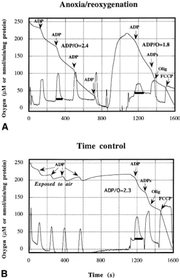
Effects of anoxia/reoxygenation on liver mitochondria The respiratory parameters of isolated liver mitochon-dria were measured before anoxia (control) and after 1 min anoxia followed by 4⫾ 1 min reoxygenation. At first glance, damage induced by anoxia/reoxygenation were important at the level of ADP pulses (e.g., Fig. 1A). There was a broadening of the peak in the slope curve (first derivative). Anoxia/reoxygenation induced a signif-icant decrease in state 3 respiration rate (V3, 98.6⫾ 5.1 to 77.5 ⫾ 3.8, p ⬍ .01), uncoupled respiration rate (VFCCP) [31], and ADP/O ratio [from 2.57⫾ 0.11 ( p ⬍ .01) to 1.75 ⫾ 0.09 ( p ⬍ .001)]. Moreover, state 4 respiration rate (V4) increased (25.5 ⫾ 1.7 to 36.8 ⫾ 4.5, p⬍ .01) with no change in oligomycin respiration rate (VOlig) [31]. The results indicated a decrease in the mitochondrial respiratory chain activities and a drop of the oxidative phosphorylation efficiency. However, the mitochondrial membrane permeability was not altered (stable VOlig). These effects were independent of anoxia duration (5 and 10 min anoxia; data not shown).
In order to verify whether these damage were due to anoxia/reoxygenation or to in vitro aging of mitochon-dria during respiratory assay, we measured the respira-tory parameters after 20 min incubation in a stirred reaction medium exposed to air (time control). To mimic preanoxic respiration, we realized 4 ADP pulses during the time control. Compared with control, mitochondrial respiratory parameters after 20 min in vitro incubation were not different from those of preanoxic respirations (Fig. 1). ADP/O was not significantly modified (from 2.44⫾ 0.12 to 2.34 ⫾ 0.11). Therefore, we can conclude that there is no in vitro time-dependent decline of liver mitochondrial function during the respiratory assay. Thus, mitochondrial damage after anoxia/reoxygenation were not due to in vitro aging.
Protective effects of EGb 761 during anoxia/reoxygenation
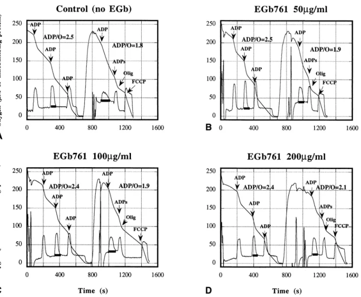
Relative to control, EGb 761 administration (50, 100, and 200 g/ml) induced no significant modification in the respiratory parameters before anoxia/reoxygenation (Figs. 2– 4). Thus, EGb 761 addition in the incubation medium did not impair the mitochondrial respiratory functions.
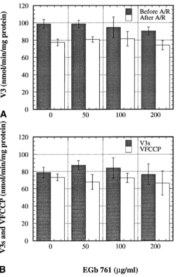
After anoxia/reoxygenation, V3, V3s, VFCCP, and VOligwere not significantly modified in the presence of
EGb 761 (Figs. 2 and 3), relative to anoxia/reoxygen-ation without EGb 761. However, V4 decreased with increasing of EGb 761 concentration (Figs. 2 and 4A) and was significantly lower at 200 g/ml EGb 761 (36.8⫾ 4.5 to 24.6 ⫾ 3.2, p ⬍ .01). Moreover, in the presence of EGb 761, the oxidative phosphorylation ef-ficiency (ADP/O ratio) was higher (1.75 ⫾ 0.09 to 2.09⫾ 0.14, p ⬍ .01) than in its absence (Figs. 2 and 4B). EGb 761 significantly protected ( p ⬍ .01) the ADP/O ratio at 50 and 100g/ml (22%), but the pro-tection was 48% at 200g/ml ( p ⬍ .001). Thus, EGb 761 protected the mitochondrial oxidative
phosphoryla-tion efficiency against damage induced by in vitro anox-ia/reoxygenation but did not improve the phosphorylat-ing and uncoupled respirations.
Effects of EGb 761 on EPR signal
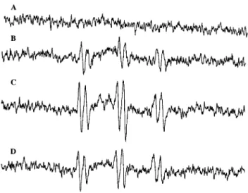
Controls were EPR spectra of the POBN/ethanol me-dium without mitochondria (Fig. 5A). After ADP pulses, mitochondria reached anoxia in state 4 and 10 min an-oxia occurred. At the end of the anan-oxia period, a first sample was harvested for EPR measurement. The EPR spectra showed that a small amount of superoxide anion was produced during preanoxic respiration (Fig. 5B). In a second sample, a significant increase of EPR signal intensity was observed after reoxygenation (Fig. 5C). This indicated an increase of superoxide anion produc-tion during postanoxic reoxygenaproduc-tion, as previously ob-served [31]. Mitochondria incubated with 50 mM POBN, 2% ethanol, and 200 g/ml EGb 761 showed a de-crease in the EPR signal intensity after anoxia/reoxy-genation (Fig. 5D). EGb 761 could decrease the EPR signal intensity because its antioxidant properties al-low to scavenge the superoxide anion produced during reoxygenation.
Effects of EGb 761 on the EPR signals of superoxide anion generated in vitro
Superoxide anion was produced in vitro by the xan-thine/xanthine oxidase system in order to verify the di-rect scavenging effect of EGb 761. Controls were EPR spectra of POBN/ethanol and DMPO without xanthine oxidase (Figs. 6A and 7A). The EPR spectra in Figs. 6B and 7B represented POBN/ethanol and DMPO adducts, respectively, resulting of the reaction of these spin traps with superoxide anion. Addition of EGb 761 (200g/ml) in the reaction mixture showed a decrease of the EPR signal intensity of POBN/ethanol adduct (Fig. 6C). Sim-ilarly, the same concentration of EGb 761 caused a decrease of the EPR signal intensity of DMPO/OOH adduct (Fig. 7C). These results demonstrated that EGb 761 actually scavenged superoxide anion as indicated in experiments with mitochondria after reoxygenation.
DISCUSSION
It is well known that ischemia/reperfusion leads to cellular damage, with among others, a decrease in the mitochondrial function [8,30,37]. Other studies have shown that mitochondria were implicated in liver and cerebral ischemia/reperfusion injury [27,28]. Moreover, mitochondria have been recognized to generate free rad-icals [25,26]. Thus in our previous experiments, we have Fig. 1. Effects of anoxia/reoxygenation and in vitro aging on the
respiratory functions of rat liver mitochondria. Oxygen-concentration traces (M) and its first derivative (nmol/min/mg protein) are shown. Mitochondria were incubated at 25°C in 2 ml of medium containing 15 mM KCl, 1 mM EDTA, 5 mM MgCl2, 50 mM Tris, and 2.5 mM
phosphate with 5 mM ketoglutarate and pyruvate as oxidizable sub-strates. Anoxia (1 min) was reached in state 4 after ADP pulses and reoxygenation was 4⫾ 1 min by exposing the stirred reaction medium to air. ADP was added at 165M (pulses) or 2.5 mM (saturating ADP). Oligomycin and FCCP concentrations were 16 g/ml and 5 M, respectively. (A) Anoxia/reoxygenation. (B) Time control, when mito-chondria were incubated 20 min in the stirred incubation medium exposed to air.
reported [31] that in vitro anoxia/reoxygenation induced mitochondrial dysfunction in liver mitochondria and generated superoxide anion.
In this study, we investigated the oxidative phosphor-ylation function in isolated liver mitochondria submitted to in vitro anoxia/reoxygenation in the presence of EGb 761. The measurements of mitochondrial respiratory pa-rameters allow us to point out dysfunctions induced by anoxia/reoxygenation. Analysis of the respiratory param-eters allows us to describe and to identify the location of the mitochondrial damage, i.e., phospholipid bilayer per-meability, enzyme activity, and substrate or ADP avail-ability. Indeed, according to the chemiosmotic coupling between respiration and ATP synthesis [38], 10 –12 pro-tons are extruded from the mitochondria for each oxygen
atom consumed [39] and 4 protons are taken up to synthesize one ATP. Such proton stoichiometry rules implicate the existence of stoichiometric ratios H⫹/O, H⫹/ATP, and ADP/O. These ratios are fully respected in tightly coupled mitochondria because their inner mem-brane is impermeable to protons (no proton leak).
According to the observed modifications of the func-tional parameters, mitochondrial damage can be located at several levels. Oligomycin resistant respiration rate (VOlig), sustained by proton leak, informs on membrane damage increasing proton permeability. Increase in VOlig indicates an increase in proton leak through the inner mitochondrial membrane which alters the chemiosmotic coupling. If V4 (nonphosphorylating respiration after complete consumption of externally added ADP) in-Fig. 2. Effects of EGb 761 on the respiratory functions of rat liver mitochondria after anoxia/reoxygenation. Oxygen-concentration traces (M) and its first derivative (nmol/min/mg protein) are shown. Mitochondria were incubated at 25°C in 2 ml of medium described in Fig. 1 with 5 mM ketoglutarate and pyruvate as oxidizable substrates. Anoxia (1 min) was reached in state 4 after ADP pulses and reoxygenation was 4⫾ 1 min. ADP, oligomycin, and FCCP concentrations as in Fig. 1. EGb 761 (50, 100, and 200g/ml) was in the incubation medium from the beginning of the respiratory assay. Anoxia/reoxygenation was performed without EGb 761 (A) or in the presence of 50g/ml (B), 100 g/ml (C), and 200 g/ml (D) of EGb 761.
creases and VOligdoes not increase, the membrane lipid bilayer is not damaged but the intrinsic stoichiometry of ATP synthase is impaired (proton slip, i.e., intrinsic uncoupling of ATP synthase). When phosphorylating (V3, V3s) and uncoupled (VFCCP) respiration rates de-crease, either the respiratory chain oxido-reductases are intrinsically damaged, or the upstream feeding of the respiratory chain with electrons is limiting (oxidizable substrate availability). Proton leak (membrane damage) and/or proton slip (modification of intrinsic proton stoi-chiometry of redox pump and/or ATP synthase) lead to a decrease in ADP/O ratio, i.e., a decrease in the oxidative phosphorylation efficiency.
In accordance with previous results [31], anoxia/ reoxygenation of liver mitochondria induced a decrease in phosphorylating and uncoupled respiration rates and a
decrease in the ADP/O ratio. The decrease in rates dem-onstrated that 1 min anoxia followed by 5 min reoxy-genation altered either the oxido-reductases of the respi-ratory chain or the oxidizable substrate availability. As the same substrate concentrations were provided in all measurements, a decrease in substrate availability could only be due to partial inactivation of ketoglutarate and/or pyruvate translocators or of their deshydrogenases. The decrease in the ADP/O ratio, indicating an alteration of the oxidative phosphorylation efficiency, was linked to a proton slip at the level of ATP synthase. Indeed, anoxia/ reoxygenation did not induce proton leak (stable VOlig) and the resting respiration (V4) increased. The absence of proton leak indicated that there was no alteration of the Fig. 3. Effects of EGb 761 on phosphorylating and uncoupled
respira-tion rates before and after anoxia/reoxygenarespira-tion (A/R). Respirarespira-tion rates were expressed in nanomoles of oxygen consumed per minute and per milligram of mitochondrial protein. Data were means⫾ SD (n ⫽ 4). Respiratory parameters were measured in the absence (0) or in the presence of EGb 761 (50, 100, and 200g/ml). (A) Phosphorylating respiration (V3) was measured before and after A/R. (B) Respiration
rate with saturating ADP (V3s) and in the presence of uncoupler FCCP
(VFCCP) was measured after A/R.
Fig. 4. Effects of EGb 761 on resting respiration rate and ADP/O ratio before and after anoxia/reoxygenation (A/R). Resting respiration rate (V4) was expressed in nanomoles of oxygen consumed per minute and
per milligram of mitochondrial protein. Data were means⫾ SD (n ⫽ 4). Statistical significance was p⬍ .01 (**) vs. data before anoxia, and
p⬍ .01 (⫹⫹) or p ⬍ .05 (⫹) vs. data after A/R (no EGb 761).
Mitochondrial parameters were measured in the absence (0) or in the presence of EGb 761 (50, 100, and 200g/ml). (A) Resting respiration rate (V4) was measured before and after A/R. (B) ADP/O ratio, i.e., the
number of moles of ADP phosphorylated by atom gram of oxygen consumed, was calculated before and after A/R.
membrane permeability. The ADP/O decrease and V4 increase were in accordance to prove that anoxia/reoxy-genation induced a proton slip at the level of the ATP synthase, as mentioned above. These latter functional
impairments supported that ATP synthase was damaged by anoxia/reoxygenation.
These results on isolated liver mitochondria showed that in vitro anoxia/reoxygenation induced mitochondrial dysfunction. In the same field, Caraceni et al. [40] related that superoxide anion generated by the respiratory chain during reoxygenation of hepatocytes could be partly re-sponsible for mitochondrial damage. Thus, anoxia/ reoxygenation induced mitochondrial dysfunction and reactive oxygen species were produced during reoxygen-ation [27,29]. Because mitochondria were damaged by anoxia/reoxygenation, it was of great interest to study the effect of antioxidants on the mitochondrial function. In the present study, we decided to use EGb 761, the Ginkgo biloba extract. EGb 761 was well known for its antioxidant properties [3,12,14,15] and its protective ef-fects against injuries induced by in vivo ischemia/reper-fusion [2,7,8] EGb 761 was used at 50, 100, and 200
g/ml, concentrations that did not impair the
mitochon-drial respiratory parameters before anoxia/reoxygenation (Figs. 3A and 4).
Respiratory parameters were measured after anoxia/ reoxygenation in the presence of EGb 761. Mitochondria were incubated with EGb 761 from the beginning of the respiratory assay, before anoxia/reoxygenation. Relative to anoxia/reoxygenation without EGb 761, V3, V3s, VFCCP, and VOligwere not modified (Figs. 2 and 3). As EGb 761 did not change the phosphorylating and uncou-pled respiration rates, there was no protective effect on Fig. 5. Effects of EGb 761 on EPR spectra obtained with POBN/
ethanol as spin trap. Mitochondria (10 mg protein/ml) were incubated in a medium containing 15 mM KCl, 1 mM EDTA, 5 mM MgCl2, 50
mM Tris, and 2.5 mM phosphate with 5 mM ketoglutarate and pyru-vate. Spectrum (A) (control) was obtained with 50 mM POBN and 2% ethanol without mitochondria, spectrum (B) with a sample harvested at the end of 10 min anoxia, and spectrum (C) after anoxia and 5 min reoxygenation. In spectrum (D), a sample of mitochondria incubated with 200g/ml EGb 761 was harvested after reoxygenation.
Fig. 6. Effects of EGb 761 on the EPR spectra of superoxide anion produced in vitro in the presence of POBN/ethanol as spin trap, xanthine (1 mM), xanthine oxidase (0.08 unit/ml), and 50 mM phos-phate buffer (pH 7.8). Spectrum (A) (control) was obtained with 100 mM POBN and 2% ethanol without xanthine oxidase; spectra (B) and (C) were obtained with xanthine oxidase without and with 200g/ml EGb 761, respectively. Instrumental settings were as described in Materi-als and Methods except receiver gain 1.105and number of scans 6.
Fig. 7. Effects of EGb 761 on the EPR spectra of superoxide anion produced in vitro in the presence of DMPO as spin trap. Superoxide anion generation system was the same as in Fig. 6. Spectrum (A) (control) was obtained with 100 mM DMPO without xanthine oxidase; spectra (B) and (C) were obtained with xanthine oxidase without and with 200g/ml EGb 761, respectively. Instrumental settings were as described in Materials and Methods except receiver gain 1.105and
the oxido-reductase activities of the respiratory chain. However, EGb 761, even at the lower concentration, protected the oxidative phosphorylation efficiency (Fig. 4) as shown by the ADP/O ratio. This protection was 22% at 50 and 100 g/ml, and 48% at 200 g/ml. Thus in the presence of EGb 761, the proton slip of the ATP synthase was reduced (V4decreased and ADP/O increased).
EGb 761 protected the mitochondrial oxidative phos-phorylation efficiency against damage induced by in vitro anoxia/reoxygenation, probably by its antioxidant action. EGb 761 was able to react with superoxide anion [12] and to scavenge free radicals. In our EPR experi-ments, we have shown that anoxia/reoxygenation in the presence of EGb 761 (200 g/ml) decreased the EPR signal intensity (Fig. 5). This implicated that EGb 761 scavenged free radicals, essentially superoxide anion, as demonstrated when superoxide anion was generated by an in vitro system with xanthine/xanthine oxidase (Figs. 6 and 7). The protection of mitochondrial function by EGb 761 is thus related to its antioxidant properties. However, it should be noted that EGb 761 and the spin trap POBN could compete to scavenge superoxide anion. In this case, the decrease of EPR signal intensity in the presence of EGb 761 underestimated the scavenging effect of EGb 761.
In conclusion, EGb 761 protects mitochondria against damage induced by anoxia/reoxygenation. In fact, super-oxide anion produced by the respiratory chain during reoxygenation impairs the ATP synthase activity (proton slip) and EGb 761 protects this enzyme by superoxide anion scavenging. Another study is in progress in our laboratory to understand the selective protection of ATP synthase by EGb 761.
Acknowledgements — Guanhua Du held a “Fonds National de la
Recherche Scientifique” postdoctoral fellowship and Katty Willet re-ceived a fellowship from the “Fonds de la Recherche pour l’Industrie et l’Agriculture.” This work was supported by IPSEN (Paris, France).
REFERENCES
[1] De Feudis, F. V. Ginkgo biloba extract (EGb 761):
pharmaco-logical activities and clinical applications. Paris: Elsevier; 1991.
[2] Clostre, F. From the body to the cell membrane: the different levels of pharmacological action of Ginkgo biloba extract. Presse
Med. 15:1529 –1538; 1986.
[3] Pincemail, J.; Deby, C. Antiradical properties of Ginkgo biloba extract. Presse Med. 15:1475–1479; 1986.
[4] Droy-Lefaix, M. T.; Cluzel, J.; Menerath, J. M.; Bonhomme, B.; Doly, M. Antioxidant effects of Ginkgo biloba extract (EGb 761) on the retina. Int. J. Tissue React. 17:93–100; 1995.
[5] Pietri, S.; Culcasi, M.; Verniere, E.; d’Ardigny, P.; Drieu, K. Effect of Ginkgo biloba extract (EGb 761) on the free radical– induced ischemia/reperfusion injury in isolated rat hearts: a he-modynamic and electron-spin-resonance investigation. In: Ferra-dini, C.; Droy-Lefaix, M. T.; Christen, Y., eds. Advances in Ginkgo biloba extract research, vol. 2, Ginkgo biloba extract
(EGb 761) as a free radical scavenger. Paris: Elsevier; 1993:163–
171.
[6] Tosaki, A.; Droy-Lefaix, M. T.; Pali, T.; Das, D. K. Effects of SOD, catalase, and a novel antiarrhythmic drug, EGb 761, on reperfusion-induced arrhythmias in isolated rat hearts. Free
Radic. Biol. Med. 14:363–370; 1993.
[7] Haramaki, N.; Aggarwal, S.; Kawabata, T.; Droy-Lefaix, M. T.; Packer, L. Effects of natural antioxidant Ginkgo biloba extract (EGb 761) on myocardial ischemia/reperfusion injury. Free
Radic. Biol. Med. 16:789 –794; 1994.
[8] Willet, K.; Vaz de Macedo, D.; Detry, O.; Evens, A.; Pereira da Silva, L.; Sluse, F. E. Mitochondrial oxidative phosphorylation injuries occurring in situ and in vitro. Transplant. Proc. 27:2827– 2828; 1995.
[9] Kose, K.; Dogan, P. Lipoperoxidation induced by hydrogen per-oxide in human erythrocyte membranes. 1. Protective effects of
Ginkgo biloba extract (EGb 761). J. Int. Med. Res. 23:1– 8; 1995.
[10] Oyama, Y.; Chikahisa, L.; Ueha, T.; Kanemaru, K.; Noda, K.
Ginkgo biloba extract protects brain neurons against oxidative
stress induced by hydrogen peroxide. Brain Res. 712:349 –352; 1996.
[11] Rong, Y. Q.; Geng, Z. H.; Lau, B. H. S. Ginkgo biloba attenuates oxidative stress in macrophages and endothelial cells. Free Radic.
Biol. Med. 20:121–127; 1996.
[12] Pincemail, J.; Dupuis, M.; Nasr, C.; Hans, P.; Haag-Berrurier, M.; Anton, R.; Deby, C. Superoxide anion scavenging effect and superoxide dismutase activity of Ginkgo biloba extract.
Experi-entia 45:708 –712; 1989.
[13] Garde`s-Albert, M.; Ferradini, C.; Sekaki, A.; Droy-Lefaix, M. T. Oxygen-centered free radicals and their interactions with EGb 761 or CP202. In: Ferradini, C.; Droy-Lefaix, M. T.; Christen, Y., eds. Advances in Ginkgo biloba extract research, vol. 2, Ginkgo biloba extract (EGb 761) as a free radical scavenger. Paris: Elsevier; 1993:1–11.
[14] Maitra, I.; Marcocci, L.; Droy-Lefaix, M. T.; Packer, L. Peroxyl radical scavenging activity of Ginkgo biloba extract EGb 761.
Biochem. Pharmacol. 49:1649 –1655; 1995.
[15] Marcocci, L.; Maguire, J. J.; Droy-Lefaix, M. T.; Paker, L. The nitric oxide scavenging properties of Ginkgo biloba extract EGb 761. Biochem. Biophys. Res. Commun. 201:748 –755; 1994. [16] Dumont, E.; Petit, E.; Tarrade, T.; Nouvelot, A. UV-C irradiation
induced peroxidative degradation of microsomal fatty acids and proteins: protection by an extract of Ginkgo biloba (EGb 761).
Free Radic. Biol. Med. 13:197–203; 1992.
[17] Kose, K.; Dogan, P. Lipoperoxidation induced by hydrogen per-oxide in human erythrocyte membranes. 2. Comparison of the antioxidant effect of Ginkgo biloba extract (EGb 761) with those of water-soluble and lipid-soluble antioxidants. J. Int. Med. Res.
23:9 –18; 1995.
[18] Jule, Y.; Droy-Lefaix, M. T. Ginkgo biloba extract (EGb 761) and the postlesion neuronal plasticity of rat sympathetic ganglia. In: Christen, Y.; Droy-Lefaix, M. T.; Macias-Nuner, J. F., eds.
Ad-vances in Ginkgo biloba extract research, vol. 5, Effects of
Ginkgo biloba extract (EGb 761) on neuronal plasticity. Paris: Elsevier; 1996:113–119.
[19] Pietri, S.; Maurelli, E.; Drieu, K.; Culeasi, M. Cardioprotective and antioxidant effects of the terpenoid constituents of Ginkgo
biloba extract (EGb 761). J. Mol. Cell. Cardiol. 29:733–742;
1997.
[20] Saris, N. E.; Eriksson, K. O. Mitochondrial dysfunction in isch-aemia-reperfusion. Acta Anaesthesiol. Scand. 107:171–176; 1995.
[21] Halestrap, A. P.; Griffiths, E. J.; Connern, C. P. Mitochondrial calcium handling and oxidative stress. Biochem. Soc. Trans. 21: 353–358; 1993.
[22] Crompton, M.; Andreeva, L. On the involvement of a mitochon-drial pore in reperfusion injury. Basic Res. Cardiol. 88:513–523; 1993.
[23] Gunter, T. E.; Gunter, K. K.; Sheu, S. S.; Gavin, C. E. Mitochon-drial calcium transport: physiological and pathological relevance.
Am. J. Physiol. 267:313–339; 1994.
[24] Nohl, H. Is redox-cycling ubiquinone involved in mitochondrial oxygen activation? Free Radic. Res. Commun. 8:307–315; 1990.
[25] Nohl, H.; Koltover, V.; Stolze, K. Ischemia/reperfusion impairs mitochondrial energy conservation and triggers O2•⫺release as by-product of respiration. Free Radic. Res. Commun. 18:127– 137; 1993.
[26] Turrens, J. F. Superoxide production by the mitochondrial respi-ratory chain. Biosci. Rep. 17:3– 8; 1997.
[27] Nishida, T.; Shibata, H.; Koseki, M.; Nakao, K.; Kawashima, Y.; Yoshida, Y.; Tagawa, K. Peroxidative injury of the mitochondrial respiratory chain during reperfusion of hypothermic rat liver.
Biochim. Biophys. Acta 890:82– 88; 1987.
[28] Almeida, A.; Allen, K. L.; Bates, T. E.; Clark, J. B. Effect of reperfusion following cerebral ischemia on the activity of the mitochondrial respiratory chain in the gerbil brain. J. Neurochem.
65:1698 –1703; 1995.
[29] Ambrosio, G.; Zweier, J. L.; Duilio, C.; Kuppusamy, P.; Santoro, G.; Elia, P. P.; Tritto, I.; Cirillo, P.; Condorelli, M.; Chiariello, M. Evidence that mitochondrial respiration is a source of potentially toxic oxygen free radicals in intact rabbit hearts subjected to ischemia and reflow. J. Biol. Chem. 268:18532–18541; 1993. [30] Detry, O.; Willet, K.; Lambermont, B.; Meurisse, M.; Pincemail,
J.; Serteyn, D.; Lamy, M.; Defraigne, J. O.; Limet, R.; Sluse, F. E. Comparative effects of university of Wisconsin and Euro-Collins solutions on pulmonary mitochondrial function after ischemia and reperfusion. Transplantation 65:161–166; 1998.
[31] Du, G. H.; Mouithys-Mickalad, A.; Sluse, F. E. Generation of superoxide anion by mitochondria and impairment of their func-tions during anoxia and reoxygenation in vitro. Free Radic. Biol.
Med. 25:1066 –1074; 1998.
[32] Davies, K. J. A. Protein damage and degradation by oxygen radicals 1. General aspects. J. Biol. Chem. 262:9895–9901; 1987. [33] Peterson, G. L. A simplification of the protein assay method of Lowri et al., which is more generally applicable. Anal. Biochem.
83:346 –356; 1977.
[34] Morel, I.; Sergent, O.; Cogrel, P.; Lescoat, G.; Pasdeloup, N.; Brissot, P.; Cillard, P.; Cillard, J. EPR study of antioxidant activity of the iron chelators pyoverdin and hydroxypyrid-4-one in iron-loaded hepatocyte culture: comparison with that of des-ferrioxamine. Free Radic. Biol. Med. 18:303–310; 1995. [35] Fridovitch, I. Quantitative aspects of the production of superoxide
anion radical by milk xanthine oxidase. J. Biol. Chem. 245:4053– 4057; 1970.
[36] Geen, M. J.; Hill, H. O. A. Oxygen radicals in biological systems. In: Packer, L., ed. Methods in enzymology, vol. 105. London: Academic Press; 1984:3–22.
[37] Kim, J. S.; Southard, J. H. Alteration in cellular calcium and mitochondrial functions in the rat liver during cold preservation.
Transplantation 65:369 –375; 1998.
[38] Mitchell, P. Coupling of phosphorylation to electron and hydro-gen transfer by chemiosmotic type of mechanism. Nature 191: 144 –146; 1961.
[39] Hinkle, P. C.; Horstman, L. L. Respiration-driven proton transport in submitochondrial particule. J. Biol. Chem. 246:6024 – 6028; 1971.
[40] Caraceni, P.; Ryu, H. S.; Van Thiel, D. H.; Bolrle, A. B. Source of oxygen radicals produced by rat hepatocytes during postanoxic reoxygenation. Biochim. Biophys. Acta 1268:249 –254; 1995.
ABBREVIATIONS
ADP/O—number of moles of ADP phosphorylated by atom gram of oxygen consumed
A/R—anoxia/reoxygenation
DMPO—5,5-dimethyl-1-pyrroline-N-oxide EGb 761—Ginkgo biloba extract
EPR— electron paramagnetic resonance
FCCP— carbonylcyanide-p-trifluoro-methoxyphenylhy-drazone
POBN—␣-(4-pyridyl-1-oxide)-N-tert-butylnitrone V3—phosphorylating respiration rate
V4—resting respiration rate VFCCP— uncoupled respiration rate