Cell dynamics and immune response to BLV infection: a unifying model
Arnaud Florins 1, Nicolas Gillet 1, Becca Asquith 2, Mathieu Boxus 1, Catherine Burteau 1, Jean-Claude Twizere 1, Patrice Urbain 1, Fabian Vandermeers 1, Christophe Debacq 3, Maria Teresa Sanchez-Alcaraz 4, Isabelle Schwartz-Cornil 5, Pierre Kerkhofs 6, Genevieve Jean 7, Andre Thewis 7, Jack Hay 8, Franck Mortreux 9, Eric Wattel 9, Michal Reichert 10, Arsène Burny 1, Richard Kettmann 1, Charles Bangham 2 and Luc Willems 1,11
1 Molecular and Cellular biology, FNRS-FUSAG, Gembloux, Belgium, 2 Department of Immunology, Imperial College, London, UK, 3
GlaxoSmithKline Biologicals, Rixensart, Belgium, 4 Department of Medical Genetics and Microbiology, University of Toronto, Toronto,
Canada, 5 U892 INRA, Jouy-en-Josas, France, 6 Department of Virology, Veterinary and Agrochemical Research Centre, Uccle, Belgium, 7
Zootechny unit, FUSAG, Gembloux, Belgium, 8 Department of Immunology, University of Toronto, Toronto, Canada, 9 Unité d'oncogenèse virale, CNRS UMR5537 Centre Leon Bérard, Lyon, France, 10 Veterinary Research Institute, Pulawy, Poland
Abbreviations: ATL: adult T cell leukemia, BLV: bovine leukemia virus, BrdU: bromodeoxyuridine, CRE:
cyclic-AMP responsive site, CFSE: carboxyfluorescein diacetate succinimidyl ester, CTL: cytotoxic T-lymphocyte, HAM/TSP: human T-lymphotropic virus-associated myelopathy/ tropical spastic paraparesis, HDAC: histone deacetylase, HTLV: human T-lymphotropic virus, LTR: long terminal repeat, MHC: major histocompatibility complex, PL: persistent lymphocytosis, TUNEL: terminal transferase-mediated DNA end labeling.
Key Words: apoptosis, BLV, expression, HTLV, homeostasis, immune response, leukemia, silencing, therapy,
trafficking, virus.
1. ABSTRACT
Bovine Leukemia virus (BLV) is the natural etiological agent of a lymphoproliferative disease in cattle. BLV can also be transmitted experimentally to a related ruminant species, sheep, in which the pathogenesis is more acute. Although both susceptible species develop a strong anti-viral immune response, the virus persists indefinitely throughout life, apparently at a transcriptionally silent stage, at least in a proportion of infected cells. Soon after infection, these humoral and cytotoxic activities very efficiently abolish the viral replicative cycle, permitting only mitotic expansion of provirus-carrying cells. Short term cultures of these infected cells initially indicated that viral expression protects against spontaneous apoptosis, suggesting that leukemia is a process of accumulation of long-lived cells. This conclusion was recently reconsidered following in vivo dynamic studies based on perfusions of nucleoside (bromodeoxyuridine) or fluorescent protein markers (CFSE). In sheep, the turnover rate of infected cells is increased, suggesting that a permanent clearance process is exerted by the immune system. Lymphocyte trafficking from and to the secondary lymphoid organs is a key component in the aintenance of cell homeostasis. The net outcome of the immune selective pressure is that only cells in which the virus is transcriptionally silenced survive and accumulate, ultimately leading to lymphocytosis. Activation of viral and/or cellular expression in this silent reservoir with deacetylase inhibitors causes the collapse of the proviral loads. In other words, modulation of viral expression appears to be curative in lymphocytic sheep, an approach that might also be efficient in patients infected with the related Human T-lymphotropic virus type 1. In summary, a dynamic interplay between BLV and the host immune response modulates a complex equilibrium between (i) viral expression driving (or) favoring proliferation and (ii) viral silencing preventing apoptosis. As conclusion, we propose a hypothetical model unifying all these mechanisms.
2. INTRODUCTION
Spleen enlargement and disruption leading to lethal hemorrhage are the most spectacular manifestations of BLV-induced leukemia in cattle. In fact, the observation in 1871 of yellowish nodules in the enlarged spleen of a cow is considered to be the first reported case of bovine leukemia (1,2). These local proliferations of B cells, called lymphosarcoma or extranodal lymphoma, can infiltrate different tissues like the spleen, liver, heart, eye, skin and lungs. In addition, transformed B cells can also induce the enlargement of lymph nodes and cause lymphoma or lymphosarcoma (Figure 1). The development of these tumors within essential organs leads to a series of functional defects which are ultimately incompatible with survival. Another clinical manifestation associated with bovine leukemia, which is characterized by an increase in the number of peripheral blood B-lymphocytes, is called persistent lymphocytosis (PL). The PL stage, which affects approximately one third of the infected cattle is considered to be a benign form of leukemia (3). During this stage, the numbers of peripheral blood B cells can remain stable over extended periods of time without any other severe clinical sign.
The BLV retrovirus is transmitted horizontally through the transfer of infected cells, natural transmission occurring via direct contact, milk and possibly biting insects (4,5). However, iatrogenic procedures like dehorning, ear tattooing and essentially, the use of infected needles largely accounted for the propagation of the virus. Although it is widespread, BLV is still highly prevalent in several regions of the world (like in the US) but almost absent nowadays in others (in the EEC). In wilds, the natural reservoir of BLV could be the water buffalo (6). Experimentally, however, the host range of BLV has been extensively studied in different animal species. Successful transmissions of BLV have been described for a number of species including rabbits, rats, goats and sheep (7-10). In contrast, humans do not seem to be susceptible to BLV, although cell lines can be infected in vitro and although there is some antigenic cross-reactivity between two related viral proteins (p24gag) of BLV and HTLV (Human T-lymphotropic virus type 1) (11,12). In addition, epidemiological studies have shown that consumption of raw milk from infected cattle did not lead to an increase in leukemia in man (13). Therefore, it is unlikely that BLV infects, replicates or induces cancer in humans.
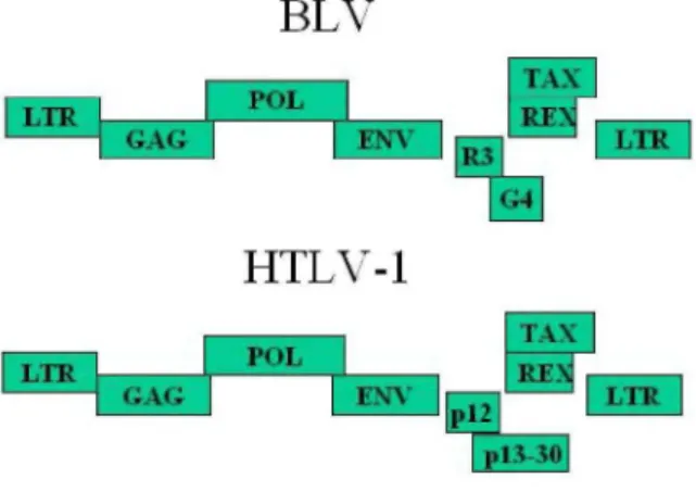
Like HTLV-1, BLV belongs to the deltaretrovirus genus. Sequence variations among different BLV isolates are very limited (14). Its genome contains the classical structural genes (gag, pol and env) coding, respectively, for the viral capsid, the RNA-dependent DNA polymerase (the reverse transcriptase) and the envelope (Figure 2). A series of other open reading frames at the 3'-end of the genome encode regulatory proteins: Tax, Rex, R3 and G4 (15-22). Tax and Rex are essential proteins required for transcriptional and post-transcriptional activation of viral expression. The R3 and G4 proteins are dispensable for infectivity but are involved in the maintenance of high viral loads (23-25). G4 harbors oncogenic potential and interacts with farnesyl pyrophosphate synthetase, an enzyme involved in prenylation of Ras. Further details on BLV genome structure, gene functions and genetic determinants are available in a series of reviews (26,27).
3. ONE PATHOGEN IN TWO DIFFERENT HOSTS DEVELOPING RELATED DISEASES
Amongst different animal species susceptible to experimental infection by BLV, only sheep and to a lesser extent, goats develop leukemia / lymphoma (28). Interestingly, the latency period before the onset of the disease is shorter in sheep than in cattle: leukemia occurs usually 1 to 4 years after infection (instead of 4 to 10 years in cows). In addition, the frequency of viral-induced pathology is much higher: almost all the infected sheep will succumb within their normal life time compared to about 5 per cent in cattle (2,29). There are many similarities but also marked differences between the diseases induced in cattle and in sheep. One of the main difference concerns the kinetics of the proviral loads. In cattle, most of the infected animals remain clinically healthy, the numbers of infected cells ranging around 1 per cent or less. A fraction of these infected animals will develop a persistent lymphocytosis during which the proviral loads remain relatively constant over extended periods of time. In sheep, the number of infected cells in the peripheral blood rise more gradually until the onset of leukemia (levels of circulating lymphocytes being above 10,000 per mm3 and up to 500,000 per mm3) (27,30), although lymphocytosis can last for several months. It should be mentioned that persistent lymphocytosis in cattle or leukemia in sheep are not mandatory steps preceding the acute phase. Indeed, in a significant number of cases, leukemia/lymphoma/lymphosarcoma can develop within the animals in the absence of any lymphocyte accumulation in the peripheral blood. However, the probability of tumor development being greater in animals harboring higher levels of circulating lymphocytes, PL in cattle or lymphocytosis in sheep may be considered as pre-neoplastic stages.
Figure 2. Schematic representation of the BLV and HTLV-1 genomic structures. Three structural genes code for the viral capsid (gag), the reverse transcriptase / integrase (pol) and the envelope (env). A series of other open reading frames (ORFs) at the 3'-end of the genome encode regulatory proteins: Tax, Rex, R3 and G4 (BLV) or pl2 and pl3/p30 (HTLV). These ORFs are translated from complex single or multiple spliced mRNAs (see 15,26,90 for further details). Tax and Rex are essential proteins required for transcriptional and post-transcriptional activation of viral expression. The R3 and G4 proteins are dispensable for infectivity but are involved in the maintenance of high viral loads.
Another difference between the pathologies associated with BLV in cattle and in sheep concerns the genetic modifications that occur during the leukemogenic process. In this context, a major regulator involved in the defense mechanisms against cell transformation, is the p53 tumor suppressor gene. In vitro, the establishment of immortal cell lines appears to be associated with frequent alterations of p53. In vivo, approximately half of the solid tumors induced by BLV in cattle contain a mutated p53 gene (31,32). These mutations interfere with essential p53 functions required for transactivation and suppression of cell growth (33). In contrast, very few mutations were found in B cells from cows with PL and none of the uninfected cattle tested harbored a mutated p53 tumor suppressor gene. Since p53 was never found to be mutated at any stage of the disease in sheep, one might speculate that p53 mutations in cattle favor cell transformation and significantly extends the latency period preceding onset of leukemia/lymphoma as well as the relatively low levels of solid tumor development.
Together, these observations illustrate a difference in the pathogeneses induced by BLV in a natural (cattle) and an experimental host (sheep), disease acuteness being more pronounced in the latter, as expected. Throughout evolution, the relationship between a pathogen and its natural host frequently evolve towards lack of pathogenicity, which represents the ultimate symbiosis (34,35).
4. STRONG BUT APPARENTLY INEFFICIENT HUMORAL AND CYTOTOXIC IMMUNE RESPONSES
Natural or iatrogenic transmission of BLV thus primarily involves the transfer of infected cells via blood or milk (5). The processes occurring after this primary infection still remain obscure. One of the earliest indications of infection is the onset of a humoral antiviral response at about 1-8 weeks post-inoculation (36,37). Antibodies recognizing epitopes from structural (envelope gp51 and capsid p24) and regulatory proteins (Tax and Rex) are synthesized at high titers. Some of these antibodies are directly lytic for BLV-producing cells (38). Almost concomitantly to the early seroconversion period, cytotoxic T-lymphocytes (CTL) specific for Tax and Envelope epitopes appear in the peripheral blood (39,40). Compared to humans, a peculiarity of cattle is that gammadelta T-lymphocytes are major players in this cytotoxic response (41). BLV infection also triggers both a virus-dependent and a virus-invirus-dependent CD4 helper T cell response (42-44). The susceptibility to the polyclonal expansion of BLV-infected B lymphocytes is associated with specific alleles of the major histocompatibility complex system (45,46). Importantly, viral infection correlates with overexpression of two key Th-1 cytokines: TNF-alpha (47-49) and interferon-gamma (50). Finally, the interleukin network is profoundly deregulated at different stages of the disease in particular IL-2, -4, -6, -10 and -12 (47,51-55) but the precise role of these cytokines during pathogenesis still remains unclear.
It thus appears that a very active humoral and cytotoxic immune response is induced soon after BLV infection. Importantly, these anti-viral activities amplify and persist throughout the animal's life indicating that the immune system is permanently stimulated by BLV antigens. Indeed, the level of antibody-mediated cytolytic activity increases with progression of the disease towards the acute phase (38). A strong CTL response is relatively unexpected for a chronic infection, at least if associated with a latent virus (see # 5 and 8 for comments on this assumption). Presently, there is no clear evidence for inhibition mechanisms that would impair virus clearance from the infected host such as, for example, downregulation of MHC class I molecules preventing CTL recognition (reviewed in 56).
5. IS THE VIRUS TRANSCRIPTIONALLY SILENT? THE CAVEATS OF A DOGMA
This question has long been, and still is, a matter of extensive debate. Many experimental arguments support a lack of expression in detectable (i.e. viable) cells harboring an integrated provirus. First, BLV virions or viral proteins cannot be directly detected in the peripheral blood by any available technique (ELISA, flow cytometry, immunoprecipitation or Western blotting). Second, viral transcripts from peripheral blood lymphocytes or tumors can only be amplified by means of very sensitive RT-PCR techniques (15,57,58). Third, using flow cytometry cell sorting and subsequent RT-PCR, only about one B lymphocyte out of 10,000 is found to express tax/rex mRNA during persistent lymphocytosis (57). Fourth, only rare cells in the peripheral blood (1 in 50,000) contain enough BLV transcripts to be identified readily by in situ hybridization (59,60). However, upon ex vivo short term culture, transcription is activated by components of fetal bovine serum and can be augmented by molecules that mimic activation of immune cells.
All these observations are thus in favor of a model postulating that the virus is latent in the very large majority of detectable cells. More, plasma from infected animals contains a fibronectin-related protein that inhibits BLV expression in peripheral blood mononuclear cell cultures (61,62). The latency of BLV in vivo and its reactivation upon ex vivo culture thus became a long-lived dogma. There are however a series of caveats in this model. Indeed, as mentioned in the previous paragraph, the maintenance of a vigorous anti-viral immune response in infected animals indicates that some degree of virus expression must occur in vivo. Furthermore, BLV transcription can even be detected in samples of whole blood upon incubation at 37°C without addition of any exogenous factor except anticoagulants (63). Then, why would this process not be ongoing permanently in infected cells in vivo? If anticoagulants do not activate viral expression, it is unlikely, although not impossible, that the simple removal of blood would be sufficient to induce BLV transcription. Alternatively, we favor the idea that viral expression occurs permanently in a subpopulation of infected cells, which are very efficiently killed by the immune system. The cytotoxic and humoral responses are however unable to destroy cells in which viral transcription is completely silenced.
6. HOW DOES THE VIRUS REPLICATE? VIRAL REPLICATION CYCLE AND CELLULAR CLONAL EXPANSION
Morphologically, the BLV virion has a diameter ranging between 60 and 125 nanometers, and contains a central electron dense nucleoid surrounded by an outer envelope. Its genome is constituted by two poly-A ribonucleic acid molecules bound to viral nucleocapsid proteins (27). This complex is surrounded by a capsid linked to the outer envelope by matrix proteins. The envelope is formed by a cellular lipid bilayer in which two viral proteins (the transmembrane gp30 and the extracellular gp51) are embedded. After infection of a target cell, the RNA genome is copied into DNA by the virally-encoded reverse transcriptase. The so-formed provirus then randomly integrates into the host genomic DNA by means of the viral integrase. As mentioned previously, this integrated provirus is by far the most commonly detectable viral form observed in persistently infected animals. Theoretically, viral spread occurs via the replicative cycle after expression of virions able to infected novel target cells. Alternatively, the integrated proviruses can expand by mitosis of the host cell by a process referred to as clonal expansion (64). Semiquantitative inverse PCR amplification of the cellular sequences flanking the BLV provirus has revealed that the viral load results almost exclusively from clonal expansion of infected cells (30). Importantly, the premalignant cellular clones from which the tumor originates can be detected as early as a few weeks after experimental infection. In fact, the latency period preceding onset of leukemia/lymphoma is characterized by a fluctuation in the abundance of different cellular clones harboring an integrated provirus. Malignancy of a given clone correlates with the accumulation of somatic mutations revealing a decrease in the genetic stability of the expanding infected cell. During the asymptomatic phase, most of the proviral load is sustained by mitosis of the infected cell (Figure 3). Efficient virus replication via the expression of virions and infection of new target cells seems to occur mostly, if not almost exclusively, during a very short period following viral inoculation (so-called primary infection). It is still however possible that the replicative cycle is ongoing continuously but the net outcome of this process does not contribute significantly to the observed viral load, very likely due to a very efficient immune response.
Two key and related questions remain to be solved: why is the abundance of the infected cell clones fluctuating? And: what is the driving force of the clonal expansion process? Based on the extensively described oncogenic properties of Tax (26,65-67), our tenet is that this virally encoded protein triggers cell proliferation. However, the selective growth advantage of the infected cells could also possibly be provided by a non-peptidic viral factor, such as for instance microRNAs (68).
7. DOES THE VIRUS PROTECT FROM APOPTOSIS? THE MOST STRAIGHTFORWARD AND THE ALTERNATIVE INTERPRETATIONS
Apoptosis is an active program leading to the destruction of cells in which abnormalities have occurred, for example, those infected by viruses or triggered by oncogenes. In principle, inhibition of this process alters the equilibrium between proliferation and cell death, modifies homeostasis within an organism and can ultimately lead to leukemia. In other words, apoptosis appears to be an essential mechanism required for the elimination of pre-transformed or normal infected cells and, it is not surprising that viruses have developed strategies to inhibit this process (56).
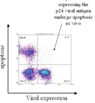
A key and apparently conclusive experiment is illustrated on Figure 4. When lymphocytes are isolated from a BLV-infected sheep and maintained for a few hours in culture, almost all cells expressing the major viral capsid protein p24gag (used as a marker for viral expression) are not stained by terminal transferase-mediated DNA end labeling (TUNEL). The most straightforward interpretation is that viral expression correlates with protection against spontaneous apoptosis. This observation fits well with previous reports showing that B-lymphocytes from infected sheep are less prone to undergo apoptosis compared to the controls (69,70). However, an alternative interpretation is that cells that spontaneously express p24gag antigen are cleared in vivo and can therefore not be detected ex vivo. If so, the low but significant percentages of p24+TUNEL+ double positive cells represent those having escaped immune destruction. Spontaneous viral expression observed ex vivo could occur at similar rates in vivo but would be hidden by the immune surveillance.
The major problem of these ex vivo experiments is that it is never possible to perfectly reproduce the situation prevaling in vivo. Even under the culture conditions that most closely reproduce the natural milieu (such as culture of heparin-containing blood), interpretations of data will always face experimental objections.
Figure 3. Two modes of viral propagation via the replicative cycle and the clonal expansion of the infected cell. Although both mechanisms likely co-exist throughout all stages of infection, the net contribution of the viral replicative cycle during the asymptomatic phase is negligible compared to the clonal proliferation of the infected cell, very likely because of the existence of a strong immune response.
Figure 4. Dot plot resulting from a flow cytometry analysis of peripheral blood mononuclear cells isolated from a lymphocytic BLV-infected sheep and transiently cultivated during 24 hours. Cells were labeled with a monoclonal antibody specific for the viral major capsid protein p24gag (X axis: viral expression) and by TUNEL (Y axis: apoptosis).
8. CELL DYNAMICS OF PATHOGENESIS IN DIFFERENT MODELS
To further gain insight into the processes mediating pathogenesis, it became interesting to define the kinetic parameters sustaining the dynamics of the different cell populations in the infected animals. Indeed, as indicated in the previous paragraph, lymphocyte homeostasis is the result of a critical balance between cell proliferation and death. Disruption of this equilibrium can lead to the onset of leukemia, an increase in the number of lymphocytes being potentially due to one or both of these parameters. The proliferation rates in BLV-infected and healthy sheep have first been determined via a method based on intravenous injection of bromodeoxyuridine (BrdU). By this in vivo approach, B-lymphocytes in BLV-positive asymptomatic sheep were shown to proliferate significantly faster than in uninfected controls (average proliferation rate of 0.020 day-1 versus 0.011 day-1), meaning that an excess of 0.9 % cells (the difference between 2 and 1.1 %) are produced by proliferation each day (71). The difference in the proliferation rates becomes even more evident at the terminal neoplastic stage of the disease (proliferation rate increased by up to tenfold). Cells in S/G2M then also appear in the peripheral blood (our unpublished results) as has been documented for acute cases of human non-viral leukemia (72). In contrast, the death rates of the BrdU-positive cells are not significantly different between aleukemic BLV-infected and control sheep (average death rate 0.089 day-1 versus 0.094 day-1, respectively).
In the natural host, BLV-infected cattle, the cell proliferation rates in asymptomatic and control animals are not significantly different (73). Surprisingly, the PL stage is characterized by a decreased B cell turnover resulting from a reduction of cell death as well as from an overall impairment of proliferation. Paradoxically, an excess of B lymphocytes in the peripheral blood in PL animals correlates with a reduction of cell proliferation, suggesting that a mechanism of feedback regulation controls lymphocyte homeostasis. The reduced dynamic parameters measured in cattle thus contrast with the kinetics observed in experimentally infected sheep. Whether these observations relate to the differences in disease acuteness in the two host species remains a tempting but still open assumption.
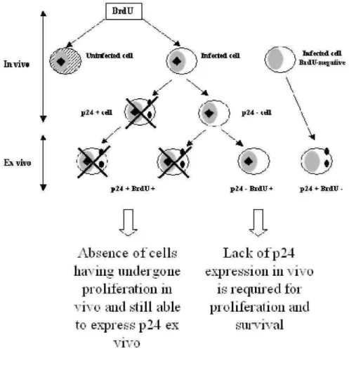
As mentioned in section 5, cells expressing viral proteins cannot be directly observed in the peripheral blood of the infected animals at any stage of the disease. However, viral expression can be induced upon transient short term culture. Surprisingly, very few (if any) cells spontaneously synthesizing p24gag antigen undergo proliferation in vivo (71,73). Indeed, dual flow cytometry labeling demonstrates an almost complete absence of p24+BrdU+ double positive cells, revealing the mutually exclusive presence of both markers. In other words, amongst all infected cells proliferating in vivo as measured by BrdU uptake, none of them are found to express viral proteins. Since lymphocytes synthesizing p24gag are spared from apoptosis ex vivo (see #7), p24+BrdU+ double positive cells are not lost during the culture but have rather been eliminated in vivo (Figure 5). If we postulate that viral expression and cell activation are closely linked, as widely illustrated in the literature, the lack of p24+BrdU+ double positive cells reveals a very efficient negative selection taking place in vivo. Another non-exclusive interpretation would be that only a subpopulation of infected cells is allowed to proliferate (i.e. incorporate BrdU) providing that no viral proteins are expressed. However, this model does not fit with the progressive accumulation of provirus-positive cells, if proliferation is triggered by a viral protein. What would indeed be the selective advantage of a cell carrying a completely silent provirus? Whatever the underlining mechanism, these kinetic studies cast light onto a very active process of immune selection directed towards proliferating infected cells that express the harbored provirus.
9. LYMPHOCYTE TRAFFICKING IN LYMPHOID ORGANS
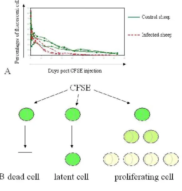
Homeostatic regulation of lymphocyte numbers in the peripheral blood results from a series of physiological factors, of which cell proliferation and death are only partial components. Indeed, kinetics of a cell population is also influenced by recirculation to lymphoid organs, in which proliferation is thought to primarily occur, at least under normal conditions. Experiments based on BrdU kinetics thus pertain mainly to cells in lymphoid tissues leading to an apparent discrepancy: the imbalance created by the net increase in proliferation in the absence of compensating cell death is estimated at 7 % growth in a day (71). Since this considerable growth rate is not reflected by an increase in the corresponding lymphocyte numbers, other regulatory mechanisms including a reduction of recirculation through the lymph nodes as well as a massive elimination of cells in other tissues could play a role. To test these hypotheses, B cell migration from blood to lymph and back from lymph to blood has been traced with the carboxyfluorescein diacetate succinimidyl ester (CFSE), a fluorescent dye that labels proteins via their NH2 terminal ends. Direct intravenous administration of CFSE into sheep achieves remarkable labeling indexes: more than 98% of peripheral blood leukocytes become fluorescent within seconds (74). Since CFSE is extremely unstable in aqueous solution, labeling is very short lived, making it feasible to track
lymphocyte migration from the periphery through the lymph node in vivo. While most studies of lymphocyte recirculation and homing have been done in rodents, the sheep model offers the opportunity to study the recirculation of lymphocytes through tissues by direct cannulation of individual lymphatic vessels (75-77). Using this approach, it has however been shown that B-lymphocytes from BLV-infected and control sheep recirculate with similar rates (Debacq et al, submitted). In contrast, the proportions of labeled B cells in the peripheral blood decrease significantly faster in infected sheep (Figure 6A). Combined with another parameter (the halving of the fluorescent dye upon cell division), it is further possible to calculate the proliferation and death rates (78) (Figure 6B). These calculations indicate that B cells labeled with CFSE in the peripheral blood undergo massive destruction during BLV chronic infection of sheep (Debacq et al, submitted).
Collectively, quantification of the dynamic parameters deduced from BrdU and CFSE kinetics shows that the excess of proliferation in lymphoid organs can be compensated by increased cell death (Figure 7). An important issue that remains to be clarified is to identify the anatomic site required for this cell destruction. In this context, our ongoing experiments reveal that the spleen is a major lymphoid tissue regulating infected cell dynamics and perhaps pathogenesis.
Figure 5. Schematic representation of BrdU incorporation in vivo (hypothetical) and p24gag labeling after short-term culture (ex vivo). The uninfected cell is hachured whereas black squares and full circles indicate BrdU and p24 markers, respectively. The crosses mean that the cells were undetectable by flow cytometry because they did not exist ore more likely because they were destroyed by the immune system.
Figure 6. In vivo CFSE kinetics. A. The CFSE fluorescent dye was injected intravenously into the jugular vein of BLV-infected (red) and control sheep (green). Blood was collected at different time intervals (days on the X axis) and the proportions of CFSE-positive B lymphocytes (Y axis) were determined by flow cytometry. At day 0, more than 98% of peripheral blood B lymphocytes are labeled with CFSE. The percentages of CFSE-positive B cells decrease faster in BLV-infected sheep. B. Since CFSE labels proteins via their NH2 terminal ends, it is assumed that halving of the fluorescent dye occurs upon cell division. The combination of these
fluorescence intensities with the cell percentages allows estimation of the proliferation and death rates (78).
Figure 7. Summary of dynamic data resulting from in vivo BrdU and CFSE kinetic experiments. Quantification of the dynamic parameters determined from the BrdU and CFSE kinetics shows that the excess of proliferation in lymphoid organs can be compensated by increased cell death of the peripheral blood compartment, thereby maintaining homeostasis.
10. MODULATION OF VIRAL EXPRESSION AS THERAPY
In the absence of ex vivo cell culture, the infected lymphocytes apparently do not express any viral protein in the peripheral blood and rest in the G0/G1 phase of the cell cycle (section 5). These apparently quiescent cells may undergo spontaneous cell proliferation and express virions upon transient short-term culture (section 7). Agents known to activate immune cells polyclonally cause an increase in the number of cells containing BLV RNA (59). Viral expression may thus be induced by activation of the host cell consecutively to immune-mediated stimulation.
It is possible that an inhibitory mechanism (or the absence of a driving force: see section 11) hampers viral gene expression in vivo. Infected cell persistence would thus be permitted under the restrictive condition that the virus is not expressed (or at least at a level undetectable by the immune system). Evidence for a very strong immune response is supported by the presence of virus specific cytotoxic T cells and by high titers of cytolytic antibodies (section 4). However, the lack of viral expression in a large proportion of infected cells does not allow efficient clearance by the immune system. Concomitantly, virus infection might also correlate with inhibition of the apoptotic processes, generating a reservoir of apparently latent cells (section 6). In this context, we aimed at evaluating the therapeutic effectiveness of a strategy based on the induction of viral and cellular gene expression. Among a number of methodological approaches, modulation of chromatin condensation, which is an essential component of the gene expression pattern, can be achieved by interference with the level of histone acetylation (79). This process results from an intrinsic balance between the activity of two families of antagonistic enzymes, histone deacetylases (HDAC) and histone acetyltransferases, respectively removing or incorporating acetyl groups into core histones. Although this model is probably oversimplified, acetyl removal by HDACs restores a positive charge to the lysine residues in the histone N-terminal tails and is thought to increase the affinity of histones for DNA, leading to transcriptional repression. Conversely, impairment of HDAC function with specific HDAC inhibitors activates both cellular and viral gene transcription.
Among a growing list of HDAC inhibitors, valproate (the sodium salt of 2-propylpentanoic acid) offers a series of advantages (80). Widely used for several decades for the treatment of epilepsy, this short-chain fatty acid has a very high bioavailability, exhibits very low toxicity in adults and, with a half life of 16-17 hours, has suitable pharmacokinetic properties in vivo. Therefore, valproate has been selected to evaluate the effectiveness of a gene activation chemotherapy in leukemic sheep. Indeed, valproate effectively activates BLV gene expression in transient transfection experiments and in short-term cultures of primary B-lymphocytes (81). In vivo, valproate administration, in the absence of any other cytotoxic drug, is efficient for the treatment of leukemia/lymphoma in sheep, demonstrating the proof-of-concept of a therapy that targets the expression of viral and/or cellular genes. Interestingly, over a long term period, valproate treatment alters neither the cell numbers in control sheep nor other lymphocyte populations in BLV-infected animals, revealing a relative innocuousness of the therapy. The mechanism by which valproate specifically decreases the number of leukemic cells remains to be determined. Amongst numerous hypotheses, we favor a model based on transient activation of cellular and/or viral expression leading to apoptosis by intrinsic (for instance dependent on mitochondrial regulated checkpoints) or extrinsic mechanisms related to membrane-bound receptors (Fas, TRAIL,...) (82-84). Alternatively, a very attractive model would include the induction of viral expression and destruction by the cytotoxic and humoral immune responses (85).
We propose that the concept of a therapy that targets the expression of viral and cellular genes might be a promising treatment of adult T cell leukemia (ATL) or human T-lymphotropic virus-associated myelopathy/tropical spastic paraparesis (HAM/TSP), diseases for which no satisfactory treatment exists so far.
11. A TENTATIVE UNIFYING MODEL AS CONCLUSION
If the latter model described in the previous section is valid, the efficiency of the valproate therapy thus indicates that it is possible to deplete BLV-infected cells via the immune response providing that viral proteins are expressed. In this context, repression of viral expression is a key element during pathogenesis. Indeed, the BLV transcriptional promoter located in the 5' long terminal repeat contains suboptimal binding sequences for the CREB transcription factor. Remarkably, the cyclic-AMP responsive site (CRE) consensus "TGACGTCA" is never strictly conserved in any viral strain and always contains a mutation (for example, AGACGTCA,
TGACGGCA, TGACCTCA). Restoring a perfect CRE sequence into the promoter increases LTR (long terminal repeat) promoter activity, as expected (86). Very surprisingly however, the proviral loads are drastically reduced in sheep infected with a virus harboring this type of mutation. It is tempting to speculate about a hiding process allowing the virus to maintain its promoter silenced while CRE-dependent pathways activate cellular promoters harboring perfect fits of CRE enhancer sequences. Together, these observations thus show that repression of viral expression is a key factor of viral persistence and spread.
BrdU kinetics and recirculation studies (sections 8 and 9) have demonstrated that proliferation is accelerated in infected sheep and that homeostasis is maintained by increased death of the peripheral blood cell population. Amongst a series of plausible hypotheses that cannot be formally excluded, one of the possible models is that the increased turnover results from an activated immune response directed towards the virus. Continuous expression of viral antigens could indeed exacerbate proliferation of virus-reactive immune cells either directly or via cytokines and potentially also expansion of BLV-infected B lymphocytes. Excessive proliferation of uninfected B-lymphocytes in response to BLV early infection has recently been clearly documented (87). In addition, uninfected B lymphocytes also accumulate above normal levels during persistent lymphocytosis. Whether a similar anti-viral process is also responsible for expansion of BLV-infected B cells is presently unknown. It would for instance be interesting to determine the IgM specificity of the infected B lymphocytes. Arguments against this hypothetical mechanism of indirect viral spread include the absence of selective growth advantage conferred to the infected cells. Why would a viral antigen-specific B cell be preferentially infected by the virus? We therefore favor a model in which the virus plays an active role by continuously expressing viral proteins, like the Tax oncogene, able to promote cell proliferation and transformation (26,49,65-67). Tax expression could be permanent providing that cells escape from immune response, which is a rare event, or initiated indirectly via cellular activation. Concomitantly, Tax expression would also stimulate the anti-viral immune response, which in turn would clear the infected cells. Shutoff of viral expression possibly by a viral accessory protein (and/or histone modification, see section 10) would be a prerequisite allowing a minority of these cells to escape from immune response. Alternatively, since the presence of the doubly spliced tax/rex transcript in the cytoplasm precedes that of other viral mRNAs (88), it is possible that a subpopulation of cells would exclusively express the Tax protein at least during a short time interval. Since very few lymphocytes expressing viral proteins can directly be observed in vivo (section 5), the frequency of infected cells surviving the host immune pressure is low. Also, this process would only marginally affect the very large majority of infected cells containing a silent virus (or a less frequently expressed virus). The net outcome of this model would be a global stability of the proviral loads with some fluctuations of individual clones, as revealed by long term follow up of proviral integration sites by ligation-mediated PCR (section 6). Although the mechanism is still unknown, variations in the abundance of provirus-positive cell clones could be due to differential antigen stimulation or to modulation of the proportions of individual progeny cells to express virus. Hence, cells isolated from sheep at terminal stages of the disease loose their ability to efficiently express virus ex vivo even in the presence of potent polyclonal activators such as phorbol esters (our unpublished results). Ultimately, a fully transformed cell clone containing a deleted replication defective provirus can even outgrow and induce leukemia (89).
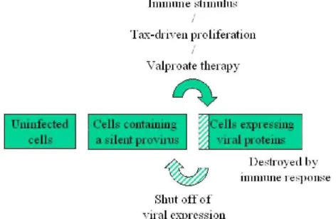
To conclude, the hypothetical model illustrated on Figure 8 best reconciles all current experimental evidences: the oncogenic potential of Tax, the permanent stimulation of the immune system, the low levels of detectable cells expressing viral proteins in vivo, the apparent stability of individual proviral clones and the dynamic parameters. During BLV chronic infection, the host-pathogen interplay is thus characterized by a very dynamic kinetics generating an equilibrium between a virus attempting to proliferate under a tight control exerted by the immune response. In this model, the virus thus permanently transits between a latent and transcriptionally active phase resulting in the progressive accumulation of viable infected cells. Occurrence of somatic mutations associated with genetic instability in these cells finally permits the outgrowth of a transformed clone, leading to leukemia.
Figure 8. Tentative unifying model conciliating immunological and molecular genetics data. Agents known to activate immune cells polyclonally cause an increase in the number of cells containing BLV RNA. Viral expression may thus be induced by activation of the host cell consecutively to immune-mediated stimulation. The selective growth advantage of BLV-infected cells is very likely provided by a virally encoded factor, probably Tax or G4. Indirectly, Tax expression also stimulates the expression of structural genes leading to the destruction of the infected cells by the anti-viral host immune response. Shutoff of viral expression possibly by viral accessory proteins then allows a minority of these cells to escape from immune response. Since very few lymphocytes expressing viral proteins can directly be observed in vivo, the frequency of infected cells surviving the host immune pressure is low. Also, this process would only marginally affect the very large majority of infected cells containing a silent provirus, accounting for the relative stability of the proviral clones. Valproate treatment would activate a proportion of cells containing a silent provirus which, upon expression of viral proteins, are destroyed by the immune system.
12. ACKNOWLEDGEMENTS
This work was supported by the Belgian Foundation against Cancer, the 6th framework program INCA project of the European Commission, the Bekales Foundation, the "Fonds national de la recherche scientifique" (FNRS) and the CGRI (Commissariat général des relations internationales). NG and FV ("Télévie" Fellows), AF (Research Fellow), MB (FRIA Fellow), JCT (Post-doctoral Fellow) and RK and LW (Research Directors) are members of the FNRS.
13. REFERENCES
1. A. Burny, Y. Cleuter, R. Kettmann, M. Mammerickx, G. Marbaix, D. Portetelle, B. A. Van den, L. Willems, and R. Thomas, Bovine leukaemia: facts and hypotheses derived from the study of an infectious cancer. Cancer Surv. 6, 139-159 (1987)
2. J. F. Ferrer, R. R. Marshak, D. A. Abt, and S. J. Kenyon, Persistent lymphocytosis in cattle: its cause, nature and relation to lymphosarcoma. Ann. Rech Vet. 9, 851-857 (1978)
3. Z. Trainin, J. Brenner, R. Meirom, and H. Ungar-Waron, Detrimental effect of bovine leukemia virus (BLV) on the immunological state of cattle. Vet. Immunol. Immunopathol. 54, 293-302 (1996)
4. S. Bech-Nielsen, C. E. Piper, and J. F. Ferrer, Natural mode of transmission of the bovine leukemia virus: role of bloodsucking insects. Am. J. Vet. Res. 39, 1089-1092 (1978)
5. S. G. Hopkins and R. F. DiGiacomo, Natural transmission of bovine leukemia virus in dairy and beef cattle. Vet. Clin. North Am. Food Anim Pract. 13, 107-128 (1997)
6. S. Meas, J. Seto, C. Sugimoto, M. Bakhsh, M. Riaz, T. Sato, K. Naeem, K. Ohashi, and M. Onuma, Infection of bovine immunodeficiency virus and bovine leukemia virus in water buffalo and cattle populations in Pakistan. J. Vet. Med. Sci. 62, 329-331 (2000)
7. M. Mammerickx, D. Portetelle, K. de Clercq, and A. Burny, Experimental transmission of enzootic bovine leukosis to cattle, sheep and goats: infectious doses of blood and incubation period of the disease. Leuk. Res. 11, 353-358 (1987)
8. V. Altanerova, J. Ban, and C. Altaner, Induction of immune deficiency syndrome in rabbits by bovine leukaemia virus. AIDS 3, 755-758 (1989)
9. V. Altanerova, D. Portetelle, R. Kettmann, and C. Altaner, Infection of rats with bovine leukaemia virus: establishment of a virus-producing rat cell line. J. Gen. Virol. 70 ( Pt 7), 1929-1932 (1989)
10. C. R. Wyatt, D. Wingett, J. S. White, C. D. Buck, D. Knowles, R. Reeves, and N. S. Magnuson, Persistent infection of rabbits with bovine leukemia virus associated with development of immune dysfunction. J. Virol. 63, 4498-4506 (1989)
11. M. Onuma, K. Tsukiyama, K. Ohya, Y. Morishima, and R. Ohno, Detection of cross-reactive antibody to BLV p24 in sera of human patients infected with HTLV. Microbiol. Immunol. 31, 131-137 (1987)
12. G. C. Buehring, S. M. Philpott, and K. Y. Choi, Humans have antibodies reactive with Bovine leukemia virus. AIDS Res. Hum. Retroviruses 19, 1105-1113 (2003)
13. R. F. DiGiacomo and S. G. Hopkins, Food animal and poultry retroviruses and human health. Vet. Clin. North Am. Food Anim Pract. 13, 177-190 (1997)
14. R. Z. Mamoun, M. Morisson, N. Rebeyrotte, B. Busetta, D. Couez, R. Kettmann, M. Hospital, and B. Guillemain, Sequence variability of bovine leukemia virus env gene and its relevance to the structure and antigenicity of the glycoproteins. J. Virol. 64, 4180-4188 (1990) 15. S. Alexandersen, S. Carpenter, J. Christensen, T. Storgaard, B. Viuff, Y. Wannemuehler, J. Belousov, and J. A. Roth, Identification of alternatively spliced mRNAs encoding potential new regulatory proteins in cattle infected with bovine leukemia virus. J. Virol. 67, 39-52 (1993)
16. D. Derse, Bovine leukemia virus transcription is controlled by a virus-encoded trans-acting factor and by cis-acting response elements. J. Virol. 61, 2462-2471 (1987)
17. D. Derse, trans-acting regulation of bovine leukemia virus mRNA processing. J. Virol. 62, 1115-1119 (1988)
18. L. Lefebvre, A. Vanderplasschen, V. Ciminale, H. Heremans, O. Dangoisse, J. C. Jauniaux, J. F. Toussaint, V. Zelnik, A. Burny, R. Kettmann, and L. Willems, Oncoviral bovine leukemia virus G4 and human T-cell leukemia virus type 1 pl3(11) accessory proteins interact with farnesyl pyrophosphate synthetase. J. Virol. 76, 1400-1414 (2002)
19. N. R. Rice, R. M. Stephens, D. Couez, J. Deschamps, R. Kettmann, A. Burny, and R. V. Gilden, The nucleotide sequence of the env gene and post-env region of bovine leukemia virus. Virology 138, 82-93 (1984)
20. N. R. Rice, R. M. Stephens, A. Burny, and R. V. Gilden, The gag and pol genes of bovine leukemia virus: nucleotide sequence and analysis. Virology 142, 357-377 (1985)
21. N. Sagata, T. Yasunaga, K. Ohishi, J. Tsuzuku-Kawamura, M. Onuma, and Y. Ikawa, Comparison of the entire genomes of bovine leukemia virus and human T-cell leukemia virus and characterization of their unidentified open reading frames. EMBO J. 3, 3231-3237 (1984)
22. L. Willems, A. Gegonne, G. Chen, A. Burny, R. Kettmann, and J. Ghysdael, The bovine leukemia virus p34 is a transactivator protein. EMBO J. 6, 3385-3389 (1987)
23. P. Kerkhofs, H. Heremans, A. Burny, R. Kettmann, and L. Willems, In vitro and in vivo oncogenic potential of bovine leukemia virus G4 protein. J. Virol. 72, 2554-2559 (1998)
24. J. C. Twizere, P. Kerkhofs, A. Burny, D. Portetelle, R. Kettmann, and L. Willems, Discordance between bovine leukemia virus tax immortalization in vitro and oncogenicity in vivo. J. Virol. 74, 9895-9902 (2000)
25. L. Willems, P. Kerkhofs, F. Dequiedt, D. Portetelle, M. Mammerickx, A. Burny, and R. Kettmann, Attenuation of bovine leukemia virus by deletion of R3 and G4 open reading frames. Proc. Natl. Acad. Sci. U. S A 91, 11532-11536(1994)
26. L. Willems, A. Burny, D. Collete, O. Dangoisse, F. Dequiedt, J. S. Gatot, P. Kerkhofs, L. Lefebvre, C. Merezak, T. Peremans, D. Portetelle, J. C. Twizere, and R. Kettmann, Genetic determinants of bovine leukemia virus pathogenesis. AIDS Res. Hum. Retroviruses 16, 1787-1795 (2000)
27. L. Willems, A. Burny, O. Dangoisse, D. Collete, F. Dequiedt, J. S. Gatot, P. Kerkhofs, L. Lefebvre, C. Merezak, D. Portetelle, J. C. Twizere, and R. Kettmann. Bovine leukemia virus as a model for human T-cell leukemia virus. Current Topics in Virology 1, 139-167. (1999)
28. M. Mammerickx, D. Portetelle, and A. Burny, Experimental cross-transmissions of bovine leukemia virus (BLV) between several animal species. Zentralbl. Veterinarmed. B 28, 69-81 (1981)
29. A. Burny, F. Bex, C. Brack, Y. Cleuter, D. Dekegel, J. Ghysdael, R. Kettmann, M. Leclercq, M. Mammerickx, and D. Portetelle, Biochemical and epidemiological studies on bovine leukemia virus (BLV) Haematol. Blood Transfus. 23,445-452 (1979)
30. V. Moules, C. Pomier, D. Sibon, A. S. Gabet, N. Reichert, P. Kerkhofs, L. Willems, F. Mortreux, and E. Wattel, Fate of premalignant clones during the asymptomatic phase preceding lymphoid malignancy. Cancer Research 65, 1234-1243 (2005)
31. F. Dequiedt, R. Kettmann, A. Burny, and L. Willems, Mutations in the p53 tumor-suppressor gene are frequently associated with bovine leukemia virus-induced leukemogenesis in cattle but not in sheep. Virology 209, 676-683 (1995)
32. W. Zhuang, S. Tajima, K. Okada, Y. Ikawa, and Y. Aida, Point mutation of p53 tumor suppressor gene in bovine leukemia virus-induced lymphosarcoma. Leukemia 11 Suppl 3, 344-346 (1997)
33. S. Tajima, W. Z. Zhuang, M. V. Kato, K. Okada, Y. Ikawa, and Y. Aida, Function and conformation of wild-type p53 protein are influenced by mutations in bovine leukemia virus-induced B-cell lymphosarcoma. Virology 243, 735-746 (1998)
34. G. G. Dimijian, Evolving together: the biology of symbiosis, part 1. Proc. (Bayl. Univ Med. Cent. ) 13, 217-226 (2000)
35. M. A. Gilchrist and D. Coombs, Evolution of virulence: Interdependence, constraints, and selection using nested models. Theor. Popul. Biol. 69, 145-153 (2006)
36. K. Radke, D. Grossman, and L. C. Kidd, Humoral immune response of experimentally infected sheep defines two early periods of bovine leukemia virus replication. Microb. Pathog 9, 159-171 (1990)
37. D. Portetelle, M. Mammerickx, and A. Burny, Use of two monoclonal antibodies in an ELISA test for the detection of antibodies to bovine leukaemia virus envelope protein gp51. J. Virol. Methods 23, 211-222 (1989)
38. D. Portetelle, C. Brack, A. Burny, D. Dekegel, M. Mammerickx, and J. Urbain, Detection of complement-dependent lytic antibodies in sera from bovine leukemia virus-infected animals. Ann. Rech Vet. 9, 667-674 (1978)
39. A. D. Hislop, M. F. Good, L. Mateo, J. Gardner, M. H. Gatei, R. C. Daniel, B. V. Meyers, M. F. Lavin, and A. Suhrbier, Vaccine-induced cytotoxic T lymphocytes protect against retroviral challenge. Nat. Med. 4, 1193-1196 (1998)
40. H. Kabeya, K. Ohashi, and M. Onuma, Host immune responses in the course of bovine leukemia virus infection. J. Vet. Med. Sci. 63, 703-708 (2001)
41. P. Lundberg and G. A. Splitter, gammadelta(+) T-Lp6phocyte cytotoxicity against envelope-expressing target cells is unique to the alymphocytic state of bovine leukemia virus infection in the natural host. J. Virol. 74, 8299-8306 (2000)
42. I. Callebaut, V. Voneche, A. Mager, O. Fumiere, V. Krchnak, M. Merza, J. Zavada, M. Mammerickx, A. Burny, and D. Portetelle, Mapping of B-neutralizing and T-helper cell epitopes on the bovine leukemia virus external glycoprotein gp51. J. Virol. 67, 5321-5327 (1993)
43. H. Kabeya, K. Ohashi, C. Sugimoto, and M. Onuma, Characterization of immune responses caused by bovine leukemia virus envelope peptides in sheep. J. Vet. Med. Sci. 61,475-480 (1999)
44. D. M. Stone, L. K. Norton, J. C. Chambers, and W. J. Meek, CD4 T lymphocyte activation in BLV-induced persistent B lymphocytosis in cattle. Clin. Immunol. 96, 280-288 (2000)
45. H. A. Lewin, G. C. Russell, and E. J. Glass, Comparative organization and function of the major histocompatibility complex of domesticated cattle. Immunol. Rev. 167, 145-158 (1999)
46. Y. Nagaoka, H. Kabeya, M. Onuma, N. Kasai, K. Okada, and Y. Aida, Ovine MHC class II DRB1 alleles associated with resistance or susceptibility to development of bovine leukemia virus-induced ovine lymphoma. Cancer Res. 59, 975-981 (1999)
47. R. Meirom, S. Moss, J. Brenner, D. Heller, and Z. Trainin, Levels and role of cytokines in bovine leukemia virus (BLV) infection. Leukemia 11 Suppl 3, 219-220 (1997)
48. H. Kabeya, A. Fukuda, K. Ohashi, C. Sugimoto, and M. Onuma, Tumor necrosis factor alpha and its receptors in experimentally bovine leukemia virus-infected sheep. Vet. Immunol. Immunopathol. 81, 129-139 (2001)
49. J. C. Twizere, V. Kruys, L. Lefebvre, A. Vanderplasschen, D. Collete, C. Debacq, W. S. Lai, J. C. Jauniaux, L. R. Bernstein, O. J. Semmes, A. Burny, P. J. Blackshear, R. Kettmann, and L. Willems, Interaction of retroviral Tax oncoproteins with tristetraprolin and regulation of tumor necrosis factor-alpha expression. J. Natl. Cancer Inst. 95, 1846-1859 (2003)
50. S. Konnai, T. Usui, K. Ohashi, and M. Onuma, The rapid quantitative analysis of bovine cytokine genes by real-time RT-PCR. Vet. Microbiol. 94, 283-294 (2003)
51. R. G. Keefe, Y. Choi, D. A. Ferrick, and J. L. Stott, Bovine cytokine expression during different phases of bovine leukemia virus infection. Vet. Immunol. Immunopathol. 56, 39-51 (1997)
52. D. Pyeon and G. A. Splitter, Regulation of bovine leukemia virus tax and pol mRNA levels by interleukin-2 and -10. J. Virol. 73, 8427-8434 (1999)
53. B. Yakobson, J. Brenner, H. Ungar-Waron, and Z. Trainin, Cellular immune response cytokine expression during the initial stage of bovine leukemia virus (BLV) infection determines the disease progression to persistent lymphocytosis. Comp Immunol. Microbiol. Infect. Dis. 23, 197-208 (2000)
54. M. Amills, V. Ramiya, J. Norimine, C. A. Olmstead, and H. A. Lewin, Reduced IL-2 and IL-4 mRNA expression in CD4+ T cells from bovine leukemia virus-infected cows with persistent lymphocytosis. Virology 304, 1-9 (2002)
55. M. Amills, J. Norimine, C. A. Olmstead, and H. A. Lewin, Cytokine mRNA expression in B cells from bovine leukemia virus-infected cattle with persistent lymphocytosis. Cytokine 28, 25-28 (2004)
56. M. B. A. Oldstone, Viral persistence: Parameters, mechanisms and future predictions. Virology 344, 111-118 (2006)
57. E. M. Gaynor, M. L. Mirsky, and H. A. Lewin, Use of flow cytometry and RT-PCR for detecting gene expression by single cells. Biotechniques 21, 286-291 (1996)
58. W. A. Jensen, J. Rovnak, and G. L. Cockerell, In vivo transcription of the bovine leukemia virus tax/rex region in normal and neoplastic lymphocytes of cattle and sheep. J. Virol. 65,2484-2490 (1991)
59. D. M. Lagarias and K. Radke, Transcriptional activation of bovine leukemia virus in blood cells from experimentally infected, asymptomatic sheep with latent infections. J. Virol. 63, 2099-2107 (1989)
60. K. Radke, T. J. Sigala, and D. Grossman, Transcription of bovine leukemia virus in peripheral blood cells obtained during early infection in vivo. Microb. Pathog. 12, 319-331 (1992)
61. P. Gupta, S. V. Kashmiri, and J. F. Ferrer, Transcriptional control of the bovine leukemia virus genome: role and characterization of a non-immunoglobulin plasma protein from bovine leukemia virus-infected cattle. J. Virol. 50, 267-270 (1984)
62. M. J. van den Heuvel, B. J. Jefferson, and R. M. Jacobs, Isolation of a bovine plasma fibronectin-containing complex which inhibits the expression of bovine leukemia virus p24. J. Virol. 79, 8164-8170 (2005)
63. S. Tajima and Y. Aida, Induction of expression of bovine leukemia virus (BLV) in blood taken from BLV-infected cows without removal of plasma. Microbes Infect. 7, 1211-1216 (2005)
64. E. Wattel, M. Cavrois, A. Gessain, and S. WainHobson, Clonal expansion of infected cells: A way of life for HTLV-I. Journal of Acquired Immune Deficiency Syndromes and Human Retrovirology 13, S92-S99 (1996)
65. S. Tajima, M. Takahashi, S. N. Takeshima, S. Konnai, S. A. Yin, S. Watarai, Y. Tanaka, M. Onuma, K. Okada, and Y. Aida, A mutant form of the tax protein of bovine leukemia virus (BLV), with enhanced transactivation activity, increases expression and propagation of BLV in vitro but not in vivo. J. Virol. 77, 1894-1903 (2003)
66. L. Willems, H. Heremans, G. Chen, D. Portetelle, A. Billiau, A. Burny, and R. Kettmann, Cooperation between bovine leukaemia virus transactivator protein and Ha-ras oncogene product in cellular transformation. EMBO J. 9, 1577-1581 (1990)
67. L. Willems, C. Grimonpont, P. Kerkhofs, C. Capiau, D. Gheysen, K. Conrath, R. Roussef, R. Mamoun, D. Portetelle, A. Burny, E. Adam, L. Lefebvre, J. C. Twizere, H. Heremans, and R. Kettmann, Phosphorylation of bovine leukemia virus Tax protein is required for in vitro transformation but not for transactivation. Oncogene 16, 2165-2176 (1998)
68. M. L. Yeung, Y. Bennasser, S. Y. Le, and K. T. Jeang, siRNA, miRNA and HJV: promises and challenges. Cell Research 15, 935-946 (2005)
69. F. Dequiedt, E. Hanon, P. Kerkhofs, P. P. Pastoret, D. Portetelle, A. Burny, R. Kettmann, and L. Willems, Both wild-type and strongly attenuated bovine leukemia viruses protect peripheral blood mononuclear cells from apoptosis. J. Virol. 71, 630-639 (1997)
70.1. Schwartz-Cornil, N. Chevallier, C. Belloc, D. Le Rhun, V. Laine, M. Berthelemy, A. Mateo, and D. Levy, Bovine leukaemia virus-induced lymphocytosis in sheep is associated with reduction of spontaneous B cell apoptosis. J. Gen. Virol. 78 ( Pt 1), 153-162 (1997) 71. C. Debacq, B. Asquith, P. Kerkhofs, D. Portetelle, A. Burny, R. Kettmann, and L. Willems, Increased cell proliferation, but not reduced cell death, induces lymphocytosis in bovine leukemia virus-infected sheep. Proc. Natl. Acad. Sci. U. SA 99, 10048-10053 (2002)
72. A. Jacobs, Myelodysplastic syndromes: pathogenesis, functional abnormalities, and clinical implications. J. Clin. Pathol. 38, 1201-1217 (1985)
73. C. Debacq, B. Asquith, M. Reichert, A. Burny, R. Kettmann, and L. Willems, Reduced cell turnover in bovine leukemia virus-infected, persistently lymphocytotic cattle. J. Virol. 77, 13073-13083 (2003)
74. B. Ristevski, A. J. Young, L. Dudler, R. N. P. Cahill, W. Kimpton, E. Washington, and J. B. Hay, Tracking dendritic cells: use of an in situ method to label all blood leukocytes. International Immunology 15, 159-165 (2003)
75. J. B. Hay and W. N. Andrade, Lymphocyte recirculation, exercise, and immune responses. Can. J. Physiol Pharmacol. 76, 490-496 (1998)
76. W. R. Hein and P. J. Griebel, A road less travelled: large animal models in immunological research. Nat. Rev. Immunol. 3, 79-84 (2003) 77. A. J. Young, J. B. Hay, and C. R. Mackay, Lymphocyte recirculation and life span in vivo. Curr. Top. Microbiol. Immunol. 184, 161-173 (1993)
78. B. Asquith, C. Debacq, A. Florins, N. Gillet, M. T. Sanchez Alcaraz, A. Mosley, and L. Willems. Quantifying lymphocyte kinetics in vivo using CFSE. Proceedings of the Royal Society of London in press. 2006.
79. C. Yoo and P. Jones. Epigenetic therapy of cancer: past, present and future. Nature reviews 5, 37-50. 2006.
80. R. A. Blaheta, H. Nau, M. Michaelis, and J. Cinatl, Valproate and valproate-analogues: Potent tools to fight against cancer. Current Medicinal Chemistry 9, 1417-1433 (2002)
81. A. Achachi, A. Florins, N. Gillet, C. Debacq, P. Urbain, G. M. Foutsop, F. Vandermeers, A. Jasik, M. Reichert, P. Kerkhofs, L. Lagneaux, A. Burny, R. Kettmann, and L. Willems, Valproate activates bovine leukemia virus gene expression, triggers apoptosis, and induces leukemia/lymphoma regression in vivo. Proc. Natl. Acad. Sci. U. SA 102, 10309-10314 (2005)
82. D. C. Drummond, C. O. Noble, D. B. Kirpotin, Z. X. Guo, G. K. Scott, and C. C. Benz, Clinical development of histone deacetylase inhibitors as anticancer agents. Annual Review of Pharmacology and Toxicology 45, 495-528 (2005)
83. A. Insinga, S. Monestiroli, S. Ronzoni, V. Gelmetti, F. Marchesi, A. Viale, L. Altucci, C. Nervi, S. Minucci, and P. G. Pelicci, Inhibitors of histone deacetylases induce tumor-selective apoptosis through activation of the death receptor pathway. Nature Medicine 11, 71-76 (2005) 84. A. Nebbioso, N. Clarke, E. Voltz, E. Germain, C. Ambrosino, P. Bontempo, R. Alvarez, E. M. Schiavone, F. Ferrara, F. Bresciani, A. Weisz, A. R. de Lera, H. Gronemeyer, and L. Altucci, Tumor-selective action of HDAC inhibitors involves TRAIL induction in acute myeloid leukemia cells. Nature Medicine 11, 77-84 (2005)
85. C. R. M. Bangham and M. Osame, Cellular immune response to HTLV-1. Oncogene 24, 6035-6046 (2005)
86. C. Merezak, C. Pierreux, E. Adam, F. Lemaigre, G. G. Rousseau, C. Calomnie, C. Van Lint, D. Christophe, P. Kerkhofs, A. Burny, R. Kettmann, and L. Willems, Suboptimal enhancer sequences are required for efficient bovine leukemia virus propagation in vivo: implications for viral latency. J. Virol. 75, 6977-6988 (2001)
87. C. Debacq, M. T. Sanchez Alcaraz, F. Mortreux, P. Kerkhofs, R. Kettmann, and L. Willems, Reduced proviral loads during primo-infection of sheep by Bovine Leukemia virus attenuated mutants. Retrovirology 1, 31 (2004)
88. M. A. Powers and K. Radke, Activation of bovine leukemia virus transcription in lymphocytes from infected sheep: rapid transition through early to late gene expression. J. Virol. 66, 4769-4777 (1992)
89. L. Willems, R. Kettmann, F. Dequiedt, D. Portetelle, V. Voneche, I. Cornil, P. Kerkhofs, A. Burny, and M. Mammerickx, In vivo infection of sheep by bovine leukemia virus mutants. J. Virol. 67, 4078-4085 (1993)
90. V. Ciminale, G. N. Pavlakis, D. Derse, C. P. Cunningham, and B. K. Felber, Complex splicing in the human T-cell leukemia virus (HTLV) family of retroviruses: novel mRNAs and proteins produced by HTLV type I. J. Virol. 66, 1737-1745 (1992)