HAL Id: hal-02324570
https://hal.archives-ouvertes.fr/hal-02324570
Submitted on 3 Jun 2020HAL is a multi-disciplinary open access
archive for the deposit and dissemination of sci-entific research documents, whether they are pub-lished or not. The documents may come from teaching and research institutions in France or abroad, or from public or private research centers.
L’archive ouverte pluridisciplinaire HAL, est destinée au dépôt et à la diffusion de documents scientifiques de niveau recherche, publiés ou non, émanant des établissements d’enseignement et de recherche français ou étrangers, des laboratoires publics ou privés.
development of the oyster Crassostrea gigas, through
parental or direct exposure
Justine Castrec, Helene Hegaret, Marianne Alunno-Bruscia, Maïlys Picard,
Philippe Soudant, Bruno Petton, Myrina Boulais, Marc Suquet, Isabelle
Queau, Dominique Ratiskol, et al.
To cite this version:
Justine Castrec, Helene Hegaret, Marianne Alunno-Bruscia, Maïlys Picard, Philippe Soudant, et al.. The dinoflagellate Alexandrium minutum affects development of the oyster Crassostrea gigas, through parental or direct exposure. Environmental Pollution, Elsevier, 2019, 246, pp.827-836. �10.1016/j.envpol.2018.11.084�. �hal-02324570�
Please note that this is an author-produced PDF of an article accepted for publication following peer review. The definitive publisher-authenticated version is available on the publisher Web site.
Environmental Pollution
March 2019, Volume 246, Pages 827-836
https://doi.org/10.1016/j.envpol.2018.11.084 https://archimer.ifremer.fr/doc/00469/58099/
Archimer
https://archimer.ifremer.frThe dinoflagellate Alexandrium minutum affects
development of the oyster Crassostrea gigas, through
parental or direct exposure
Castrec Justine 1, Hégaret Helene 4, Alunno-Bruscia Marianne 2, Picard Maïlys 1, Soudant Philippe 1, Petton Bruno 2, Boulais Myrina 1, 3, Suquet Marc 2, Quéau Isabelle 2, Ratiskol Dominique 2,
Foulon Valentin 1, Le Goïc Nelly 1, Fabioux Caroline 1, *
1 LEMAR UMR 6539 CNRS/UBO/IRD/Ifremer, IUEM, rue Dumont d’Urville, 29280, Plouzané, France 2
Ifremer, LEMAR UMR 6539 CNRS/UBO/IRD/Ifremer, Centre de Bretagne, CS 10070, 29280, Plouzané, France
3
University of North Carolina Wilmington, Center for Marine Science, 5600 Marvin K. Moss Lane, Wilmington, NC, 28409, USA
* Corresponding author : Caroline Fabioux, email address : caroline.fabioux@univ-brest.fr
Abstract :
Harmful algal blooms are a threat to aquatic organisms and coastal ecosystems. Among harmful species, the widespread distributed genus Alexandrium is of global importance. This genus is well-known for the synthesis of paralytic shellfish toxins which are toxic for humans through the consumption of contaminated shellfish. While the effects of Alexandrium species upon the physiology of bivalves are now well documented, consequences on reproduction remain poorly studied. In France, Alexandrium
minutum blooms have been recurrent for the last decades, generally appearing during the reproduction
season of most bivalves including the oyster Crassostrea gigas. These blooms could not only affect gametogenesis but also spawning, larval development or juvenile recruitment. This study assesses the effect of toxic A. minutum blooms on C. gigas reproduction. Adult oysters were experimentally exposed to A. minutum, at environmentally realistic concentrations (102 to 103 cells mL−1) for two months during their gametogenesis and a control group, not exposed to A. minutum was fed with a non-toxic dinoflagellate. To determine both consequences to next generation and direct effects of A. minutum exposure on larvae, the embryo-larval development of subsequent offspring was conducted with and without A. minutum exposure at 102 cells mL−1. Effects at each stage of the reproduction were investigated on ecophysiological parameters, cellular responses, and offspring development. Broodstock exposed to A. minutum produced spermatozoa with decreased motility and larvae of smaller size which showed higher mortalities during settlement. Embryo-larval exposure to A. minutum significantly reduced growth and settlement of larvae compared to non-exposed offspring. This detrimental consequence on larval growth was stronger in larvae derived from control parents compared to offspring from exposed parents. This study provides evidence that A. minutum blooms, whether they occur during gametogenesis, spawning or larval development, can either affect gamete quality and/or larval development of C. gigas, thus potentially impacting oyster recruitment.
Please note that this is an author-produced PDF of an article accepted for publication following peer review. The definitive publisher-authenticated version is available on the publisher Web site.
Graphical abstract
Highlights
► Two-month exposure of adult oysters to Alexandrium minutum decreased sperm motility. ► Oocytes and spermatozoa of exposed oysters contained paralytic shellfish toxins. ► Larvae derived from these gametes showed reduced growth and survival. ► Exposure of oyster larvae to A. minutum altered larval growth and settlement. ► Adult exposure influenced offspring response to A. minutum exposure.
Keywords : Harmful algal bloom (HAB), Paralytic shellfish toxin (PST), Crassostrea gigas, Gametes,
M
AN
US
CR
IP
T
AC
CE
PT
ED
each stage of the reproduction were investigated on ecophysiological parameters, cellular 29
responses, and offspring development. Broodstock exposed to A. minutum produced 30
spermatozoa with decreased motility and larvae of smaller size which showed higher 31
mortalities during settlement. Embryo-larval exposure to A. minutum significantly reduced 32
growth and settlement of larvae compared to non-exposed offspring. This detrimental 33
consequence on larval growth was stronger in larvae derived from control parents compared 34
to offspring from exposed parents. This study provides evidence that A. minutum blooms, 35
whether they occur during gametogenesis, spawning or larval development, can either affect 36
gamete quality and/or larval development of C. gigas, thus potentially impacting oyster 37
recruitment. 38
Capsule: The toxic dinoflagellate Alexandrium minutum affected larval development of the
39
oyster Crassostrea gigas, directly through exposure of larvae and indirectly through parental 40
exposure during gametogenesis. 41
Keywords: Harmful Algal Bloom (HAB), Paralytic Shellfish Toxin (PST), Crassostrea
42
gigas, Gametes, Larvae.
43
Introduction
44
Harmful algal blooms (HABs) have increasingly disrupted coastal ecosystems for the last 45
few decades (Kudela et al., 2015). Among toxic species, blooms of microalgae producing 46
paralytic shellfish toxins (PST) represent an important threat for marine ecosystems, human 47
health and the economy in coastal areas. Blooms of Alexandrium affect marine ecosystems by 48
disrupting community and food web structures (Hallegraeff, 2010), and causing death of 49
seabirds and mammals (Hattenrath-Lehmann et al., 2017). The resulting PST can affect 50
humans via shellfish poisoning events (Anderson et al., 2012a), as filter-feeding bivalves 51
accumulate PST in their flesh by feeding on PST-producing microalgae (Bricelj and 52
M
AN
US
CR
IP
T
AC
CE
PT
ED
Shumway, 1998). Prohibition of shellfish harvesting, impairment of tourism and recreational 53
activities are direct socio-economic consequences of PST-producing microalgae blooms in 54
coastal regions (Anderson et al., 2012b). Detrimental effects of those phenomena are expected 55
to intensify as the geographical distribution of HABs and the use of coastal waters for 56
aquaculture are increasing worldwide (Lassus et al., 2016). 57
In marine ecosystems, outbreaks of Paralytic Shellfish Poisonings (PSPs) are mainly 58
caused by the globally distributed dinoflagellate genus Alexandrium (Rossini, 2014). Blooms 59
of Alexandrium spp. affect the physiology of bivalves including exploited species such as 60
Mytilus edulis, Pecten maximus, Venerupis philippinarum, and Crassostrea gigas (Borcier et
61
al., 2017; Bricelj et al., 1993; Haberkorn et al., 2010a; Lassudrie et al., 2014). Alexandrium 62
minutum modifies valve behavior, disrupts biological rhythms, affects defense responses and
63
digestion of C. gigas (Haberkorn et al., 2010b, 2011; Mat et al., 2013; Mello et al., 2013; 64
Payton et al., 2017; Tran et al., 2010). 65
In France, A. minutum blooms usually occur from April to November and can reach high 66
concentrations (> 107 cells L-1) (Guallar et al., 2017, Chapelle et al., 2015). In French coastal 67
waters, A. minutum blooms may last several months (Guallar et al., 2017) and are often 68
concomitant with breeding period of bivalves, including the oyster C. gigas (Pouvreau et al., 69
2016; Suquet et al., 2016; Ubertini et al., 2017). Marine bivalves are ecosystem engineers and 70
a key resource in coastal areas (Ekstrom et al., 2015). In France, 64,200 tons of Pacific oysters 71
were produced in 2016, representing a value of 360,400 USD (FAO, 2018). In France, the 72
natural recruitment of oyster spat constitutes a large part of cultivated stocks and repeated 73
recruitment failures could have negative impacts on local oyster aquaculture. Given the 74
economic importance of oysters, an understanding of the effects of Alexandrium blooms on 75
the reproduction of C. gigas is critically important. Adult Pacific oysters which reproduce 76
during the summer are frequently exposed to the harmful algae during gametogenesis. 77
M
AN
US
CR
IP
T
AC
CE
PT
ED
Gametes, embryos and larvae, the free-living stages of oysters, also directly experience the 78
toxicity of Alexandrium with potential consequences on recruitment. 79
Short-term exposure of mature Pacific oysters to Alexandrium spp. affected spermatozoa 80
quality, and in vitro exposure of oocytes to A. minutum increased reactive oxygen species 81
production in oocytes (Haberkorn et al., 2010b; Le Goïc et al., 2013). Knowledge regarding 82
potential effects of adult oyster exposure to ecological concentrations of HAB species on the 83
next generation remains scarce (Rolton et al., 2018, 2016; Vasconcelos et al., 2010). Adult 84
eastern oysters Crassostrea virginica exposed to a natural bloom of the brevetoxin producer, 85
Karenia brevis, produced smaller larvae with higher mortality than non-exposed oysters
86
(Rolton et al., 2016). Northern quahogs Mercenaria mercenaria exposed to K. brevis showed 87
reduced gonadal allocation and fertilization success, and the early development of the 88
subsequent offspring was also affected (Rolton et al., 2018). Concerning direct exposure of 89
larvae, acute short-term exposures of Alexandrium spp. negatively affected survival, growth, 90
activity, and settlement of bivalve larvae, including C. gigas, Pinctada fucata martensii, 91
Mercenaria mercenaria, Chlamys farreri, and Argopecten irradians concentricus (Basti et al.,
92
2015; Matsuyama et al., 2001; Mu and Li, 2013; Tang and Gobler, 2012; Yan et al., 2003, 93
2001). However, the effects of A. minutum blooms on oyster larvae remain unknown. 94
The present study investigated the effects of a 3-month exposure to toxic A. minutum at 95
concentrations similar to natural bloom events on the gametogenesis of adult C. gigas and on 96
their offspring. We assessed the weight (total, shell, and flesh), feeding, sex and 97
gametogenesis stage of adult oysters, the quality and cellular, molecular and biochemical 98
characteristics of gametes, and the growth, survival and settlement of larvae. The hypotheses 99
tested were: (1) Gamete quality will be negatively affected by a long-term exposure to A. 100
minutum, (2) Parental exposure may adversely affect subsequently produced offspring, and
101
(3) Direct exposure of larvae to A. minutum will alter larval development. 102
M
AN
US
CR
IP
T
AC
CE
PT
ED
1. Materials and methods
103
1.1 Biological materials
104
Oysters. Adult oysters Crassostrea gigas (Magallana gen. nov.; Salvi and Mariottini,
105
2017) were produced and cultured in controlled conditions according to a standardized 106
protocol (Petton et al., 2015), in Ifremer experimental facilities (Argenton and Bouin, France). 107
Oysters were never exposed to any harmful algal bloom. Histological inspection at the start of 108
the experiment in April 2016 showed that oysters (12 months old, mean total weight 18.0 ± 109
0.4 g) were in early gametogenesis (stage 1), according to Steele and Mulcahy (1999). 110
Algal cultures. Tisochrysis lutea (formerly Isochrysis sp., T. iso strain, CCAP 927/14) and
111
Chaetoceros sp. (formerly Chaetoceros neogracile, strain CCAP 1010-3) were used as
non-112
toxic food for oysters. They were cultured with continuous light (200 µmol photons m-2 s-1) in 113
separated 300-L cylinders enriched with Conway medium (Walne, 1970), and with silicium 114
for Chaetoceros sp. The dinoflagellates Alexandrium minutum (Halim, AM89BM strain) and 115
Heterocapsa triquetra (Ehrenberg, 1840; HT99PZ strain) were grown in filtered seawater (0.2
116
µm) supplemented with L1 medium (Guillard and Hargraves, 1993) and kept in exponential 117
growth phase. Cultures were maintained at 17 ± 1 °C. This A. minutum strain produced a 118
quantity of Paralytic Shellfish Toxins (PST) equivalent to 1.3 ± 0.1 pg eq. STX per cell in the 119
exponential growth phase (Haberkorn et al., 2010 a, b). This strain also produces bioactive 120
extracellular compounds (BECs), which have allelopathic and cytotoxic activities (Borcier et 121
al., 2017; Castrec et al., 2018; Lelong et al., 2011). The non-toxic H. triquetra was chosen as 122
a control because of its similarity to A. minutum in terms of size and shape (Tran et al., 2015). 123
1.2 Experimental exposure of adult oysters to A. minutum
124
The flow chart of experimental procedure is presented in Fig. 1. After acclimation, the 125
oysters were placed in six experimental 50-L tanks (46 oysters per tank) supplied with filtered 126
running seawater (1-µm filtered and UV-treated (FSW); 17.0 ± 0.1 °C, pH 8.3 ± 0.1, and 35.1 127
M
AN
US
CR
IP
T
AC
CE
PT
ED
± 0.1 PSU; 12.5 L h-1), and fed continuously on an algal mixture (1:1 equivalent volume) of T. 128
lutea and Chaetoceros sp. (Tiso/Chaeto) at a daily ratio equal to 6 % dry mass algae/dry mass
129
oyster (Petton et al., 2015). The toxic A. minutum was continuously added to Tiso/Chaeto in 3 130
replicate tanks (exposed treatment) at a concentration of 102 cells mL-1, except during the 131
seventh week of the experiment where the concentration was gradually increased to 103 cells 132
mL-1 for 4 days, mimicking an A. minutum bloom event (Chapelle et al., 2015). The 3 other 133
tanks (control treatment) were exposed to the same concentrations of the non-toxic 134
dinoflagellate H. triquetra. Algal concentrations and exposure time correspond to 135
environmentally realistic conditions (Chapelle et al., 2015). To prevent algal sinking, the 136
water inflow was pressurized to create recirculating flow in the tank and air bubbling was 137
used. The oysters were conditioned for 8 weeks to reach ripeness. 138
1.3 Algal consumption of adult oysters
139
For each algal treatment, a fourth tank was deployed without oysters to evaluate algal 140
sinking. Once a day, inflow and outflow seawater was sampled from each tank (20 mL). 141
Phytoplankton counts were made using an electronic particle counter (Multisizer 3 equipped 142
with a 100-µm aperture tube) to provide 56 days of continuous data. Algal sinking (S) was 143
evaluated in percentage of microalgae retained in the tank without oysters: S = [(Vo−Vi) / Vi]
144
× 100; Vi being the number of algae at the inlet of the control tank, and Vo the number of
145
algae at the outlet. Algal sinking was low (< 4 %) and similar in the two treatments. Total 146
algal consumption, i.e. dinoflagellate and Tiso/Chaeto, was expressed in algal cell volume per 147
oyster per hour (µm3 oyster−1 h−1), as in Sussarellu et al. (2016). 148
1.4 Sampling
149
At 2, 4, 6, and 8 weeks after the beginning of exposure (corresponding to T2, T4, T6 and 150
T8, respectively), five animals per tank were sampled for flesh mass, and a transversal section 151
of the gonadic area for histological examination (Fig. 1). The remainder of the digestive gland 152
M
AN
US
CR
IP
T
AC
CE
PT
ED
was flash frozen in liquid nitrogen and stored at -20 °C for subsequent PST quantification. At 153
the beginning (T0) and at the end (T8) of the experiment, 15 oysters per algal treatment were 154
sampled to measure biometric parameters (total wet mass (TWM), wet shell mass (WSM), 155
and dry flesh mass (DFM)). Condition index was calculated as: DFM × 100 / (TWM - WSM). 156
Six control and 6 exposed oysters of each sex were collected after 7 weeks of broodstock 157
conditioning (T7) for gamete quality measurements (see section 1.6). 158
Gametes were collected at T8 in the remaining control and exposed animals (45 oysters per 159
treatment, 15 oysters per tank) by stripping the gonad, and pooling gametes of oysters from 160
the same tank, for each sex. Oocytes were suspended in FSW, filtered in a 100-µm sieve, and 161
oocyte concentrations were determined by flow cytometry (FCM) according to Le Goïc et al. 162
(2014). Oocytes were transferred in 2 mL tubes for PST quantification and biochemical 163
composition measurements (2 × 10⁶ oocytes for each analysis, stored at -20 °C), and for RNA 164
analyses (4 × 10⁶ oocytes, stored in liquid nitrogen). Sperm samples for PST content and 165
biochemical composition were obtained by direct pipetting the incised male gonad. For larval 166
rearing, gametes were pooled for each treatment and each sex (Fig. 1). 167
1.5 Histology
168
A 3-mm cross section of the visceral mass was excised in front of the pericardial region and 169
immediately fixed in modified Davidson’s solution (Latendresse et al., 2002) for 24 h at 4 °C. 170
Tissues were processed, stained with Harris' hematoxylin–eosin, and observed as described by 171
Hermabessiere et al. (2016). Tissue sections were examined under a light microscope (Leica 172
DMIRB) equipped with a digital camera (Imaging RETIGA 2000R) to determine the sex and 173
gametogenesis stage of each oyster according to the reproductive scale reported by Steele and 174
Mulcahy (1999). In addition, measurement of reproductive effort was determined by image 175
software (Adobe® Photoshop®): gonadal occupation index is the percentage of whole gonadal 176
area in relation to the total transverse section area (Fabioux et al., 2005). 177
M
AN
US
CR
IP
T
AC
CE
PT
ED
1.6 Gamete cellular analyses
178
Gamete collection. Oocytes and spermatozoa were collected at T7 for each oyster by
179
stripping entirely the gonad, according to Boulais et al. (2017) for spermatozoa and Le Goïc et 180
al. (2014) for oocytes. Spermatozoa were collected in 10 mL of FSW at 19 °C and sieved 181
through 60 µm mesh. Oocytes were suspended in FSW, sieved through 100 µm to remove 182
pieces of gonad tissue and concentrated on a 20-µm sieve. Oocyte and spermatozoa 183
concentrations were determined by FCM according to Le Goïc et al. (2013, 2014) and 184
adjusted to 5 × 10⁴ oocytes mL-1 and 1 × 10⁷ spermatozoa mL−1, for FCM and cellular 185
analyses. 186
Cellular parameters by flow cytometry. FCM measurements were performed using an
187
EasyCyte Plus cytometer (Guava Millipore) equipped with standard optics and a 488 nm 188
argon laser. Analyses of gamete relative size and complexity, viability, mitochondrial 189
membrane potential (MMP), and reactive oxygen species (ROS) production were performed 190
according to Le Goïc et al. (2013, 2014) for spermatozoa and oocytes. 191
Oocyte morphological measurements. Oocyte circularity (ranging from 0 to 1, where a
192
value of 1 indicates a perfect circle) and Feret’s diameter (Ferreira and Rasband, 2012) were 193
measured under microscope, using ImageJ software (n = 30 oocytes for each female) 194
according to Boulais et al. (2015a). 195
Characterization of spermatozoa movement. Spermatozoa movement was triggered using a
196
two-step dilution in an activating solution (FSW, 5 g L−1 bovine serum albumin, Tris 20 mM, 197
pH 8.1, dilution rate 1:30) and analyzed under microscope using a CASA plug-in for ImageJ 198
software. The percentage of motile spermatozoa and their velocity (VAP: Velocity of the 199
Average Path) were assessed on a minimum of 30 spermatozoa for each male, according to 200
Boulais et al. (2015b). 201
M
AN
US
CR
IP
T
AC
CE
PT
ED
Quantification of ATP content in spermatozoa. Intracellular ATP content of spermatozoa
202
was estimated in triplicates using 5 × 10⁶ spermatozoa for each male as described in Boulais 203
et al. (2015b), by bioluminescence (kit ATPlite, Perkin Elmer) using a plate reader 204
(EnSpire™ 2300 Multilabel Reader, PerkinElmer). 205
1.7 Quantification of total RNA in oocytes
206
Total RNA was isolated using Tri-reagent (Sigma), treated with rDNase (Macherey-207
Nagel), purified using affinity chromatography (Nucleospin RNA kit, Macherey-Nagel) 208
according to the manufacturer's instructions, and assayed for concentration using a ND-1000 209
spectrophotometer (Nanodrop Technologies). 210
1.8 Protein, lipid, and carbohydrate compositions of gametes
211
Total lipids and carbohydrates were analyzed as described by Bligh and Dyer (1959), and 212
Dubois et al. (1956), respectively. Total protein content was assayed as described by Da Costa 213
et al. (2016). Dry weights were determined with a 400 µL aliquot of the first fraction 214
distributed in pre-weighed capsules and dried at 80 °C for 48 h. Content of each constituent 215
was expressed as: biochemical content of each constituent in mg × 100 / dry mass of gonad in 216
mg. 217
1.9 Toxin quantification
218
PST extraction was performed individually following manufacturer instructions: digestive 219
gland tissue was homogenized in HCl 0.1 M (1:1, w:v) using a Precellys®24 beads-grinder, 220
then boiled for 5 min. For the sampling times T2, T4, and T8, individual homogenates were 221
pooled for each tank (n = 3 pools of 5 oysters for each sampling time). The PST digestive 222
gland content was analyzed individually just before (T6, n = 15) and after (T7, n = 12) the 223
high dinoflagellate bloom simulated in this experiment. Toxin quantification was performed 224
by spectrophotometry using the Abraxis ELISA PSP kit (Novakits, France; see methods in 225
M
AN
US
CR
IP
T
AC
CE
PT
ED
Lassudrie et al. (2015)). Toxin load was expressed in µg STX 100 g-1 of wet digestive gland 226
weight (DG) or gonad weight. 227
1.10 Larval rearing
228
To test the influence of parental exposure on offspring, fertilization was performed for 229
each treatment; a pool of 15 × 106 oocytes (from 33 control females or 29 exposed females) 230
were fertilized separately in a beaker using a pool of sperm (from 12 control males or 16 231
exposed males) using a non-limiting sperm to oocyte ratio (100:1) (Fig. 1). Embryos were 232
transferred 1.5 hours post-fertilization (hpf) in 150-L tanks in UV-treated FSW at a 233
concentration of 50 embryos mL−1 and maintained 48 h at 21 °C. D-larvae were then 234
transferred to 5-L cylindrical triplicate tanks at the density of 50 larvae mL-1, and maintained 235
in a flow-through rearing system (100% seawater renewal h−1, 21 °C, 35 PSU) (Fig. 1). The 236
larvae were fed continuously with Tiso/Chaeto as described by Asmani et al. (2016). 237
To test for influence of offspring exposure, fertilization and larval rearing were performed 238
for each algal treatment as described above, but the subsequent offspring were continuously 239
exposed to A. minutum (102 cells mL-1), from 4-cell embryos (2.5 hpf) to veliger larvae (22 240
days post-fertilization, dpf) (Fig. 1). 241
Larvae were sampled every 2–3 days and fixed in a 0.1% formaldehyde-seawater solution 242
until image analysis for size monitoring. Larval size was assessed by measuring shell length 243
using image analysis on at least 30 larvae per tank per day of sampling (WinImager 2.0 and 244
ImageJ software for image capture and analysis, respectively). 245
A small complementary experiment was carried out on oyster larvae raised on the same 246
conditions as the control larvae to test the capacity of larvae to ingest A. minutum cells. 247
Umbonate larvae (mean shell length 150 ± 22 µm) and eyed larvae (mean shell length 304 ± 248
15 µm) were exposed to A. minutum (AM89BM strain) and the presence of A. minutum cells 249
in larvae digestive tract and feces was checked under light microscope. 250
M
AN
US
CR
IP
T
AC
CE
PT
ED
1.11 Larval survival and settlement
251
All the tanks were drained and the total number of larvae was determined at 22 dpf (Fig. 252
1), when ≥ 50% of larvae reached the eyed larvae stage (morphological competence for 253
metamorphosis) in the control tanks (non-exposed oysters derived from control parents). Each 254
larval population was transferred to a PVC container with a 125-µm nylon mesh base (in 255
triplicate per treatment), maintained in a flow-through system (9 L h−1, 30% h−1 seawater 256
renewal, 21 °C, 35 PSU) and fed the Tiso/Chaeto diet as described previously. After 7 days 257
(29 dpf), the number of remaining swimming larvae, dead larvae, and larvae settled on tank 258
walls and sieve were counted (Fig. 1). The percentage of survival during the settlement step 259
was evaluated as: total number of alive larvae at 29 dpf × 100 / total number of larvae initially 260
stocked at 22 dpf. The larval settlement was evaluated as: number of settled larvae at 29 dpf × 261
100 / total number of alive larvae at 29 dpf. 262
1.12 Statistical analyses
263
Statistical analyses were performed using R version 3.2.2 (R Core Team, 2012). All values 264
are expressed as mean ± standard error (SE). Differences were considered significant when p 265
< 0.05. Differences in oyster algal consumption were evaluated using repeated measures 266
ANOVA, where ‘algal exposure’ and ‘day’ were fixed factors (Huvet et al., 2015). 267
Comparison for oyster gonadal maturation and occupation index between the two algal 268
treatments were investigated for each sampling time using Mann-Whitney U test and t-test, 269
respectively. Results of oyster condition index, gonadal maturation and occupation were 270
pooled for the 3 tanks of each algal treatment, after verifying there were no statistical 271
differences between tanks using one-way ANOVA, Fisher’s exact test, and t-test, 272
respectively. Number of oocytes, and gamete parameters were compared using t-test. 273
Comparison for PST content in oyster digestive glands between sampling times was assessed 274
using a non-parametric Kruskal-Wallis test with Mann-Whitney pairwise as post hoc test, as 275
M
AN
US
CR
IP
T
AC
CE
PT
ED
homoscedasticity assumption was not met. To determine any significant differences between 276
‘parental exposure’, and ‘larval exposure’ on larvae, larval length, survival, and settlement 277
were analyzed using a two-way ANOVA, where ‘parental exposure’ and ‘larval exposure’ 278
were fixed and orthogonal factors. Levene's test was used to determine any heterogeneity of 279
variances and data were transformed if significant. In case of a significant interaction between 280
the two factors, a Tukey HSD was used to detect differences among means. 281
2. Results
282
2.1 Oyster algal consumption, gonad development and biochemical composition
283
No mortality was observed in adult oysters during the experiment. A significantly higher 284
algal consumption (+10 %, F = 66.93, df = 1, p < 0.01; two-way ANOVA) was observed over 285
the whole experiment for oysters exposed to A. minutum (3.32 × 10⁹ ± 5.72 × 107 µm3 of 286
algae oyster−1 h−1) compared to control oysters (3.02 × 10⁹ ± 5.87 × 107 µm3 of algae oyster−1 287
h−1) when averaged over the whole experiment. There was a significant effect of date (F = 288
114.88, df = 47, p < 0.001; two-way ANOVA), and a date-exposure interaction on algal 289
consumption (F = 4.92, df = 47, p < 0.001; two-way ANOVA). No difference (t = 0.39, df = 290
28; t-test) in condition index was observed between exposed and control oysters at the end of 291
the conditioning (10.8 ± 0.5 and 10.6 ± 0.4, respectively). 292
Gonadal maturation was not different between exposed and control oysters during 293
broodstock conditioning (Table S1). At T6, all oysters were ripe (stage 3) in both treatments 294
and gonadal occupation index was not significantly different between exposed (61.5 ± 3.0 %) 295
and control (55.1 ± 3.1 %) oysters (Table S1). The total number of oocytes collected by 296
stripping at the time of breeding was not significantly different (t = -1.06, df = 4; t-test) in 297
exposed (5.4 × 10⁶ ± 0.4) and control (6.5 × 10⁶ ± 0.9) females. 298
Gonad lipid content was higher (+55 %, t = 3.98, df = 4, p < 0.05; t-test) in exposed males 299
(17.0 ± 1.5 %) than in controls (10.9 ± 0.4 %), but protein and carbohydrate contents were not 300
M
AN
US
CR
IP
T
AC
CE
PT
ED
different (Table S2). No significant difference in oocyte biochemical composition was 301
observed between both treatments (Table S2). 302
2.2 Gamete quality
303
Significantly lower percentage of motile spermatozoa (−36 %, t = -2.94, df = 9, p < 0.05; t-304
test) was observed in exposed oysters (30 ± 5 %) compared to control (48 ± 3 %) oysters 305
(Table S3). Spermatozoa velocity and ATP content were similar between the two treatments 306
(Table S3). Oocyte diameter and circularity did not differ between treatments (Table S3). 307
Oocyte and spermatozoa cellular characteristics measured by flow cytometry were similar in 308
exposed and control oysters (Table S3). 309
2.3 Paralytic shellfish toxin content
310
The PST content in the digestive glands of exposed oysters was 10.9 ± 0.5, 26.7 ± 1.6, and 311
19.3 ± 3.5 µg STX 100 g-1 of DG after 2 (T2), 4 (T4), and 6 weeks of conditioning (T6), 312
respectively. The PST content in the digestive glands of exposed oysters was higher (p < 0.05; 313
Kruskal-Wallis) at T7 (131.3 ± 22.4 µg STX 100 g-1 of DG) and T8 (52.5 ± 5.0 µg STX 100 314
g-1 of DG) (i.e. after the mimicked A. minutum bloom at 103 cells per mL) than at T2, T4 and 315
T6. 316
The PST content in exposed oyster gonads measured at T8 was 1.8 ± 0.1 µg STX 100 g-1 317
wet gonad weight (n = 3 pools of 4-6 oysters) for exposed males, and 0.3 ± 0.1 µg STX 100 318
g-1 wet oocytes weight (n = 3 pools of 9-10 oysters) for exposed females, corresponding to 3.3 319
% and 0.6 % of the PST content measured in the digestive glands, respectively. 320
2.4 Larval length, survival and settlement
321
Larval length was significantly affected by both parental and larval exposures to A. 322
minutum (Fig. 2). Larval and parental exposures had significant independent (F = 39.04, df =
323
1, p < 0.001, and F = 7.134, df = 1, p < 0.05, respectively; two-way ANOVA) and interactive 324
effects (F = 15.22, df = 1, p < 0.01; two-way ANOVA) upon larval length at 22 dpf (Fig. 3Fig.
M
AN
US
CR
IP
T
AC
CE
PT
ED
3A). At 22 dpf, non-exposed larvae derived from exposed parents (248.3 ± 11.3 µm) were 326
significantly smaller (-15 %, p < 0.01; Tukey HSD) compared to non-exposed larvae derived 327
from control parents (293.2 ± 3.7 µm). 328
Exposed larvae derived from control parents (223.9 ± 4.4 µm) were smaller (-24 %, p < 329
0.001; Tukey HSD) compared to non-exposed larvae derived from control parents, whereas 330
the length of exposed larvae derived from exposed parents (232.3 ± 5.1 µm) was not 331
significantly different (-6 %) from non-exposed larvae derived from exposed parents (Fig. 332
3A). The length of exposed larvae derived from exposed parents was, however, lower (-20 %, 333
p < 0.01; Tukey HSD) than non-exposed larvae derived from control parents (Fig. 3A).
334
Parental exposure, but not larval exposure, significantly affected larval survival during the 335
settlement step (Fig. 3B, F = 8.21, df = 1, p < 0.05; two-way ANOVA). Larvae derived from 336
exposed parents had reduced survival (33.6 ± 10.8 % and 32.0 ± 7.1 % for non-exposed and 337
exposed larvae, respectively) compared to larvae derived from control parents (67.3 ± 4.2 % 338
and 41.2 ± 6.3 % for non-exposed and exposed larvae, respectively; Fig. 3B). 339
Larval exposure, but not parental exposure, significantly reduced larval settlement (F = 340
8.77, df = 1, p < 0.05; two-way ANOVA, Fig. 3C). Settlement rates were 3.9 % ± 0.3 and 5.1 341
% ± 1.8 for exposed larvae, and 11.9 % ± 1.1 and 30.0 % ± 10.9 for non-exposed larvae 342
derived from control and exposed parents, respectively (Fig. 3C). 343
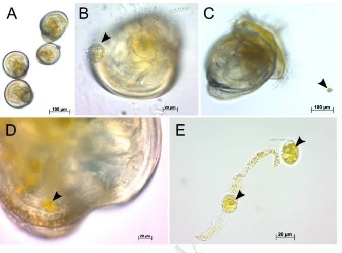
Ingestion tests on control larvae indicated that eyed larvae fed on A. minutum, as these 344
algal cells were observed in the intestine and feces of eyed larvae (Fig. 4C-E). Conversely, 345
ingestion was not observed with early umbonate larvae due to the relative large cell size of A. 346
minutum (23–29 µm) (Fig. 4A, B).
347
3. Discussion
348
Coastal areas regularly experience few days to several weeks of HAB during bivalve 349
reproductive season (Guallar et al., 2017). This experiment was designed to assess the 350
M
AN
US
CR
IP
T
AC
CE
PT
ED
consequences of the toxic dinoflagellate Alexandrium minutum upon reproduction, early 351
development, and settlement of Crassostrea gigas oysters, an environmentally and economic 352
important species. 353
3.1 Presence of A. minutum modifies feeding of maturing oysters
354
Algal consumption was significantly higher for oysters exposed to A. minutum than for 355
control oysters over the whole experiment. Pousse et al. (2018) applied a mechanistic model 356
based on Dynamic Energy Budget theory to the data of the present study, coupling the 357
kinetics of PST accumulation and bioenergetics in C. gigas. They evidenced that toxicant 358
stress provoked by A. minutum affected the energy balance of oysters, more energy being 359
needed for tissue damage repair and detoxification of toxic substances produced by A. 360
minutum. Exposed adult oysters would therefore increase their food consumption to adjust
361
energy intake. Such modification of feeding activity was observed in oysters exposed to 362
polystyrene microspheres to compensate digestive interference caused by plastic particles 363
(Sussarellu et al., 2016) and has been proposed for female copepods Acartia (Acartiura) 364
clausi exposed to A. minutum (previously A. lusitanicum) (Dutz, 1998).
365
Under our experimental conditions of dual feeding with A. minutum and non-toxic 366
Tiso/Chaeto algae, the higher feeding rate seemed to partly counterbalance the higher energy 367
demand due to A. minutum. Coupled with active detoxification of toxins (Fabioux et al., 368
2015), this could explain the absence of major visible effects on gonadal maturation and 369
reproductive effort. However, the lower percentage of motile spermatozoa in oysters exposed 370
to A. minutum still suggests that this response might not be sufficient to overcome PST 371
toxicity on broodstock. 372
3.2 Broodstock exposure to A. minutum affected quality of gametes
373
In male oysters, exposure to A. minutum decreased the percent of motile spermatozoa. 374
Haberkorn et al. (2010b) evidenced that a short acute exposure of mature oysters to A. 375
M
AN
US
CR
IP
T
AC
CE
PT
ED
minutum reduced spermatozoa motility and ATP content, and altered structural and reserve
376
lipids of the digestive gland. In the present study, neither decreased ATP content nor sperm 377
mortality can explain this reduced motility. Total lipid content in male gonads of exposed 378
oysters increased maybe reflecting modifications in lipid metabolism. Lipids are key 379
component of cellular membranes of spermatozoa and modifications in lipid metabolism 380
could be associated with changes in gamete features. PST bind to voltage-gated Na+ channels 381
with high affinity and interact, to a lesser extent, with Ca2+ and K+ channels, modifying ionic 382
fluxes into cells (Llewellyn, 2006) and associated metabolic pathways (Mat et al., 2018). 383
Spermatozoa motility in Pacific oysters is a key factor for reproduction and notably depends 384
on concentrations of ions including K+, Ca2+ and Na+ (Alavi et al., 2014). Indeed, the 385
percentage of motile sperm is drastically reduced in Na+-free seawater (Boulais et al., 2018). 386
In the present study, PST could be responsible for the decreased motility observed in 387
spermatozoa through membrane alterations or ionic fluxes changes. In the field, the 388
fertilization rate of oysters could be impaired by fewer motile spermatozoa with negative 389
consequences on recruitment, as proposed for another free-spawning invertebrate, the sea 390
urchin exposed to high doses of cadmium (Au et al., 2001). The present experimental 391
conditions probably hide this negative effect, as spermatozoa are put in excess compared to 392
oocytes for in vitro fertilization, in a small limited volume, increasing pairing probability 393
compared to natural conditions. 394
3.3 Broodstock exposure to A. minutum affected offspring growth and survival
395
Broodstock exposure to A. minutum decreased offspring growth and induced higher 396
mortality in larvae during the settlement period. Intracellular PST initially accumulate in the 397
digestive gland of bivalves following algal cell lysis and are then transferred into other 398
organs, including gonads (Bricelj and Shumway, 1998). In this study, PST was detected in 399
oocytes (0.3 ± 0.1 µg STX 100 g-1 wet oocytes weight) and male gonad (1.8 ± 0.1 µg STX 400
M
AN
US
CR
IP
T
AC
CE
PT
ED
100 g-1 wet gonad weight), which is consistent with PST content observed in oocytes of 401
mature oysters exposed to a natural A. minutum bloom (Hermabessiere et al., 2016). 402
Crassostrea virginica larvae derived from oysters naturally exposed to Karenia brevis showed
403
significantly higher mortalities and smaller length than larvae derived from non-exposed 404
oysters, suggesting that these negative effects on larval development may be due to the 405
presence of brevetoxins in oocytes (Rolton et al., 2018, 2016). In the present study, toxic 406
effects on the next generation could thus originate from deleterious effect of PST transferred 407
to offspring via gametes. Maternal effects in eggs influence embryogenesis and larval 408
development (Bayne, 2017). PST may also have resulted in functional cell damage during the 409
process of gametogenesis with consequences on development of offspring. Larval physiology 410
could be affected even later during development and settlement like in the present study. 411
3.4 Larval growth and settlement are affected by direct exposure to A. minutum
412
A substantial alteration of larval growth was observed for larvae exposed to A. minutum, 413
both derived from control and exposed adult oysters. Alexandrium toxicity comes from 414
intracellular PST but also from bioactive extracellular compounds (BECs) produced and 415
excreted in the surrounding water by some Alexandrium strains, such as the A. minutum strain 416
tested in this study (Borcier et al., 2017; Castrec et al., 2018; Long et al., 2018). These BECs 417
can be allelopathic, cytotoxic, haemolytic, or ichthyotoxic (Arzul et al., 1999; Ford et al., 418
2008; Lelong et al., 2011; Mardones et al., 2015), however, their molecular structures remain 419
largely unknown (Ma et al., 2011). This detrimental effect of A. minutum on larvae could be 420
attributed sequentially to BEC and PST during development. 421
Toxic effects on small C. gigas larvae (D-larvae to larvae < 150 µm) which are unable to 422
feed on A. minutum could not be related to PST toxicity arising from algal cell consumption, 423
but rather arise from BECs. These bioactive substances produced by Alexandrium minutum 424
mainly exert their action by direct contact with external tissues, e.g. the gills (Borcier et al., 425
M
AN
US
CR
IP
T
AC
CE
PT
ED
2017; Castrec et al., 2018) or cells, e.g. gametes (Le Goïc et al., 2014). These BECs could be 426
cytotoxic to the velum, the feeding and swimming organ of the larvae, thereby reducing 427
energy uptake and subsequent larval growth. Similarly, the toxic effects of A. minutum and A. 428
ostenfeldii on M. edulis larvae observed by De Rijcke et al. (2016) could mainly result from
429
extracellular bioactive substances, as suggested by the authors. This hypothesis was also 430
suggested by Banno et al. (2018) who identified some unknown bioactive compounds as 431
responsible for the decrease of sperm mobility and egg viability of oysters P. fucata martensii 432
exposed to two Alexandrium species. 433
The PST probably become harmful from the moment larvae are able to ingest A. minutum 434
cells. In this study, C. gigas eyed larvae (mean length ± SD: 304 ± 15 µm) ingested A. 435
minutum cells. Veliger larvae (> 200 µm) of the oyster C. virginica fed preferably on large
436
food material (22 to 30 µm) in the presence of large cell dinoflagellate bloom (Baldwin, 437
1995), suggesting that oyster larvae (> 200 µm) likely fed on A. minutum in the present study. 438
Thus, both the PST accumulated through A. minutum consumption and the BECs could have 439
contributed to the adverse effects on growth at the end of the larval development. This 440
hypothesis supports the findings of Mu and Li (2013) who suggested that the reduced growth 441
of early umbonate larvae of C. gigas following a 4-day exposure to 3 × 10² cells mL-1 of A. 442
catenella might relate to both PST and unknown toxins produced by A. catenella.
443
Larval exposure to A. minutum also altered the settlement of oyster larvae. Larval mortality 444
does not explain the reduced settlement in exposed larvae. The decreased settlement could 445
either result from the lagged growth observed after 22 days of exposure, as most exposed 446
larvae did not reach the competence for settlement and metamorphosis, and/or from altered 447
physiology caused by A. minutum toxins. Similarly, the activity of Japanese pearl oyster (P. 448
fucata martensii) pre-settling larvae was decreased when exposed to Alexandrium affine and
449
A. catenella, at 2.5 × 10² cells mL-1 and 10 cells mL-1, respectively (Basti et al., 2015). This 450
M
AN
US
CR
IP
T
AC
CE
PT
ED
effect was attributed to PST metabolites with potent lytic activity produced by the non-451
PST A. affine or to the PST produced by A. catenella, following ingestion of algal cells, 452
leading to paralysis and/or altered cellular homeostasis (Basti et al., 2015). 453
3.5 Broodstock conditioning influenced larval response to A. minutum exposure
454
In the present study, growth of larvae derived from exposed parents was less affected by A. 455
minutum exposure than growth of larvae derived from non-exposed oysters. This result
456
suggests that parental exposure to A. minutum may have led to an improved capacity to cope 457
with the stress caused by A. minutum exposure. Similarly, exposure of adult Sydney rock 458
oysters to elevated pCO2 improved the capacity of their offspring to regulate extracellular pH
459
at elevated pCO2 (Parker et al., 2012). Boullot et al. (2018) revealed that the sensitivity of C.
460
gigas nerves to saxitoxin was decreased when oysters had been previously exposed to
PST-461
producing A. minutum. It can be hypothesized that larvae derived from PST-containing 462
gametes produced by exposed parents may be less sensitive to PST during larval 463
development. 464
4. Conclusions
465
Successful reproduction is essential for the sustainability of marine populations. This study 466
demonstrates that long term exposure of adult oysters to A. minutum during gametogenesis 467
affected spermatozoa motility, and reduced growth and survival of the subsequent offspring. 468
The present laboratory experiment also evidenced that direct A. minutum exposure during 469
oyster embryo-larval development significantly altered growth and settlement of larvae. These 470
effects of A. minutum blooms on oyster reproduction are likely to compromise recruitment of 471
benthic post-larvae of C. gigas by slowing down growth, prolonging the time larvae remain in 472
the seawater column, thus making them more vulnerable to predation. Further research is 473
needed to investigate potential long term effects on marine bivalve populations by studying 474
the consequences of recurrent Alexandrium blooms over multiple generations. 475
M
AN
US
CR
IP
T
AC
CE
PT
ED
Acknowledgments 476This project was supported by the National Research Agency ANR CESA, which founded 477
the ACCUTOX project ANR-13-CESA-0019 (2013–2017). This work was also co-funded by 478
grants from the Regional Council of the Région Bretagne and Brest Métropole. The authors 479
gratefully acknowledge all the colleagues who provided a valuable help during the 480
experiment, dissections, discussions and advices: Guillaume Rivière, Christian Mingant, 481
Matthias Huber, Jacqueline Le Grand, Ashley Taylor Demey, Emilien Pousse, and Claudie 482
Quéré. 483
M
AN
US
CR
IP
T
AC
CE
PT
ED
Figures 484Fig. 1. Flow chart of the experiment. For broodstock conditioning, oysters in red are exposed to the
485
toxic Alexandrium minutum (A. m.), whereas oysters in white are fed with non-toxic Heterocapsa
486
triquetra (H. t. = control treatment). During adult oyster exposure, oysters were sampled every two
487
weeks (T0, T2, T4, T6 and T8) for PST accumulation and histological analyses to study
488
gametogenesis. Gamete cellular analyses were conducted on oysters sampled after 7 weeks of
489
exposure (T7). For larval rearing, embryos and larvae in red are exposed to the toxic A. minutum,
490
whereas stages in white are non-exposed. Tiso/Chaeto feeding: Tisochrysis lutea and Chaetoceros sp.
491
feeding ad libitum; hpf: hours post-fertilization; dpf: days post-fertilization. (For interpretation of the
492
references to color in this figure legend, the reader is referred to the web version of this article.)
M
AN
US
CR
IP
T
AC
CE
PT
ED
Fig. 2. Larval length from D-larvae up to metamorphosis of the controls (non-exposed larvae derived
494
from control parents) and the three other combination of parental and larval exposures. Larval groups
495
were obtained by crossing gametes collected from adult Crassostrea gigas exposed to the non-toxic
496
Heterocapsa triquetra (control parents) and from oysters exposed to Alexandrium minutum (exposed
497
parents), and then exposing the offspring continuously to A. minutum (exposed larvae) or not
(non-498
exposed larvae). Mean ± SE, n = 3.
M
AN
US
CR
IP
T
AC
CE
PT
ED
Fig. 3. Larval length 22 days post-fertilization (dpf) (A), survival (B) and settlement (C) of C. gigas
500
larvae, non-exposed or exposed to A. minutum, derived from A. minutum exposed parents or control
501
parents. Survival is estimated as the total number of alive larvae 29 dpf divided by number of larvae
502
initially stocked at 22 dpf. Settlement is calculated as the number of settled larvae at 29 dpf divided by
503
the total number of live larvae at 29 dpf. Mean ± SE, n = 3.Letters denote significant groupings (p <
504
0.05; two-way ANOVA and Tukey HSD).
M
AN
US
CR
IP
T
AC
CE
PT
ED
Fig. 4. Capacity of control C. gigas larvae to ingest A. minutum. Light micrographs. Umbonate larvae
506
(A, B; mean shell length 150 ± 22 µm) were unable to ingest A. minutum cells due to their relative
507
large size (23–29 µm), whereas eyed larvae (C-E; mean shell length 304 ± 15 µm) fed on A. minutum:
508
algal cells were observed in the intestine (D) and fecal pellets (E). Black arrows indicate A. minutum
509
cells.
M
AN
US
CR
IP
T
AC
CE
PT
ED
ReferencesAlavi, S.M.H., Matsumura, N., Shiba, K., Itoh, N., Takahashi, K.G., Inaba, K., Osada, M., 2014. Roles of extracellular ions and pH in 5-HT-induced sperm motility in marine bivalve. Reproduction 147, 331–345. https://doi.org/10.1530/REP-13-0418
Anderson, D.M., Alpermann, T.J., Cembella, A.D., Collos, Y., Masseret, E., Montresor, M., 2012a. The globally distributed genus Alexandrium: Multifaceted roles in marine ecosystems and impacts on human health. Harmful Algae 14, 10–35. https://doi.org/10.1016/j.hal.2011.10.012
Anderson, D.M., Cembella, A.D., Hallegraeff, G.M., 2012b. Progress in understanding harmful algal blooms (HABs): Paradigm shifts and new technologies for research, monitoring and management. Annu. Rev. Mar. Sci. 4, 143–176. https://doi.org/10.1146/annurev-marine-120308-081121
Arzul, G., Seguel, M., Guzman, L., Erard-Le Denn, E., 1999. Comparison of allelopathic properties in three toxic Alexandrium species. J. Exp. Mar. Biol. Ecol. 232, 285–295. https://doi.org/10.1016/S0022-0981(98)00120-8
Asmani, K., Petton, B., Le Grand, J., Mounier, J., Robert, R., Nicolas, J.-L., 2016. Establishment of microbiota in larval culture of Pacific oyster, Crassostrea gigas. Aquaculture 464, 434–444. https://doi.org/10.1016/j.aquaculture.2016.07.020
Au, D.W.T., Lee, C.Y., Chan, K.L., Wu, R.S.S., 2001. Reproductive impairment of sea urchins upon chronic exposure to cadmium. Part I: Effects on gamete quality. Environ. Pollut. 111, 1–9. https://doi.org/10.1016/S0269-7491(00)00035-X
Baldwin, B.S., 1995. Selective particle ingestion by oyster larvae (Crassostrea virginica) feeding on natural seston and cultured algae. Mar. Biol. 123, 95–107. https://doi.org/10.1007/BF00350328
Banno, K., Oda, T., Nagai, K., Nagai, S., Tanaka, Y., Basti, L., 2018. Deleterious effects of harmful dinoflagellates and raphidophytes on egg viability and spermatozoa swimming velocity in the Japanese pearl oyster Pinctada fucata martensii. J. Shellfish Res. 37, 41–48. https://doi.org/10.2983/035.037.0103
Basti, L., Nagai, S., Go, J., Okano, S., Nagai, K., Watanabe, R., Suzuki, T., Tanaka, Y., 2015. Differential inimical effects of Alexandrium spp. and Karenia spp. on cleavage, hatching, and two larval stages of Japanese pearl oyster Pinctada fucata martensii. Harmful Algae 43, 1–12. https://doi.org/10.1016/j.hal.2014.12.004
Bayne, B., 2017. Reproduction, in: Bayne, B. (Ed.), Biology of Oysters, Developments in Aquaculture and Fisheries Science. Elsevier, San Diego, CA, pp. 565–701. https://doi.org/10.1016/B978-0-12-803472-9.00009-1
Bligh, E.G., Dyer, W.J., 1959. A rapid method of total lipid extraction and purification. Can. J. Biochem. Physiol. 37, 911–917. https://doi.org/10.1139/o59-099
Borcier, E., Morvezen, R., Boudry, P., Miner, P., Charrier, G., Laroche, J., Hegaret, H., 2017. Effects of bioactive extracellular compounds and paralytic shellfish toxins produced by Alexandrium minutum on growth and behaviour of juvenile great scallops Pecten
maximus. Aquat. Toxicol. 184, 142–154.
https://doi.org/10.1016/j.aquatox.2017.01.009
Boulais, M., Corporeau, C., Huvet, A., Bernard, I., Quéré, C., Quillien, V., Fabioux, C., Suquet, M., 2015a. Assessment of oocyte and trochophore quality in Pacific oyster,
Crassostrea gigas. Aquaculture 437, 201–207. https://doi.org/10.1016/j.aquaculture.2014.11.025
Boulais, M., Soudant, P., Le Goïc, N., Quéré, C., Boudry, P., Suquet, M., 2017. ATP content and viability of spermatozoa drive variability of fertilization success in the Pacific
M
AN
US
CR
IP
T
AC
CE
PT
ED
oyster (Crassostrea gigas). Aquaculture 479, 114–119. https://doi.org/10.1016/j.aquaculture.2017.05.035
Boulais, M., Soudant, P., Le Goïc, N., Quéré, C., Boudry, P., Suquet, M., 2015b. Involvement of mitochondrial activity and OXPHOS in ATP synthesis during the motility phase of spermatozoa in the Pacific oyster, Crassostrea gigas. Biol. Reprod. biolreprod.115.128538. https://doi.org/10.1095/biolreprod.115.128538
Boulais, M., Suquet, M., Arsenault-Pernet, E.J., Malo, F., Queau, I., Pignet, P., Ratiskol, D., Grand, J.L., Huber, M., Cosson, J., 2018. pH controls spermatozoa motility in the Pacific oyster (Crassostrea gigas). Biol. Open 7, bio031427. https://doi.org/10.1242/bio.031427
Boullot, F., Fabioux, C., Hegaret, H., Soudant, P., Boudry, P., Benoit, E., 2018. Assessment of saxitoxin sensitivity of nerves isolated from the Pacific oyster, Crassostrea gigas, exposed to Alexandrium minutum. Toxicon 149, 93. https://doi.org/10.1016/j.toxicon.2017.12.025
Bricelj, V.M., Greene, M., Lee, J., Cembella, A., 1993. Growth of the blue mussel Mytilus
edulis on toxic Alexandrium fundyense and effects of gut passage on dinoflagellate
cells, in: T.J. Smayda, Y. Shimizu (Eds.), Toxic Phytoplankton Blooms in the Sea, Elsevier Science Publishers B. V. pp. 371–376.
Bricelj, V.M., Shumway, S.E., 1998. Paralytic shellfish toxins in bivalve molluscs: Occurrence, transfer kinetics, and biotransformation. Rev. Fish. Sci. 6, 315–383. https://doi.org/10.1080/10641269891314294
Castrec, J., Soudant, P., Payton, L., Tran, D., Miner, P., Lambert, C., Le Goïc, N., Huvet, A., Quillien, V., Boullot, F., Amzil, Z., Hégaret, H., Fabioux, C., 2018. Bioactive extracellular compounds produced by the dinoflagellate Alexandrium minutum are highly detrimental for oysters. Aquat. Toxicol. 199, 188–198. https://doi.org/10.1016/j.aquatox.2018.03.034
Chapelle, A., Le Gac, M., Labry, C., Siano, R., Quere, J., Caradec, F., Le Bec, C., Nezan, E., Doner, A., Gouriou, J., 2015. The Bay of Brest (France), a new risky site for toxic
Alexandrium minutum blooms and PSP shellfish contamination. Harmful Algae News
51, 4–5.
Da Costa, F., Petton, B., Mingant, C., Bougaran, G., Rouxel, C., Quéré, C., Wikfors, G. h., Soudant, P., Robert, R., 2015. Influence of one selected Tisochrysis lutea strain rich in lipids on Crassostrea gigas larval development and biochemical composition. Aquac. Nutr. 22, 813–836. https://doi.org/10.1111/anu.12301
De Rijcke, M., Van Acker, E., Nevejan, N., De Schamphelaere, K.A.C., Janssen, C.R., 2016. Toxic dinoflagellates and Vibrio spp. act independently in bivalve larvae. Fish Shellfish Immunol. 57, 236–242. https://doi.org/10.1016/j.fsi.2016.08.027
Dubois, M., Gilles, K.A., Hamilton, J.K., Rebers, P.A.T., Smith, F., 1956. Colorimetric method for determination of sugars and related substances. Anal. Chem. 28, 350–356. Dutz, J., 1998. Repression of fecundity in the neritic copepod Acartia clausi exposed to the
toxic dinoflagellate Alexandrium lusitanicum: Relationship between feeding and egg production. Mar. Ecol. Prog. Ser. 175, 97–107.
Ekstrom, J.A., Suatoni, L., Cooley, S.R., Pendleton, L.H., Waldbusser, G.G., Cinner, J.E., Ritter, J., Langdon, C., van Hooidonk, R., Gledhill, D., Wellman, K., Beck, M.W., Brander, L.M., Rittschof, D., Doherty, C., Edwards, P.E.T., Portela, R., 2015. Vulnerability and adaptation of US shellfisheries to ocean acidification. Nat. Clim. Change 5, 207–214. https://doi.org/10.1038/nclimate2508
Fabioux, C., Huvet, A., Le Souchu, P., Le Pennec, M., Pouvreau, S., 2005. Temperature and photoperiod drive Crassostrea gigas reproductive internal clock. Aquaculture 250, 458–470. https://doi.org/10.1016/j.aquaculture.2005.02.038
M
AN
US
CR
IP
T
AC
CE
PT
ED
Fabioux, C., Sulistiyani, Y., Haberkorn, H., Hégaret, H., Amzil, Z., Soudant, P., 2015. Exposure to toxic Alexandrium minutum activates the detoxifying and antioxidant systems in gills of the oyster Crassostrea gigas. Harmful Algae 48, 55–62. https://doi.org/10.1016/j.hal.2015.07.003
FAO, 2018. Fisheries and aquaculture information and statistics service, global aquaculture production statistics 1950-2016. http://www.fao.org/fishery/statistics/global-aquaculture-production/query/en. (Accessed 25 Mai 2018).
Ferreira, T., Rasband, W., 2012. ImageJ user guide. ImageJ/Fiji 1.
Ford, S.E., Bricelj, V.M., Lambert, C., Paillard, C., 2008. Deleterious effects of a nonPST bioactive compound(s) from Alexandrium tamarense on bivalve hemocytes. Mar. Biol. 154, 241–253. https://doi.org/10.1007/s00227-008-0917-z
Guallar, C., Bacher, C., Chapelle, A., 2017. Global and local factors driving the phenology of
Alexandrium minutum (Halim) blooms and its toxicity. Harmful Algae 67, 44–60.
https://doi.org/10.1016/j.hal.2017.05.005
Guillard, R.R.L., Hargraves, P.E., 1993. Stichochrysis immobilis is a diatom, not a chrysophyte. Phycologia 32, 234–236. https://doi.org/10.2216/i0031-8884-32-3-234.1 Haberkorn, H., Lambert, C., Le Goïc, N., Guéguen, M., Moal, J., Palacios, E., Lassus, P.,
Soudant, P., 2010a. Effects of Alexandrium minutum exposure upon physiological and hematological variables of diploid and triploid oysters, Crassostrea gigas. Aquat. Toxicol. 97, 96–108. https://doi.org/10.1016/j.aquatox.2009.12.006
Haberkorn, H., Lambert, C., Le Goïc, N., Moal, J., Suquet, M., Guéguen, M., Sunila, I., Soudant, P., 2010b. Effects of Alexandrium minutum exposure on nutrition-related processes and reproductive output in oysters Crassostrea gigas. Harmful Algae 9, 427–439. https://doi.org/10.1016/j.hal.2010.01.003
Haberkorn, H., Tran, D., Massabuau, J.-C., Ciret, P., Savar, V., Soudant, P., 2011. Relationship between valve activity, microalgae concentration in the water and toxin accumulation in the digestive gland of the Pacific oyster Crassostrea gigas exposed to
Alexandrium minutum. Mar. Pollut. Bull. 62, 1191–1197. https://doi.org/10.1016/j.marpolbul.2011.03.034
Hallegraeff, G.M., 2010. Ocean climate change, phytoplankton community responses, and harmful algal blooms: A formidable predictive challenge. J. Phycol. 46, 220–235. https://doi.org/10.1111/j.1529-8817.2010.00815.x
Hattenrath-Lehmann, T.K., Ossiboff, R.J., Burnell, C.A., Rauschenberg, C.D., Hynes, K., Burke, R.L., Bunting, E.M., Durham, K., Gobler, C.J., 2017. The role of a PSP-producing Alexandrium bloom in an unprecedented diamondback terrapin (Malaclemys terrapin) mortality event in Flanders Bay, New York, USA. Toxicon 129, 36–43. https://doi.org/10.1016/j.toxicon.2017.02.006
Hermabessiere, L., Fabioux, C., Lassudrie, M., Boullot, F., Long, M., Lambert, C., Le Goïc, N., Gouriou, J., Le Gac, M., Chapelle, A., Soudant, P., Hégaret, H., 2016. Influence of gametogenesis pattern and sex on paralytic shellfish toxin levels in triploid Pacific oyster Crassostrea gigas exposed to a natural bloom of Alexandrium minutum. Aquaculture 455, 118–124. https://doi.org/10.1016/j.aquaculture.2016.01.001
Huvet, A., Béguel, J.-P., Cavaleiro, N.P., Thomas, Y., Quillien, V., Boudry, P., Alunno-Bruscia, M., Fabioux, C., 2015. Disruption of amylase genes by RNA interference affects reproduction in the Pacific oyster Crassostrea gigas. J. Exp. Biol. 218, 1740– 1747. https://doi.org/10.1242/jeb.116699
Kudela, R.M., Berdalet, E., Bernard, S., Burford, M., Fernand, L., Lu, S., Roy, S., Usup, G., Tester, P., Magnien, R., Anderson, D., Cembella, A.D., Chinain, M., Hallegraeff, G., Reguera, B., Zingone, A., Enevoldsen, H., Urban, E., 2015. Harmful Algal Blooms. A scientific summary for policy makers. IOC/UNESCO, Paris, France.