Copyright © 2000, American Society for Microbiology. All Rights Reserved.
Discordance between Bovine Leukemia Virus Tax Immortalization
In Vitro and Oncogenicity In Vivo
JEAN-CLAUDE TWIZERE,
1PIERRE KERKHOFS,
2ARSE`NE BURNY,
1DANIEL PORTETELLE,
1RICHARD KETTMANN,
1ANDLUC WILLEMS
1*
Department of Applied Biochemistry and Biology, Faculty of Agronomy, Gembloux,
1and Veterinary and Agrochemical Research Centre, Uccle,
2Belgium
Received 22 May 2000/Accepted 17 July 2000
Bovine leukemia virus (BLV) Tax protein, a transcriptional activator of viral expression, is essential for viral
replication in vivo. Tax is believed to be involved in leukemogenesis because of its second function,
immor-talization of primary cells in vitro. These activities of Tax can be dissociated on the basis of point mutations
within specific regions of the protein. For example, mutation of the phosphorylation sites at serines 106 and
293 abrogates immortalization potential in vitro but maintains transcriptional activity. This type of mutant is
thus particularly useful for unraveling the role of Tax immortalization activity during leukemogenesis
inde-pendently of viral replication. In this report, we describe the biological properties of BLV recombinant
pro-viruses mutated in the Tax phosphorylation sites (BLVTax106
ⴙ293). Titration of the proviral loads by
semi-quantitative PCR revealed that the BLV mutants propagated at wild-type levels in vivo. Furthermore, two
animals (sheep 480 and 296) infected with BLVTax106
ⴙ293 developed leukemia or lymphosarcoma after 16
and 36 months, respectively. These periods of time are within the normal range of latencies preceding the onset
of pathogenesis induced by wild-type viruses. The phenotype of the mutant-infected cells was characteristic of
a B lymphocyte (immunoglobulin M positive) expressing CD11b and CD5 (except at the final stage for the
lat-ter marker), a patlat-tern that is typical of wild-type virus-infected target cells. Inlat-terestingly, the transformed B
lymphocytes from sheep 480 also coexpressed the CD8 marker, a phenotype rarely observed in tumor biopsies
from chronic lymphocytic leukemia patients. Finally, direct sequencing of the tax gene demonstrated that the
leukemic cells did not harbor revertant proviruses. We conclude that viruses expressing a Tax mutant unable
to transform primary cells in culture are still pathogenic in the sheep animal model. Our data thus provide a
clear example of the discordant conclusions that can be drawn from in vitro immortalization assays and in vivo
experiments. These observations could be of interest for other systems, such as the related human T-cell
leukemia virus type 1, which currently lack animal models allowing the study of the leukemogenic process.
Bovine leukemia virus (BLV) and human T-cell leukemia
virus type 1 (HTLV-I) are members of the Deltaretrovirus
ge-nus in the Retroviridae family (6, 18, 28, 30, 54, 69, 78, 81, 82).
In addition to the structural genes required for the synthesis of
the viral particle (gag, pol, and env), these viruses also contain
a region X located at the 3⬘ end of their genome. This region
encodes a series of proteins involved in the regulation of viral
expression (Tax, Rex, R3, and G4 for BLV). Among these, the
Tax protein is a 34- to 38-kDa transcriptional activator which
increases the synthesis of all viral mRNAs (15, 71).
Transacti-vation by Tax requires 21-bp imperfect repeats located in the 5⬘
long terminal repeat (LTR). In fact, Tax does not bind directly
to DNA but interacts with the CREB/ATF cellular proteins
and increases their affinity for the 21-bp enhancer elements (1,
2, 8). Although some limited variation might be compatible
with function, tax is an essential gene that is absolutely
re-quired for infectivity in vivo (77). Besides its transactivation
activity, the Tax protein also exhibits another property in cell
culture: its expression induces immortalization of primary rat
embryo fibroblasts (REF) (74). In addition, coexpression of tax
and the Ha-ras oncogene fully transforms REF cells yielding
tumors in nude mice. These Tax activities can be dissociated by
mutations within specific regions of the protein. For example,
transcriptional activity requires an amino-terminal zinc finger
structure and an internal leucine-rich activation domain (72,
76). Conversely, phosphorylation of Tax at serines 106 and 293
is required for in vitro transformation but not for
transactiva-tion (73). These phosphorylatransactiva-tion-deficient Tax mutants should
thus provide a unique opportunity to correlate in vitro
trans-formation assays with pathogenicity in vivo.
During the last decades, several methods have been
de-signed to unravel the oncogenic potential of selected viral
proteins. One of the earliest-developed techniques, which was
described in 1983 (36, 37), is based on the immortalization of
primary REF. This method also allowed the characterization
of two types of oncogenes: those that indefinitely prolong the
cellular lifespan (like Myc), and others that induce
transfor-mation by altering cell morphology, impeding contact
inhibi-tion, and decreasing growth factor requirements (such as Ras).
Both kinds of oncogenes are able to cooperate in order to yield
fully transformed cells that induce tumors in nude mice.
Sim-ilar studies in the BLV-HTLV field have shown that one of the
viral regulatory proteins, called Tax, is able to functionally
substitute for Myc in this type of assay (53, 74). A slightly
modified version of this protocol utilizes murine cell lines
(Rat-1 or -2) in which the tax gene provokes the formation of
transformed foci upon transfection (19, 42, 63, 66, 79, 80). The
main objection against these approaches concerns the cell type
(fibroblast versus lymphocyte) and the origin of the species
(murine instead of human, ovine, or bovine). Therefore, other
cell culture systems utilizing T or B lymphocytes have been
developed (3, 16, 22, 23, 47, 55–58, 70). Among these,
proto-cols using recombinant proviruses and primary lymphocytes
* Corresponding author. Mailing address: Biologie mole´culaire,
Faculte´ universitaire des Sciences agronomiques (FUSAG), 13 ave.
Mare´chal Juin, B5030 Gembloux, Belgium. Phone: 32-81-622157. Fax:
32-81-613888. E-mail: willems.l@fsagx.ac.be.
probably provide the most relevant information.
Unfortu-nately, this type of technique has not been established for
BLV. The conclusions drawn from these different studies have
been a matter of dispute, in particular those concerning the
pathways involved in transformation. For instance, the ability
of HTLV-1 Tax to transform primary rat embryo fibroblasts
requires its potential to activate the CArG element, whereas
NFB activity is essential in Rat-1 cells (42). The situation
appears to be far more complex in cell culture systems based
on T lymphocytes. Indeed, the NFB function of Tax-I appears
to be sufficient to promote growth response to interleukin-2,
but clonal expansion of CD4
⫹cells requires the CREB/ATF
and SRF pathways (3).
To further understand the role of Tax during pathogenesis,
extensive efforts have been made to establish animal models in
mice, rats, rabbits, and monkeys (7, 12, 13, 17, 24–27, 29, 31, 33,
38, 48, 51, 55, 59, 60, 62, 65, 68, 83), and indeed, these systems
yielded valuable information in various aspects of viral
infec-tivity and pathogenesis. Despite this extensive progress, the
main objection of these models is that the virus is not in the
context of its natural host species environment and that none
of them perfectly conciliates all the different phenomena
oc-curring during leukemogenesis. In this context, an alternative
approach based on viruses related to HTLV, like BLV, might
provide very useful additional information.
MATERIALS AND METHODS
Animals.All sheep were maintained under controlled conditions at the Vet-erinary and Agrochemical Research Centre (Uccle, Belgium). At regular inter-vals of time, total leukocyte counts were determined by using a Coulter counter ZN, and the corresponding lymphocyte numbers were calculated using the blood formula after examination under the microscope. In parallel, the corresponding sera were analyzed for BLV seropositivity using immunodiffusion and enzyme-linked immunosorbent assay (ELISA) techniques. Sheep were infected with a wild-type strain (plasmid pBLV344 in animal 235), with viruses propagating with equivalent efficiencies and inducing pathogenesis after similar latency periods (plasmid pBLVIX in 8, 11, 247, 292, and 293 and pBLVgag150 in 175), or with the Tax mutant (pBLVTax106⫹293 in 103, 104, 296, and 480). The construction of the pBLV344, pBLVIX, pBLVgag150, and pBLVTax106⫹293 recombinant proviruses has been described elsewhere (73, 75, 77). Of note, the pBLVgag150 mutant, which was initially referred to as attenuated (75), appeared to induce pathogenesis at later times in sheep 175. Finally, three sheep (113, 114, and 115) were used as uninfected controls. The procedures used for infection have been described (77). Briefly, 100g of circular plasmid DNA was mixed with 200 g of Dotap (Roche Diagnostics) and injected intradermally into the back of the sheep.
PCRs.Aliquots of peripheral blood were collected by jugular venipuncture at 4, 6, 15, and 30 months postinfection, and crude cell lysates were prepared as described (77). Briefly, 500l of blood sample was mixed with an equal volume of lysis buffer (0.32 M sucrose, 10 mM Tris-HCl [pH 7.5], 5 mM MgCl2, 1%
Triton X-100). The samples were centrifuged for 20 s, and the pellets were washed at least four times with 1 ml of the same buffer. The samples were then resuspended in 500l of PCR buffer (10 mM Tris-HCl, 1.5 mM MgCl2, 50 mM
KCl [pH 8.3]) and incubated with 6l of proteinase K (5 mg/ml) for 1 h at 50°C. Five microliters of these lysates was then amplified by PCR in the presence of 200 M each of the four deoxynucleoside triphosphates, 200 ng of primers PCRTB (5⬘-CGGGGCGGTGGCGGCGCCTAGG-3⬘) and PCRTD (5⬘-TAACGACAA AATTAT-TTCTTGTC-3⬘), and 2 U of Taq DNA polymerase (Roche Diagnos-tics). Since PCRTD is located upstream of the splice acceptor site of the tax and
rex sequences, the oligonucleotides used do not amplify DNA corresponding to
reverse-transcribed double-spliced cDNA. The reaction mixtures were dena-tured for 5 min at 94°C and amplified by 22 cycles of PCR (30 s at 94°C, 30 s at 57°C, and 1 min at 72°C). After PCR, the amplicons were analyzed by Southern blotting hybridization using a tax probe (1-kb insert from plasmid pSGTax).
For sequencing, the tax amplicons were prepared as described above except for the number of cycles (36 cycles of PCR). The amplification products were purified with Gene Elute columns (Sigma) and sequenced by PCR with primers CAT3 (5⬘-CCTCAGGCCTTACAACGCTTC-3⬘) and CAT1C (5⬘-TCCGAGG ACAGGATGCGTTAC-3⬘) using the double-stranded DNA Cycle Sequencing system (Life Technologies).
Isolation of PBMCs.Peripheral blood mononuclear cells (PBMCs) were iso-lated by Percoll gradient centrifugation as described previously (14). Briefly, blood samples were collected by jugular venipuncture, and PBMCs were purified by Percoll density gradient centrifugation (Amersham-Pharmacia). Cells were then extensively washed with phosphate-buffered saline (PBS) supplemented
with 0.075% EDTA and with PBS alone (three times each). Cell viability was next estimated by trypan blue dye exclusion.
Titration of the major capsid protein by ELISA.Purified PBMCs were culti-vated for 24 h at 2⫻ 106cells/ml in RPMI 1640 medium supplemented with 10%
fetal bovine serum, 2 mM glutamine, 100 U of penicillin, and 100 ng of strep-tomycin (Life Technologies) per ml. The cell supernatants were recovered and analyzed for p24 protein expression using an ELISA procedure. Briefly, 96-well microtiter plates (Maxisorb immunoplate; Nunc) were coated for 4 h at room temperature with the 4⬘G9 monoclonal antibody (300 ng in PBS per well). After three washes with PBS-Tween 80 (0.2%), the cell culture supernatants were added and incubated overnight at 4°C in the presence of bovine serum albumin (0.67%) and Tween 80 (1.33%). After three washes, the presence of the p24 antigen was revealed by using two monoclonal antibodies (2⬘C1 and 4⬘F5) con-jugated with horseradish peroxidase.
Flow cytometry analysis.After isolation, the PBMCs were labeled with dif-ferent monoclonal antibodies specific for surface immunoglobulin M (IgM) (1H4 and PIg45), CD5 (CC17), CD11b (CC125), or CD8 (CC63). Optimal antibody concentrations were determined by serial dilutions of the ascites or the hybrid-oma cell culture supernatants. The cells were incubated in the presence of the monoclonals for 30 min at 4°C, washed with PBS containing 10% fetal calf serum, and labeled with isotype-specific secondary antibodies (Caltag Laborato-ries) conjugated with fluorescein isothiocyanate or phycoerythrin. Flow cytom-etry analyses were performed with a Becton Dickinson FACScan using the CELLQUEST software. Ten thousand events were collected, and the results were presented in dot plots.
A slightly different protocol was performed to determine the number of cells expressing the p24 major capsid protein. To trigger viral expression, the PBMCs first had to be cultivated for 1 day as described above. In addition, to label intracellular p24 antigen with the 4⬘G9 antibody, the cells had to be fixed in 1% paraformaldehyde (15 min at 4°C) and permeabilized with 70% ethanol for 1 h at⫺20°C.
RESULTS
Evolution of the proviral loads in sheep infected with the
BLV Tax mutants.
In a previous work, we reported the
iden-tification of the major phosphorylation sites of the BLV Tax
protein at serine residues 106 and 293 (73). These two
phos-phoserines appear to be dispensable for transcriptional
activa-tion of the viral promoter and for infectivity in vivo. Indeed,
their replacement by alanines still allows transactivation of
LTR-based reporter plasmids during transient-transfection
ex-periments. In addition, inhibition of the kinase which
phos-phorylates Tax does not alter transcriptional activation in cell
culture (73). Finally, recombinant proviruses harboring
serine-to-alanine mutations at positions 106 and 293 (BLVTax106⫹
293) were infectious in the sheep animal model. Since tax is an
essential gene, this observation is perhaps the best evidence for
the dispensability of phosphoserines 106 and 293 during the
viral life cycle.
In order to further characterize the role of these residues in
vivo, we estimated the efficiency of viral propagation of the
BLVTax106⫹293 mutant in four sheep (103, 104, 296, and
480). The proviral loads were measured at regular intervals
after seroconversion by semiquantitative PCR of the tax gene.
As a control for quantification, serial 10-fold dilutions of a
positive control were analyzed in parallel (1⫻, 10⫻, 100⫻, and
1,000⫻; Fig. 1). At 4 months, the proviral loads in the animals
infected with the Tax mutant or with the wild-type virus were
similar. As a negative control, no tax sequences were amplified
using lysates from an uninfected sheep (NI 113). It thus
ap-pears that the Tax mutants are infectious and propagate at
wild-type levels, extending our previous observations
per-formed at earlier times after seroconversion (73). At 6 and 15
months, the proviral loads rose gradually, indicating
continu-ous viral spread within all the animals. Viral expansion
ap-peared to be particularly fast in sheep 11 despite the low levels
of virus determined at 4 months in this animal. A similar
evolution also occurred in one of the sheep infected with the
BLV Tax mutant (sheep 480). Three sheep (11, 103, and 480)
died soon after this period and could not be analyzed at later
times. In the remaining animals, the proviral loads were very
similar at 30 months independently of the type of virus (292,
293, 104, and 296).
We conclude that the BLVTax106⫹293 mutant is infectious
in vivo and propagates at wild-type levels in four different sheep.
BLV Tax recombinant is leukemogenic in sheep.
Among the
three sheep that succumbed during the clinical survey, two of
them (11 and 480) exhibited very high proviral loads at 15
months (Fig. 1). In contrast, sheep 103, which was infected with
the BLV Tax mutant, only yielded low viral levels. At post
mortem autopsy, this animal did not present any clinical sign
that could be characteristic of leukemia (lymphocyte counts
above 10,000/mm
3). In fact, this sheep died from an accidental
cause linked to enterotoxemia. In contrast, sheep 480 and 11,
infected with BLVTax106⫹293 and wild-type virus,
respec-tively, harbored very high proviral loads. These animals also
contained tremendous levels of lymphocytes within the
periph-eral blood (940,500 and 402,930 cells/mm
3) (Fig. 2). It thus
appears that both sheep developed leukemia independently of
the type of infecting virus. Another animal (296) infected with
BLVTax106⫹293 also died with leukemia after a latency
pe-riod of 3 years (Fig. 2). In contrast to sheep 480, which only
exhibited an expansion of B lymphocytes in the blood stream
(leukemia), animal 296 developed in addition a
lymphoma-lymphosarcoma characterized by tumor infiltrations in the
lymph nodes, the liver, and the kidney. Finally, sheep 104,
infected with the Tax mutant, was still alive 44 months after
seroconversion. We conclude that, during this period of
time, two animals (480 and 296) that were injected with the
BLVTax106⫹293 recombinant developed a pathology
charac-teristic of BLV-associated leukemia.
Wild-type and Tax mutant viruses infect and transform
sim-ilar cell types.
Although BLV can infect many different cell
types in vitro, the main cell target for BLV is the B lymphocyte
in both sheep and cattle species. Elegant experiments based on
single-cell PCR have indeed revealed that BLV infects only B
cells in vivo (44). Besides surface IgM, BLV-infected B
lym-phocytes are characterized by the expression of several
pro-teins at the cell membrane (5, 43, 45, 64). The most frequently
encountered markers are CD5 and the CD11b integrin
mole-cule (41, 61). Although the virus can also infect CD11b cells,
the CD11b
⫹lymphocytes preferentially expand during
patho-genesis (10). In contrast, the CD5 marker is frequently lost at
the final stages of BLV transformation in the sheep host (46).
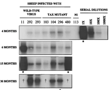
FIG. 1. Evolution of proviral loads in sheep infected with the BLV Taxmutants. Three sheep (11, 292, and 293) were injected with plasmid pBLVIX, which contains an infectious and pathogenic BLV provirus (clone 344). Four other animals (103, 104, 296, and 480) were infected with pBLVTax106⫹293, which is isogenic to pBLVIX except for two serine-to-alanine mutations in the
tax gene. Blood was extracted by jugular venipuncture at regular times after
seroconversion (4, 6, 15, and 30 months), and partially purified DNA was pre-pared from the corresponding lysates. A fraction corresponding to 5l of blood was amplified by 22 cycles of PCR using two primers flanking the tax gene, and the resulting DNAs were analyzed by Southern blotting using a tax probe. Under these conditions, the PCRs were semiquantitative, as shown by 10-fold dilutions (1⫻, 10⫻, 100⫻, and 1,000⫻) of lysate 480 at 15 months. In some lanes (ⴱ), the DNAs had to be isolated from smaller volumes of blood (50l instead of 500) because of the very high lymphocyte counts. Sheep 113 is an uninfected (NI) animal used as a negative control for PCR contaminations. Three samples are lacking at 30 months (†) because sheep 11, 103, and 480 died at about 19 to 20 months after seroconversion. Sheep 103 died because of enterotoxemia, whereas the other animals succumbed with leukemia or lymphosarcoma.
FIG. 2. Evolution of lymphocyte counts in BLV-infected sheep. Sheep were infected with the pBLVTax106⫹293 recombinant (TAX) (animals 103, 104, 296, and 480) or with viruses exhibiting wild-type (WT) behavior during pathogenesis (plasmid pBLVIX in sheep 8, 11, 247, 292, and 293; pBLV344 in 235; and pBLVgag150 in 175). Blood samples were extracted at regular intervals (routinely every month), and the number of leukocytes per microliter was determined by using a Coulter counter ZN. The lymphocyte counts (in parentheses) were deduced from these numbers after microscopic determination of the blood formula.
It thus appears that the phenotype of the BLV target cell is a
B lymphocyte potentially harboring CD5 and CD11b markers.
The presence of these molecules on cells infected by the
BLV Tax mutant was assessed by flow cytometry. To this end,
PBMCs were isolated from sheep 104, 296, and 480 by using
the Percoll gradient centrifugation procedure. Labeling these
cells with monoclonal antibody 1H4, which binds to surface
IgM, and their subsequent analysis by flow cytometry revealed
that the majority of the cells within the PBMC population from
sheep 296 and 480 were B lymphocytes (respectively 80 and
93%; Fig. 3A). In contrast, sheep 104 exhibited normal B-cell
counts (32% versus 22 to 29% in uninfected sheep 113, 115,
and 116). The phenotypes of these lymphocytes were
com-pared with those isolated from wild-type virus-infected sheep
exhibiting either high (animals 175, 235, and 247) or low
(an-imals 8, 292, and 293) B-cell counts within their peripheral
blood. The B-lymphocyte concentrations paralleled the
provi-ral loads, as determined by semiquantitative PCR (Fig. 3A). Of
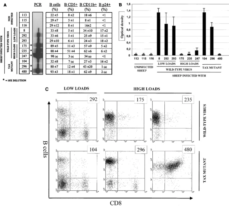
FIG. 3. (A) Phenotype of B cells in BLV-infected sheep. A series of 12 sheep were analyzed to determine and compare the phenotypes of the B-lymphocyte populations within the bloodstream of animals 104, 296, and 480 infected with pBLVTax106⫹293 (Tax mutant). Three sheep (113, 115, and 116) that were seronegative for BLV were used as controls, whereas six others were infected with viruses exhibiting wild-type behavior during pathogenesis (8, 292, 293, 247, 175, and 235). The different samples were classified in the figure on basis of the proviral loads as determined by semiquantitative PCR. In some lanes (ⴱ), the lysates were diluted 10-fold prior to PCR. PBMCs were isolated form the bloodstream and purified by Percoll gradient centrifugation. The cells were then labeled with monoclonal antibodies 1H4, CC17, and CC125, which recognize surface IgM, CD5, and CD11b, respectively. A similar protocol was applied for labeling the major capsid protein p24 with 4⬘G9 except that the cells were first cultivated for 24 h to trigger viral expression. Discrimination of the different cell populations was performed by two-color flow cytometry. The data, represented as percentage of the total PBMC population (⫾ the corresponding standard deviation), were deduced from three independent experiments performed over a period of several weeks. When the standard deviation is not indicated (a), the results are the mean values of only two analyses. (B) Titration of the major capsid protein p24 after short-term culture. PBMCs were isolated from the sheep indicated and cultivated for 24 h. Then, the p24 antigen was titrated in the cell culture supernatants by using the ELISA procedure. The data, represented as optical densities, derive from three independent experiments. (C) Expression of CD8 marker on B lymphocytes from sheep 480. PBMCs from six representative sheep infected either with wild-type viruses (292, 175, and 235) or the Tax mutant (104, 296, and 480) were double-labeled with monoclonal antibodies 1H4 and CC63, specific for surface IgM B lymphocytes and CD8, respectively. The cells were then analyzed by two-color flow cytometry, and results from a representative experiment (out of three) are shown as dot plots.note, the samples corresponding to sheep harboring very high
viral loads (marked with an asterisk) were diluted 10-fold prior
to amplification. Among the B-cell population from sheep 104
infected with the Tax mutant, a minority of lymphocytes
har-bored the CD5 marker (32% B versus 7% B CD5
⫹), but most
of them were CD11b positive (32% B versus 27% B CD11
⫹).
These values are within the normal range observed in wild-type
virus-infected animals at similar viral loads (sheep 8, 292, and
293). At the leukemic stage, when the circulating blood
con-tains almost pure populations of B cells (around 90% or more),
expression of the CD5 molecule was only poorly associated
with the transformed lymphocytes. Indeed, only one animal
(235) contained high levels of B CD5
⫹cells (51%; Fig. 3A). In
contrast, CD11b appeared to be a far better marker for the
transformed B lymphocytes both in Tax mutant- and in
wild-type virus-infected sheep (between 34 and 62%). There was,
however, no significant and systematic difference between
these two categories of infected animals.
We next analyzed the ability of the wild-type and Tax mutant
viruses to be expressed during ex vivo cell cultivation. In vivo,
BLV is a hiding pathogen which is rarely expressed within the
infected lymphocyte population, but isolation and cultivation
of the infected PBMCs permits the evaluation of viral protein
synthesis (34, 52). BLV expression was estimated by two
com-plementary techniques, ELISA and flow cytometry, based on
the synthesis of the major capsid protein p24. In the
asymp-tomatic sheep, the B-cell population expressing the p24
anti-gen (i.e., double-positive B
⫹p24
⫹cells) accounted for 15 to
18% of the PBMCs independently of the type of infecting virus
(Fig. 3A, compare 8, 292, 293, and 104). Among the animals
harboring high viral loads, ex vivo p24 synthesis becomes
in-efficient, particularly at the final stages of leukemogenesis.
Despite tremendous levels of B lymphocytes (around 90% of
the PBMCs), less than 5% of the cells were p24 positive both
in wild-type and in Tax mutant cell populations (Fig. 3A, sheep
175, 235, 247, 296, and 480). The total amount of p24 expressed
in the culture supernatants, as measured by ELISA, generally
paralleled nicely the percentages of cells revealed by flow
cy-tometry (Fig. 3B). The sole exception was sheep 296, infected
by the Tax mutant, whose PBMCs expressed significant levels
of p24 protein in the culture medium despite low numbers of
p24-positive cells as revealed by flow cytometry. It should be
mentioned, however, that the total amounts of p24
corre-sponding to this particular animal also dropped just before
death (data not shown). We conclude that the mean levels of
p24 and their evolution at different stages of pathogenesis are
similar in all the infected sheep, independently of the type of
virus.
Interestingly, during the characterization of the cell
pheno-types, we observed high numbers of CD8-positive cells in sheep
480, which was infected by the Tax mutant virus. In fact, most
of the B-cell population harbored this marker, as revealed by
double staining and flow cytometry (Fig. 3C, 480). The
expres-sion of the CD8 molecule was confirmed by using two
inde-pendent antibodies (CC63 and ST8), and transcription of the
corresponding gene was verified by RNA hybridization (data
not shown). In addition, two independent antibodies (1H4 and
PIg45) confirmed that the leukemic cells were B lymphocytes.
Such a B/CD8 phenotype was not associated with cells from
other animals harboring either high (175, 235, and 296) or low
(104 and 292) viral loads. More specifically, the CD8 molecule
was not expressed at the surface of the leukemic B lymphocytes
from sheep 296 infected by the BLV Tax mutant.
To summarize, it appears that, with the exception of a
pe-culiar B/CD8 phenotype in sheep 480, B lymphocytes from
animals infected either by wild-type virus or by the Tax mutant
are indistinguishable. In other words, both types of viruses can
infect and transform similar cell types.
Tax mutant viruses in the transformed cells are not
rever-tants.
Since both the evolution of pathogenesis and the cellular
phenotypes associated with the Tax mutant and wild-type virus
were almost identical, our experiments needed an essential
control demonstrating the lack of reversion in the tumor cells.
It was indeed possible that the pathogeneses observed in sheep
296 and 480 were induced by viruses in which the two alanine
mutations at positions 106 and 293 had reverted to a wild-type
serine codon. Therefore, cell lysates were prepared from blood
isolated by jugular venipuncture of sheep 103, 104, 296, and
480. The tax gene fragments were amplified by PCR, and the
corresponding amplicons were subjected to direct sequencing.
As illustrated in Fig. 4, the alanine codons 106 and 293 were
perfectly conserved in all the lysates, demonstrating lack of
reversion of the tax sequences. These analyses were performed
at different time points, including at the terminal stage with
fully transformed tumor cells. In addition, six independent
amplicons were also completely sequenced over a region
en-compassing the entire tax gene. No mutation within all these
samples could ever be identified (data not shown).
We conclude that the pathogeneses observed in sheep 296
and 480 infected by the BLV Tax mutant did not result from a
reversion of the recombinant to a wild-type virus.
DISCUSSION
In this report, we have shown that mutations of the BLV tax
gene that hamper immortalization of primary REFs still allow
the occurrence of leukemogenesis in sheep. These
observa-tions cast light onto contradictory conclusions that might be
drawn from transformation assays performed in cell culture
and experiments in vivo.
A first critique to be answered concerns the lack of reversion
of the recombinants in vivo. In fact, we have shown that the tax
and rex sequences are not mutated after leukemogenesis in two
different sheep (480 and 296; Fig. 4). However, it is possible
that unidentified compensatory mutations occurred in other
parts of the viral genome, for example, in the R3/G4 accessory
genes. Although we have not formally ruled out this possibility
FIG. 4. Direct sequencing of codons surrounding alanines 106 and 293 of thetax gene in four sheep infected by the BLV Tax mutant. Blood was extracted by
jugular venipuncture of sheep 103, 104, 296, and 480 infected with provirus pBLVTax106⫹293. After lysis, partially purified DNA was amplified by PCR using two primers flanking the tax gene. The resulting amplicons were then subjected to direct sequencing by PCR and migrated onto a denaturing poly-acrylamide gel. The sequences surrounding alanines 106 and 293 are indicated.
by sequencing the entire virus, we think that this is unlikely
because deletion of R3 or G4 provokes a drastic reduction in
the proviral loads (78).
A second point to be discussed concerns the immortalization
assay itself. In our previous study on Tax phosphorylation, we
reported that changing serine residues 106 and 293 into
ala-nines abrogated its immortalization potential in primary REFs.
It remains possible, however, that this type of experiment lacks
sensitivity and reveals only the oncogenes harboring very high
immortalization activity. Alternatively, the REF assay could
be oversensitive, and slight differences in the oncogenic
poten-tial of Tax would generate large phenotypic variations during
transformation in cell culture. However, in our previous
stud-ies (72; unpublished results), systematic screening of a serstud-ies of
mutants revealed that Tax oncogenic activity remains very
fre-quently conserved, whereas its transactivation potential is
de-stroyed. In fact, the Tax phosphorylation recombinant was the
sole immortalization-deficient mutant that is still able to
acti-vate viral transcription. In other words, most Tax mutants are
negative for transactivation and positive for immortalization,
indicating that the REF assay is not sensitive to various
mod-ifications within Tax.
Another straightforward interpretation of the results would
concern the differential pathways involved in transformation of
REF cells or B lymphocytes. This has been illustrated in the
HTLV system by specific Tax mutants that display various
phenotypes depending on the cell type (3, 42, 57). In particular,
the abilities of some mutants to activate transcription via the
CRE, NFB, or CArG enhancer elements were different in
Rat-1, REF, and T cells. For example, clone 703 was
compe-tent for the NFB pathway in the three cell types but displayed
a mixed pattern for activation via the CRE: negative in T
lymphocytes, intermediate in Rat-1 cells, and positive in REFs
(3, 42). This mutant was unable to cooperate with the ras
on-cogene in REF fibroblasts but induced the formation of foci in
soft agar in Rat-1 cells. Furthermore, the same mutant induced
a higher proliferative response and allowed long-term
expan-sion of CD8-positive T lymphocytes. Although the
T-lympho-cyte system appears to be the best mimic of the in vivo
situa-tion, this example illustrates the difficulties encountered during
interpretation of the data. In this context, the availability of an
animal model, such as the infection of sheep by BLV
recom-binants, might cast some light on our understanding of the
leukemogenic process induced by these related viruses. In fact,
there is an intriguing parallel between the phenotypes
associ-ated with HTLV mutant 703 and the BLV Tax recombinant,
allowing us some speculation. Both clones are indeed unable to
transform REF cells in cooperation with the ras oncogene but
exhibit immortalization potential in cell culture and in vivo. In
addition, a peculiar phenotype characterized by the expression
of the CD8 marker occurred in 90% of the T cells transduced
with Tax-I 703 and sheep 480 infected with the BLV Tax
mutant. Although the CD8 marker was not present on the B
lymphocytes from the other sheep (animal 296 infected with
the same mutant) and was not identified at the early stages of
infection (data not shown), the coincidence is appealing.
Any-way, the modification of the target cell phenotype suggests
that the metabolic pathways leading to full transformation are
somehow perturbed. In fact, the emergence of CD4
⫹CD8
⫹double-positive cells has been reported in patients infected
with HTLV-1 (11, 20, 39, 50). Similarly, CD8
⫹T lymphocytes
appear to be the main but not the sole target for the related
HTLV-II virus (9, 21, 35, 40, 67). In contrast, the presence of
a T-cell marker has only very rarely been observed on B
lym-phocytes from patients with large granular or chronic
lympho-cytic leukemia (4, 32).
The generation of B cells harboring T-specific markers in
sheep 480 is a matter of speculation. First, it is possible that a
rare IgM
⫹CD8
⫹subset already exists in seronegative animals
and that this population is expanded after BLV infection. The
two-color flow cytometry data presented in Fig. 3C would
in-deed suggest that such double-positive cells could be present
among the PBMCs of other sheep. According to this
hypoth-esis, the mutation of the Tax phosphorylation sites could
some-how alter the viral target specificity and yield B lymphocytes
with a CD8 marker. Modification of the cell preference
has been illustrated in cultures of T lymphocytes infected by
HTLV mutants (3, 55). An increased tropism for a given
cel-lular phenotype might be due to better viral replication
con-secutive to, for instance, enhanced expression, receptor
recog-nition, or escape from immune surveillance. In this context, the
ability of Tax to activate the HTLV-1 LTR is greatly increased
in CD4
⫹cells compared to CD8-positive lymphocytes (49).
From our data, it does not appear that the BLV Tax
phosphor-ylation mutant mediates enhanced transcription during
tran-sient-transfection experiments (73) and ex vivo (Fig. 3B). In
addition, based on the evolution of the proviral loads, viral
replication of the Tax mutant also appears unaffected (Fig. 1).
Another hypothesis underlining the generation of IgM
⫹CD8
⫹cells would be the ability of the Tax mutant to induce the
ex-pression of the CD8 protein. We think that this assumption is
unlikely because (i) transient transfection of the pSGtax106⫹
293 vector does not augment CD8 RNA transcription in
cul-ture (data not shown) and (ii) the presence of the CD8
mol-ecule is not always associated with the Tax mutant virus (in the
case of sheep 296). We therefore favor the hypothesis based on
alteration of cell target specificity.
A fact that merits some comment is the imperfect
concor-dance between the presence of the CD8 marker and infection
by the Tax mutant. The CD8 molecule indeed appeared to be
expressed only at the final stage of leukemogenesis (data not
shown), and this phenotype was not observed at any time in
another animal (sheep 296; Fig. 3C). A similar situation holds
true for T lymphocytes immortalized in cell culture by
HTLV-1 Tax mutants, the cell lines generated in vitro being either
CD4
⫹, CD8
⫹, or double positive (3, 55). In contrast, cells
immortalized by the wild-type Tax-I protein were, in their great
majority, pertaining to CD4
⫹lymphocyte subtypes.
Altogeth-er, these observations indicate that the frequency of an altered
phenotype is increased when the tax gene is mutated.
Deter-mining whether this is a general rule in sheep infected with the
BLV Tax mutant is hampered by experimental restrictions,
such as limited numbers of animals and long latency periods. It
should, however, be mentioned that, in a series of eight tumor
biopsies from sheep infected by wild-type viruses, none of them
were CD8 positive (data not shown). In addition, this marker
has never been associated with BLV-infected cells in naturally
infected cattle (reviewed recently in reference 78). The
pres-ence of the CD8 molecule at the surface of the B lymphocytes
of sheep 480 thus appears to be a very rare event, but the
analogy with other viral systems like HTLV (3) or even other
leukemias in humans (32) is striking (4).
ACKNOWLEDGMENTS
R.K. and L.W. are Research Directors of the “Fonds national de la
Recherche scientifique” (FNRS). We thank the “Fe´de´ration Belge
contre le Cancer,” the “Action de Recherche Concerte´e du Ministe`re
de la Communaute´ Franc¸aise,” the Fortis Bank, the FNRS, the
“Ser-vice de Programmation pour la Politique scientifique” (SSTC P4/30),
and the “Bekales Foundation” for financial support.
Antibodies or plasmids were kindly provided by S. Alberti (Institute
Mario Negri Sud, S. Maria Imbaro, Italy), C. Howard (Institute of
Animal Health, Compton, U.K.), J. J. Letesson (FUNDP, Namur,
Belgium), I. Schwartz-Cornil (INRA, Paris, France), and K. Walravens
(VARC, Uccle, Belgium). We are grateful to F. Dequiedt and E.
Hanon for advice in flow cytometry. We thank J. M. Londes, T.
Pere-mans, G. Vandendaele, and M. Verhoeven for excellent technical help.
REFERENCES
1. Adam, E., P. Kerkhofs, M. Mammerickx, A. Burny, R. Kettman, and L.
Willems.1996. The CREB, ATF-1, and ATF-2 transcription factors from bovine leukemia virus-infected B lymphocytes activate viral expression. J. Vi-rol. 70:1990–1999.
2. Adam, E., P. Kerkhofs, M. Mammerickx, R. Kettmann, A. Burny, L.
Droog-mans, and L. Willems.1994. Involvement of the cyclic AMP-responsive element binding protein in bovine leukemia virus expression in vivo. J. Virol.
68:5845–5853.
3. Akagi, T., H. Ono, H. Nyunoya, and K. Shimotohno. 1997. Characterization of peripheral blood T-lymphocytes transduced with HTLV-I Tax mutants with different trans-activating phenotypes. Oncogene 14:2071–2078. 4. Akashi, K., T. Shibuya, M. Nakamura, A. Oogami, M. Harada, and Y. Niho.
1998. Large granular lymphocytic leukaemia with a mixed T-cell/B-cell phe-notype. Br. J. Haematol. 100:291–294.
5. Birkebak, T. A., G. H. Palmer, W. C. Davis, D. P. Knowles, and T. F.
McElwain.1994. Association of GP51 expression and persistent CD5⫹ B-lymphocyte expansion with lymphomagenesis in bovine leukemia virus in-fected sheep. Leukemia 8:1890–1899.
6. Blattner, W. A. 1999. Human retroviruses: their role in cancer. Proc. Assoc. Am. Physicians 111:563–572.
7. Boris-Lawrie, K., V. Altanerova, C. Altaner, L. Kucerova, and H. M. Temin. 1997. In vivo study of genetically simplified bovine leukemia virus derivatives that lack tax and rex. J. Virol. 71:1514–1520.
8. Boros, I. M., F. Tie, and C. Z. Giam. 1995. Interaction of bovine leukemia virus transactivator Tax with bZip proteins. Virology 214:207–214. 9. Casoli, C., A. Cimarelli, and U. Bertazzoni. 1995. Cellular tropism of human
T-cell leukemia virus type II is enlarged to B lymphocytes in patients with high proviral load. Virology 206:1126–1128.
10. Chevallier, N., M. Berthelemy, D. Le Rhun, V. Laine, D. Levy, and I.
Schwartz-Cornil.1998. Bovine leukemia virus-induced lymphocytosis and increased cell survival mainly involve the CD11b⫹B-lymphocyte subset in
sheep. J. Virol. 72:4413–4420.
11. Ciminale, V., M. Hatziyanni, B. K. Felber, J. Bear, A. Hatzakis, and G. N.
Pavlakis.2000. Unusual CD4⫹CD8⫹ phenotype in a Greek patient diag-nosed with adult T-cell leukemia positive for human T-cell leukemia virus type I (HTLV-I). Leukocyte Res. 24:353–358.
12. Cockerell, G. L., J. Rovnak, P. L. Green, and I. S. Chen. 1996. A deletion in the proximal untranslated pX region of human T-cell leukemia virus type II decreases viral replication but not infectivity in vivo. Blood 87:1030–1035. 13. Coscoy, L., D. Gonzalez-Dunia, F. Tangy, S. Syan, M. Brahic, and S. Ozden.
1998. Molecular mechanism of tumorigenesis in mice transgenic for the human T cell leukemia virus Tax gene. Virology 248:332–341.
14. Dequiedt, F., E. Hanon, P. Kerkhofs, P. P. Pastoret, D. Portetelle, A. Burny,
R. Kettmann, and L. Willems.1997. Both wild-type and strongly attenuated bovine leukemia viruses protect peripheral blood mononuclear cells from apoptosis. J. Virol. 71:630–639.
15. Derse, D. 1987. Bovine leukemia virus transcription is controlled by a virus-encoded trans-acting factor and by cis-acting response elements. J. Virol. 61: 2462–2471.
16. Derse, D., J. Mikovits, and F. Ruscetti. 1997. X-I and X-II open reading frames of HTLV-I are not required for virus replication or for immortaliza-tion of primary T-cells in vitro. Virology 237:123–128.
17. Fang, J., S. Kushida, R. Feng, M. Tanaka, T. Kawamura, H. Abe, N. Maeda,
M. Onobori, M. Hori, K. Uchida, and M. Miwa.1998. Transmission of human T-cell leukemia virus type 1 to mice. J. Virol. 72:3952–3957. 18. Franchini, G. and H. Streicher. 1995. Human T-cell leukaemia virus.
Bail-liere’s Clin. Haematol. 8:131–148.
19. Fujita, M., and H. Shiku. 1995. Differences in sensitivity to induction of apoptosis among rat fibroblast cells transformed by HTLV-I tax gene or cellular nuclear oncogenes. Oncogene 11:15–20.
20. Furukawa, S., K. Sasai, J. Matsubara, K. Yabuta, K. Hiramatsu, J.
Yama-moto, T. Shirai, and K. Okumura.1992. Increase in T cells expressing the gamma/delta receptor and CD4⫹CD8⫹ double-positive T cells in primary immunodeficiency complicated by human T-cell lymphotropic virus type I infection. Blood 80:3253–3255.
21. Gongora-Biachi, R. A., P. Gonzalez-Martinez, C. Castros-Sansores, N.
Pavia-Ruz, D. L. Rudolph, and R. B. Lal.1993. Human T lymphotropic virus type II (HTLV-II) infection among female prostitutes in Yucatan, Mexico. Am. J. Med. Sci. 306:207–211.
22. Grassmann, R., S. Berchtold, I. Radant, M. Alt, B. Fleckenstein, J. G.
Sodroski, W. A. Haseltine, and U. Ramstedt.1992. Role of human T-cell leukemia virus type 1 X region proteins in immortalization of primary human lymphocytes in culture. J. Virol. 66:4570–4575.
23. Grassmann, R., C. Dengler, I. Muller-Fleckenstein, B. Fleckenstein, K.
McGuire, M. C. Dokhelar, J. G. Sodroski, and W. A. Haseltine.1989.
Trans-formation to continuous growth of primary human T lymphocytes by human T-cell leukemia virus type I X-region genes transduced by a herpesvirus saimiri vector. Proc. Natl. Acad. Sci. USA 86:3351–3355.
24. Grossman, W. J., J. T. Kimata, F. H. Wong, M. Zutter, T. J. Ley, and L.
Ratner.1995. Development of leukemia in mice transgenic for the tax gene of human T-cell leukemia virus type I. Proc. Natl. Acad. Sci. USA 92:1057– 1061.
25. Habu, K., J. Nakayama-Yamada, M. Asano, S. Saijo, K. Itagaki, R. Horai, H.
Yamamoto, T. Sekiguchi, T. Nosaka, M. Hatanaka, and Y. Iwakura.1999. The human T cell leukemia virus type I-tax gene is responsible for the development of both inflammatory polyarthropathy resembling rheumatoid arthritis and noninflammatory ankylotic arthropathy in transgenic mice. J. Immunol. 162:2956–2963.
26. Hall, A. P., J. Irvine, K. Blyth, E. R. Cameron, D. E. Onions, and M. E.
Campbell.1998. Tumours derived from HTLV-I tax transgenic mice are characterized by enhanced levels of apoptosis and oncogene expression. J. Pathol. 186:209–214.
27. Hinrichs, S. H., M. Nerenberg, R. K. Reynolds, G. Khoury, and G. Jay. 1987. A transgenic mouse model for human neurofibromatosis. Science 237:1340– 1343.
28. Hollsberg, P. 1999. Mechanisms of T-cell activation by human T-cell lym-photropic virus type I. Microbiol. Mol. Biol. Rev. 63:308–333.
29. Kazanji, M., J. P. Moreau, R. Mahieux, B. Bonnemains, R. Bomford, A.
Gessain, and G. de The. 1997. HTLV-I infection in squirrel monkeys (Saimiri sciureus) using autologous, homologous, or heterologous HTLV-I-transformed cell lines. Virology 231:258–266.
30. Kettmann, R., A. Burny, I. Callebaut, L. Droogmans, M. Mammerickx, L.
Willems, and D. Portetelle.1994. Bovine leukemia virus, p. 39–81. In J. A. Levy (ed.), The Retroviridae, vol. 3. Plenum Press, New York, N.Y. 31. Kimata, J. T., F. H. Wong, J. J. Wang, and L. Ratner. 1994. Construction and
characterization of infectious human T-cell leukemia virus type 1 molecular clones. Virology 204:656–664.
32. Koelliker, D. D., P. E. Steele, P. E. Hurtubise, H. C. Flessa, Y. P. Sheng, and
S. H. Swerdlow.1994. CD8-positive B-cell chronic lymphocytic leukemia: a report of two cases. Am. J. Clin. Pathol. 102:212–216.
33. Kucerova, L., V. Altanerova, C. Altaner, and K. Boris-Lawrie. 1999. Bovine leukemia virus structural gene vectors are immunogenic and lack pathoge-nicity in a rabbit model. J. Virol. 73:8160–8166.
34. Lagarias, D. M., and K. Radke. 1989. Transcriptional activation of bovine leukemia virus in blood cells from experimentally infected, asymptomatic sheep with latent infections. J. Virol. 63:2099–2107.
35. Lal, R. B., S. M. Owen, D. L. Rudolph, C. Dawson, and H. Prince. 1995. In vivo cellular tropism of human T-lymphotropic virus type II is not restricted to CD8⫹ cells. Virology 210:441–447.
36. Land, H. 1995. Transformation of primary rat embryo cells. Methods Enzy-mol. 254:37–41.
37. Land, H., L. F. Parada, and R. A. Weinberg. 1983. Tumorigenic conversion of primary embryo fibroblasts requires at least two cooperating oncogenes. Nature 304:596–602.
38. Lydy, S. L., M. E. Conner, and S. J. Marriott. 1998. Relationship between anti-Tax antibody responses and cocultivatable virus in HTLV-I-infected rabbits. Virology 250:60–66.
39. Macchi, B., G. Graziani, J. Zhang, and A. Mastino. 1993. Emergence of double-positive CD4/CD8 cells from adult peripheral blood mononuclear cells infected with human T cell leukemia virus type I (HTLV-I). Cell. Immunol. 149:376–389.
40. Martin, M. P., R. J. Biggar, G. Hamlin-Green, S. Staal, and D. Mann. 1993. Large granular lymphocytosis in a patient infected with HTLV-II. AIDS Res. Hum. Retroviruses 9:715–719.
41. Matheise, J. P., M. Delcommenne, A. Mager, C. H. Didembourg, and J. J.
Letesson.1992. CD5⫹ B cells from bovine leukemia virus infected cows are activated cycling cells responsive to interleukin 2. Leukemia 6:304–309. 42. Matsumoto, K., H. Shibata, J. I. Fujisawa, H. Inoue, A. Hakura, T.
Tsuka-hara, and M. Fujii.1997. Human T-cell leukemia virus type 1 Tax protein transforms rat fibroblasts via two distinct pathways. J. Virol. 71:4445–4451. 43. Meirom, R., S. Moss, and J. Brenner. 1997. Bovine leukemia virus-gp51 antigen expression is associated with CD5 and IgM markers on infected lymphocytes. Vet. Immunol. Immunopathol. 59:113–119.
44. Mirsky, M. L., C. A. Olmstead, Y. Da, and H. A. Lewin. 1996. The prevalence of proviral bovine leukemia virus in peripheral blood mononuclear cells at two subclinical stages of infection. J. Virol. 70:2178–2183.
45. Murakami, K., Y. Aida, R. Kageyama, S. Numakunai, K. Ohshima, K.
Okada, and Y. Ikawa.1994. Immunopathologic study and characterization of the phenotype of transformed cells in sheep with bovine leukemia virus-induced lymphosarcoma. Am. J. Vet. Res. 55:72–80.
46. Murakami, K., K. Okada, Y. Ikawa, and Y. Aida. 1994. Bovine leukemia virus induces CD5-B cell lymphoma in sheep despite temporarily increasing CD5⫹ B cells in asymptomatic stage. Virology 202:458–465.
47. Murata, K., M. Fujita, T. Honda, Y. Yamada, M. Tomonaga, and H. Shiku. 1996. Rat primary T cells expressing HTLV-I tax gene transduced by a retroviral vector: in vitro and in vivo characterization. Int. J. Cancer 68:102– 108.
48. Nerenberg, M., S. H. Hinrichs, R. K. Reynolds, G. Khoury, and G. Jay. 1987. The tat gene of human T-lymphotropic virus type 1 induces mesenchymal tumors in transgenic mice. Science 237:1324–1329.
49. Newbound, G. C., J. M. Andrews, J. P. O’Rourke, J. N. Brady, and M. D.
Lairmore.1996. Human T-cell lymphotropic virus type 1 Tax mediates en-hanced transcription in CD4⫹T lymphocytes. J. Virol. 70:2101–2106.
50. Ohata, J., M. Matsuoka, T. Yamashita, A. Tojo, K. Tani, and S. Asano. 1999. CD4/CD8 double-positive adult T cell leukemia with preceding cytomega-loviral gastroenterocolitis. Int. J. Hematol. 69:92–95.
51. Ohya, O., U. Tomaru, I. Yamashita, T. Kasai, K. Morita, H. Ikeda, A.
Wakisaka, and T. Yoshiki. 1997. HTLV-I induced myeloneuropathy in WKAH rats: apoptosis and local activation of the HTLV-I pX and TNF-alpha genes implicated in the pathogenesis. Leukemia 11(Suppl. 3):255–257. 52. Powers, M. A., and K. Radke. 1992. Activation of bovine leukemia virus transcription in lymphocytes from infected sheep: rapid transition through early to late gene expression. J. Virol. 66:4769–4777.
53. Pozzatti, R., J. Vogel, and G. Jay. 1990. The human T-lymphotropic virus type I tax gene can cooperate with the ras oncogene to induce neoplastic transformation of cells. Mol. Cell. Biol. 10:413–417.
54. Ressler, S., L. M. Connor, and S. J. Marriott. 1996. Cellular transformation by human T-cell leukemia virus type I. FEMS Microbiol. Lett. 140:99–109. 55. Robek, M. D., and L. Ratner. 1999. Immortalization of CD4(⫹) and CD8(⫹) T lymphocytes by human T-cell leukemia virus type 1 Tax mutants expressed in a functional molecular clone. J. Virol. 73:4856–4865.
56. Rosin, O., C. Koch, I. Schmitt, O. J. Semmes, K. T. Jeang, and R.
Grass-mann.1998. A human T-cell leukemia virus Tax variant incapable of acti-vating NF-kappaB retains its immortalizing potential for primary T-lympho-cytes. J. Biol. Chem. 273:6698–6703.
57. Ross, T. M., M. Narayan, Z. Y. Fang, A. C. Minella, and P. L. Green. 2000. Human T-cell leukemia virus type 2 tax mutants that selectively abrogate NF-B or CREB/ATF activation fail to transform primary human T cells. J. Virol. 74:2655–2662.
58. Ross, T. M., S. M. Pettiford, and P. L. Green. 1996. The tax gene of hu-man T-cell leukemia virus type 2 is essential for transformation of huhu-man T lymphocytes. J. Virol. 70:5194–5202.
59. Saggioro, D., D. M. D’Agostino, and L. Chieco-Bianchi. 1999. Analysis of Tax-expressing cell lines generated from HTLV-I tax-transgenic mice: cor-relation between c-myc overexpression and neoplastic potential. Exp. Cell Res. 247:525–533.
60. Schatzl, H., M. Tschikobava, D. Rose, A. Voevodin, H. Nitschko, E. Sieger,
U. Busch, K. von der Helm, and B. Lapin.1993. The Sukhumi primate monkey model for viral lymphomogenesis: high incidence of lymphomas with presence of STLV-I and EBV-like virus. Leukemia 7(Suppl. 2):S86–S92. 61. Schwartz, I., A. Bensaid, B. Polack, B. Perrin, M. Berthelemy, and D. Levy.
1994. In vivo leukocyte tropism of bovine leukemia virus in sheep and cattle. J. Virol. 68:4589–4596.
62. Seto, A., T. Isono, and K. Ogawa. 1991. Infection of inbred rabbits with cell-free HTLV-I. Leukocyte Res. 15:105–110.
63. Smith, M. R. and W. C. Greene. 1991. Type I human T cell leukemia virus tax protein transforms rat fibroblasts through the cyclic adenosine mono-phosphate response element binding protein/activating transcription factor pathway. J. Clin. Investig. 88:1038–1042.
64. Stone, D. M., A. J. Hof, and W. C. Davis. 1995. Up-regulation of IL-2 receptor alpha and MHC class II expression on lymphocyte subpopulations from bovine leukemia virus infected lymphocytotic cows. Vet. Immunol. Immunopathol. 48:65–76.
65. Sun, B., J. Fang, K. Yagami, S. Kushida, M. Tanaka, K. Uchida, and M.
Miwa.1999. Age-dependent paraparesis in WKA rats: evaluation of MHC k-haplotype and HTLV-1 infection. J. Neurol. Sci. 167:16–21.
66. Tanaka, A., C. Takahashi, S. Yamaoka, T. Nosaka, M. Maki, and M.
Ha-tanaka.1990. Oncogenic transformation by the tax gene of human T-cell
leukemia virus type I in vitro. Proc. Natl. Acad. Sci. USA 87:1071–1075. 67. Tarsis, S. L., M. T. Yu, E. S. Parks, D. Persaud, J. L. Munoz, and W. P.
Parks.1998. Human T-lymphocyte transformation with human T-cell lym-photropic virus type 2. J. Virol. 72:841–846.
68. Tomaru, U., H. Ikeda, O. Ohya, M. Abe, T. Kasai, I. Yamasita, K. Morita, A.
Wakisaka, and T. Yoshiki.1996. Human T lymphocyte virus type I-induced myeloneuropathy in rats: implication of local activation of the pX and tumor necrosis factor-alpha genes in pathogenesis. J. Infect. Dis. 174:318–323. 69. Uchiyama, T. 1997. Human T cell leukemia virus type I (HTLV-I) and
human diseases. Annu. Rev. Immunol. 15:15–37.
70. Van Den Broeke, A., C. Bagnis, M. Ciesiolka, Y. Cleuter, H. Gelderblom, P.
Kerkhofs, P. Griebel, P. Mannoni, and A. Burny.1999. In vivo rescue of a silent Tax-deficient bovine leukemia virus from a tumor-derived ovine B-cell line by recombination with a retrovirally transduced wild-type tax gene. J. Virol. 73:1054–1065.
71. Willems, L., A. Gegonne, G. Chen, A. Burny, R. Kettmann, and J. Ghysdael. 1987. The bovine leukemia virus p34 is a transactivator protein. EMBO J. 6: 3385–3389.
72. Willems, L., C. Grimonpont, H. Heremans, N. Rebeyrotte, G. Chen, D.
Portetelle, A. Burny, and R. Kettmann.1992. Mutations in the bovine leu-kemia virus Tax protein can abrogate the long terminal repeat-directed transactivating activity without concomitant loss of transforming potential. Proc. Natl. Acad. Sci. USA 89:3957–3961.
73. Willems, L., C. Grimonpont, P. Kerkhofs, C. Capiau, D. Gheysen, K.
Conrath, R. Roussef, R. Mamoun, D. Portetelle, A. Burny, E. Adam, L. Lefebvre, J. C. Twizere, H. Heremans, and R. Kettmann.1998. Phosphory-lation of bovine leukemia virus Tax protein is required for in vitro transfor-mation but not for transactivation. Oncogene 16:2165–2176.
74. Willems, L., H. Heremans, G. Chen, D. Portetelle, A. Billiau, A. Burny, and
R. Kettmann.1990. Cooperation between bovine leukaemia virus transacti-vator protein and Ha-ras oncogene product in cellular transformation. EMBO J. 9:1577–1581.
75. Willems, L., P. Kerkhofs, L. Attenelle, A. Burny, D. Portetelle, and R.
Kettmann.1997. The major homology region of bovine leukaemia virus p24gag is required for virus infectivity in vivo. J. Gen. Virol. 78:637–640. 76. Willems, L., R. Kettmann, and A. Burny. 1991. The amino acid (157–197)
peptide segment of bovine leukemia virus p34tax encompass a leucine-rich globally neutral activation domain. Oncogene 6:159–163.
77. Willems, L., R. Kettmann, F. Dequiedt, D. Portetelle, V. Voneche, I. Cornil,
P. Kerkhofs, A. Burny, and M. Mammerickx.1993. In vivo infection of sheep by bovine leukemia virus mutants. J. Virol. 67:4078–4085.
78. Willems, L., A. Burny, D. Collete, O. Dangoisse, J. S. Gatot, P. Kerkhofs, L.
Lefe`bvre, M. Merezak, D. Portetelle, J. C. Twizere, and R. Kettmann.1999. Bovine leukemia virus as a model for human T-cell leukemia virus. Curr. Top. Virol. 1:139–167.
79. Yamaoka, S., H. Inoue, M. Sakurai, T. Sugiyama, M. Hazama, T. Yamada,
and M. Hatanaka.1996. Constitutive activation of NF-kappa B is essential for transformation of rat fibroblasts by the human T-cell leukemia virus type I Tax protein. EMBO J. 15:873–887.
80. Yamaoka, S., T. Tobe, and M. Hatanaka. 1992. Tax protein of human T-cell leukemia virus type I is required for maintenance of the transformed phe-notype. Oncogene 7:433–437.
81. Yoshida, M. 1996. Molecular biology of HTLV-I: recent progress. J. Acquir. Immune Defic. Syndr. Hum. Retrovirol. 13(Suppl. 1):S63–S68.
82. Yoshida, M. 1997. Howard Temin Memorial Lectureship: molecular biology of HTLV-1: deregulation of host cell gene expression and cell cycle. Leuke-mia 11(Suppl. 3):1–2.
83. Zhao, T. M., M. A. Robinson, F. S. Bowers, and T. J. Kindt. 1996. Infectivity of chimeric human T-cell leukemia virus type I molecular clones assessed by naked DNA inoculation. Proc. Natl. Acad. Sci. USA 93:6653–6658.