O
pen
A
rchive
T
OULOUSE
A
rchive
O
uverte (
OATAO
)
OATAO is an open access repository that collects the work of Toulouse researchers and
makes it freely available over the web where possible.
This is an author-deposited version published in :
http://oatao.univ-toulouse.fr/
Eprints ID : 11518
To link to this article :
URL :
http://mires-and-peat.net/pages/volumes/map07/map0704.php
To cite this version : Le Roux, Gaël and De Vleeschouwer, François
Preparation of peat samples for inorganic geochemistry used as
palaeoenvironmental proxies. (2010) Mires and Peat, vol. 7 (n°4). pp.
1-9. ISSN
1819-754X
Any correspondance concerning this service should be sent to the repository
administrator:
staff-oatao@listes-diff.inp-toulouse.fr
Preparation of peat samples for inorganic geochemistry
used as palaeoenvironmental proxies
G. Le Roux1 and F. De Vleeschouwer2
1
EcoLab UMR 5245 CNRS, Université de Toulouse, France
2
Department of Ecology and Environmental Sciences, Umeå University, Sweden
_______________________________________________________________________________________
SUMMARY
This article provides a brief review of protocols used in peat inorganic geochemistry. We emphasise the key issues that could lead to inter-comparison problems. For each section (drying, grinding, non-destructive analyses, acid digestions and destructive analyses), recommendations are provided to guide the reader through an idealised protocol, which is the only workable approach for studies incorporating long-term comparisons.
KEY WORDS: acid digestion, freeze-drying, grinding, X-ray fluorescence.
_______________________________________________________________________________________
1. INTRODUCTION
During recent decades, numerous techniques have been used to concentrate and analyse the inorganic components of peat samples (e.g. Kempter et al. 1997, Shotyk et al. 1998, Martinez-Cortizas et al. 1999, Mighall et al. 2002). These studies have generally aimed to reconstruct past human activities (e.g. Kempter & Frenzel 2000, Mighall et al. 2006), atmospheric pollution (e.g. Shotyk et al. 1998, Novák et al. 2003, De Vleeschouwer et al. 2010b) or environmental changes (e.g. Shotyk et al. 1998, Klaminder et al. 2003, Roos-Barraclough et al. 2004, De Vleeschouwer et al. 2009, Kylander et al. 2010). However, while these studies contribute greatly to accurately retrieving and better understanding the inorganic geochemical signals contained in peat records, no comprehensive review of the various existing geochemical protocols has been undertaken. Such an idealised protocol is much needed because some fluctuations in geochemical records may be artefacts of the analytical procedures used to obtain the information. A protocol paper dedicated to peat geochemistry, which focuses on metal contamination and minimising peat compression during coring, has already been published by Givelet et al. (2004). This paper also provides some methods for improving sampling resolution, and any researcher interested in the inorganic geochemistry of peat is encouraged to read it.
Since 2004, multidisciplinary studies, as opposed to investigations focusing solely on the geochemistry of major and trace elements in peat,
have been achieved; and new projects combining biological and geochemical proxies have been launched. This article presents refinements of the protocol proposed by Givelet et al. (2004), but also different strategies depending on the inorganic geochemical proxies being studied.
2. PEAT DRYING
For all inorganic geochemical analyses, the samples must be dried before analysis. Usually, simple overnight oven drying at 105°C is performed. However, there is a major issue concerning such hot drying, as some elements are potentially volatile even at low temperatures. Therefore, we recommend freeze-drying following the procedure described below:
1. Freeze the sub-samples in their bags overnight at -20°C.
2. Also prepare ice cubes.
3. Place a series of bags upright in a plastic box that fits inside the freeze dryer.
4. Open the bags in such a way that they will remain slightly open during freeze-drying. 5. If some sub-samples are beginning to thaw after
this operation, close the bags and place the whole box in the freezer for one hour, then retrieve the box and re-open the bags.
6. Start the freeze dryer.
7. Place a couple of ice cubes in visible positions around the sample bags within the box, then place the box in the freeze dryer. The purpose of the ice cubes is to monitor the completeness of freeze-drying.
8. When the ice cubes have totally disappeared, stop the freeze dryer, remove the box and close the bags.
9. Inspect the sub-samples whilst still inside their closed bags. They should be easily crushable. If any of them (especially large ones) still have solid cores, it may be that some ice is still present. In that case the sub-samples should be re-processed, starting from Step 1 above.
Note that it is possible to measure peat density in conjunction with freeze-drying. If this is required, before Step 1 of the freeze-drying process:
a) Weigh the empty bags that will contain the sub-samples.
b) Precisely measure the volume of each sub-sample, following the instructions given by De Vleeschouwer et al. (2010a) and Chambers et al. (this volume).
c) Put the fresh sub-samples inside their individual bags and weigh again.
Then, after Step 9 of the freeze-drying process: d) Weigh the freeze-dried sub-samples in their
bags.
3. PEAT GRINDING
An environmental sample may be crushed in several different ways. The most widely used technique employs an automated planetary agate mortar ("ball mill"), which can crush rocks that have previously been reduced to a gravel grain-size. Each sample is placed in a separate agate jar, together with several small agate marbles. The jars are then rotated at high speed so that the movement of the marbles crushes the samples. This crushing method is highly efficient for non-fibrous peat, and in this case the crushing time can be reduced to ten minutes at 400 r.p.m. However, fibrous peat requires longer crushing times, sometimes up to one hour. The samples must be absolutely dry because even small
quantities of residual water will cause the powder to clump and damage the device. After every crushing and before the next, each jar should be carefully cleaned with tap water and a synthetic brush, rinsed with ultra-pure water or acetone, and thoroughly dried using compressed air. Ideally, jars used for peat samples should not be used for other sample types such as rock, which may have very different elemental concentrations, in order to prevent any possibility of cross-contamination. An advantage of this crushing method is that one can control the theoretical grain size, which may be important for some non-destructive geochemical analyses such as X-ray fluorescence. A limitation is the amount of material needed; the pre-crushed sample should almost cover the agate marbles, which may be problematic depending on the size of the jars. For small samples, hand crushing using an agate mortar and pestle can be used as an alternative, but will not deliver the fine grain size obtained using the ball mill. Moreover, fibrous peat is difficult to crush by hand, and the overall procedure is time-consuming.
Because peat is composed mainly of plant remains, mills are good tools for pulverising peat samples. They can be crushed simply and quickly in an inexpensive coffee mill or herb grinder with an aluminium cup. However, possible contamination must be taken into account in a systematic way, especially for early Holocene peat with very low elemental contents. To our knowledge, the possibility of contamination from commercial mills has not been investigated.
A homogenous and fine preparation of the sample is normally necessary for geochemical analyses. However, first results on pre-industrial Belgian peat indicate quite homogenous trace element concentrations regardless of the method of preparation (Table 1). For investigation of volatile organic and metal pollutants, peat may be prepared using similar methods with the modification that liquid nitrogen is involved (Becker et al. 2006, Pugh
et al. 2008).
4. NON-DESTRUCTIVE CHEMICAL ANALYSES BY X-RAY FLUORESCENCE
X-ray fluorescence (XRF) is an inexpensive, rapid and non-destructive technique which is used widely in geology to characterise the bulk geochemical composition of a variety of materials. Samples may be analysed as compressed powder, pellets or fused glass discs (see Potts 1987 and references therein). In the case of peat, the last of these is excluded because the quantity of ash residue remaining after fusion would be too small to be measurable. The
Table 1. Measured concentrations (μg g-1) of selected elements in three sets of replicate peat samples which were crushed using two different techniques. The variation between replicates is quite similar regardless of the crushing method used.
Sample Name Al K Ti V Fe Sb La Pb U
in powder
MIS20a 2146 1003 295 22.3 1899 9.21 3.83 632 0.607 MIS20b 1795 1006 297 22.1 1884 9.20 3.71 623 0.598 MIS20c 1763 986 300 21.5 1838 9.11 3.58 614 0.592
only hand crushed
MIS135a 1216 190 125 1.32 247 0.07 0.77 1.97 0.068 MIS135b 1184 174 122 1.36 282 0.03 0.84 1.71 0.069
only hand crushed
MIS140a 599 40 30 0.71 310 0.03 0.31 2.99 0.024 MIS140b 586 17 29 0.70 295 0.03 0.32 2.93 0.023
interaction of the primary X-ray with the sample causes ionisation (i.e. ejection) of discrete inner orbital electrons, causing re-arrangement of the remaining electrons accompanied by an emission of X-ray fluorescence whose characteristic wavelength is given by Moseley’s law:
)² (
1
σ
λ
=k Z − (1)where λ is the wavelength, Z is the atomic number of the element, k is the orbital (K,L,M) constant and
σ is a shielding constant.
According to Beer-Lambert’s law, the intensity of the secondary X-ray is given by:
µx
e
I
I
=
0 − (2)where x is the finite thickness of the sample and µ is the mass attenuation coefficient. This coefficient is density dependent. In other words, the first limiting variable in XRF analyses of peat will be the density of the sample. A ~1.5 g dry pellet of a low-density material such as peat would require 3–5 g of powder for homogenisation reasons, and this in turn would be derived from 30–50 g of fresh peat on account of its high (~90 %) water content. This quantity of fresh material is difficult to obtain in conjunction with high-resolution sampling (i.e. 1 cm thick peat samples). However, Richardson et al. (1995) noted that quantity is not a limiting factor for peat mosses, as 3 g of dry peat is relatively homogenous (owing to compaction during the burial process) compared to, for example, 3 g of single plant leaves. Therefore, it is possible to sub-sample at depth intervals of one centimetre and analyse the resulting
dry powder. Depending on the set-up of the XRF device, it may be necessary to press the powder into a pellet. This may require addition of a coherence agent (e.g. lucite), but our experience has shown that most peat powders can be pressed successfully without any additive. Moreover, many modern XRF devices can provide measurements on both pellets and powder. For organic samples, measurements on powder are generally preferred, especially as the samples can then be re-used for other analyses.
Quantitative analyses of theoretically infinitely thick samples bring to the fore a second problem, namely the matrix effects caused by the mineralogy and thickness of the pellet or the powder layer. This is especially critical when analysing low-density materials (e.g. plant, food, lichen or peat samples). Amongst these effects are the attenuation of the primary X-ray beam, the attenuation of the fluorescence radiation, the secondary fluorescence, and the tertiary fluorescence (Potts 1987). In rock analyses, the secondary absorption effect is dominant, primary absorption is less significant, and the last two effects are often ignored in correction procedures (Potts 1987). Thus, there is a critical maximum depth below the sample surface where X-rays can no longer penetrate and be detected by the spectrometer. However, in peat samples (and indeed more generally in organic samples), an inverse effect can be expected because, being composed primarily of C, H, and O, their mass absorption coefficients are very low. In fact, bulk organic material is so transparent to X-rays that minor concentrations of elements other than C, H, and O could bias the absorbency of the pellet. Again, it must be emphasised that the powder weight and pellet thickness should be sufficient for the bulk analyses required, especially in the case of heavy metals.
To avoid transparency problems in plant leaves or lichen samples, Richardson et al. (1995) recommend the addition of a moderately light element such as Al or Si in order to increase the absorbency of the pellet, which they postulate renders XRF suitable for determining low levels (i.e. a few μg g-1) of heavy metals such as Zn, Ni and Pb in plant material. Conversely, we do not recommend adding anything to the samples, as this amounts to contamination making them unusable for other purposes, and thus cancels out one of the main advantages of non-destructive XRF analyses. The most satisfactory method for circumventing the matrix effect is to calibrate the XRF properly by repeatedly measuring an adequate series of standards (i.e. with the same matrix as the samples). These standards are generally plant materials such as leaves, lichens, or sometimes coal. By calibrating the XRF using this series of standards, and by analysing the sample using the same procedure, the matrix effects can be prevented and good data can be acquired. Several environmental geochemists have now designed and optimised XRF devices for peat so that dry peat powder can be routinely analysed for major and trace elements (Cheburkin & Shotyk 1996, 1999, 2005). The detection limits and standard errors are variable but generally higher than for destructive techniques such as ICP-MS. They are suitable for a wide range of peat samples, and XRF has the great advantage of not requiring sample digestion. Also, new commercial XRF devices give promising detection limits which may possibly broaden their ranges to encompass low concentrations.
5. DESTRUCTIVE PEAT ANALYSES: PEAT DIGESTION FOR ELEMENTAL AND ISOTOPIC GEOCHEMISTRY
5.1 Microwave system
The different protocols for peat digestion have been comparatively described and discussed in detail by Krachler et al. (2001, 2002a, 2002b, 2004) and others (Weiss et al. 1999). Recently developed microwave ovens and autoclaves enable the digestion of peat samples under high pressure and high temperature. The advantages of microwave systems are speed and the smaller volumes of acid required. Mixture recipes include ultrapure HNO3
and HF or HBF4 to digest minerals and HCl and/or
H2O2 to dissolve organic matter through oxidation.
Krachler et al. (2001) show that HBF4 can be used
instead of the more harmful HF in microwave systems.
5.2 Mixed ashing-acid digestion procedure
When a microwave oven or autoclave is not available for digesting the peat samples, it is still possible to process them with some minor adaptations to the digestion protocol. Direct digestion of 200 mg samples of peat powder can be carried out in dedicated 15 ml vials made from Teflon® or carbon glass using a mixture of HNO3
-HCl and HF, possibly with H2O2, on a hotplate or in
a warm waterbath. The main problem that arises in digesting peat in concentrated acid is that the high organic matter content causes a strongly exothermic reaction. Moreover, the reaction of bulk peat with acids is relatively ‘dirty’ and this often leads to incomplete digestion, reagent consumption, or a long time requirement. An easy way to circumvent these problems is to ash the peat before acid digestion. The ashing should ideally be performed in ceramic crucibles topped with ceramic caps, under oxidative environment, at 550°C for four hours. The ash residue needs to be handled with great care and its transfer to the Teflon® vials should be performed in a room without air turbulence; firstly because the ash is very volatile, and secondly because only a small fraction of the initial volume of powder will remain as residue. Moreover, the reaction of organic matter with HNO3 is strongly exothermic and causes
a lot of degassing, leading to potential health hazards. A step-by-step procedure for digesting peat samples without an autoclave or microwave oven is given below:
1. Mark the bottom of each crucible using a graphite pen (graphite will survive the high ashing temperature).
2. Weigh the crucibles with their caps.
3. Weigh one crucible without its cap and reset the scale to zero.
4. Pour and weigh precisely 100 mg of dry peat powder into this crucible, and put the cap on it. 5. Alternatively, as 100 mg of dry peat powder will
yield 1–10 mg of ash, and therefore a very small quantity of material to be transferred into the Teflon® vial, 1–2 g can be weighed and burned, and then an aliquot of the resulting ash can be weighed into the Teflon® vial.
6. Repeat the operation for all the samples.
7. Put the crucible in a muffle furnace and set the time to four hours and the temperature to 550°C.
8. Once the oven has cooled, place the crucibles in a desiccator or desiccator cabinet (this will remove any water adsorbed by the ash during cooling of the oven).
9. Re-weigh each closed crucible and calculate the loss on ignition by deducting the weight of the empty crucible; Chambers et al. (this volume) give a precise protocol for this measurement. 10. Mark all the Teflon® vials, on both vials and
caps, with waterproof pen (this operation may be repeated during the digestion process as the markings will progressively disappear).
11. Carefully transfer the ash into the Teflon® vials. 12. The remaining steps should be performed in a
high-quality clean laboratory (at least class 100) using high-purity reagents (bidistilled or commercial suprapur®, or ultrapur®);
13. Add 1 ml of distilled cleaned (or commercially
suprapur or AnalaR ) HNO3 and 4 ml suprapur
or AnalaR HF;
14. Put the closed vials on a hotplate at 120°C for 48 hours;
15. Remove the vials from the hotplate and let them cool down;
16. Open the vials and slowly evaporate at 90°C; 17. Add 2 ml of suprapur HCl and close the vials; 18. Put the closed vials on a hotplate at 100°C for
24h;
19. Remove the vials from the hotplate and let them cool down;
20. Open the vials and slowly evaporate at 75°C (the low temperature guarantees that the residue will not burn and will therefore be easily dissolved again);
21. Add a known amount of diluted suprapur HNO3
(or the appropriate carrier for the further analysis);
22. Put the closed vials on a hotplate at 75°C for six hours;
23. Remove the vials from the hotplate and let them cool down;
24. Proceed to analysis.
25. If the analytical session is not scheduled to immediately follow digestion, it is better to store the residues dry and dissolve them in diluted HNO3 at the last moment before analysis.
26. The precise powder weight and the precise diluted HNO3 weight will allow calculation of
the dilution factor, which must be applied to the results obtained subsequently.
5.3 Lead isotope analysis
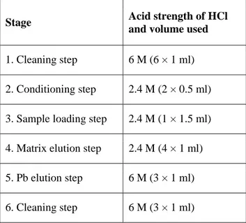
Lead (Pb) isotopes are commonly measured in digested peat samples, either directly (Krachler et al. 2004) or indirectly (Weiss et al. 2004) after resin extraction (Table 2). Measurements using multi-collection instruments (MC-ICP-MS, TIMS) need preliminary Pb purification (Weiss et al. 2004). This purification can be affected if the digestion is not complete. Using digested peat samples directly, some problems have been encountered during resin extraction and filament loading for TIMS, perhaps because of remaining soluble organic material. Thus, in order to get better measurements on early Holocene peat, we suggest digesting the peat samples again using microwave systems or after ashing.
Table 2. Ion exchange column procedure for separating Pb from the sample matrix (Weiss et al. 2004). The columns used here were made in-house from Teflon tubing and packed with 600 µl of EiChrom Sr-resin, resulting in a bed height of about 0.5 cm.
Stage Acid strength of HCl and volume used
1. Cleaning step 6 M (6 × 1 ml)
2. Conditioning step 2.4 M (2 × 0.5 ml)
3. Sample loading step 2.4 M (1 × 1.5 ml)
4. Matrix elution step 2.4 M (4 × 1 ml)
5. Pb elution step 6 M (3 × 1 ml)
6. TECHNIQUES IN PROSPECT
6.1 Impregnated peat
Resin impregnation is classically used in micromorphology of soils (e.g. Murphy 1986, Tippkotter & Ritz 1996) and to stabilise lake sediments (e.g. Lamoureux 1994, Lotter & Lemke 1999, Boës et al. 2005). A review of the different polymer impregnation techniques is given by Boës & Fagel (2005). The main laboratory steps are: (1) water removal (i.e. dehydration), (2) polymer impregnation and (3) drying and thin-section preparation. Dehydration is achieved by various methods, namely: full water substitution by acetone in liquid (e.g. Pusch 1999) or vapour phase (e.g. Camuti & McGuire 1999), water evaporation by oven-drying or freeze-drying (e.g. Francus 1998, Boës & Fagel 2005, De Vleeschouwer et al. 2008b). Impregnation is generally achieved by acetone-resin-exchange under vacuum (e.g. Boës & Fagel 2005). Several polymers have been used to accomplish impregnation (e.g. Tippkotter & Ritz 1996), depending on the purpose of the analyses; the most widely used are epoxy polymers (e.g. Francus 1998).
The high water and organic contents of peat render it especially difficult to impregnate, and few laboratories have developed specific techniques (e.g. Mackenzie & Dawson 1961, Takeda 1988, De Vleeschouwer et al. 2008b). Water removal must be performed by freeze-drying because simple oven drying will lead to shrinkage; and full water-acetone exchange cannot be performed, as acetone causes physical stress within the peat by dissolving organic matter and weakening plant cells. As this technique is described prospectively here, we do not report the laboratory steps in full; instead, we direct the reader to De Vleeschouwer et al. (2008b), where the methodology is described step-by-step along with the main problems encountered (sample size, type of peat, freeze-drying of large sections, polymer viscosity, acetone content).
The impregnation of peat deposits can reveal information concerning their state of preservation, vegetation type, and the presence of charcoal or mineral particles. More specifically, their subsequent study in thin section using various techniques allows a broad range of observations which are useful for better understanding peat accumulation, compression and decomposition; and also to give a visual counterpart to geochemical analyses.
6.2 Continuous core scanning
X-ray fluorescence (XRF) core scanning has been widely used in studies of marine and lacustrine
sediments (Jansen et al. 1998, Haug et al. 2001, Brown et al. 2007, Francus et al. 2009) and provides rapid, non-destructive analyses of split sediment cores for a broad range of major, minor and trace elements (Jansen et al. 1998, Richter et al. 2006). A mean spatial resolution of 1 mm can be achieved, potentially allowing for detection of fine structures and tackling of high-frequency signals. The XRF logging results are presented as number of XRF counts, tracing down-core geochemical variability in the sediment column. The first weakness of XRF core scanning of peat sections is that quantitative geochemical interpretation of bulk sediment data has not yet been attempted for this type of deposit. This is a major problem because further geochemical calculations, e.g. of elemental fluxes or dust concentrations, are often performed on the basis of elemental concentrations. The calibration of an XRF core scanner would require (1) corrections for sediment porosity, which is high in peat deposits and likely to be variable at a small spatial scale; (2) calibration based on standards with a similarly organic-rich matrix; and (3) validation by destructive quantitative analyses at selected depths. Attempts have been made to calibrate XRF core scanners (Weltje & Tjallingii 2008), but this is a time-consuming procedure requiring careful checks which is applicable to only one type of sediment and selected chemical elements, so that the calibration is more a case-by-case problem than an easily applicable procedure. It is hoped that further efforts will be made to solve this major difficulty. The second major problem of applying this technique to peat cores is that the split cores must be carefully flattened in order to reduce high noise:signal ratio arising from surface irregularities. Flattening is difficult in the case of peat, which is composed of various plant macrofossils, roots and fine material. Nonetheless, XRF scanning of fresh peat cores has sometimes been conducted successfully (e.g. Caseldine et al. 1999). Surface roughness problems may be avoided by stabilising the peat sections in resin (see Swindles et al. this volume) and polishing each impregnated block to obtain a perfectly smooth surface. However, the chemical composition of the resin must be critically assessed. The limited response depth of elements to incoming X-ray radiation should also be taken into account (Richter
et al. 2006 and references therein), because the XRF
scanning method will not ‘detect’ grains far below the surface of the impregnated core section. An example of successful XRF core scanning of impregnated peat sections is described in De Vleeschouwer et al. (2008a), who applied the technique to investigate geochemical trends in mineral-rich peat sections from Iceland and to
attempt to detect cryptotephras. The major conclusions were that, although substantial tephra layers leave a clear signature in XRF logging records, cryptotephras cannot be detected; and that minor variability in elemental geochemical records can be interpreted only if microscopic observations of corresponding thin sections are carried out. Therefore, XRF core scanning of impregnated peat sections has potential for a wide range of chemical investigations, but there are problems regarding data acquisition.
6.3 Non-traditional stable isotopes and platinoids
Non-traditional stable isotopes (Hg, Cu, Zn, Sn etc.) are promising tracers of environmental processes and human influences, and of sources of metal in the environment (Weiss et al. 2007). Also, Rauch et al. (2004, 2010) have recently described a protocol specifically dedicated to the characterisation of platinoid and Os isotopes in peat. However, owing to the complexity of the peat matrix and because of possible volatility, further research is needed before a clear protocol can be provided for these measurements.
7. REFERENCES
Becker, P.R., Gunter, E.W., Schlüter, C., Shibata, Y. & Wise, S.A. (2006) The U.S. national biomonitoring specimen bank and the marine environmental specimen bank. Journal of
Environmental Monitoring, 8, 795–799.
Boës, X. & Fagel, N. (2005) Impregnation method for detecting annual laminations in sediment cores: an overview. Sedimentary Geology, 179, 185–194.
Boës X., Piotrowska N. & Fagel, N. (2005) High-resolution diatom/clay record in Lake Baikal from grey scale, and magnetic susceptibility over Holocene and Termination I. Global and
Planetary Change, 46, 299–313.
Brown, E.T., Johnson, T.C., Scholz, C.A., Cohen, A.S. & King, J.W. (2007) Abrupt change in tropical African climate linked to the bipolar seesaw over the past 55,000 years. Geophysical
Research Letters, 34, L20702.
Camuti, K.S. & McGuire, P.T. (1999) Preparation of polished thin sections from poorly consolidated regolith and sediment materials.
Sedimentary Geology, 128, 171–178.
Caseldine, C., Baker, A. & Barnes, W. (1999) A rapid, non-destructive scanning method for detecting distal tephra layers in peats. The
Holocene, 9, 635–638.
Chambers, F.M., Yu, Z. & Beilman, D. (this volume) Measurements of humification, bulk density, loss on ignition, total, organic and inorganic carbon in peat: a review. Mires and
Peat, 7, in prep.
Cheburkin, A.K. & Shotyk, W. (1996) An Energy-dispersive Miniprobe Multielement Analyzer (EMMA) for direct analysis of Pb and other trace elements in peats. Fresinus Journal of Analytical
Chemistry, 354, 688–691.
Cheburkin, A.K. & Shotyk, W. (1999) High-sensitivity XRF analyzer (OLIVIA) using a multi-crystal pyrographite assembly to reduce the continuous background. X-Ray Spectrometry, 28, 145–148.
Cheburkin, A.K. & Shotyk, W. (2005) Energy-dispersive XRF spectrometer for Ti determination (TITAN). X-ray Spectrometry, 33, 69–72.
De Vleeschouwer, F., Chambers, F.M. & Swindles, G. (2010a) Coring and sub-sampling of peatlands for palaeoenvironmental research. Mires and
Peat, 7(01), 1–10.
De Vleeschouwer, F., Le Roux, G. & Shotyk, W. (2010b) Peat as an archive of atmospheric metal pollution: the example of Pb in Europe. In: Jackson, S. & Charman, D. (eds.) PAGES News, 18, 20–22.
De Vleeschouwer, F., Piotrowska, N., Sikorski, J., Pawlyta, J., Cheburkin, A., Le Roux, G., Lamentowicz, M., Fagel, N. & Mauquoy, D. (2009) Multiproxy evidence of ‘Little Ice Age’ palaeoenvironmental changes in a peat bog from northern Poland. The Holocene, 19, 625–637. De Vleeschouwer, F., van Vliët-Lanoé, B. & Fagel,
N. (2008a) Long-term mobilisation of chemical elements in tephra-rich peat (NE Iceland).
Applied Geochemistry, 23, 3819–3839.
De Vleeschouwer, F., van Vliët-Lanoé, B., Fagel, N., Richter, T. & Boës, X. (2008b) Development and application of high-resolution petrography on resin-impregnated Holocene peat columns to detect and analyse tephras, cryptotephras, and other materials. Quateranry International, 178, 54–67.
Francus, P. (1998) An image-analysis technique to measure grain-size variation in thin-sections of soft clastic sediments. Sedimentary Geology, 121, 289–298.
Francus, P., Kamb, H., Nakagawa, T., Marshall, M., Brown, E. & Suigetsu 2006 Project members (2009) The potential of high-resolution X-ray fluorescence core scanning: applications in paleolimnology. PAGES News, 17, 93–96.
Givelet, N., Le Roux, G., Cheburkin, A., Chen, B., Frank, J., Goodsite, M., Kempter, H., Krachler,
M., Noernberg, T., Rausch, N., Rheinberger, S., Roos-Barraclough, F., Sapkota, A., Scholz, C. & Shotyk, W. (2004) Suggested protocol for collecting, handling and preparing peat cores and peat samples for physical, chemical, mineralogical and isotopic analyses. Journal of
Environmental Monitoring, 6, 481–492.
Haug, G.H., Hughen, K.A., Sigman, D.M., Peterson, L.C. & Röhl, U. (2001) Southward migration of the intertropical convergence zone through the Holocene. Science, 293, 1304–1308. Jansen, J.H.F., Van der Gaast, S.J., Koster, B. &
Vaars, A.J. (1998) CORTEX, a shipboard XRF-scanner for element analyses in split sediment cores. Marine Geology, 151, 143–153.
Kempter, H. & Frenzel, B. (2000) The impact of early mining and smelting on the local tropospheric aerosol detected in ombrotrophic peat bogs in the Harz, Germany. Water, Air, and
Soil Pollution, 121, 93–108.
Kempter, H., Görres, M. & Frenzel, B. (1997) Ti and Pb concentrations in rainwater-fed bogs in Europe as indicators of past anthropogenic activities. Water, Air, and Soil Pollution, 100, 367–377.
Klaminder, J., Renberg, I., Bindler, R. & Emteryd, O. (2003) Isotopic trends and background fluxes of atmospheric lead in northern Europe: analyses of three ombrotrophic bogs from south Sweden.
Global Biogeochemical Cycles, 17, 1019–1029.
Krachler, M., Emons, H., Barbante, C., Cozzi, G., Cescon, P. & Shotyk, W. (2002a) Inter-method comparison for the determination of antimony and arsenic in peat samples. Analytica Chimica
Acta, 458, 387–396.
Krachler, M., Le Roux, G., Kober, B. & Shotyk, W. (2004) Optimising accuracy and precision of lead isotope measurement (206Pb, 207Pb, 208Pb) in acid digests of peat with ICP-SMS using individual mass discrimination correction. Journal of
Analytical Atomic Spectrometry, 19, 354–361.
Krachler, M., Mohl, C., Emons, H. & Shotyk, W. (2002b) Influence of digestion procedures on the determination of rare earth elements in peat and plant samples by USN-ICP-MS Journal of
Analytical Atomic Spectrometry, 17, 844–851.
Krachler, M., Shotyk, W. & Emons, H. (2001) Digestion procedures for the determination of antimony and arsenic in small amounts of peat samples by hydride generation-atomic absorption spectrometry. Analytica Chimica Acta, 432, 303– 310.
Kylander, M.E., Klaminder, J., Bindler, R. & Weiss, D.J. (2010) Natural lead isotope variations in the atmosphere. Earth and Planetary Science
Letters, 290, 44–53.
Lamoureux, S. (1994) Embedding unfrozen lake sediments for thin section preparation. Journal of
Paleolimnology, 10, 141–146.
Lotter, A.F. & Lemcke, G. (1999) Methods for preparing and counting biochemical varves.
Boreas, 28, 243–252.
Mackenzie, A.F. & Dawson, J.E. (1961) The preparation and study of thin sections of wet organic soil materials. Journal of Soil Science, 12, 142–144.
Martínez-Cortizas, A., Pontevedra-Pombal, X., García-Rodeja, E., Nóvoa-Muñoz, J.C. & Shotyk, W. (1999) Mercury in a Spanish peat bog: archive of climate change and atmospheric metal deposition. Science, 284, 939–942.
Mighall, T.M., Abrahams, P.W., Grattan, J.P., Hayes, D., Timberlake, S. & Forsyth, S. (2002) Geochemical evidence for atmospheric pollution derived from prehistoric copper mining at Copa Hill, Cwmystwyth, mid-Wales. The Science of
the Total Environment, 292, 69–80.
Mighall, T.M., Timberlake, S., Jenkins, D.A. & Grattan, J.P. (2006) Using peat bog archives to reconstruct paleopollution and vegetation change during the late Holocene. In: Martini, P., Martínez Cortizas, A. & Chesworth, W. (eds.)
Evolution and Records of Environmental and Climatic Changes. Elsevier, Amsterdam, 409–
429.
Murphy, C.P. (1986) Thin Section Preparation of
Soils and Sediments. AB Academic Publishers,
Berkhamsted, Herts, UK, 149 pp.
Novák, M., Emmanuel, S., Vile, M.A., Erel, Y., Véron, A., Paes, T., Wieder, R.K., Vaněek, M., Štěpánová, M., Břízová, E. & Hovorka, J. (2003) Origin of lead in eight central European peat bogs determined from isotope ratios, strengths, and operation times of regional pollution sources. Environmental Science and Technology, 37, 437–445.
Potts, P.J. (1987) A Handbook of Silicate Rock
Analysis. Blackie & Son Limited,
Glasgow-London, UK, 622 pp.
Pugh, R.S., Becker, P.R., Porter, B.J., Ellisor, M.B., Moors, A.J. & Wise, S.A. (2008) Design and applications of the National Institute of Standards and Technology's (NIST’s) environmental specimen banking programs. Cell
Preservation Technology, 6, 59–72.
Pusch, R. (1999) Experience from preparation and investigation of clay microstructure. Engineering
Geology, 54, 187–194.
Rauch, S., Hemond H.F. & Peucker-Ehrenbrink, B. (2004) Recent changes in platinum group element concentrations and osmium isotopic composition in sediments from an urban lake.
Environmental Science & Technology, 38, 396–
402.
Rauch, S., Peucker-Ehrenbrink, B., Kylander, M.E., Weiss, D.J., Martinez-Cortizas, A., Heslop, D., Olid, C., Mighall, T. & Hemond, H. (2010) Anthropogenic forcings on the surficial osmium cycle. Environmental Science & Technology, 44, 881–887.
Richardson, D.H.S., Shore, M., Hartee, R. & Richardson, R.M. (1995) The use of X-ray fluorescence spectrometry for the analysis of plants, especially lichens, employed in biological monitoring. The Science of the Total Environment, 176, 97–105.
Richter, T.O., van der Gaast, S., Koster, B., Vaars, A., Gieles, R., de Stigter, H.C., de Haas, H. & van Weering, T.C.E. (2006) The Avaatech XRF core scanner: technical description and applications to NE Atlantic sediments. In: Rothwell, G. (ed.) New Techniques in Sediment
Core Analysis. Geological Society of London,
UK, Special Publications No. 267, 39–50.
Roos-Barraclough, F., van der Knaap, W.O., van Leeuwen, J.F.N. & Shotyk, W. (2004) A Late Glacial and Holocene record of climate and possible modern anthropogenic influences from a Swiss peat humification profile. The Holocene, 14, 7–19.
Shotyk, W., Weiss, D., Appleby, P.G., Cheburkin, A.K., Frei, R., Gloor, M., Kramers, J.D., Reese, S. & van der Knaap, W.O. (1998) History of atmospheric lead deposition since 12,370 14C yr BP recorded in a peat bog profile, Jura Mountains, Switzerland. Science, 281, 1635– 1640.
Swindles, G.T., De Vleeschouwer, F. & Plunkett, G.
(this volume) Dating peat profiles using tephra: stratigraphy, geochemistry and chronology.
Mires and Peat, 7, accepted.
Takeda, H. (1988) A rapid method for preparing thin sections of soil organic layers. Geoderma, 42, 159–164.
Tippkotter, R. & Ritz, K. (1996) Evaluation of polyester, epoxy and acrylic resins for suitability in preparation of soil thin sections for in situ biological studies. Geoderma, 69, 31–57.
Weiss, D.J., Kober, B., Dolgopolova, A., Gallaghera, K., Spiro, B., Le Roux, G., Mason, T.F.D., Kylander, M. & Coles, B.J. (2004) Accurate and precise Pb isotope ratio measurements in environmental samples by MC-ICP-MS. International Journal of Mass Spectrometry, 232, 205–215.
Weiss, D.J., Rausch, N., Mason, T.F.D., Coles, B.J., Wilkinson, J.J., Ukonmaanaho, L., Arnold, T. & Nieminen, T.M. (2007) Atmospheric deposition and isotope biogeochemistry of zinc in ombrotrophic peat. Geochimica et
Cosmochimica Acta, 71, 3498–3517.
Weiss, D.J., Shotyk, W., Schafer, J., Loyall, U., Grollimund, E. & Gloor, M. (1999) Microwave digestion of ancient peat and determination of Pb by voltammetry. Fresenius Journal of Analytical
Chemistry, 363, 300–305.
Weltje, G.J. & Tjallingii, R. (2008) Calibration of XRF core scanners for quantitative geochemical logging of sediment cores: theory and application. Earth and Planetary Science Letters, 274, 423–438.
Submitted 28 April 2010, revision 14 June 2010 Editor: Olivia Bragg
_______________________________________________________________________________________ Author for correspondence:
Dr Gaël Le Roux, EcoLab UMR 5245 CNRS-UPS-INPT, Université de Toulouse, France. Tel: +32 (0)4 366.22.10; Fax: +32 (0)4 366.22.02; E-mail: gael.leroux@ensat.fr