UNIVERSITÉ DE MONTRÉAL
EXPRESSION AND REGULATION 0F PROTEASE NEXIN-1 AND PLASMINOGEN ACTIVATORS IN BOVINE OVARIAN FOLLICLES
Par
MINGJU CAO
Centre de recherche en reproduction animale (CRRA) Département de biomédecine vétérinaire
Faculté de médecine vétérinaire
Thèse presentée à la Faculté des études supérieures en vue de 1’ obtention du grade de Philosophiae Doctor (Ph.D.)
en sciences vétérinaires (Option: Reproduction)
Avril, 2005
cJP
-E
Université
(ll’b
de Montréal
Direction des bibliothèques
AVIS
L’auteur a autorisé l’Université de Montréal à reproduire et diffuser, en totalité ou en partie, par quelque moyen que ce soit et sur quelque support que ce soit, et exclusivement à des fins non lucratives d’enseignement et de recherche, des copies de ce mémoire ou de cette thèse.
L’auteur et les coauteurs le cas échéant conservent la propriété du droit d’auteur et des droits moraux qui protègent ce document. Ni la thèse ou le mémoire, ni des extraits substantiels de ce document, ne doivent être imprimés ou autrement reproduits sans l’autorisation de l’auteur.
Afin de se conformer à la Loi canadienne sur la protection des renseignements personnels, quelques formulaires secondaires, coordonnées ou signatures intégrées au texte ont pu être enlevés de ce document. Bien que cela ait pu affecter la pagination, il n’y a aucun contenu manquant. NOTICE
The author of this thesis or dissertation has granted a nonexclusive license allowing Université de Montréal to reproduce and publish the document, in
partor in whole, and in any format, solely for noncommercial educational and research purposes.
The author and co-authors ifapplicable retain copyright ownership and moral rights in this document. Neither the whole thesis or dissertation, nor substantial extracts from it, may be printed or otherwise reproduced without the author’s permission.
In compliance with the Canadian Privacy Act some supporting forms, contact information or signatures may have been removed from the document. While this may affect the document page count, it does flot represent any loss of content from the document.
Université de Montréal Faculté des études supérieures
Cette thèse intitulée:
EXPRESSION AND REGULATION 0F PROTEASE NEXIN-1 AND PLASMINOGEN ACTIVATORS IN BOVINE OVARIAN FOLLICLES
Présentée par
MINGJU CAO
a été évaluée par un jury composé des personnes suivantes:
Président du jury: Dr. Bruce D. Murphy Directeur de recherche: Dr. Christopher A. Price Codirecteur de recherche: Dr. Jacques G. Lussier
Membre du jury: Ur. Alan K. Goff
Examinateur externe: Dr. James J. Ireland Répresentant du doyen: Dr. Bruce D. Murphy
111
Résumé
Les changements dans la composition de la matrice extracellulaire et la membrane basale surviennent lors de la croissance folliculaire, possiblement par l’intemédiaire des cascades d’enzymes protéolytiques, incluant les activateurs du plasminogène (PA) et leurs inhibiteurs. Un tel inhibiteur est la protéine nexine-1 (PN-1), une protéine secrétée par les cellules de la granulosa (GC). Notre connaissance sur la régulation et sur l’expression des PA et la PN-1 dans les GC est très limitée. L’objectif général de cette étude était de tester l’hypothèse que l’expression de PN-1 est régulée par des gonadotrophines et les facteurs de croissance au cours de la croissance folliculaire.
L’expression des PA et de la PN-1 a été étudiée dans des GC bovine non lutéinisée. L’activité de PA de type tissulaire (tPA) était plus élevée dans les GC issues de petits follicules comparée aux follicules plus gros, et le taux de sécrétion du PN-1 était plus élevé dans les GC de gros follicules. Dans les cellules provenant de petits follicules, les taux de sécrétion de tPA et PN-1 augmentaient en fonction du temps de culture. Dans les cellules provenant de gros follicules, l’activité de tPA augmentait de façon significative en fonction du temps de culture, alors que la sécrétion de PN- 1 diminuait.
Dans les GC en culture, la FSH stimulait l’expression du gène codant pour la PN 1, ainsi que la sécrétion de PN-1. La FSH augmentait de façon dose-dépendante l’expression de l’ARNm de la tPA mais n’a pas affecté le taux de sécrétion de la protéine. L’IGF-l a stimulé l’ARNm codant pour la PN-1, et la sécrétion d’uPA mais a diminué la sécrétion de la tPA et le taux de son ARNm. La protéine morphogénétique osseuse 7 (BMP-7) a augmenté la sécrétion de PN-1 des cellules stimtilées à l’IGF-l ou la FSH et a
augmenté la sécrétion de tPA par des cellules stimulées à l’IGF-l. Le facteur de croissance fibroblastique-2 (FGF-2) était généralement inhibiteur, diminuant la sécrétion de tPA des cellules stimulées à l’IGF-l ou à la FSH, et diminuant la sécrétion de PN-1 des cellules stimulées à l’IGf-l. Les effets d’EGF étaient variés puisque la sécrétion de PN-1 était inhibée mais la sécrétion de tPA était augmentée.
Finalement, nous avons examiné l’expression génique de PN-1 dans les follicules à divers stades de développement. Dans les follicules périovulatoires, le taux de l’ARNm pour la PN-l était stimulé par hCG, tout comme ceux pour les tPA et uPA. Deuxièmement, les follicules prélevés à l’abattoir ont été classifiés comme étant oestrogénique ou non-oestrogénique basé sur la concentration d’oestradiol dans le liquide folliculaire (FF). La concentration de PN-l dans le FF et l’expression de l’ARNm de PN 1 dans les CG étaient significativement plus élevées dans les follicules possédant les concentrations d’oestradiol élevées. Finalement, la concentration de PN-1 dans le FF des follicules dominants durant la déviation de la première vague folliculaire n’a démontré aucun changement significatif, suggérant que le PN-1 n’est pas un bon indicateur du processus de la sélection folliculaire.
En résumé, l’expression et la sécrétion de PN-1 des CG bovines sont sous contrôle hormonale. Il est probable que PN-1 joue un rôle physiologique dans le remodelage tissulaire au cours de la croissance folliculaire et de la rupture de la paroi folliculaire au moment de l’ovulation.
Mots-clés protéase nexine- 1, activateur du plasminogène, follicule, cellule de la granulosa, matrice extracellulaire, remaniement tissulaire, FSH, facteur de croissance.
V
ABSTRACT
Understanding follicle development leads to practical control of reproduction in agriculturally important species sucli as cattie. Changes in the composition of the extracellular matrix (ECM) and the basement membrane occur during follicle growth, likely through proteolytic enzyme cascades, including plasminogen activators (PA) and their inhibitors. One such inhibitor is protease nexin-1 (PN-1), a granulosa ceil-specific secreted protein. Regulation and expression of PN-1 and the PAs in granulosa ceils is poorly understood.
The expression of PAs and PN-1 was examined in a non-luteinizing bovine granulosa ceils culture model. Secreted tPA activity was higher in cultures of ceils from small follicles compared to large follicles, and secreted PN- 1 levels were higher in cultures of celis from large follicles. In cultures of celis from small follicles, secreted tPA and PN-l levels increased with time of culture. In cultures of granulosa ceils from large follicles, tPA activity increased significantly with tirne of culture, whereas PN-1 rnRNA and protein levels decreased. Cell-associated uPA activity decreased with tirne in ceils from medium and large follicles.
To study the regulation of PN-1, granulosa ceils were cultured with doses of FSH and growth factors. PN-l mRNA and protein levels and uPA secretion by cultured GCs were stirnulated by fSH in a biphasic manner, with maximum levels at Ing. FSH caused a dose-dependent increase in tPA gene expression but not secreted enzyme activity. IGF-I stirnulated PN-l and uPA secretion. However, IGF-I decreased secreted tPA activity and tPA gene. In addition, bone morphogenetic protein 7 (BMP-7) increased PN-l secretion
in FSH- and IGF-I stimulated ceils, and secreted tPA activity in IGf-I stimulated but flot FSH stimulated celis. In contrast, fibroblast growth factor 2 (FGF-2) was generally inhibitory, decreasing tPA secretion in FSH- and IGF-I- stirnulated celis, and decreasing PN-1 secretion in IGF-I stimulated but flot FSH stimulated ceils. The effects ofEGF were diverse, as PN-l secretion were inhibited, but secreted tPA activity was increased.
As PN-1 secretion differs with follicle stage, we examined PN-l gene expression levels in follicles at defined stages in vivo. firstly, the regulation of PN-l gene during ovulation was measured following administration of hCG. There was an initial upregulation of gene expression, followed by a marked inhibition nearer the expected time of ovulation. Secondly, follicles collected from the abattoir were classified as nonatretic or atretic based on ff estradiol content. PN-1 protein in FF and PN-1 mRNA expression in GC was significantly higher in nonatretic than atretic follicles. In contrast, FF plasmin activity was correspondingly higher in the atretic follicles. finally, No significant changes in PN-l levels in FF were observed during the growth of pre deviation follicles early in a follicle wave, suggesting PN-1 is not a good marker for the process of follicle selection. These resuits indicate that PN-1 may be involved in the process of atresia in nonovulatory dominant follicles and the prevention of precocious proteolysis in periovulatory follicles.
In summary, PN-1 expression and secretion from bovine granulosa celis is under hormonal regulation. PN-l likely plays a physiological role in growing follicles and the process of follicle wall rupture at ovulation.
Key words: protease nexin-l, plasminogen activator, follicle, granulosa cell, extracellular matrix, tissue remodelling, FSH, growth factor
vii CONTENTS JURY IDENTIFICATION ii RÉSUMÉ ABSTRACT y CONTENTS vii LIST 0F TABLES xi
LIST 0F FIGURES xii
LI$T 0F ABBREVIATIONS xvi
DEDICATORY xxi
AKNOWLEDGEMENTS xxii
NTRODUCTION .1
LITERATURE REVIEW 4
FOLLICULAR DEVELOPMENT AND STEROIDOGENESIS 4
Morphological changes in follicular development 4
Foïlicular dynamics in the cow 7
follicular Steroidogenesis 9
Biochernical changes infollicular development 13
EXTRACELLULAR MATRIX IS DYNAMIC IN FOLLICULAR DEVELOPMENT 15
fl
Collagen and larninin are structural proteins in ECM components 16Other ECM components 18
Interaction ofECM proteins and granulosa ceils function 21
PROTEASES AND THEIR INHIBITORS REGULATE ECM REMODELING 22
The MMP system 23
The cysteine proteases 28
PLASMINOGEN ACTIVATOR SYSTEM 29
Plasminogen 30
Plasmin 31
ix
Urokinase plasminogen activator .32
uPA receptor 33
Plasminogen activator inhibitor-1 34
Plasminogen activator inhibitor-2 35
u2-antiplasmin 36
PROTEASE NEXTN-1: MOLECULAR ANI BIOCHEMICAL
CHARACTERISTICS 38
EXPRESSION AND REGULATION 0F THE PA SY$TEM IN OVARIAN
FOLLICLE 41
Biosynthesis and secretion ofPA system in ovarian follicle 41 Ovulation requires coordinated expression ofPAs and PAIs in ovary 43 Expression and regulation ofPA and PAIs in the srnall growing follicles 48
ROLES 0F PA SYSTEM IN CORPUS LUTEUM AND OVARIAN
ANGIOGENSIS 49
HORMONAL REGULATION ON THE PA SYSTEM IN GRANULOSA CELLS 51
Gonadotropins 51
GnRH 53
Growth factors 54
OVERALL HYPOTHESIS AND OBJECTIVES .58
ARTICLE 1 59
Plasminogen Activator and Serine Protease li±ibitor-E2 (Protease nexin-1)
Expression by Bovine Granulosa Celis in Vitro 60
ARTICLE 2 99
Regulation of Serpin-E2 and Plasminogen Activator Expression and $ecretion by Growth factors in Non-luteinizing Bovine Granulosa Ceils in Vitro 100
ARTICLE 3 140
Expression of Protease Nexin-1 and Plasminogen Activators during Follicle
Growth and the Periovulatory Period in Cattie 141
GENERAL DISCUSSION 181
FUTURE STUDY 192
GENERAL CONCLUSION 193
GENERAL REFERENCES 195
xi
LIST 0F TABLES
Article 3
Table I. Surnmary on RT-PCR protocol in Expt 1&2 162
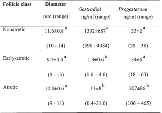
TableIL Oestradiol and progesterone concentrations, and mean diarneters of follicle in non-atretic, early-atretic and atretic follicles in Experiment 2 163
Table III. Mean (±SEM) diameter and follicular fluid steroid content ofthe dorniant (Fi) and two largest subordinate follicles (f2, F3) collected early in a follicle wave, when the dominant follice reached approximately 6.5, 7.5, 8.5 or 9.5 mm
LIST 0F FIGURES
Literature review
Fig 1. The follicular growth continuum 5
Fig 2. Follicular phase steroid biosynthesis in the ovary with the illustration
ofthe two-ceÏl/two-gonadotropin theory 12
Fig 3. A schernatic Proteolytic Cascade 37
Article 1
Fig 1. Zyrnographic dernonstration ofPA activity in bovine granulosa celis
cultured for 6 days in serum-free medium $0
Fig 2. Secreted PA activity from cultured bovine granulosa cells from (A) srnall (2-5 mm diarneter), (B) medium (6-8 mm) and (C) large (>8mrn) follicles. . . .$2
Fig 3. Cellular PA activity from cultured bovine granulosa ceils from (A) small (2-5 mm diameter), (B) medium (6-8 mm) and (C) large (>$mm) follicles 84 Fig 4. Western analysis of secreted SERPIN-E2 from cultured bovine antral and
basal granulosa ceils from small, medium and large follicles 86 Fig 5. SERFIN-E2 rnRNA levels in cultured bovine antral and basall GC from
small, medium and large follicles 8$
xiii
basal granulosa celis from small, medium and large follicles .90
Article 2.
Fig 1. Dose-dependent stimulation of A) estradiol and B) progesterone production, and C) cell proliferation by fSH in bovine granulosa cells cultured in
serurn-free medium 119
Fig 2. Effect ofFSH on A) secreted tPA activity and gene expression, B) secreted uPA activity and gene expression, and C) serpin-E2 protein and gene expression in bovine granulosa celis culturedin serum-free medium 121 Fig 3. Dose-dependent stimulation of A) estradiol and B) progesterone production,
and C) celi proliferation by IGF-I analog (LR3) in bovine granulosa ceils
cultured in serum-ftee medium 123
Fig 4. Effect of IGf-I analog (LR3) on A) secreted tPA activity and gene expression, B) secreted uPA activity and gene expression, and C) serpin-E2 protein
secretion and gene expression 125
Fig 5. Effect of BMP-7 on A) estradiol secretion, B) ceil proliferation, C) secreted PA activity and D) serpin-E2 secretion from bovine granulosa ceils cultured in
serum-free medium 127
Fig 6. Effect of FGF-2 onA) estradiol secretion, B) ce!! proliferation, C) secreted PA activity and D) serpin-E2 protein secretion from granulosa ce!ls cu!tured in
senirn-free medium 129
activity and D) serpin-E2 protein secretion from granulosa celis cultured in
serum-free medium 131
Article 3.
Fig. 1. Analysis of A) PN-1, B) PAT-1, C) uPA, and D) tPA mRNA expression in follicular wall lysates ofpreovulatory follicles by RT-PCR (experirnent 1) 165 Fig. 2. Analysis of A) PN-1 protein content and B) proteolytic enzyme activities in
GC lysates from periovulatory bovine follicles at 0, 12, and 24 hours after hCG
(experiment 1) 167
Fig. 3. Analysis of A) PN-1 protein content and B) proteolytic enzyme activity in FF collected from periovulatory follicles at 0, 6, 12, 18, and 24 hours after
hCG (experiment 1) 169
Fig. 4. Plasmin and uPA enzyme activities (A) and PN-1 protein (B) in fF from nonatretic (n=7), early-atretic (n=7) and atretic (n=4) follicles
(experiment 2) 171
Fig. 5. RT-PCR analysis ofmRNA expression ofPA and PA inhibitors in
(A) granulosa and (B) theca celis from nonatretic (n=7), early-atretic (n=7)
and atretic (n=4) follicles (experiment 2) 173
Fig. 6. PN- 1 protein (A) and uPA enzyme activity (B) in follicular ftuid samples collected from growing dominant (Fi) and subordinate (F2, F3) follicles
General discussion
Fig 1. A working model for the PA system in follicular growth 190 Fig 2. A working model for the PA system during ovulation 191
LIST 0f ABBREVIATIONS
ActR-TI activin type II receptor
ADAMT$-1 a disintegrin and metalloproteases with thrombospondin motifs-1 Œ2-antiplasrnin
Œ2-M Œ2-macroglobulin
Œ2-MR a2-macrogÏobulin receptoi BCEC bovine capillary endothelial ceils BMP-7 bone morphogenetic protein 7 BMP-15 bone morphogenetic protein 15 BMPR-II BMP type II receptor
cAMP cyclic adenosine 3’ ,5 ‘monophosphate cDNA cornp lementary deoxyribonucleic acic COC cumulus-oocyte-complex
CRE cAMP-response element
CREBP cAMP response element-binding protein
CL corpus luteum
DES diethylstilbestrot
DEX dexamethasone (glucocorticoid agonist) DHT dihydrotestosterone
DMSO dimethyl sulfoxide DNA deoxyribonucleic acid
xvii
ECM extracellular matrix EGF epidermal growth factor
EGFR epidermal growth factor receptor ER endoplasmic reticulum
ERK extracellular signal-regulated kinase FF follicular fluid
fGf-2 fibroblast growth factor 2 (bfGf) fSH follicle-stinnilating hormone
FSHr follicle-stimulating hormone receptor GAG glycosaminoglycans
GAPDH glyceraldehyde-3-phosphate dehydrogenase
GC granulosa celi
GDf-9 growth differentiation factor 9 GDN glia-derived nexin
GnRH gonadotropin releasing hormone GPI anchor glycosyl phosphatidylinositol linkage
HA hyaluronan
HDL high density lipoproteins
3 f3-HSD 3 f3-hydroxysteroid dehydrogenase 1 7f3-HSD 1 7f3-hydroxysteroid dehidrogenase HSPG heparin suiphate proteoglycan perlecan IGF-1 insulin-like growth factor 1
IL-1f3 interleukin- 1 beta
IP3K inositol trisphosphate 3 kinase
KL kit ligand
KO knock-out
LDL low density lipoproteins LH luteinizing hormone
LHr luteinizing hormone Tee eptor LOX lysyl oxidase
LRP the LDL receptor-related proteins MAPK mitogen-aetivated protein kinase MEK mitogen-activated protein kinase kinase MIX 1 -rnethyl-3 -isobutylxanthine
MMP matrix metalloproteinase
MT-MMP membrane type-matrix metalloproteinase NF 1 nuclear factor 1
ORF open reading frame
P45°aIom cytochromeP450 aromatase
P45O1701 cytochrome P450 l7hydroxylase P4500 cytochrome P450 side chain cleavage
PACAP pituitary adenylate cyclase-activating polypeptide PAl- 1 plasminogen activator inhibitor- 1
PAT-2 plasminogen activator inhibitor-2 PAR-1 protease activated receptor
xix
PDGF platelet derived growth factor PGF2a prostaglandin F2 alpha PKA protein kinase A PKC protein kinase C
PMA phorbol myristate acetate
PMSG pregnant mare serum gonadotropin (eCG) PN-1 protease nexin-1 (Serpin-E2)
PRL prolactin
RNA ribonucleic acid RIA radioirnrnunoassay SERPIN serine protease inhibitor
Serpin-E2 serine protease inhibitor-E2 (PN-1) SF1 steroidogenic factor 1
StAR steroidogenic acute regulatory protein TGFa transforming growth factor alpha TGF- transforming growth factor beta TIIVIP tissue inhibitors ofmetalloproteinases TNTΠtumor necrosis factor alpha
tPA tissue type plasminogen activator
TSG-6 tumour necrosis factor Œ-stimulated gene 6 uPA urokinase type plasminogen activator uPAR uPA receptor
VEGF vascular endothelial growth factor VIP vasoactive intestinal peptide VLDL the very-low-density lipoprotein
xxi
n
DEDICATORY
To ail my family members both in China and Canada, to rnymother, my father, my grandfather, my brother and sisters for their support and encouragement.
To my wife Ling Yin, and our lovely daughter Annie Cao and son Bright Cao for the love and happiness they give me.
ACKNOWLED GEMENTS
I want to thank my supervisor Dr. Christopher A. Price for his excellent direction, valuable time, availability, patience and friendship. Over the past four years and eight months I’ve experienced many unforgettable things with Dr. Price not only atwork in bis lab but also traveling on the way between Montreal and St-Hyacinthe.
I want to thank my co-supervisor Dr. Jacques G. Lussier for his availability, kind help and advice, which have ail made this collaborating project productive and pleasant.
I want to thank Dr. Bruce D. Murphy for his professional orientation on scientific research whenever I attend Atelier de recherche, and his group, in particular Mme Mira Dobias, for the availability of laboratory and kind help.
I want to thank Mme. Micheline Sicotte and other secretaries at CRRA for their professional and efficient secretary service.
I want to thank Dr. Maiha Sahrni, Dr. Edmir Nicola, Mélanie Hamel and Daniel Sylvain for their tecfmical help in the lab and friendship.
I want to thank Dr. Alan K. Goff, Dr. Lawrence C. Smith, Dr. Paul Carrière and Dr. Jean Sirois for their help and equipments availability. Special thanks give to Mrne Clarisse Desautels at GREMIP, Faculty of Veterinary Medicine, for her help with sample lyophilization.
I want to thank ail members of CRRA for their friendships and the good environrnent in which I was immersed, as well as for the moments of fun we shared.
Finally, I want to thank Drs. Bingtuan Wang, Yuanyi Li, Yule Pan and Xiaofeng Zheng and other Chinese friends in St-Hyacinthe and Montreal, for their help and friendship.
xxiii
AVANT-PROPOS (PREFACE)
Cette thèse est présentée à la Faculté des études supérieures de l’Université de Montréal pour l’obtention du grade de Philosophiae Doctor en sciences vétérinaires, option reproduction. Elle est composée d’une introduction générale, d’une revue de littérature générale; trois articles comprenant chacun: une introduction, une section matériel et méthodes, des résultats, une discussion et des références; une discussion générale, une conclusion générale ainsi que des references générales.
This thcsis is presented to the Faculté des études supérieures de l’Université de Montréal for the obtention of the Philosophiae Doctor degree in veterinary sciences, option reproduction. It composes a general introduction, a general literature review; three published or submitted articles, each of which contains a specific introduction, materials and rnethods, resuits, discussion and references; a general discussion, a general conclusion and general references.
INTRODUCTION
Ovarian follicular development begins with the initial recruitment of primordial follicles into the pool ofgrowing follicles (Fortune et al., 2000). follicles that are destined to ovulate pursue their development into the preantral and antral phases, become selected as the dominant follicle (deviation) (Ginther et al., 1996; Fortune et aÏ., 2001), then undergo ovulation (Richards et al., 2002) and luteinization (Murphy, 2004). However, most follicles degenerate by atresia throughout the antral phase (Markstrôrn et al., 2002). The mechanisms for the recruitment, deviation and ovulation of follicles are flot fully understood (Fortune et al., 2001).
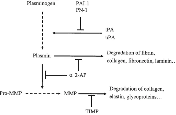
In cattie, follicles typically increase in size 400-fold between preantral and preovulatory stages (Lussier et al., 1987). The surface area of preovulatory follicles doubles 19 tirnes compared with primordial follicles (Rodgers et al., 1999). During follicle growth, there is expansion of basal lamina and changes in composition of follicular extracellular matrix (ECM) (Rodgers et aÏ., 2003). The follicular basal lamina is formed of specialized sheets of ECM, which separates the epithelial cells (membrana granulosa) from adjoining stroma (including theca interna and externa, and vasculature). Previous studies suggest that the follicular basal lamina is extremely dynamic during follicular development; the follicular basal lamina becornes less collagenous and more larninin rich, such that it becomes more expandable to meet the requirement for follicle enlargernent (Rodgers et al., 2000). These changes likely occur through proteolytic enzyme cascades, such as the plasminogen activators (PA) and their inhibitors (Ny et al., 2002).
2 Plasminogen activators are serine proteases that convert the abundant extracelluiar zymogen piasminogen into piasmin, an active protease that promotes degradation of components of the ECM as well as activating the matrix metalloproteinase (MMP) protease cascade (reviewed by Ny et al., 2002; Liu, 2004). The PA system contains the proteolytic enzymes plasmin, tissue type (tPA) and urokinase type (uPA) piasminogen activators, and regulatory components including inhibitors, cofactors, ccii surface receptors and binding proteins. The activity ofproteoiytic enzymes is regulated by inhibitors such as plasminogen activator inhibitor-1 (PAl-1), piasminogen activator inhibitor-2 (PAl-2), protease nexin-1 (PN-1) (Kruithof, 1988; Roberts et al., 1995), and the plasmin specific inhibitor Œ2-antiplasmin (a2-AP). PAl-1, PAl-2, PN-1, and Œ2-AP ail belong to the serine protease inhibitor (SERP1N) superfamily (Silverman et al., 2001). PN-1, also referred to as serine protease inhibitor-E2 (SERPIN-E2), and giia-derived nexin (GDN), is a secreted 43 kDa giycoprotein, and is a broad spectrum, trypsin-like inhibitor that rapidly inhibits a number of target proteases inciuding uPA, tPA, plasmin, trypsin, and thrornbin (Siiverman et al., 2001).
The expression and regulation of PN-1 during foiiicie deveioprnent is poorly understood. In the ovary, PAl-1 (SERPIN-El) mRNA and activity are predorninantly synthesized by theca-interstitial ceiis in the rodent (Liu et al., 19$7b; Hggiund et al., 1996), cattie (Dow et aL, 2002b) and monkey (Liu et al., 2004). Stimulation with hCG upreguiated PAT-1 expression in theca celis, and induced PAl-1 mRNA expression in GC in rats (Chun et al., 1992). In contrast to PAl-I, PN-1 is exclusively expressed in GC in mammals including mice (Hagglund et cil., 1996), rats (Hasan et al., 2002) and catt]e (Bédard et al., 2003). Furthennore, studies show that the ievel of PN-l mRNA is high in
GC throughout the periovulatory period, and decrease in ovulated follicles of mice (Higg1und et aÏ., 1996) and rats (Hasan et aÏ., 2002). The expression of PN-1 mRNA is also demonstrated in bovine GC, and is regulated in a spatio-temporal pattem with highest steady state levels in GC of growing dominant bovine follicles cornpared with small follicles (Bédard et al., 2003). However, the physiological role of PN-1 during follicle growth, follicle deviation and ovulation is unknown.
Ovarian follicular growth and development are integrated processes controlled by both extraovarian signais, such as gonadotropins, and intraovarian factors. Follicle stimulating honrione (fSH) is an essential factor in the regulation of follicle development from primary follicles through to dominant preovulatory follicles. A number of growth factors are also involved in follicle deveioprnent, including insulin-like growth factor-I (IGF-I), bone morphogenetic proteins (BMPs), fibroblast growth factors (FGF) and epidenrial growth factor (EGF). However, littie is known that whether or not these growth factors regulate PAs and their inhibitors in GC, in particular, nothing is known about their regulation of PN- 1.
Using a non-luteinizing bovine GC culture model, as well as in vivo approaches, we performed a number of studies to elucidate the regulation ofPN-1 expression and secretion in ovarian GC. Our findings provided new insiglits on the role of PN-1 during antral follicle growth and ovulation.
4 LITERATURE REVIEW
FOLLICULAR DEVELOPMENT AND STEROIDOGENESIS
The release of an ovum that is ready for fertilization is one of the major functions of ovarian follicular development. Ovarian follicular growth and development are integrated processes controlled by both extraovarian signais such as gonadotropins and metabolic hormones, and intraovarian factors. Follicular developrnent is classified into gonadotropin independent and gonadotropin-dependent phases (Webb et al., 1999).
In mammals, follicles develop continuously from the pool of primordial follicles throughout the reproductive lifespan of the animal. The development of follicles involves the recruitment of primordial follicles from the resting pool (follicle activation), the continued growth from primary follicies to small antral (Fortune, 2003; McNatty et al., 1999; Webb et al., 1999), selection ofa dominant follicle (deviation) (Ginther et aï., 1996; Fortuneet aL, 2001; Zeleznik, 2001; Fortune et al., 2004), ovulation (Richards et al., 2002) and luteinization (Murphy, 2004). Foilicular development is illustrated in Figure 1.
Morphological changes in follicular development
Follicular development is morphologicaily characterized by an increase in the diameter of the oocyte, and a synchronous proliferation of GC, resulting in multiple layers of cells that surround each oocyte. The earliest stage of foilicular growth is initiaiiy characterized by the transition of GC from flattened to cuboidal cells. This phase of
Selection and Ovulation dominance Emergence Initial Recruitment
N
Primarvft)llicles formation Atresi’t
Primordial lollicles
—3 monttis—* —2 estrous cycles
Preantral follicle Antral follicle growth groth
t
t
t
t
Growth factor dependent Gondotropin dependent TGFI3 and KW families FSH LH
t
t tGonadotropin Growth tactor influenccd in ticnced
Fig 1. The follicular growth continuum
Schematic representation ofthe requirement for growth factors, such as the TGFB and IGF families, and gonadotropins at different stages of ovarian follicle development in cattie. Growth factors seem to be important in both the initiation of and in early follicle growth, whereas gonadotropins are essential for the final stages of follicle growth. In this regard, the dominant follicle switches its requirernent from FSH to LH. There is also increasing evidence that gonadotropins can influence follicle development before antrum formation and growth factors can influence follicle development throughout the follicular growth cofltifluuiTl.
6 preantral growth is relatively slow, comprising about 85% of the total duration of follicle growth in some species (Vanderhyden, 2002). The regulation of primordial follicle and preantral growth in cattie bas been well reviewed (McNatty et al., 1999; fortune et al., 2000; Fortune, 2003).
During preantral (primordial, primary, and secondary) follicular growth, although theca ceils remain separated from GC by a basement membrane, they become associated with the growing follicle in this stage. Continued growth features both an increase in oocyte diameter and proliferation of granulosa ceils. Granulosa celis of preantral follicles are a relatively homogenous population of proliferating ceils that acquire receptors for follicle-stimulating hormone (FSH) and steroid hormones (Oxberry & Greenwald, 1982; Richards, 1975). Under the influence ofFSH, cyclin D2 expression is induccd in granulosa celis (Sicinski et al., 1996) and the follicle continues to grow.
Transition to an antral follicle is associated with the formation of a fluid-fflled cavity, and the granulosa cells differentiate into two sub-populations: cumulus granulosa cells, which are those most closely associated with the oocyte and are ovulated with it; and mural granulosa celis, which forma multi-layered wall against the basement membrane and acquire differentiated functions, including steroidogenesis (Zlotkin et al., 1986) and the expression of luteinizing hormone (LH) receptors (Oxbeny & Greenwald, 1982; Bortolussi et al., 1979).
In cattie, follicular antrum formation begins at a diameter of 0.2 mm, and there is a large pooi of mostly healthy, growing follicles from 0.2 to 2 mm in size (Lussier et al., 1987). A critical physiological stage is reached at 3 to 4 mm diarneter in size, when most follicles are lost by atresia (Lussier et al., 1987). In the absence of sufficient FSH, or by the
natural process of follicle selection, most follicles will fail to reach ovulatory size and will undergo apoptosis or atresia (Lussier et al., 1994; Gong et al., 1996; Murdoch, 2000; Asselin et al., 2000). Jndeed, more than 99% of ovarian follicles present at birth neyer reach ovulation due to follicular atresia (Ireland, 1987).
Atresia is reguÏated by endocrine factors, notably fSH and LH, and mediated by intraovarian factors such as IGF-I (insulin-like growth factor-I), EGf (epidermal growth factor) and fGf-2 (fibroblast growth factor-2) (Markstn5m et al., 2002). The fate of follicle development versus atresia largely depends on the crosstalk between oocyte and granulosa ceils and theca ceils. For example, oocyte secreted factors including bone morphogenetic protein 15 (BMP-15) and growth differentiation factor 9 (GDF-9) act on the granulosa ceils to enhance follicle development in mice and the inhibition of luteinization, in tum, granulosa celis produce Kit Ligand (KL) that acts through Kit receptors to promote oocyte growth (Vanderhyden, 2002; Gilchrist et al., 2004).
Follicular dynarnics in the cow
Follicular growth occurs in distinct waves in cattie (Ireland et al., 2000; Mihm et al., 2002). Early studies suggest that two waves of follicular growth occur during the cycle, the first wave emerging a few days after estms, and the second follicular wave beginning around day 12-14 of the estrous cycle (Rajakoski, 1960). The two-wave hypothesis is not tested for more than 20 years. Studies involving measurements of follicles and steroid assays of blood and follicular fluid, lead to the conclusion that there are three follicular waves (Ireland & Roche, 1983). Monitoring of antral follicles in cattle by transrectal ultrasonic imaging technology (Pierson & Ginther, 1987) show that most (81%) estrous
8 cycles consist of two follicular waves (Ginther et aÏ., 1989). This technology is exploited by other groups, who find mostiy (80%) three-wave cycles (Savio et aÏ., 1988; Sirois & Fortune, 1988). The number of follicle waves can change from one estrous cycle to the next in Hoistein heifers (Price & Carrière, 2004). Most recently, evidence shows that numbers of antral follicles during follicular waves in cattle are highly variable among animais, very highly repeatable in individuals, and are inversely associated with serum FSH concentrations (Bums et aÏ., 2005).
The appearance and regression of follicle waves is termed follicular dynamics. It is characterized by the initiation of growth of a cohort of 3-6 srnall antrai follicles (2-4 mm), which are recruited from the pool of smaller antral follicles (<2 mm in diameter) (Lucy et al., 1992). Selection is the process by which the appropriate number of follicles is seiected from a cohort of growing follicles to develop to ovulatory competence. In monovular species such as cattle, a single follicle is selected to continue growth affer recruitment and has the potential to achieve ovulation (Fortune, 1994). Foilicular dominance is the process by which a single selected follicle exerts an inhibitory effect on the other follicles of the wave, which cease growing and undergo atresia (Lucy et al., 1992; Fortune, 1994). The dominant follicle also inhibits the recruitment of a new cohort of follicles (Ireland, 1987). Foiiicie waves aiso occur during pregnancy (Ginther et al., 1989) and during the prepuberal period (Adams et ai., 1994). When the dominant follicle coincides with the presence of an
active corpus luteum, the fate of this follicle is usually atresia, and a new follicular wave emerges. If luteal regression occurs when there is a dominant follicle present, this follicle will usually ovulate.
Follicular Steroidoenesis
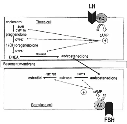
Steroid production is one of the most important functions for ovarian follicles. According to the biological activity and the numbers of carbon atoms, steroid hormones can be classified into progestins, androgens and estrogens, comprising 21, 19 and 17 carbons respectively, also designated as C21, Ci9 and C17 steroids. They comprise a ring compex, forrned of three cyclohexane rings (A, B, C) and a cyclopentane ring (D) (Gore Langton & Armstrong, 1994). Steroidogenesis invoives a long and complex biosynthetic pathway. The biosynthesis of steroids is mediated by steroidogenic enzymes, with each enzyme responsible for the conversion of one steroid to another. The major enzymes include three enzyme cytochrome P450s, inciuding P450 cholesterol side-chain cleavage (P450scc or CYP1 lAi), P450 17Πhydroxylase (P450170H or CYP17) and P450 aromatase (P45Oarom or CYP19A1), and two hydroxysteroid dehydrogenase (HSD) enzymes e.g.
3f3-HSD (or 3f3-HSD3B2) and 17Ç3-3f3-HSD (or 3f3-HSD17B1). Most of these enzymes have essential co-enzyrnes as electron donors or acceptors (Gore-Langton & Armstrong, 1994).The precursor of ail steroids is cholesterol. Cholesterol is imported into the ccii through internalization of blood-bome lipoproteins. The predominant foi-ni used for steroidogenesis appears to be tow-density-lipoprotein (LDL), which binds to the LDL receptor on foliicle ceils. Within the cell, cholesterol is maintained within lipid droplets as cholesterol esters. The enzyme choiesterol ester hydrolase converts the cholesterol esters to free cholesterol, which is intensely hydrophobic (Murphy & Silavin, 1989). free cholesterol within the cytoplasm is mobilized to the mitochondria, and then intemalized. This internalization of cholesterol by the mitochondria is the rate-lirniting step for the general steroidogenic pathway, and is mediated by steroidogenic acute regulatory protein
10 (StAR) (Stocco & Clark, 1996). The major ligand-regulated step in luteal and theca celis is StAR. This protein is acutely regulated by LH in these ccli types. The application of LH causes a rapid and transient production of StAR rnRNA and protein. Once the protein is forrned in the cytosol, it is rapidly directed to the mitochondria where it binds to a recognition site on the outer mitochrondial membrane. Whiie StAR is bound to the mitochondria, there is a transfer of cholesterol from the cytosol to the inside of the mitochondria (Stocco et aÏ., 2001). Once inside the mitochondria, cholesterol is converted
to pregnenolone by the enzyme cytochrome P45Oscc.
Pregnenolone is the first steroid in the pathway and is the common precursor for ail species and ail tissues, and from this point the converted cholesterol is committed to becoming a steroid. Pregnenolone in the microsornes can then be rnetaboiized using two different pathways, either converting to progesterone by the enzyme 33-HSD (A4 pathway), or to 17Œ-hydroxypregnenolone by P45O17.oH
(g
pathway) (Gore-Langton & Armstrong, 1994). It bas been suggested there are differences between species in the utilization of steroidogenic pathways, and that theg
pathway is the preferred pathway in ruminants (Zuber et al., 1986). In cattie, as in other ruminants, the five separate enzymes mentionedabove are required for the production of estradiol.
In bovine luteal and granulosa celis, the enzyme P450170H is flot expressed, and so steroidogenesis goes through to progesterone; this progesterone is not metaboiized further, and is secreted. In theca ceils, however, there is abundant P45017-OH activity, and so
pregnenolone is converted to 17a hydroxypregnenolone. This 17a-hydroxypregnenoione then undergoes sequentiai conversion to androstenedione by P450170H and 3-HSD activities (Bosc & Nicolle, 1998). Bovine theca celis convert iimited amounts of
androstenedione to testosterone with the enzyme 1 7f3-HSD and both androstenedione and testosterone are secreted. A good portion of these secreted androgens are absorbed by the neighbouring granulosa celis and are further converted to estrogens. Bovine granulosa ceils prefer to metabolize androstenedione to estrone by the enzyme cytochrome P45Oarorn, and then the estrone is rnetabolized to estradiol by 17-HSD (Conley & Bird, 1997). Alternatively, testosterone can be metabolized to estradiol by P45Oarom (Conley & Bird, 1997). The major steroidogenic pathways inespective of species and tissues are illustrated in the Figure 2.
The expression of the steroidogenic enzymes is regulated. The enzymes expressed in luteal and theca celis are in general regulated by LH, as these celis possess LH receptors. Thus it is fairly well recognized (mainly in rodent but in sorne ruminant models) that LH stimulates expression!activity of P45Oscc, P450170H and 173-HSD. LH also acutely
upregulates StAR gene expression and LDL receptor mRNA levels (Sekar et al., 2000). In granulosa celis of smaller follicles, only the FSH receptor is expressed, although both gonadotropin receptors are expressed in follicles >$rnm diameter in cattie. In granulosa celis, FSH stirnulates LDL receptor levels, P45Oscc and P45Oarom activity (Soumano & Price, 1997). In addition, FSH upregulates aromatase (Silva & Price, 2002) and 17f3-HSD (Sahrni et al., 2004). As consequences, LH stirnulates progesterone secretion from luteal ceils and androgen secretion from theca celis, whereas FSH stimulates progesterone and estradiol secretion from granulosa celis (Mihrn & Bleach, 2003).
The gonadotropins are not the only regulators of steroidogenesis, as a number of growth factors also alter steroid production (Armstrong & Webb, 1997). Insulin and/or IGF-1 stirnulate progesterone and estradiol secretion from bovine granulosa ceils in vitro,
12
LI-I
cho€s[eroI Th&ca oeil ‘‘-_.2
J
I SiA I CYP1IA pregnn.oIona -•.-. cAMP
j
CYP1 17OH-pregernoIorij
DHA indrostnedoneI
HSDI7B1 CP1estradiol ‘ estton androtnedione
FSH
Fig.
2. Follicular phase steroid biosynthesis in the ovary with
the illustration of the two-cell/two-gonadotropin theory.
and increase expression of P45Oarom mRNA in bovine granulosa ceils (Gutiérrez et al., 1997; Silva & Price, 2002), and stimulate progesterone and androstenedione secretion from theca celis (Allegrucci et al., 2003). Components of the insulinllGF system also act as
modulators of follicular ceil responses to gonadotropins. Another major group of growth factors is the transforming growth factor-t3 (TGF-) family. TGF-E3 enhances gonadotropin stirnulated steroidogenesis (Ke et al., 2004). Two other growth factors have the opposite effect on steroidogenesis: the epidermal growth factor (EGF) and fibroblast growth factor 2 (FGF-2) inhibit steroidogenesis (Armstrong & Webb, 1997).
Biochemical changes in follicular development
Steroidogenic activity changes during follicle developrnent. Estradiol concentration is a key biochemical marker for the degree of healthlatresia of follicles. Data from the older literature shows that morphologically healthy ruminant follicles contain higlier estradiol and lower progesterone concentrations than atretic follicles (Ireland & Roche, 1982; Sunderland et al., 1994; Price et aÏ., 1995). It is now known that small follicles contain relatively littie estradiol, and that follicular fluid estradiol concentrations increase with follicle size in healthy growing follicles. Estradiol concentrations decrease in subordinate follicles while the dominant follicle is growing. Once the dominant folticle reaches maximum diameter, follicular fluid estradiol concentrations fali dramatically, and decrease further once the follicle starts regressing (Price et al., 1995; Mihm et al., 2000).
The secretion of one steroid can be affected by a number of steps in the steroidogenic cascade, which limit or increase precursor supply. To determine which point in the pathway is responsible for increased or decreased estradiol secretion by follicles, a
14 number of studies have examined steroidogenic enzyme mRNA levels in bovine follicles at different stages ofdevelopment (Bao & Garverick, 1998).
Preantral foliicles express FSH receptor mRNA, but other aspects of the steroidogenic rnachinery do flot appear until early antral stage. In early antral follicles, the theca celis start to express mRNA coding for LH receptors, P45Oscc, P45O170H and
33-HSD, thus these celis are able to make progesterone and androgens in rats (Zlotkin et al., 1986). Granulosa celis continue to express only fSH receptors, and are thus steroidogenically inactive. As small antral follicles are recruited into a follicle wave, granulosa celis express P45Oscc and P45Oarom, and are thus able to synthesize pregnenolone and to convert androstenedione to estrone. They cannot in principle convert pregnenolone to progesterone as they lack 3f3-HSD at this stage. The theca ceils of these srnall recruited follicles continue to express mRNA for ail thecal steroidogenic enzymes, and also start to express StAR. These celis are thus fully and actively steroidogenic (Bao et
al., 1998).
As a growing follicle becomes a dominant follicie, a key change occurs in granulosa ceils. They start to express mRNA for LH receptors and 3f3-HSD. The ceils are thus able to convert pregnenolone to progesterone, and are able to respond to LH, considered to be essential for dominant follicle maturation. As the dominant follicÏe grows, there are also increases in rnRNA for P450 arom in granulosa celis and for StAR in theca celis (Bao et al., 1998). The subordinate follicles regress, and this is associated with decreases in ail steroidogenic enzymes in granulosa celis. If the dominant follicie also undergoes atresia, the first changes in steroidogenesis occur as the follicle reaches the ‘static’ phase of its growth phase. There is a reduction in mRNA for P450scc, P45017.OH
and LH receptor in theca celis, and P45Oscc in granulosa ceils. These follicles secrete significantly less estradiol than growing follicles, although there are no changes in P45Oarom mRNA (Bao & Garverick, 1998). The decrease in estradiol secretion is most likely due to the decrease in theca P45O170H and LH receptor levels, thus reducing
androgen precursor supply to GC. There is no ftirther loss of mRNAs encoding steroidogenic enzymes in the theca as the dominant follicle starts to regress, but GC suffer a loss ofP45Oscc, P45Oarom, LH receptor and 3f3-HSD mRNA (Bao & Gaiwerick, 1998).
The growth of bovine follicles from primordial to preovulatory stage is characterized by an approximately 360,000-fold increase in surface area, and several hundred-fold increase in follicle size (Lussier et al., 1987). Ovarian follicular growth and development involves extensive tissue rernodeling (Smith et al., 1999). Overali, normal ovarian function depends on cyclical rernodeling ofthe ECM. The next section will discuss the composition, the changes and the roles ofECM in the mammalian ovary.
EXTRACELLULAR MATRIX IS DYNAMIC N FOLLICULAR DEVELOPMENT
During follicle growth there is extensive cellular proliferation and remodeling of the ECM. This process is characterized by proliferation of GC, differentiation of the granulosa and theca cornpartments, and the deposition of a basement membrane separating the avascular granulosa ceils from the vascularized theca layer (Ny et al., 2002).
The extracellular matrix has many different roles (Rodgers et al., 2000; Rodgers et al., 2003; Rodgers et al., 1999). First, the ECM affects celi shape and behavior, such as migration, division, differentiation, cell death and cell anchorage. All these behaviors occur
16 in follicle development. Second, the ECM can play a role in the fluid dynamics ofa tissue, either providing osmotic forces or filtering soluble materials including nutrients and hormones. Third, the ECM can provide rigid or elastic mechanical support for tissues. Fourth, follicular growth factors can bind to the ECM directly or indirectly. For instance, FGF-2 can directly bind to the ECM, and IGF-I or activin can indirectly bind to the matrix through their binding proteins IGFBP-2 and -5, or follistatin, respectively. Collectively, the ECM defines or provides a specialized microenvironment for ceils and tissues.
Col1aen and laminin are structural proteins inECM components
The ECM provides a structural tissue support, and forms barriers between tissue cornpartrnents. The matrix is known to contain three major fibre forming proteins— collagen, elastin, and fibronectin, which are interwoven in a hydrated gel formed by a network of glycosaminoglycans (GAG) dornains. All of these macromolecules are locally secreted by the celis in contact with the matrix (Alberts, 1983). The collagens are ropelike, triple-stranded, helical molecules that aggregate in long cable-like fibrils or sheets in the extracellular space. Elastin molecules form an extensive cross-linked network of fibres and sheets that can stretch and recoil, imparting elasticity to the matrix. fibronectin molecules fonri fibres that prornote ceil adhesion, and the GAGs are a heterogeneous group of long, negative charged polysaccharide chains (except for hyaluronic acid) covalently linked to protein to fomi proteoglycan (Alberts, 1983). Follicular growth requires the ECM to be rernodeled to incorporate the increasing volume of tissue and follicular fluid. hi ovarian
include the follicular basal lamina, the membrana granulosa, and theca interna and extema (Rodgers et aÏ., 2000).
11e follicular basal lamina is forrned of specialized sheets of ECM, which separate the epithelial cells (membrana granulosa) from adjoining stroma (including theca interna and externa, and vasculature). The basal lamina is composed of a lattice-type network of collagen type IV interwound with a network of laminin. This structure is stabilized by the binding of entactin or nidogen to the collagen and laminin. The heparin sulphate proteoglycan perlecan (HSPG) and other molecules such as fibronectin are associated with the collagen type W-laminin backbone (Rodgers et al., 2003). Furthermore, each molecule of collagen type IV comprises three u chains; there are six different chains (cil- u6) of collagen type IV (Rodgers et aï., 199$). Sirnilarly, each laminin molecule is composed of three chains, the u, (3, & -y chain, there are five different u chains, three (3 chains and one y chain, thus, many potential different combinations of collagen type IV and larninin are possible, and many of these combinations have been observed in nature (van Wezel et al., 199$). The ECM undergoes cyclic changes in its composition (Greenwald, 1994); (Moirniaux et al., 1997). Interestingly, the composition of follicular basal lamina changes during follicle development. Cyclic expression pattems of the mRNA encoding type III, IV, and VI collagens as well as the proteoglycans have been observed in mouse ovary, suggesting that the ovarian ECM changes during follicular growth (Oksjoki et al., 1999). For instance, in cattie collagen type IV u3, u4, u5 and u6 levels are lower in antral follicles cornpared to primordial and preantral follicles, and larninin cii, (32 and -y1 are higher in antral follicles than in primordial and preantral follicles (Rodgers et al., 2003). Thus, during follicle development, the follicular basal lamina becornes less collagenous and more
1$ laminin rich, such that it becomes more expandable to meet the requirement for follicle enlargernent. In addition, perlecan and nidogen are absent in primordial follicles, but become components of the follicular basal lamina in antral follicles, and atretic but not healthy antral follicle express laminin a2 (van Wezel et al., 1998). In the sheep, laminin 1
(Œ1f31-yl structure) and different collagen I chains have been irnmunolocalized in the basal lamina, and the levels of type I collagen increase in granulosa layers during terminal follicular growth (Huet et al., 1997). Ail these studies suggest that the follicular basal
lamina is extremely dynamic during follicular development (Rodgers et al., 2000).
ECM components in theca layers (named the theca matrix) slightly differ from those in the follicular basal lamina. Even though laminin 1 components (al or f32 or y1) (van Wezelet al., 1998) and collagen W ai, a2 (Rodgers et al., 1998) are present in bovine follicles throughout the theca interna, other structural proteins such as collagen type 1(2 al and 1 a2) (Luck et al., 1995; Zhao & Luck, 1995) and collagen type VI (Iwahashi et al., 2000) have been identified in the theca interna.
Other ECM components
Apart from collagen and laminin described above, there are a number of other extraceltular matrix proteins in ovarian follicles such as gelatin, elastin, fibronectin, integrins, vitronectin, and proteoglycans including versican, hyaluronan.
Gelatin contains a large number of glycine, proline and 4-hydroxyproline residues. Gelatin is a heterogeneous mixture of single or multi-stranded polypeptides, each with extended left-handed proline helix conformations and containing between 300 - 4000
two types of gelatin, acid pretreatment (type A gelatin) and aikaline treatment (type B gelatin) dependent on whether or flot the preparation involves an aikaline pretreatment.
Elastin molecules forrn an extensive cross-linked network of fibres and sheets that can stretch and recoil, imparting elasticity to the matrix (Alberts, 1983). Elastin and collagen forrns a cross-link structure in the ECM. Lysyl oxidase (LOX) initiates cross-link formation of the collagen and elastin, and therefore has a crucial foie fl the regulation the formation and maintenance of the ECM in the ovary (Henmi et al., 2001; Harlow et al., 2003).
Fibronectin is a common ECM compound in stroma and it is important for ccli migration, which occur in theca expansion during follicular development (Rodgers et al., 2003). Due to alternative spiicing of mRNA at three separate sites, there are at least 20 different isofornis of fibronectin in humans (De Candia & Rodgers, 1999). Fibronectin exists as a homo- or heterodirner ofthese spiice variants, a number ofthe spiice variants are expressed in bovine foilicles in vivo (De Candia & Rodgers, 1999). Fibronectin is mitogenic in granulosa celis in vitro (Colman-Lerner et al., 1999), and fibronectin synthesis by granulosa celis can be upregulated by FGF-2 (Rodgers et al., 1996). further experiments are needed to assess the physiological importance of the different fibronectin isoforms, as well as the respective roles of the soluble forms present in follicular fluid and the insoluble fonTis deposited in basal lamina and on celi membranes.
Integrins are ECM receptors on the celi surface. Ceils interact with the matrix through celi-surface adhesion receptors including the integrins. Integrins are heterodimeric glycoproteins composed ofΠand [3 subunits. Over 17 ci and 8 f3subunits can make over 23 different heterodimeric combinations (Belkin & Stepp, 2000). Only a few integrins have
20 been localized to granulosa ceils: Œ6131 in non-luteinized granulosa ceils (Fujiwara et al., 1997; Le Beliego et al., 2002) and u2 and Œ5 in luteinizing celis (Yamada et al., 1999). Moreover, the a6f3lintegrin serves as a larninin receptor, the a5f3lintegrin serves as a fibronectin receptor, and the Œ2f3 1 integrin serves as a collagen type I receptor in various species such as mouse (Fujiwara et al., 199$), human (Nardo et al., 2003; Yamada et al., 1999), and sheep (Le Bellego et al., 2002).
Versican (also named chondroitin sulfate proteoglycan-2, CSPG2) is a soluble ECM molecule. Granulosa ceils in antral follicles are bathed in follicular fluid containing proteoglycans. Proteoglycans consist of a core protein with attached GAGs. Belonging to the proteoglycan family, versican is Hkely to be synthesizedin the granulosa cells (and also theca). Versican was identified in human follicular fluid (Eriksen et al., 1999) and in ail follicular layers in small bovine follicies (McArthur et aÏ., 2000), and may participate in celi-matrix and celi-celi interactions. Versican plays a key role in cumulus oocyte expansion and fertility (Russeli et al., 2003), together with tumour necrosis factor u stimulated gene 6 (TSG-6) (Mukhopadhyay et al., 2001; Ochsner et al., 2003), inter-u trypsin inhibitor (Carrette et al., 2001; Ochsner et al., 2003), and hyaluronan (Mahoney et al., 2001). There are four isofoms ofversican (VO, Vi, V2, and V3), ofwhich VO and VI expression is localized to granulosa ceils (Russeli et al., 2003). Versican VO and Vi rnRNA are differentially expressed in GC of actively growing dominant follicles compared to small follicies (2-4mm) in cattie (Fayad et al., 2004b). In addition, a disintegrin and metalloproteases with thrombospondin motifs (ADAMTS- 1) can proteolytically cleave versican (Sandy, 2001).
Hyaluronan (HA), a glycosaminoglycan polymer, is synthesized by cumulus celis surrounding oocytes before ovulation. Along with other factors, hyaluronan makes a gelatinous matrix, and plays a role in cumulus expansion (Salustri et aÏ., 1999; Mahoney et al., 2001).
Interaction of ECM proteins and granulosa celi function
In terms of their origins, most components of the follicular basal lamina are probably synthesized by GC. Both fibronectin and lamininyl chain are expressed by GC in rats (Camegie, 1990) and cows (Zhao & Luck, 1995). Luteinized granulosa cells in culture express ECM proteins (collagen I and collagen IV) and their regulators, matrix metalloproteinase 9 (MMP-9) and tissue inhibitors of metalloproteinases (TIMP-1) (Zhao & Luck, 1996). Bovine GC in culture can also synthesize a basal lamina containing collagen IV and fibronectin (Rodgers et al., 1995; Rodgers et al., 1996), providing a possible model to study the origin of ECM proteins and as well as the interaction of ECM proteins and GC function. furthermore, TGFa stimulates the production and deposition of fibronectin by chicken GC (Asem & Novero, 1994).
In vitro studies show that the ECM modulates GC function in various mammalian species. For example, ECM stirnulates bovine GC proliferation and progesterone secretion in response to FSH (Savion et aÏ., 1981). Similarly, rat and human luteinized GC require ECM in order to retain their structural and functional characteristics in culture (Amsterdam et al., 1998). To test the role of ECM proteins in GC survival, proliferation and steroidogenesis, Huet and colleagues carry out an experirnent in which various pure ECM components (type I collagen, fibronectin and laminin) are added to ovine GC in vitro. They
22 observe that coflagen I is able to maintain estradiol secretion in GC derived from large follicles (4-7 mm in diameter), whereas fibronectin and larninin dramatically increase the
proliferation rate and enhance survival ofGC from both small (1-3 mm) and large follicles
(Huet et al., 2001). In addition, the authors also report that heparin treatment changes ccli morphology (induces celi rounding), reduces celi proliferation, enhances estradiol but
inhibits progesterone secretion (Huet et al., 2001). However, it rernains to be determined
whether changes in GC function resulting from heparin treatment are directed by the
change in celi shape, or invoïve other rnechanisms. One possible explanation is that the
addition of excess heparin to the cultured celis likely disturbs the action of endogenous
heparin-binding growth factors such as FGf-2, by regulating its bioavaiiability (Ruosiahti
& Yarnaguchi, 1991). Immunostaining studies show that laminin and fibronectin are
mainly localized to vascular walls, the outer layer of GC, and the basement membrane of
the rat ovary (Akkoyuniu et al., 2003). Taken together, these data indicate ECM proteins,
together with growth factors, are invoived in follicle deveiopment during the estrous
(menstntal) cycle. In conclusion, the ECM influences basic cellular process such as proliferation, differentiation, migration and adhesion, are invoived in the control of ovarian follicular deveiopment, and modulate interactions between growing foilicies and
surrounding connective tissue.
PROTEASES AND THER IMUBITORS REGULATE ECM REMODELING
Proteases and their inhibitors are regulators of ECM remodeling during follicular development. Based on their evolutionary structure, proteases and their inhibitors can be
classified into three grotips (three proteolytic systems), the plasminogen activator (PA) system (Liu, 1999; Liu et al., 2004), the matrix metalloproteinase (MMP) system (Ny et al., 2002; Curry & Osteen, 2003) and the cysteine protease system (Sriraman & Richards, 2004). This review will focus on the plasminogen activator system (see the PA section in detail), and describe briefty the MMP system and cysteine protease system.
The MMP system
Currently, the MMP family encompasses at least 25 reÏated proteolytic enzymes that include four broad classes: collagenases, gelatinases, strornelysins, and membrane type enzymes (MT-MMPs) (reviewed by Curry & Osteen, 2003). Common features ofthe MMP family include: 1) the presence of zinc in the active site of the catalytic dornain; 2) synthesis of the MMPs as preproenzymes that are secreted in an inactive form; 3) activation of the latent zyrnogen in the extracellular space; 4) recognition and cleavage of the ECM by the catalytic domain of the enzyme; and 5) inhibition of enzyme action by metalloproteinase inhibitors in the extracellular environment (Curry & Osteen, 2003). Although simiÏarities exist in the structure of the MMPs, there are also distinct differences in the recognition and specificity for the ECM components (Nagase & Woessner, 1999); (Murphy et al., 1999b). for instance, collagenases (MMP-l, MMP-8, MMP-13) cleave fibrillar collagens sucli as collagen types I, II, III, V, and XI, as well as nonfibrillar collagens. Cleavage of the triple helical collagen by collagenases resuits in denaturation of collagen molecules into gelatin by changing the stability and solubility of collagen. The gelatinases (MMP-2 and MMP-9) contain a fibronectin-like sequence within their catalytic domain, which results in a potent ability for these MMPs to bind and cleave gelatin. The
24 stromelysins (MMP-3, MMP-7, MMP-10 and MMP-11) act on a diverse array of ECM substrates, including collagen type IV, laminin, and fibronectin. The MT-MMPs (Mil or MMP-14, Mi-2 or MMP-15, MT-3 or MMP-16, and MT-4 or MMP-17) contain a transmembrane domain near their C-terminal region, and their extracellular region contains the catalytic domain (Curry & Osteen, 2003). One important role of the MT-MMPs is activation of MMP-2 (Strongin et al., 1995). In addition to degrading ECM, MMPs and especially strornelysins exhibit activity toward other MMPs, growth factors, and cytokines such as IGF binding proteins, epidermal growth factor (EGF), TNF-a, and substance P (Sternlicht & Werb, 2001), and subsequently modulate celi growth. The regulatory action on cell growth occurs either directly by controlling ceil-matrix interactions or indirectly by controlling growth factor bioavailability.
Apart from the MMPs discussed above, there are a number of family members that are classified outside of the four broad classes of MMPs. ADAMTS-1 is a member of the ADAMTS family of metalloproteinases (termed the adamalysins, a different metalloproteinase farnily) that degrades members of the lectican family of proteoglycans (Kuno & Matsushima, 199$). In the ovary, ADAMTS-1 is involved in ovulation (Robkeret aï., 2000a).
MMP activity in the extracellular environment is rigorously controlled by MMP inhibitors. Two major classes of MMP inhibitors are generally distinguished, serum-bome and tissue-derived inhibitors (reviewed by Gomez etal., 1997; Brew et al., 2000). Alpha 2-macroglobulin (a2-M), a 720 kDa tetrameric glycoprotein, belongs to serum-bome inhibitor of metalloproteinases, and ct2-M is present in hurnan follicular fluid whereas a2-M rnRNA is virtually undetectable in GC (Curry et al., 1990). The tissue inhibitors of
metalloproteinase (TIMP) are locally produced and specifically inhibit MMPs, and are highly expressed and hormonally regulated in reproductive tissues. The abiÏity of TIMPs to inhibit MMP action occurs through the interaction of the N-terminal dornain of TIMP with the active site on the catalytic domain ami the substrate-binding groove of the MMP. TIMPs act selectively on different MMPs (Gomez et al., 1997). Currently, there are four TllvIP members. TIMP-1 is a secreted glycoprotein, which preferentially binds to MMP-9 but caimot act on MT1-MMP (MMP-14). TIMP-2 is also a secreted glycoprotein, and has a high affinity for MMP-2 (Gomez et al., 1997). Unlike TIMP-1 or TIMP-2, TEvfP-3 is secreted and then bound to the ECM, and has been suggested to act as an additional regulatory stop point as opposed to being free in the extracellular fluid. TEVIP-3 also exhibits a differentiai preference for the MMPs, having a high affinity for MMP-9 ami being able to inhibit MT1-MMP (Leco et al., 1994). TIIvIP-4 is cloned and expressed in reproductive tissues (Leco et al., 1997). TIMP-4 also acts on nurnerous MMPs (Stratmann et al., 2001), suggesting that this TIMP may be a good non-specific inhibitor for ah classes ofMMPs.
The regulation of MMP and TIMP synthesis and activation in ovarian follicular development and ovulation are reviewed ($mith et al., 1999; Curry & Osteen, 2003). Most studies in different species show that MMP and TIMP mRNA leveis or protein activities are increased during ovulation (Srnith et al., 1999). For example, in the rat, mRNA levels ofMMP-1, MMP-2 (Reich et al., 1991), MMP-13 (Balbin et al., 1996) and MT1-MMP (Jo et aï., 2002b) ail increase after the endogenous LH surge or hCG administration, similar to previous reports for enzyme activity (Smith et al., 1999). The gene expression ofTLMP-1 and TIMP-3 also increases (Reich et al., 1991), whereas TIMP-4 expression decreases in
26 rat ovarian follicles during ovulation (Sirnpson et aÏ., 2003). Although there are many similarities in expression patterns of MMPs between the rat and mouse, there are differences in MMP-2 and MMP-9 expression. In the rat, MMP-9 mRNA does flot change, whereas MMP-2 mRNA increase during ovulation (Curry et al., 2000). In contrast to the rat, MMP-9 mRNA increases after LH stimulus in the mouse, whereas MMP-2 is unchanged (Robker et al., 2000b). After investigation of eleven different MMPs during the periovulatory period in mice ovaries (Higglund et al., 1999), only MMP-19 mRNA increases. The other MMP members are either unchanged or undetectable. TIMP-1 and TIMP-3 mRNA expression increases in the mouse, whereas TIMP-2 is unchanged as in the rat (Higg1und et al., 1999; Inderdeo et al., 1996). The parallel up-regulation ofMMPs and their inhibitors is postulated to maintain proteolytic homeostasis (Nagase & Woessner, 1999), and therefore act to regulate the location and extent of ECM remodeling of the follicular apex during ovulation.
There are potentially important species differences in rnRNA expression pattems of the MMP system during ovulation. hi the rat, MT1-MMP mRNA levels increase (Jo et al., 2002a), but TIMP-2 mRNA levels do not change (Curry & Nothnick, 2000). hi cattle, the expression of mRNA for MTY-MMP and TEVIP-2 increases at 24 h and 6 h after the preovulatory LH surge, respectively (Bakke et al., 2002). However, MMP-2 mRNA in follicular tissue and enzyme activity in follicular fluid is unchanged. The MT1-MMP mRNA is localized prirnarily to the theca layer before the gonadotropin surge but is expressed in GC at 12 h and 24 h after the surge (Bakke et al., 2002), suggesting that this enzyme is required more for follicle wall degradation in bovine. In sheep, MMP-2 appears a pivotai MMP member in ovulation, as the injection of MMP-2 antibody into the antral
cavity of preovulatory follicles in the ewe resuit in the formation of luteinized, unruptured follicles (Gottsch et aÏ., 2002).
There is considerable evidence that the MMPs and TIIVIPs function at earlier stages of follicular growth. In the neonatal rat ovary, MMP-2 is immunostained in GC and the surface epithelium, whereas MMP-9 is absent at earlier stages of follicular development (Bagavandoss, 199$). In the same study, follicular growth induced by PMSG increases the cellular expression of MMP-2 and MMP-9, which is consistent with protein expression pattems (Bagavandoss, 1998). The cellular activity of the MMPs rneasured by in-situ zymography dernonstrate a pattern of gelatinolytic activity that corresponds with the oca1ization ofMMP-2 and MMP-9 mRNA around developing foïlicles (Curry et aÏ., 2001). MMP-2 and MMP-9 activity also increases during follicular growth in sheep, pigs, cattie, horses and hurnans (reviewed by Curry & Osteen, 2003).
The ernerging data for TIMP mRNA expression pattems during follicular growth suggest that the expression ofTEVIP-I parallels changes in MMPs. Curry and Osteen (2003) summarized that TIMP-1 mRNA levels increase after PMSG administration, but TIMP-2 rnRNA does not change, and TTMP-3 rnRNA level even decline slightly in the rat (Curry & Osteen, 2003). However, the mRNA and protein for TIMP-4 increase after PMSG treatment (Simpson et al., 2003). Thus, the parallel regulation of MMPs and TIIVIP-1 and TIIvIP-4, as well as the basal expression of TLMP-2, and TIIVIP-3, may act to maintain a proteolytic balance during follicular growth that provides localized control of ECM degradation, thereby regulating the location and extent of follicular remodeling (Curry & Osteen, 2003).
2$ In surnrnary, the MMPs would facilitate remodeling of the granulosa ceil basement membrane and the theca ECM and allow follicular expansion. The THVIPs may provide control for the location and extent of MMP action. It is readily clear that the MMPs and TIMPs are in the appropriate cellular compartments, and are regulated by the hormonal signais that regulate follicular developrnent and atresia.
The cysteine proteases
There are lirnited studies on cysteine proteases cornpared to other major protease farnilies. Cysteine proteases are lysosornal enzymes that act at acid pH, whereas most serine proteases and the metalloproteinases act at neutral pH. The best-known cysteine proteases are cathepsin B and cathepsin L (Salamonsen, 1999). Cathepsin L is a lysosomal cysteine protease expressed in many endocrine tissues and celi types including granulosa celis (Oksjoki et al., 2001; Robker et al., 2000a). Cathepsin L is a secreted protein indicating that this protease functions at both intracellular and extracellular sites (Ishidoh & Kominami, 199$). In the ovary, cathepsin L is expressed in GC of follicles at different stages of growth suggesting that this protease may play diverse roles in this tissue. Along with other proteases, cathepsin L may impact the extensive rernodeling of the ECM during ovulation (Robker et al., 2000a). Specifically, since cathepsin L is activated when cornpïexed with glycosaminoglycans (GAG) present in follicular fluid, and since it can degrade coilagen (I and W), elastin and fibronectin, cathepsin L is likely a modifier of ECM in preovulatory follicles (Salustri et al., 1999; Robker et aï., 2000a). Although no endogenous cysteine protease inhibitor has been identified, TIMP- I forms a complex with procathepsin L in steroidogenic ceils, including Leydig cefls and ovarian GC, indicating