Development of an animal experimental model to study the effects of
levonorgestrel on the human endometrium
M.-L. Alvarez Gonzalez1, C. Galant3, F. Frankenne1, M. Nisolle1,2, S. Labied1, J.-M. Foidart1,2, E. Marbaix3,†, and A. Béliard1,2,4†
Laboratory of Tumor and Development Biology, GIGA-Research, B23, University de Liège, Sart-Tilman, B-4000 Liège, Belgium
2Department of Gynecology, CHU, University of Liège, B-4000 Liège, Belgium 3Cell Biology Unit and Department of Pathology, Université
Catholique de Louvain, B-1200 Bruxelles, Belgium.†Equal senior authors.
Background: This study was designed to develop an animal model to test the response of endometrium to local
progestin delivery.
Methods: Proliferative human endometrium was subcutaneously grafted in two groups of SCID mice that
received, 2 days before, a subcutaneous estradiol (E2) pellet and, for half of them, an additional implant of levonorgestrel (LNG). Mice were sacrificed 1, 2, 3 or 4 weeks after endometrial implantation and grafts were histologically analysed. Proliferation, steroid hormone receptors, blood vessels and stromal decidualization in both groups (E2 and LNG) were immunohistologically evaluated and compared with proliferative endometrium and endometrium from women with an LNG intrauterine device.
Results: Grafts presented normal morphological endometrial characteristics. The expression of progesterone
receptors was significantly decreased in glands and stroma of the LNG group as compared with the E2 group at all times. A significant decrease was also observed in the stromal expression of estrogen receptor-α in the LNG group. At 4 weeks, the mean cross-sectional area of vessels was significantly higher after LNG treatment.
Conclusions: These morphological and immunohistochemical characteristics are similar to those observed in
women treated with local LNG. This mouse model might facilitate further investigations needed to understand the mechanisms responsible for the breakthrough bleeding frequently observed in progestin users.
Key words: levonorgestrel ⁄ endometrium ⁄ mouse model ⁄ endometrial transplants
INTRODUCTION
The levonorgestrel-releasing intrauterine system (LNG-IUS) is considered as an efficient contraceptive.
However, it is commonly associated with vaginal bleeding and spotting (breakthrough bleeding, BTB) in the first few months of use. Irregular bleeding is the most common reason for discontinuation of progestin-only
contraception (Findlay, 1996; Hickey et al., 1999). LNG delivered locally by the LNG-IUS is known to alter the morphology and function of endometrium (Critchley et al., 1998a). Morphological changes include
pseudodecidualization in the stromal compartment associated with leukocytes infiltration, atrophy of glandular and surface epithelium, and alterations in the vasculature (Jones and Critchley, 2000). These latter morphological modifications include dilatation and changes in shape, basement membrane components and pericyte support (Hickey et al., 2000; Hickey and Fraser, 2003). Furthermore, an increase in blood vessel number, size and density associated with a decrease in the pericyte coating have been shown after short-term exposure to LNG-IUS (Stephanie et al., 2007)) and hypothesized to contribute to vascular fragility and BTB (Hickey et al., 2000; Rogers et al., 2000; Jondet et al., 2005). Functional alterations include down-regulation of oestrogen and progesterone receptors as well as cell proliferation (Critchley et al., 1998b; Salmi et al., 1998). This alteration in sex steroid receptor expression may affect endometrial cytokine release (Jones and Critchley, 2000). The paracrine mechanisms implicated in bleeding after intrauterine LNG delivery remains to be elucidated. Endometrium obtained from women using LNG-IUS provides an opportunity to study the effect of a high dose of local progestin delivery upon endometrial development. Nevertheless, because of obvious practical and ethical limitations in humans, the animal models provide an invaluable tool to study in vivo early events through the examination of morphology of transplanted tissue. In the future, events associated with the pathogenic process
underlying BTB should be identified and the possible efficacy of various therapeutic modalities should be assessed. Uterine bleeding does not occur in animals except in monkeys. The high costs of monkey handling limit the use of this animal model for research. As rodents do not have uterine bleeding, transplantation of endometrial tissue is needed. For this purpose, severe combined immunodefi-cient mice (SCID) were used as they are characterized by a combined congenital deficiency in T and B lymphocyte function and therefore successfully host various heterotransplants (Phillips et al., 1989). Previous studies have described the success of human tissue grafts which retain their morphological and biochemical characteristics after transplantation in different mice strains (Shimosato et al.,1976; Kim et al., 1978; Satyaswaroop et al., 1983; Awwad et al., 1999). The aim of this study was to validate the suitability of SCID mice grafted with human endometrium as an experimental model to mimic the effects of LNG in women. The morphology of both groups of endometrial implants was compared with that of proliferative eutopic endometrium and eutopic endometrium treated with local delivery of LNG. Specific features of these implants such as cell proliferation, steroid hormone receptors and the vasculature were also characterized.
MATERIALS AND METHODS Collection of human endometrium
The use of human tissue for this study was approved by the Ethical Committee of the University of Liege. Proliferative endometrium (5-9 cycle days) was obtained from 6 reproductive aged women (aged 31-38 years) without endometriosis undergoing surgery for benign purposes (myoma, infertility). They had regular menstrual cycles and did not receive any hormonal treatment for at least 3 months before surgery.
The endometrial biopsy was taken with a Cornier Pipelle suction curette (C.C.D. International, Paris, France), placed in sterile phosphate-buffered saline (PBS) solution, pH 7.3, and immediately transported to the laboratory. Endometrial biopsies were cut into pieces of 1-2 mm3; some fragments were fixed in 4% buffered formaldehyde, dehydrated and embedded in paraffin. Sections were stained with haematoxylin-eosin for histological confirmation of the menstrual phase (Noyes et al., 1950).
Eight endometrial biopsies were also obtained from women (mean age 39 years) exposed to LNG-IUS (20 µg/day, Mirena®, Bayer Schering Pharma, Berlin, Germany) for a period of 1 month to compare their morphology with the grafts of the LNG group at 4 weeks.
Animals
Female SCID/SCID CB17 mice (n = 64) of 8-10 weeks were obtained from Charles River Laboratories
(Brussels, Belgium) and housed under a barrier husbandry in a well-controlled, pathogen-free environment with a 12 h light/12 h dark cycle. All housing materials, food and water were autoclaved before use. Mice were fed ad
libitum on laboratory chow and water. Experiments were approved by the Liege University Animal Ethics
Committee.
Transplantation into SCID mice and tissue processing
All surgical procedures were conducted under parenteral anaesthesia with an injection of a solution containing 10mg/kg of Xylalin® (Ceva Sante Animale, Libourne, France) and 80 mg/kg body weight of Ketamine 1000 Ceva® (Ceva Sante Animale) in a sterile laminar flow hood.
One group of 32 mice was implanted subcutaneously in the neck with a pellet of 17β-estradiol (E2; 1.72 mg/60 day release; Innovative Research of America) (= E2 group). A second group of 32 mice was implanted
simultaneously with a pellet of 17β-estradiol and an implant of LNG (20 µg/ day) (LNG group). After 2 days, 10 fragments of fresh human proliferative endometrium (1-2 mm3) were grafted subcutaneously in the dorsal region of each animal next to the implant of LNG. Depending on the size of the endometrial biopsy, we grafted
simultaneously at least four mice of the same hormonal treatment group, one at each time point Eight mice per group were sacrificed at 1, 2, 3 or 4 weeks after endometrium transplantation. To evaluate the endometrial lesions, the dorsal skin was largely opened in order to perform a resection of grafts adherent to the skin. All specimens were fixed in 4% buffered formalin, and embedded in paraffin. Sections were stained with
Immunohistochemistry
Cell proliferation was evaluated using a monoclonal mouse anti-Ki-67 antibody (MIB-1, M7240, DAKO, Glostrup, Denmark, dilution 1:100) and human steroid hormone receptors were evaluated using monoclonal mouse anti-human oestrogen receptor alpha (ERα, NCL-ER-6F11, Novocastra Laboratories Ltd, UK, dilution 1:50), progesterone A-form receptor (PRA,NCL-L-PRG-312, Novocastra Laboratories Ltd, dilution 1:100) and
progesterone B-form receptor (PRB,NCL-PRG-B,Novocastra Laboratories Ltd, dilution 1:100).
Stromal decidualization was confirmed by a monoclonal mouse anti-human insulin-like growth factor binding protein-1 (IGFBP-1, 100056, Medix Biochemica, Kauniainen, Finland, dilution 1:100).
Vascular smooth muscle cells were stained using a monoclonal mouse antibody against α- Smooth Muscle Actin (α-SMA, F3777, Sigma-Aldrich Chemie Gmbh, Munich, Germany, dilution 1:1000) and blood vessels were stained using an antibody against human and mouse collagen IV (SIF 100, guinea pig polyclonal antibody produced in our laboratory, dilution 1:750).
To unmask antigens on paraffin sections (5 µm thick), slides were incubated 40 min in 0.025% Trypsine (S2012, DAKO) for α-SMA and SIF 100, or autoclaved for 11 min at 126°C and 1.4 bar in citrate buffer for the others antibodies. Endogenous peroxidase was subsequently blocked by 3% H2O2/H2O for 20 min and non-specific binding was prevented by incubation in PBS/bovine serum albumin 10% (Fraction V, Acros Organics, NJ, USA) for 30 min (for Ki-67, α-SMA and SIF 100) or in normal goat serum (for PRA, PRB, ERα and IGFBP-1).
Sections were incubated for 90 min at room temperature with the primary antibody. Slides were then incubated 30 min at room temperature with an appropriate secondary antibody: rabbit anti guinea pig Ig-HRP (P0141, DAKO; dilution 1:50) or a biotinylated goat anti mouse Ig (E433, DAKO, dilution 1:400) followed by incubation with a streptavidin/HRP complex (P0397, DAKO, dilution 1:500) at room temperature for 30 min. Staining was done with 3-3'-diaminobenzidine hydrochloride (K3468, DAKO) for 3 min. Slides were finally counterstained with haematoxylin and mounted with Eukitt medium for microscope observation. Negative controls were performed both by omission of the primary antibody and by replacement of this primary antibody by an appropriate isotype control (mouse IgG1, X 0931, DAKO).
Scoring and analysis of immunoreactivity
The immunostaining intensity of receptors (PRA,PRB,ERα) and the distribution of specific staining were
assessed in a semi-quantitative manner by an HS-score (Huang et al., 1996; Vereide et al., 2006). The HS-score has been evaluated as follows: HS = ∑ (Pi x i)/100, where Pi denotes the percentage of stained cells and ⁄ denotes the intensity of the staining ranging from 0 to 3. A score of 0 indicated an absence of staining and a score of 1, 2 and 3 indicated weak, moderate and intense immunostaining, respectively. The HS-score evaluation was performed blinded by a trained biologist. Glandular and stromal endometrial cells were analysed separately. At least five representative fields (x40 objective on an Olympus Vanox AHBT3 microscope, Aartselaar, Belgium) within each section were selected for assessment.
The cell proliferation of glandular and stromal endometrial cells was evaluated on the complete field by giving a score according to the percentage of positive stained cells: 0 = no immunostaining, 1 = <25% immunostaining, 2 = from 25 to 75% immunostaining and 3 = >75% immunostaining.
Analysis of endometrial vasculature
For each graft, two successive sections were stained either for SIF 100 or for α-SMA to analyse the vascular morphological changes induced at 4 weeks by LNG compared with E2. All blood vessels were delineated manually at x400 magnification using Photoshop software (version 7.0.1) throughout the totality of each section. Images were therefore binarized and analysed using the NIH ImageJ analysis free software
(http://rsb.info.nih.gov/ij/) to determine the number of blood vessels per stromal surface unit as well as their mean area. All these parameters were calculated for each image individually. Then, the mean value of these parameters was determined for each mouse. Finally, the mean + SEM vessel number per unit of tissue area and the relative vascular area for all patients of the same group were determined.
Statistics
Values were presented as mean + SEM. Scoring results were analysed using GraphPad Prism (version 5.00) by an ANOVA test followed by the Bonferroni's multiple comparison post hoc test on E2 groups in function of time
and compared with proliferative eutopic endometrium (Prolif.). A two-way ANOVA test followed by a
Bonferroni post hoc test was used to compare the two different treatment mice groups (E2 and LNG) in function of time. Finally, a t-test was used to compare the proliferative eutopic endometrium (Prolif.) or the LNG group at 4 weeks to LNG-IUS samples. The vessel number per unit of stromal tissue area and the mean vascular area were compared by a Mann-Whitney U-test. Differences were considered significant when P ≤ 0.05.
RESULTS
From a total of 64 SCID mice, 52 survived the procedure (27 in the E2 group and 25 in the LNG group) and 12 died after anaesthesia. Endometrial transplants were recovered from all surviving mice.
Histological morphology
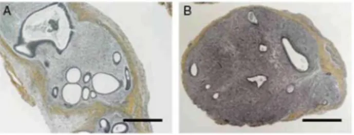
All endometrial biopsies used for graft displayed normal proliferative features as described by Noyes et al. (1950). The morphology of human endometrial transplants was well preserved. Necrosis was only observed in transplants removed after 1 week from E2 group. Transplants consisted of an endometrium-like tissue with typical glands and stroma surrounded by a thin capsule of connective tissue (Fig. 1). In the E2 group, we observed after 1 week, small and round glands with pseudostratification of the nuclei surrounded by a dense stroma. At 2 weeks, the glands became dilated with a persistence of the pseudostratified structure of the
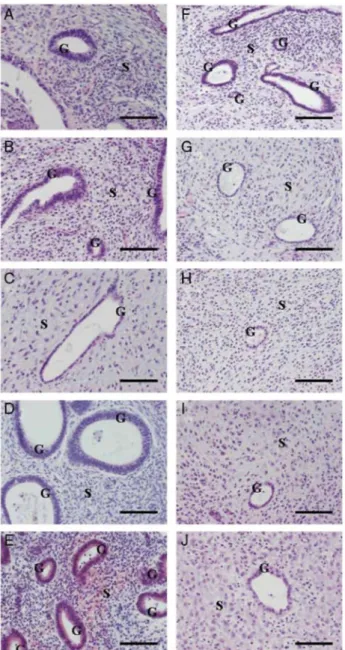
glandular epithelium and a less dense stroma. After 3 weeks, pseudostratification was no longer observed and the epithelial cells were cuboidal. After 4 weeks (Fig. 2A-D), we observed dilated glands with pseudostratified nuclei and a dense stroma.
In the LNG group, pseudostratification of the nuclei and dense stroma were observed after I week. As early as the second week, the pseudostratified glandular epithelium was replaced by cuboidal cells and the stromal density decreased. This morphology persisted at week 3 and 4 (Fig. 2F-I). Heterogeneous decidualization was observed in all mice in the LNG group after 4 weeks (Fig. 3). A typical pseudodecidualized stroma associated with atrophic glands was observed in eutopic endometrium in the LNG-IUS group (Figs 2J and 3C).
Immunohistochemistry
All the quantitative results are presented in detail in Figs 4 and 5 for the mouse model compared with eutopic endometrium. Eutopic proliferative endometrium and endometrium treated for 1 month with LNG served as comparisons for the E2 group and LNG group, respectively (Fig. 6). We observed in mice in both glands and stroma a significant decrease in PRA and PRB immunoreactivity after LNG administration, as it is also observed following insertion of a LNG-IUS in women. ERα immunoreactivity in stromal cells was also significantly reduced by LNG treatment in both human and mouse model. In glandular cells, the fall in ERα became significant only at weeks 2 and 4 in mouse model. Cell proliferation was significantly lower in glands in mouse and in women after LNG treatment than in the E2 group and eutopic proliferative endometrium.
Figure 1 Haematoxylin-safran staining at 4 weeks after (A) endometrial graft in SCID mouse treated with E2;
Figure 2 Haematoxylin-eosin staining at 1, 2, 3 and 4 weeks after endometrial graft in SCID mouse treated with
E2 (A-D); endometrial graft in SCID mouse treated with E2 + LNG (F-l); (E) proliferative eutopic endometrium;
(J) eutopic endometrium with 1 month LNG-IUS. S, stroma G, gland. Scale bar = 100 µm.
Vessels
The results are presented in detail in Fig. 7 for E2 compared with LNG mice at 4 weeks. Considering both markers (SIF 100 or α-SMA), the number of blood vessels did not differ between the two groups (Fig. 7A), whereas the mean area was significantly higher after LNG treatment (Fig. 7B).
DISCUSSION
Most studies analysing the effects of LNG on endometrium are descriptive and have been performed on human endometrial biopsies.
In humans, intrauterine delivery of LNG induces profound morphological and functional effects on the
endometrium, such as extensive decidualization of stromal cells and atrophy of glandular epithelium associated with altered vascular morphology (Maruo et al., 2001). Down-regulation of ER and PR has also been observed in all cellular components of the endometrium (Critchley et al., 1998b; Guttinger and Critchley, 2007). One study used a three-dimensional endometrial stromal and epithelial cell co-culture to evaluate the effects of
antiprogestin and LNG on the blastocyst implantation (Lalitkumar et al., 2007; Meng et al., 2008). This model is interesting to analyse the early events linked to embryo implantation such as apposition, adhesion and
attachment of the embryo to the decidua, but presents limitations in analysing the endometrial effect of LNG because of the absence of vessels. Furthermore, we do not know whether this system keeps endometrial
characteristics for a period of up to 4 weeks as it was used only for short-time culture (5 days to reach confluence and 5 more days for blastocyst attachment).
Several animal models have been developed to study the pathophysiology of endometriotic disease and ectopic implantation of endometrial cells using either endometrial tissue or endometriotic biopsies (Grummer, 2006). However, few of these models have been used to mimic the endometrial effects of progestin-only contraceptives and to understand their endometrial side effects. Some researchers analysed the effect of LNG on autologous mouse endometrium in C57BL/6J mice (Girling et al., 2004; Morison et al., 2007). However, since phylogenetic differences between species limit our understanding of the diseases in human (in this case, side effects associated with the use of local endometrial LNG), investigating the effects of LNG on human endometrium implanted in animals seems to be more promising than the study of the effects of LNG on mouse endometrium.
Immunocompromized animals do not possess the ability to reject foreign tissue and therefore appear ideal candidates for such studies. According to the strain of mouse used to implant human endometrium
subcutaneously, the graft can present various morphological aspects. In nude mice, various degree of fibrosis developed around the endometrial graft. In contrast, human endometrial grafts in SCID mice retained their normal histological features with no fibrotic reaction. The main difference between the two strains of mice is that nude mice lack a thymus and cannot regenerate mature T lymphocytes, while SCID mice lack both T and B lymphocytes. Other researchers transplanted human endometrial tissue into RAG-2/γ(c) mice lacking
lymphocytes T and B, as well as NK cells (Greenberg and Slayden, 2004) or into NOD/SCID/γcnul mice, which are also defective in cytokine production (Matsuura-Sawada et al., 2005; Masuda et al., 2007). In the study of Greenberg, the RAG-2/γ(c) mice were used to avoid graft rejection in long-term studies. These mice were treated for <4 artificial cycles, each consisting of 14 days of E2 treatment followed with 14 days E2+Progesterone. The non-obese diabetic (NOD/SCID/γcnull) mice are known to have a higher acceptance rate for human tissue. These last two models are interesting when grafts are maintained for more than 4 weeks. In our study, we evaluated the effect of LNG on the human endometrium grafted in mice for a maximum period of 4 weeks to reproduce the effect of the first month of LNG-IUS use. Using SCID mice, we observed no rejection of the graft during this short period of time and, as the endometrial transplants were recovered from 100% of mice, SCID mice were considered a suitable model to investigate the effects of LNG on the endometrium.
Figure 3 Immunoperoxidase staining with monoclonal species-specific anti-human IGFBP1 antibody to
visualize the decidualized cells. Comparison between (A) endometrial graft in SCID mouse treated with E2
during 4 weeks; (B) endometrial graft in SCID mouse treated with E2 + LNG during 4 weeks; (C) eutopic
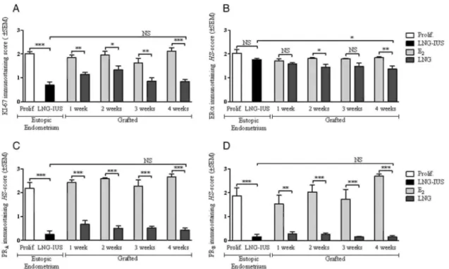
Figure 4 Immunostaining score (mean ± SEM) in function of time in glands for (A) proliferation (Ki67), (B)
oestrogen receptor α (ERα), (C) progesterone receptor A (PRA) and (D) progesterone receptor B (PRB). Prolif, proliferative eutopic endometrium; LNG- lUS, 1 month LNG-IUS eutopic endometrium; E2, endometrial
grafts in SCID mice treated with E2; LNG, endometrial grafts in SCID mice treated with E2 + LNG. NS, not
significant; *P< 0.05; **P< 0.01; ***P< 0.001.
In the present study, we used two groups of mice. The first one was treated with E2 to maintain the proliferative endometrium and to demonstrate that the grafts present the same morphology as the eutopic endometrium. The second group was treated with E2 and LNG for comparison to eutopic endometrium of women treated with LNG-IUS. We showed that human endometrial transplants retained histological characteristics as well as reactivity to hormonal manipulation similar to normal eutopic endometrium. At 4 weeks, all grafts from mice treated with LNG presented similar morphology to that observed in women exposed to LNG-IUS during 4 weeks, i.e. features of extensive decidualization of stromal cells and atrophy of glandular epithelium. In the LNG group, cellular proliferation at 4 weeks was reduced in glandular cells as previously described in women (Salmi et al., 1998; Hurskainen et al., 2000; Maruo et al., 2001). Hurskainen et al. and Maruo et al. described a fall in stromal cell proliferation by evaluating endometrial proliferation 3-12 months after LNG-IUS insertion. We evaluated cell proliferation within a shorter period of time: one month after lUS insertion and after a maximum of 4 weeks LNG treatment in mice. In the study of Salmi et al., more than half of the patients retained some degree of stromal endometrial proliferation. In this study, the cell proliferation analysis was performed as early as 1 month after lUS insertion up to 10 months after insertion of the device. The authors did not specify how many patients had the lUS for 1 month. The persistence of stromal proliferation in our model and in the patients evaluated in our study as well as in the study of Salmi et al. could be explained by difference in the design of these studies, with the two latter studies being performed within a shorter time-lapse by comparison with the studies of Hurskainen et al. and Maruo et al.
We also observed down-regulation of ERα, PRA and PRB in all cellular components of the grafted endometrium. This down-regulation confirmed what we observed in eutopic endometrium treated with LNG-IUS and was in good agreement with previously published data (Critchley et al., 1998b).
Figure 5 Immunostaining score (mean ± SEM) in function of time in stromal cells for (A) proliferation (Ki67),
(B) oestrogen receptor α (ERα), (C) progesterone receptor A (PRA) and (D) progesterone receptor B (PRB). Prolif, proliferative eutopic endometrium; LNG- lUS, 1 month LNG-IUS endometrium; E2, endometrial grafts in
SCID mice treated with E2; LNG, endometrial grafts in SCID mice treated with E2+LNG. NS, not significant;
*P < 0.05; **P< 0.01; ***P< 0.001.
Finally, at 4 weeks, we observed no significant difference in the number of blood vessel sections between the two experimental groups, but a higher mean cross-sectional area of these vessels in the LNG-treated animals. These data support the work of Jondet et al. (2005) and McGavigan et al. (2003). Jondet et al. deducted from their experiments and from additional observations of clinical data that LNG has a dose-dependent action on the number and dilatation of the blood vessels: the higher the plasmatic concentration, the lower the number of blood vessel section and the higher their mean area. One could consider that in our experimental design, the subcutaneous administration of LNG close to the endometrial grafts led to a high local dose of the drug and, consequently, to the observed effects on the vasculature. Finally, the morphological changes observed in the endometrial vasculature induced by a treatment of 4 weeks with LNG were similar to those published by Stephanie et al. (2007).
In conclusion, the use of an animal model presents advantages in comparison with descriptive studies in human: biopsy specimens from each patient can be grafted into several mice, which amplifies the number of samples and allows studies with multiple purposes. In the present experiment, we demonstrated that the graft of endometrium in SCID mice treated with E2 allows one to reproduce the main characteristics of eutopic endometrium and to investigate the local effects of LNG on human endometrium such as a decrease in cell proliferation and in steroid hormone receptor scoring. This model could therefore be used to further study pathophysiological mechanisms underlying the onset of unscheduled BTB that impairs the use of this hormonal therapeutic device and decreases the quality of life of patients. The model represents also a useful tool to develop a therapeutical strategy to overcome the problem of endometrial bleeding before testing in humans.
Author's contribution
M.-L.A.G.: major contribution to conception and design; acquisition, analysis and treatment of data; drafting article; final approval of the version to be published. C.G.: contribution to the design of the model; revising of the manuscript. F.F., M.N. and J.-M.F.: substantial contribution to conception and design; revising critically the article; final approval of the version to be published. S.L.: acquisition of data; revising critically the article; final approval of the version to be published. E.M.: contribution to the conception of the model; revising of the manuscript. A.B.: substantial contribution to conception and design; drafting and revising critically the article; final approval of the version to be published.
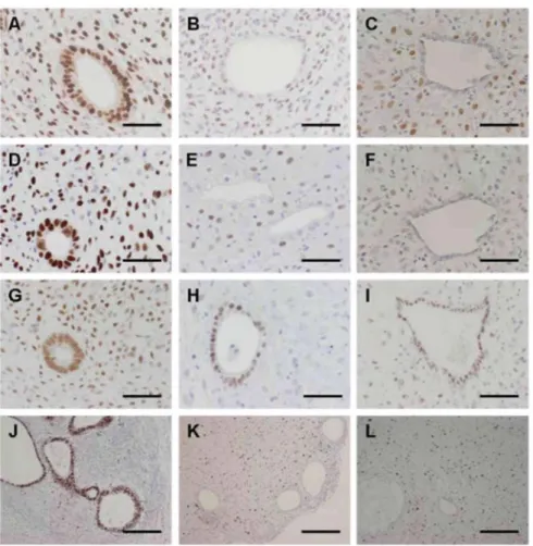
Figure 6 Immunoperoxidase staining with monoclonal species-specific anti-human PRA (A-C), PRB (D-F),
ERα (G-I) and Ki67 (J-L) antibodies. Comparison between mice treated with E2 (A, D, G and J), mice treated
with E2+LNG (B, E, H and K) and eutopic endometrium with 1 month LNG-IUS (C, F, I and L). Scale bar = 25
µm (A-I) or 200 µm (J-L).
Figure 7 Proportion of vessel number/mm (A) and mean vessel surfaces (B) in mice grafted for 4 weeks.
E2, endometrial grafts in SCID mice treated with E2; LNG, endometrial grafts in SCID mice treated with
E2 + LNG; NS, not significant. *P < 0.05; **P < 0.01.
Acknowledgements
The authors thank Fabrice Olivier, Isabel Dasoul, Emilie Seyereisen, Patricia Gavitelli and Laurence Poma for their technical assistance.
Funding
This work was supported by grants from the European Union Framework Programme 6 projects: LSHC-CT-2004-503224 'BRECOSM', Framework Programme 6-NOE no. LSHM-CT-2004-512040 'EMBIC, the Fonds de la Recherche Scientifique Médicale, the Fonds National de la Recherche Scientifique (F.N.R.S., Belgium), the C.G.R.I.-F.N.RS.-INSERM Coopération, the Fonds spéciaux de la Recherche (University of Liège), the Fonds Léon Fredericq (University of Liège), the D.G.T.R.E. from the 'Région Wallonne', the F.S.E. (Fonds Social Européen), the Fonds d'Investissements de la Recherche Scientifique (F.I.RS., CHU, Liège, Belgium), the Interuniversity Attraction Poles Programme - Belgian Science Policy (Brussels, Belgium).
References
Awwad JT, Sayegh RA, Tao XJ, Hassan T, Awwad ST, Isaacson K. The SCID mouse: an experimental model for endometriosis. Hum Reprod 999;14:3107-3111.
Critchley HO, Wang H, Jones RL, Kelly RW, Drudy TA, Gebbie AE, Buckley CH, McNeily AS, Glasier AF. Morphological and functional features of endometrial decidualization following ong-term intrauterine
levonorgestrel delivery. Hum Reprod 1998a; 13:1218-1224.
Critchley HO, Wang H, Kelly RW, Gebbie AE, Glasier AF. Progestin receptor isoforms and prostaglandin dehydrogenase in the endometrium of women using a levonorgestrel-releasing intrauterine system. Hum Reprod 1998b; 13:1210-1217.
Findlay JK. Future directions for research on endometrial bleeding. Hum Reprod 1996; 11 (Suppl. 2): 179- 183. Girling JE, Heryanto B, Patel N, Rogers PA. Effect of long-term progestin treatment on endometrial
vasculature in normal cycling mice. Contraception 2004;70:343-350.
Greenberg LH, Slayden OD. Human endometriotic xenografts in immunodeficient RAG-2/gamma(c) KO mice.
Am J Obstet Gynecol 2004;190:1788-1795.
Grummer R. Animal models in endometriosis research. Hum Reprod Update 2006
Guttinger A, Critchley HO. Endometrial effects of intrauterine levonorgestrel. Contraception 2007;75:S93-S98. Hickey M, Fraser I. Human uterine vascular structures in normal and diseased states. Microsc Res Tech 2003;60:377-389.
Hickey M, Simbar M, Young L, Markham R, Russell P, Fraser IS. A longitudinal study of changes in endometrial microvascular density in Norplant implant users.Contraception 1999;59:123-129.
Hickey M, Dwarte D, Fraser IS. Superficial endometrial vascular fragility in Norplant users and in women with ovulatory dysfunctional uterine bleeding. Hum Reprod 2000; 15:1509-1514.
Huang A, Pettigrew NM,Watson PH. Immunohistochemical assay for oestrogen receptors in paraffin wax sections of breast carcinoma using a new monoclonal antibody. J Pathol 1996; 180:223-227.
Hurskainen R, Salmi A, Paavonen J, Teperi J, Rutanen E. Expression of sex steroid receptors and Ki-67 in the endometria of menorrhagic women: effects of intrauterine levonorgestrel. Mol Hum Reprod 2000; 6:1013-1018. Jondet M, Letellier B, Verdys MT. Endometrial vascularization in levonorgestrel intrauterine device users; computerized microvessel measurement study. Contraception 2005; 71:60-64.
Jones RL, Critchley HO. Morphological and functional changes in human endometrium following intrauterine levonorgestrel delivery. Hum Reprod 2000; 15(Suppl. 3): 162-172.
Kim W, Takahashi T, Nisselbaum JS, Lewis JL Jr. Heterotransplantation of human choriocarcinoma in nude mice. I. Morphologic and biologic characteristics. Gynecol Oncol 1978; 6:165-182.
Mifepristone, but not levonorgestrel, inhibits human blastocyst attachment to an in vitro endometrial three-dimensional cell culture model. Hum Reprod 2007; 22:3031-3037.
Maruo T, Laoag-Fernandez JB, Pakarinen P, Murakoshi H, Spitz IM, Johansson E. Effects of the levonorgestrel-releasing intrauterine system on proliferation and apoptosis in the endometrium. Hum Reprod 2001;16:2103-2108.
Masuda H, Maruyama T, Hiratsu E, Yamane J, Iwanami A, Nagashima T, Ono M, Miyoshi H, Okano HJ, Ito M et al. Noninvasive and real-time assessment of reconstructed functional human endometrium in
NOD/SCID/{gamma} Formula immunodeficient mice. Proc Natl Acad Sci USA 2007;104:1925-1930. Matsuura-Sawada R, Murakami T, Ozawa Y, Nabeshima H, Akahira J, Sato Y, Koyanagi Y, Ito M, Terada Y, Okamura K. Reproduction of menstrual changes in transplanted human endometrial tissue in immunodeficient mice. Hum Reprod 2005;20:1477-1484.
McGavigan CJ, Dockery P, Metaxa-Mariatou V, Campbell D, Stewart CJ, Cameron IT, Campbell S. Hormonally mediated disturbance of angiogenesis in the human endometrium after exposure to intrauterine levonorgestrel.
Hum Reprod 2003;18:77-84.
Meng CX, Andersson KL, Bentin-Ley U, Gemzell-Danielsson K, Lalitkumar PG. Effect of levonorgestrel and mifepristone on endometrial receptivity markers in a three-dimensional human endometrial cell culture model.
Fertil Steril 2008; in press.
Morison NB, Zhang J, Kaitu'u-Lino TJ, Fraser IS, Salamonsen LA. The long-term actions of etonogestrel and levonorgestrel on decidualized and non-decidualized endometrium in a mouse model mimic some effects of progestogen-only contraceptives in women. Reproduction 2007;133:309-321.
Noyes RW, Hertig AT, RocK J. Dating the endometrial biopsy. Fertil Steril 950;l:3-25.
Phillips RA, Jewett MA, Gallie BL. Growth of human tumors in immune-deficient scid mice and nude mice.
Curr Top Microbiol Immunol 1989:152:259-263.
Rogers PA, Plunkett D, Affandi B. Perivascular smooth muscle alpha-actin is reduced in the endometrium of women with progestin-only contraceptive breakthrough bleeding. Hum Reprod 2000; l5(Suppl. 3):78-84. Salmi A, Pakarinen P, Peltola AM, Rutanen EM. The effect of intrauterine levonorgestrel use on the expression of c-JUN, oestrogen receptors, progesterone receptors and Ki-67 in human endometrium. Mol Hum Reprod 1998;4:1110-1115.
Satyaswaroop PG, Zaino RJ, Mortel R. Human endometrial adenocarcinoma transplanted into nude mice: growth regulation by estradiol. Science 1983;219:58-60.
Shimosato Y, Kameya T, Nagai K, Hirohashi S, Koide T, Hayashi H, Nomura T Transplantation of human tumors in nude mice. J Natl Cancer Inst 1976;56:1251-1260.
Stephanie R, Labied S, Blacher S, Frankenne F, Munaut C, Fridman V, Beliard A, Foidart JM, Nisolle M. Endometrial vessel maturation in women exposed to levonorgestrel-releasing intrauterine system for a short or prolonged period of time. Hum Reprod 2007;22:3084-3091.
Vereide AB, Kaino T, Sager G, Arnes M, Orbo A. Effect of levonorgestrel lUD and oral medroxyprogesterone acetate on glandular and stromal progesterone receptors (PRA and PRB), and estrogen receptors (ER-alpha and ER-beta) in human endometrial hyperplasia. Gynecol Oncol 2006;101:214-223.