Reproductive Biology 88 (2000) 75–80 www.elsevier.com / locate / ejogrb
Original Article
Effect of grade on disease-free survival and overall survival in FIGO
stage I adenocarcinoma of the endometrium
a b c
Jean F. Delaloye (M.D.) , Sandro Pampallona (D.Sc.) , Philippe A. Coucke (M.D.) ,
a a
Alexandre Megalo (M.D.) , Pierre De Grandi (M.D. Prof.)
a´ ´ ´
Departement de Gynecologie-Obstetrique, Centre Hospitalier Universitaire Vaudois, 1011 Lausanne, Switzerland
b
`
forMed, 1983 Evolene, Switzerland
c
Service de Radio-Oncologie, Centre Hospitalier Universitaire Vaudois, 1011 Lausanne, Switzerland Received 30 July 1998; accepted 20 April 1999
Abstract
Objective: To analyse the effect of differentiation on disease-free survival (DFS) and overall survival (OS) in patients with stage I adenocarcinoma of the endometrium. Patients and methods: From 1979 to 1995, 350 patients with FIGO stage IA–IC with well (G1), moderately (G2) or poorly (G3) differentiated tumors were treated with surgery and high dose-rate brachytherapy with or without external radiation. Median age was 65 years (39–86 years). Results: The 5-year DFS was 8863% for the G1 tumors, 7764% for the G2 tumors, and 6767% for the G3 tumors (P50.0049). With regard to the events contributing to DFS, the 5-year cumulative percentage of local relapse was 4.6% for the G1 tumors, 9.0% for the G2 tumors, and 4.6% (P50.027) for the G3 tumors. Cumulative percentage of metastasis was 1.4, 6.3 and 7.2% (P,0.001), respectively, whereas percentages of death were 6.0, 7.9 and 20.7% (P,0.001). The 5-year OS was 9163, 8364 and 7667%, respectively (P50.0018). In terms of multivariate hazard ratios (HR), the relative differences between the three differentiation groups correspond to an increase of 77% of the risk of occurrence of either of the three events considered for the DFS (HR51.77, 95% Cl [0.94–3.33]), (P50.078) for the G2 tumors and of 163% (HR52.63, 95% Cl [1.27–5.43]), (P50.009) for the G3 tumors with respect to the G1 tumors. The estimated relative hazards for OS are, respectively, in line with those for DFS: HR51.51 (P50.282) for the G2 tumors; and HR53.37 (P50.003) for the G3 tumors. Conclusion: Patients with grade 1 tumors are those least exposed to either local relapse, metastasis, or death. In contrast patients with grade 2 tumors seem to be at higher risk of metastasis, whereas patients with grade 3 tumors appear at higher risk of death. Since we have looked at the first of three competing events (local relapse, metastasis and death), this suggests that patients with grade 3 tumors probably progress to death so fast that local relapse, if any, cannot be observed. 2000 Elsevier Science Ireland Ltd. All rights reserved.
1. Introduction based on the myometrial invasion only, independently of
cell differentiation. Cell differentiation has been shown to be one of the
most important prognostic factors in carcinoma of the
endometrium [1–4]. In this retrospective analysis we 2. Patients and methods
aimed to investigate the effect of grade on disease control
in FIGO stage I patients [5], who were treated with a 2.1. Patient population
combination of surgery and postoperative radiotherapy
From April 1979 to December 1995, 350 patients with primary FIGO stage IA–IC pure adenocarcinoma of the
*Corresponding author. Tel.: 141-21-3143269; fax: 141-21-3143263. endometrium were treated in the Centre Hospitalier
Uni-0301-2115 / 00 / $ – see front matter 2000 Elsevier Science Ireland Ltd. All rights reserved. P I I : S 0 3 0 1 - 2 1 1 5 ( 9 9 ) 0 0 1 2 4 - 4
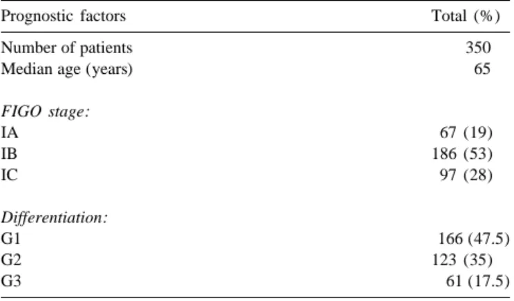
Table 1 local recurrence, metastasis or death, whichever occurred Patients characteristics first. The secondary end-point was overall survival, defined Prognostic factors Total (%) as time from surgery to death.
Statistical analyses were carried out on the software
Number of patients 350
Median age (years) 65 package Stata [9]. Survival percentages over time have been calculated according to the Kaplan–Meier method
FIGO stage: [10], and their corresponding standard errors (SEs) with
IA 67 (19)
Greenwood’s formula [11]. For the univariate analysis the
IB 186 (53)
P values from the log-rank test are reported [12].
Esti-IC 97 (28)
mated hazard ratios (HR) for DFS and OS, their 95%
Differentiation: confidence intervals (95% Cl) and P values were
calcu-G1 166 (47.5) lated with a multivariate proportional hazard-regression
G2 123 (35)
model [13]. Appropriate binary indicators have been
G3 61 (17.5)
defined to identify the categories of the following vari-ables: age (50, 51–60, 61–70, 71), FIGO stage IA–IC, grades 1–3. Variables describing duration and intensity of
versitaire Vaudois, Lausanne, Switzerland. Patients charac- radiation therapy have not been considered given the
teristics are summarized in Table 1. The median age was homogeneity of treatment delivered. HRs quoted in the
65 years (range 36–86 years). The tumors were classified Section 3 are from a model including all above variables.
in three grades: well (G1), moderately (G2) or poorly Values of HR greater than unity indicate increased rates of
(G3), according to the WHO cellular differentiation [5]. any of the events contributing to death (or of death for OS)
Slides were reviewed by two pathologists. with respect to the chosen reference category (for which
HR51 by definition). The probability that local
progres-2.2. Treatment sion or metastasis or death occurs before any specified
time has been estimated by means of cumulative incidence
All patients underwent total extrafascial hysterectomy functions [14]. For the sake of interpretation we point out
and bilateral salpingo-oophorectomy. Postoperative treat- that one minus the sum of such probabilities (local relapse,
ment strategy was planned on the pathology report of the metastasis, death), at any given point in time, corresponds
myometrial invasion only, independently of the cellular to the probability of being alive and disease-free as
differentiation. Peritoneal washing was not routinely ob- obtained from the Kaplan–Meier estimate of the DFS.
tained and lymph nodes dissection was very uncommon. Appropriate statistical methods have been used for
com-All 350 patients received vaginal vault irradiation. In paring cumulative incidences allowing for stratification
addition 56 / 186 patients with stage IB greater than 30% factors [15]. All probability values are for two-sided tests.
myometrial infiltration, and all 97 patients with stage IC For the purpose of the analyses, observations have been
received external irradiation. censored at 5 years in order to limit the effect of
competing causes of mortality and also to avoid having
2.3. Radiation therapy only patients registered during the earlier periods
contrib-ute to the right tail of the curves. Four weeks after surgery, external irradiation was given
on the pelvic volume with a linear accelerator (6–18 MeV)
for a total dose of 4500 cGy fractionated at a daily dose of 3. Results
180–200 cGy [6–8]. The reference point for dose
prescrip-tion was at the intersecprescrip-tion of the four beams. The upper 3.1. Association among patient characteristics
limit of the pelvic volume was defined by a line passing
through the L5–S1 interspace. The lateral borders were Age was associated with tumor stage. Patients with
located 1 cm beyond the bony pelvis. The inferior border FIGO stage IA were younger (63 years) than those with
covered two-thirds of the vagina. stage IB (65 years) or stage IC (67 years) (P50.0036). No
Brachytherapy delivered a boost of 45 Gy at the surface other association among prognostic factor was found.
of the vaginal cuff over 2 weeks (three fractions) with a
137
high dose-rate ( Cs) remote afterloading system. 3.2. Disease-free survival analysis
2.4. Statistical methods The median follow-up was in excess of 7 years. As
explained in Section 2, DFS and OS have been censored at
The primary endpoint of this retrospective study was 5 years. A total of 55 events have been observed by the
disease-free survival (DFS), which was defined as time fifth year. The 5-year DFS was 8068%. When considering
Table 3
7764% for the G2 group, and 67.7% for the G3 group
a
Cumulative incidences (%) of three events contributing to DFS
(P50.0049). In terms of multivariate hazard ratio (HR),
Grade Local relapse Metastasis Death
the relative difference between the three groups
corre-sponds to an increase of 77% of the risk of occurrence of 1 4.6 1.4 6.0
either of the three events considered (local relapse, metas- 2 9.0 6.3 7.9
3 4.6 7.2 20.7
tasis and death) (HR51.77, 95% Cl [0.94–3.33]) (P5
P value 0.027 ,0.001 ,0.001
0.078) for the G2 group, and of 163% (HR52.63, 95% Cl
a
Standard errors are in all cases less than 3%. The reported P value are
[1.27–5.43]) (P50.009) for the G3 group with respect to
from an adjusted test, stratifed by FIGO stage.
G1 group. These and additional multivariate results are displayed in Table 2. Local relapse was defined as vaginal
and / or latero-pelvic recurrence. 3.4. Overall survival analysis
The 5-year overall survival was 84.862%. When
consid-3.3. Cumulative incidence analysis ering sub-groups, the 5-year OS was 9163% for the G1
group, 8364% for the G2 group, and 67.7% for the G3
In order to better investigate the effect of grade on the group (P50.0018). In terms of multivariate hazard ratio
pattern of occurrence of the events contributing to DFS (HR), the relative difference between the three groups
(local relapse, metastasis and death), we have estimated the corresponds to a non-significant increase of 51% of the
cumulative incidence of each of these competing events risk of death for the G2 group (HR51.51, 95% Cl [0.71–
individually. The results are summarized in Table 3. With 3.19]) (P50.282), and of 237% for the G3 group (HR5
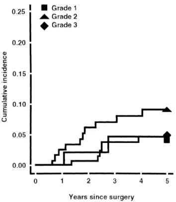
regard to cell differentiation, the 5-year cumulative per- 3.37, 95% Cl [1.52–7.44]) (P50.003) with respect to the
centage of local relapses was 4.6% for grade 1, 9.0% for G1 group (Table 2).
grade 2, and 4.6% for grade 3 (P50.027). Similarly, such percentages were 1.4, 6.3 and 7.2% (P,0.001) for
metas-tasis, and 6.0, 7.9 and 20.7% (P,0.001) for deaths. The 4. Discussion
cumulative incidence curves are displayed in Figs. 1–3.
Fig. 1 for local recurrence shows a rapid increase of the Our study confirms the prognostic importance of cell
curve of the grade 2 tumors during the first year after differentiation on disease-free survival (DFS) and overall
treatment, and a slower increase and subsequent leveling survival (OS) of adenocarcinoma of the endometrium. The
off of the curves in the G1 and G3 groups by the third relative shapes of the cumulative incidence curves suggest
year. Fig. 2 for distant metastasis shows a slow increase that the strong reduction in DFS beyond the first year after
and subsequent levelling off of the curves in the G2 and treatment for patients with G3 differentiation can be
G3 groups at three years. As concern deaths, Fig. 3 attributed to death. As in other series, well-differentiated
suggests that they continue to accumulate over time in all (G1) tumors were easily locally controlled, resulting in a
differentiation groups, but again much faster for G3. The longer survival. In contrast, moderately (G2) and poorly
5-year percentage of deaths is almost triple compared to (G3) differentiated tumors were characterised by
metas-what has been observed in the G1 and G2 groups. tases and death, respectively [1,16–22]. G2 patients
pro-Table 2
Cox’s analysis fitted to the overall survival (OS) and disease-free survival (DFS)
OS P value DFS P value
HR (95% CI) HR (95% CI)
Age(years):
50 Reference category Reference category
51–60 1.85 (0.21–16.85) 0.577 0.86 (0.16–4.77) 0.871
61–70 2.51 (0.33–19.05) 0.371 1.77 (0.41–7.57) 0.438
70 4.86 (0.64–36.45) 0.124 3.22 (0.76–13.67) 0.112
FIGO stage:
I Reference category Reference category
II 0.99 (0.36–2.73) 0.996 0.71 (0.31–1.64) 0.430
III 2.20 (0.82–5.89) 0.116 1.84 (0.83–4.09) 0.130
Grade:
1 Reference category Reference category
2 1.51 (0.71–3.19) 0.282 1.77 (0.94–3.33) 0.078
Fig. 3. Cumulative percentages of death according to differentiation. Fig. 1. Cumulative percentages of local relapse according to
differentia-tion.
gressed at a relatively slow pace, possibly allowing for the detection of local relapse and metastases. On the other hand G3 patients, in whom a high mortality has been observed as opposed to a low rate of local or distant metastases, were probably prone to a rather fast disease progression, in which only the final stage of the evolution, death, was unfortunately detected.
Factors associated with the high grade histological variant that may have a bearing on prognosis include a tendency for higher mean age at diagnosis, a greater incidence of both deep myometrial invasion and lymph node involvement [3], and a high risk of clinical understag-ing as well as a higher rate of vaginal recurrences [4]. Age at diagnosis did not appear as a significant prognostic factor, though for both OS and DFS the estimated HRs showed a trend in the expected direction. This contrasts with other papers showing that age has a prognostic impact [18,23]. Our small number of events might not have allowed us to detect this effect, possibly of small mag-nitude. One might not exclude a bias in the results due to competing causes of death especially acting in older age groups. Our observations cover a time span of over 15 years during which cause of death attribution has not been performed systematically and according to consistent standards. Even if cause of death was available for all patients, we would question its precision and especially so for older patients. Additionally, the focus of the report is
[4] Price JJ. Vaginal involvement in endometrial carcinoma. Am J
with age. Since the aim of this contribution was to
Obstet Gynecol 1965;91:1060–5.
compare the pattern of occurrence of the events
contribut-[5] Announcement. Changes in gynecologic cancer staging by the
ing to disease-free survival among tumor grade subgroups, International Federation of Gynecology and Obstetrics. Am J Obstet
the results observed should at least qualitatively, if not in Gynecol 1990;162:610–11.
magnitude, be considered. In particular, the possible bias [6] Aalders J, Abeler V, Kolstad P, Onsrud M. Postoperative external
irradiation and prognostic parameters in stage I endometrial
car-introduced by death due to other causes than cancer should
cinoma. Clinical and histopathologic study of 540 patients. Obstet
equally affect all subgroups of grade. Furthermore, the
Gynecol 1980;56:419–26.
tests comparing the cumulative incidence of either local [7] Ahmad NR, Lanciano R, Corn BW, Schultheiss T. Postoperative
relapse or metastasis or death among tumor grade sub- radiation therapy for surgically staged endometrial cancer: impact of
groups was stratified by stage, which was found to be time factor (overall treatment time and surgery-to-radiation interval)
on outcome. Int J Radiat Oncol Biol Phys 1995;33:837–42.
significantly associated with age. This approach should
[8] Rush S, Gal D, Potters L, Bosworth J, Lovecchio J. Pelvic control
control the potential bias introduced by causes of death
following external beam radiation for surgical stage I endometrial
other than cancer possibly occurring more frequently in
adenocarcinoma. Int J Radiat Oncol Biol Phys 1995;33:851–4.
older age. [9] Stata. Computing Resource Center Reference Manual (1991).
Although pelvic and para-aortic lymphadenectomy are [10] Kaplan EL, Meier P. Nonparametric estimation from incomplete
observations. J Am Stat Assoc 1958;53:457–81.
now mandatory, especially for grade 3 patients, the
pro-[11] Greenwood M. Reports on public health and medical subjects: the
cedure was not systematically performed in this series.
natural duration of cancer. HMSO 1926;33:1–16.
Only the depth of myometrial invasion was taken into
[12] Peto R, Pike MC, Armitage P, Breslow NE, Cox DR, Howard SV,
account for prescribing adjuvant radiation therapy. We Mantel N, McPherson K, Peto J, Smith PG. Design and analysis of
have not seen an effect of myometrial infiltration on DFS randomized clinical trials requiring prolonged observation of each
or on OS, which contrasts with previous reports [2,18– patient. II. Analysis and examples. Br J Cancer 1977;35:1–39.
[13] Cox DR. Regression models and life tables. J R Stat Soc B
21,23]. There might be two explanations. On the one hand,
1972;34:187–220.
it is possible that as the difference in prognosis being
[14] Kalbfleisch JD, Prentice RL. In: The statistical analysis of failure
relatively small between stage IA, IB and IC, we have not time data, New York: Wiley, 1980.
been able to detect it with our sample. On the other hand, [15] Pepe MS, Mori M, Kaplan-Meier EL. Marginal or conditional
therapy was tailored to stage and the two factors may be probability curves in the analysis of competimg risk failure time
data. Stat Med 1993;12:737–51.
confounded with each other in such a way to mask their
[16] Piver MS, Vongtama V. Parameters adversely affecting survival in
respective effect. The effect of stage was not of specific
women with stage I endometrial carcinoma. Am J Obstet Gynecol
interest, and our analytic approach (multivariate analysis 1965;91:1060–5.
and stratified testing) has in any case allowed for its role, [17] Malkasian GD, Annegers JF, Fountain KS. Carcinoma of the
whether as a prognostic factor or as a confounder. endometrium: Stage I. Am J Obstet Gynecol 1980;136:872–88.
[18] Connelly PJ, Alberhasky RC, Christopherson WM. Carcinoma of
It is well established that postoperative irradiation
the endometrium. III. Analysis of 865 cases with adenocarcinoma
reduces the incidence of vaginal recurrences and distant
and adenoacanthoma. Obstet Gynecol 1982;59:569–75.
metastases, and improves survival rates in patients with [19] Hendrickson M, Ross J, Eifel P, Cox RS, Martinez A, Kempson R.
more than one-half myometrial invasion and / or grade 2 or Adenocarcinoma of the endometrium: analysis of 256 cases with
3 stage I endometrial adenocarcinoma [24–32]. One carcinoma limited to the uterine corpus. Gynecol Oncol
1982;13:373–92.
wonders then if a more aggressive adjuvant radiation
[20] Boronow Rc, Morrow CP, Creasman WT, DiSaia PJ, Silverberg SG,
therapy (more than 45 Gy on the pelvic volume or
Miller A, Blessing JA. Surgical staging in endometrial cancer. I.
irradiation of the whole abdominal cavity) or a more Clinical-pathologic findings of a prospective study. Obstet Gynecol
intensive follow-up of such patients would be justified. A 1984;63:825–32.
strong evidence is available in favour of the first practice [21] Burke TW, Heller PB, Woodward JE, Davidson SA, Hoskins WJ,
Park RC. Treatment failure in endometrial carcinoma. Obstet
[33]. However the option of a more intensive follow-up
Gynecol 1990;75:96–101.
would be appropriate only if the efficacy of a second line
[22] Morrow CP, Bundy BN, Kurman RJ, Creasman WT, Heller P,
treatment could be demonstrated. Homesley HD, Graham JE. Relationship between surgical–
pathological risk factors and outcome in clinical stage I and II carcinoma of the endometrium: a Gynecologic Oncology Group Study. Gynecol Oncol 1991;40:55–65.
References [23] Christopherson WM, Connelly PJ, Alberhasky RC. Carcinoma of
the endometrium: an analysis of prognosticators in patients with [1] DiSaia PJ, Creasman WT, Boronow RC, Blessing JA. Risk factors favorable subtypes and stage I disease. Cancer 1983;51:1705–9.
and recurrent patterns in stage I endometrial cancer. Am J Obstet [24] Vangtoma J, Kurohara S, Badihi AO, Webster JH. The value of Gynecol 1985;151:1009–15. adjuvant irradiation in the treatment of endometrial carcinoma Stage [2] Mammoliti S, Bruzzone M, Chiara S et al. Clinical stage I and II I, Group I. Cancer 1970;25:45–9.
endometrial carcinoma: multivariate analysis of prognostic factors. [25] Joslin CA, Vaishampayan GV, Mallik A. The treatment of early Anticancer Res 1992;12:1415–8. cancer of the corpus uteri. Br J Radiol 1977;50:38–45.
[3] Creasman WT, Boronow RC, Morrow CP, DiSaia PJ, Blessing J. [26] Reddy S, Lee MS, Henderson FR. Patterns of recurrences in Adenocarcinoma of the endometrium: its metastatic lymph node endometrial carcinoma and their management. Radiol potential. Gynecol Oncol 1976;4:239–43. 1979;133:737–40.
[27] Aalders J, Abeler V, Kolstad P, Onsrud M. Postoperative external [31] Sorbe BG, Smeds AC. Postoperative vaginal irradiation with high irradiation and prognostic parameters in stage I endometrial car- dose rate afterloading technique in endometrial carcinoma stage I. cinoma. Obstet Gynecol 1980;56:419–26. Int J Radiat Oncol Biol Phys 1990;18:305–14.
[28] Marchetti L, Piver S, Tsukoda Y, Reese P. Prevention of vaginal [32] Elliott P, Green D, Coates A, Krieger M, Russel P, Coppleson M, recurrence of stage I endometrial adenocarcinoma with postopera- Solomon J, Tattersall M. The efficacy of postoperative vaginal tive vaginal radiation. Obstet Gynecol 1986;67:399–402. irradiation in preventing vaginal recurrence in endometrial cancer. [29] Nori D, Hilaris BS, Tome M, Lewis Jr. JL, Birnbaum S, Fuks Z. Int J Gynecol Cancer 1994;4:84–93.
Combined surgery and radiation in endometrial carcinoma: an [33] Gibbons S, Martinez A, Schray M, Podratz K, Stanhope R, Garton analysis of prognostic factors. Int J Radiat Oncol Biol Phys G, Weiner S, Brabbins D, Malkasian G. Adjuvant whole ab-1987;13:489–97. dominopelvic irradiation for high risk endometrial carcinoma. Int J [30] Meerwaldt JH, Hoekstra CJM, van Putten WLJ, Subandono Tjok- Radiat Oncol Biol Phys 1991;21:1019–25.
rowardojo AJ, Koper PCM. Endometrial adenocarcinoma adjuvant radiotherapy tailored to prognostic factors. Int J Radiat Oncol Biol Phys 1990;18:299–304.