Développement de techniques avancées de microscopie électronique à transmission pour la cartographie à l'échelle nanométrique
Texte intégral
Figure


Documents relatifs
Domain wall images at A and wall stray field images at B are shown in figure 2a whilst the stray fields between antiparallel domains in coba1,t ptoduce edge images of
a-emitting and highly irradiated samples of nuclear fuels. The instrument is being used in programmes of research into the swelling and transient behaviour of advanced nuclear
COMPUTER TECHNIQUES FOR TOPOGRAPHICAL ANALYSIS IN THE SCANNING ELECTRON MICROSCOPE... JOURNAL
The geometrical loci of the points for which IT(s), Is(s) are minima (as function of s) are inclination extinction contours. They will move rapidly over the image if the
Heating tests inside Environmental Scanning Electron Microscope (ESEM), tensile tests inside Scanning Electron Microscope (SEM) (dedicated to nanowires and nanotubes), heating
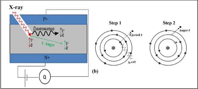
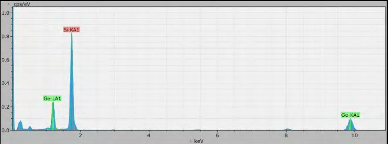
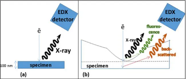
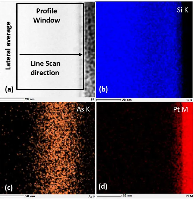
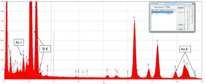
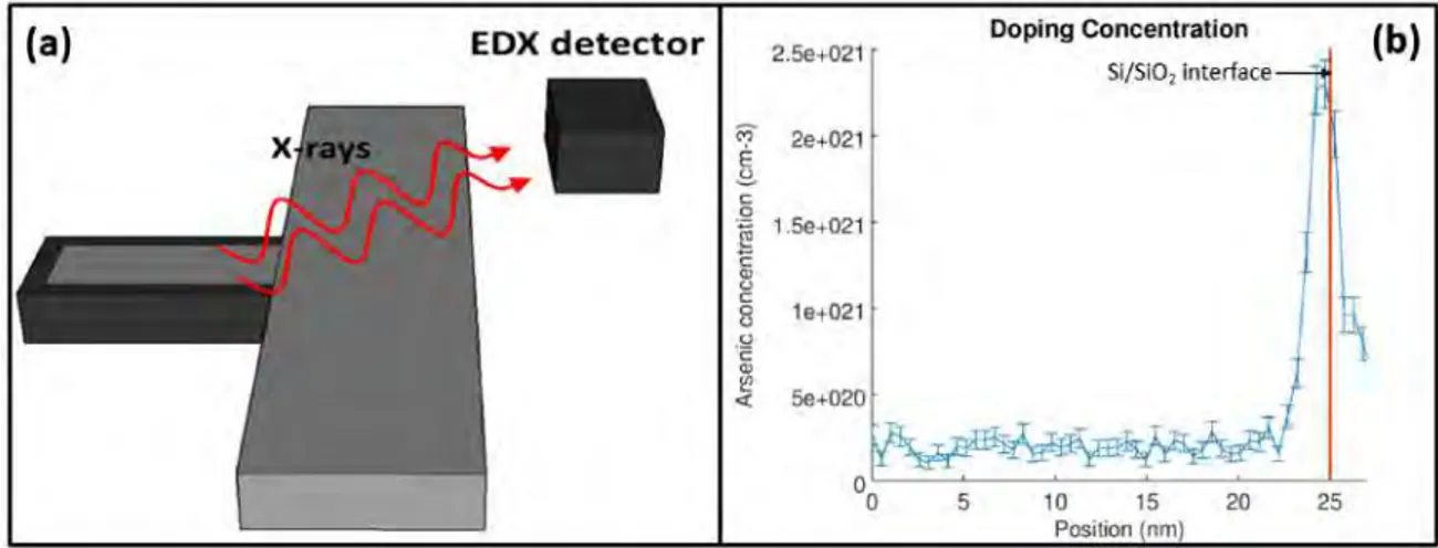
L’analyse de la composition chimique est réalisée par les techniques de spectroscopie des Rayons X émis par l’échantillon (EDS) et également par perte
On peut, réciproquement, utiliser le faisceau diffracté pour faire l’image, le faisceau transmis étant éliminé ; dans ce cas, il faut remarquer que l’image obtenue n’est
This study was conducted on the composi- tion and relative abundance of the species belonging to the genus Culicoides (Diptera: Ceratopogonidae) in certain regions of Romania
