Decrease in Systemic Tolerance to Fed Ovalbumin in
Indomethacin-Treated Mice
E. Louis a,*, D. Franchimont b, M. Deprez b, A. Lamproye a, N. Schaaf c, P. Mahieu c, J. Belaiche a
Departments of a Gastroenterology b Pathology and c Immunology, CHU of Liège, Belgium
Abstract
The oral administration of non-steroidal anti-inflammatory drugs (NSAID) to animals induces a quick increase in intestinal permeability and secondary inflammatory lesions of the intestine. The mechanisms leading to the inflammatory lesions are hypothetical. The increased intestinal permeability could allow a greater mucosal and systemic penetration of fed antigens and bacterial products leading to an abnormal mucosal and systemic immune and inflammatory response toward these materials. We examined the effect of oral dosing with indomethacin on ovalbumin serum levels and the systemic immune response to ovalbumin in mice fed with ovalbumin. The ovalbumin serum level was higher in indomethacin-treated mice and the increase was
proportional to the dose of indomethacin. It was associated with epithelial and subepithelial lesions. Moreover, the systemic humoral and, to a lesser extent, the cellular tolerance were partially abrogated in the treated mice. These findings suggest that the oral administration of indomethacin in mice induces an increased passage of fed antigen through the intestinal epithelium associated with a decrease in systemic tolerance to this antigen. The reason for this decrease remains unclear. Besides a disequilibrium between systemic and mucosal immune responses, a loss of integrity of the intestinal epithelial cells and a direct immunomodulating effect of
indomethacin may also be involved. This decrease in systemic tolerance to luminal antigen could be involved in the development of NSAID enteropathy.
Keywords : NSAID enteropathy ; Immune tolerance ; Immunization ; Intestinal permeability
Introduction
Non-steroidal anti-inflammatory drug (NSAID) enteropathy is a well-documented entity in humans [1]. It is characterized by an increased intestinal permeability, modifications of intestinal absorption and motility and jejuno-ileal inflammation. Sometimes, ulcerative lesions, bleeding, perforations or the more specific diaphragm disease can occur. Studies in animals [2] have shown two steps in this NSAID enteropathy: first, microvascular damages, imbalance in eicosanoids, mitochondrial damage and increased intestinal permeability within hours following the administration of one dose of NSAID; secondly, oedema, ulcers and inflammatory reaction developing 12-24 h after NSAID. This inflammatory reaction may become chronic if the NSAID administration is prolonged. This second step is relatively non-specific and depends on luminal agents. It could be diminished or even suppressed in germ-free animals [3], after antibiotic therapy [4] or in case of fasting [5]. The
mechanisms leading from an increased intestinal permeability and early mucosal modifications to lesions and more chronic inflammation are hypothetical. An increased mucosal and systemic penetration of antigens, superantigens, bacterial cell wall fragments and infectious agents is suspected to lead to a breakdown in systemic immune tolerance, to a perturbation of the mucosal immune response and to a nonspecific inflammatory
response in the mucosa. The aim of our study was to clarify whether oral administration of indomethacin is associated with an increased penetration of fed soluble antigen and with a decrease in systemic tolerance toward this fed antigen.
Material and Methods Animals
Female Balb/c mice, ranging from 4 to 8 weeks of age, were obtained from the Iffa Credo Laboratories in
Brussels, Belgium. Their mean weight was 20±2.7 g. Antigen
Crystallized and lyophilized ovalbumin grade V was obtained from Sigma Chemical Co., St. Louis, Mo., USA. Study Design
At day 0,90 mice were administered an oral dose of indomethacin. Eighteen hours' later, they were administered a single oral dose of ovalbumin. Three hours after ovalbumin administration, 30 of them were killed by cervical dislocation. They were used for serum ovalbumin measurement and for histological examination of the intestine. Other mice were immunized subcutaneously with ovalbumin at day 7, and 30 of them were killed by cervical dislocation at day 17 for measurements of anti-ovalbumin antibody levels and an in vitro lymphocyte proliferation test. The last 30 mice were used for a specific delayed hypersensitivity test in vivo. Mice were divided into six groups representing different experimental conditions (table 1). Five mice were used for each experimental condition.
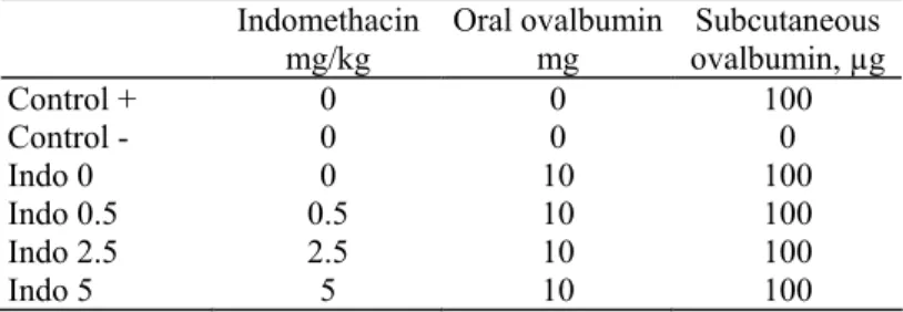
Table 1. Study design
Indomethacin mg/kg Oral ovalbumin mg Subcutaneous ovalbumin, µg Control + 0 0 100 Control - 0 0 0 Indo 0 0 10 100 Indo 0.5 0.5 10 100 Indo 2.5 2.5 10 100 Indo 5 5 10 100 Indomethacin Administration
Natrii indomethacinum (Merck, Sharp and Dohme) was diluted in saline to different concentrations: 100 µg/ml, 500 µg/ml and 1 mg/ml. It was administered by an intragastric tube under light ether anesthesia. One group of mice (Indo 0) was injected with saline only.
Oral Pre-Immunization
Mice were fed a single dose of 10 mg ovalbumin in 100 µl of saline by an intragastric tube. Subcutaneous Immunization
Ovalbumin was precipitated in Al(OH)3 at 2 mg/ml. Fifty microlitres of this suspension were injected subcutaneously at the base of the tail with a 30-gauge needle.
Measurement of Serum Ovalbumin
Mice were bled by cardiac puncture. Fifty to 200 µl of serum were obtained from each mouse and stored at -20 °C. Serum ovalbuminaemia was determined by RIA. We used an inhibition dosage with a double-antibody method. Briefly, sera were incubated for 16 h at 20°C in tubes with 125I-ovalbumin (grade V, Sigma) and a rabbit anti-ovalbumin. IgG (Organon Teknika). Then a goat antirabbit antibody was added and mixed slightly during 4 h at 20°C. Tubes were then centrifuged and the supernatants were adsorbed. The radioactivity of the pellets were counted (cpm). A reference curve was made simultaneously with different concentrations of ovalbumin and the ovalbuminaemia of the different sera was calculated from this reference curve.
Histology
Jejunal and ileal segments were fixed in Bouin's solution and embedded in paraffin. Five-micrometre sections were routinely stained with haematoxylin-eosin. Periodic acid-Schiff staining was performed when necessary.
Immune Evaluation
Systemic humoral response to ovalbumin was determined by an antibody assay. Anti-ovalbumin serum levels were measured by ELISA. Mice were bled by cardiac puncture. Fifty to 200 µl of serum were obtained from each mouse and stored at -20°C. Microtitre plates were coated with 100 µl of an ovalburnin solution (100 µg/rnl) per well at 4°C for 24 h. Plates were rinsed 3 times with a washing solution (PBS + Tween 0.5%). Plates were then incubated for 1 h at room temperature with a bovine powdered milk solution (PBS + Tween 0.5% + powdered milk 1%) to block non-specific sites. Plates were rinsed 3 times with the washing solution. Sera diluted at 1/10, 1/40, 1/160 and 1/640 were added (50 µl/well) for 1 h at 37°C. Reference serum was obtained by serum pooling from mice immunized several times subcutaneously with ovalbumin; it was diluted at 1/500, 1/2,000, 1/8,000 and 1/32,000. Plates were rinsed 3 times with the washing solution. The immunoglobulin total was detected with peroxidase-conjugated goat antimouse immunoglobulin (Dako) used at 1/500. Plates were incubated at 37°C for 1 h and then rinsed 3 times with die washing solution. The reaction was developed with O-phenylenediamine dihydrochloride for 5 min (50 µl/well). The reaction was stopped with H2SO4 (25 µl/well). Plates were read with a spectrophotometer type LP 200 at 405 nm. The level of anti-ovalbumin antibody in the serum for each mouse was expressed as units of reference serum antibody level.
Systemic cellular response to ovalbumin was determined by a lymphocyte proliferation test in vitro and by a specific delayed-type hypersensitivity (DTH) in vivo. Inguinal lymph nodes (draining lymph nodes of the tail) were used as a source of cells for an antigen-induced proliferation: 400,000 cells were cultured in flat-bottom microwell plates in 0.2 ml of culture medium (RPMI 1640 Gibco + decomplemented fetal calf serum 10% + non-essential amino acids 1% + soduim pyruvate 1% + penicillin and streptomycin + mercapto-ethanol 0.1%). Cells were cultured in culture medium alone or with ovalbumin 100 µg/well. Moreover, a control of the non-specific mitogenic activity was made for each condition using pokeweed mitogen 1%. Cultures were triplicated for each condition. The plates were incubated in a humidified 5% CO2, 95% air atmosphere for 72 h and were pulsed with 1 µCi (in 10 µl) of 3H-thymidine for the final 24 h of culture. Cultures were harvested with an automated sample harvester and radioactive measurement was determined in a Packard liquid scintillation analyser. The result was expressed, for each mouse, as the ratio between the mean proliferation, in counts per minute, of the triplicated culture with and without ovalbumin (stimulation indices). DTH to ovalbumin was evaluated by a footpad testing. Fifty microlitres of physiological saline or heat-aggregated ovalbumin diluted in saline (100 µg/50 µl) were injected intradermally, respectively, into the right and left footpads. Volumes of the footpads were measured with a Mitutoyo micrometer before and 48 h after the injection. The volume variations between the left and the right footpads were compared. The results were recorded as the difference between the volume variations of the right and the left footpads (µm).
Statistical Analysis
Results are expressed as means ± SEM of the results obtained with the 5 mice for each experimental condition. Comparisons of data were made using a two-tailed t test.
Results
Serum Ovalbuminaemia
Results are shown in table 2. There was a significant increase between non-fed mice (negative and positive controls) and ovalbumin-fed mice. There was also a significant increase between non-treated mice and indomethacin-treated mice. This increase was more pronounced for higher doses of indomethacin.
Table 2. Serum ovalbuminaemia (ng/ml)
Control (+and-) Indo 0 Indo 0.5 Indo 2.5 Indo 5
6.06 ± 0.42 22.2 ± 2.67 58.6 ± 17.32 134 ± 25.8 192 ± 58.5
Histology
Jejunal and ileal specimens from mice treated with indomethacin 0.5 mg/kg did not show any significant difference with those from untreated mice. After treatment with indomethacin 2.5 mg/kg, mucosal lesions
appeared, characterized by a basal interstitial oedema detaching epithelial cells from the underlying basement membrane. Specimens from mice treated with indomethacin 5 mg/kg showed basal interstitial oedema and necrosis and shedding of epithelial cells located at the top of the villi (fig. 1).
Fig. 1. Jejunum 21 h after oral dosing with 5 mg/kg of indomethacin. Subepithelial oedema and focal epithelial necrosis and desquamation Periodic acid-Schiff. × 150.
Humoral Systemic Immune Response
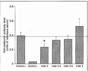
Results are shown in figure 2. There was a significant decrease in the serum anti-ovalbumin antibody level induced by subcutaneous immunization with ovalbumin in ovalbumin-fed mice. However, this decrease was not significant in indomethacin-treated, ovalbumin-fed mice any more, and there was even an increase in the serum anti-ovalbumin antibody level in mice treated with indomethacin 5 mg/kg.
Cellular Systemic Immune Response Evaluated by a Lymphocyte Proliferation Test
Results are shown in figure 3. There was a significant decrease in the specific lymph node cell proliferation rate induced by subcutaneous immunization with ovalbumin in ovalbumin-fed mice. This decrease was specific because the mitogenic activity elicited by pokeweed mitogen 1% was unchanged. This significant decrease was still present in indomethacin-treated mice, even in those treated with 5 mg/ kg indomethacin. Although the decrease was weaker in indomethacin-treated mice, it was not significantly different from the non-treated mice.
Fig. 2. Serum anti-ovalbumin antibody levels, 10 days after subcutaneous immunization, in the different experimental conditions. There was a significant decrease in previously ovalbumin-fed mice (indo 0, p < 0.05). However, the decrease in indomethacin-treatedmice was no longer significant and there was even an increase in the mice treated with indomethacin 5 mg/kg.
Fig. 3. Inguinal lymph node cell proliferation rates in the presence, of ovalbumin, 10 days after subcutaneous immunization at the base of the tail, in the different experimental conditions. There was a significant decrease in the lymphocyte proliferation rate in previously ovalbumin-fed mice both in non-treated (p < 0.004) and
indomethacin-treated mice (p < 0.05).
Cellular Systemic Immune Response Evaluated by DTH
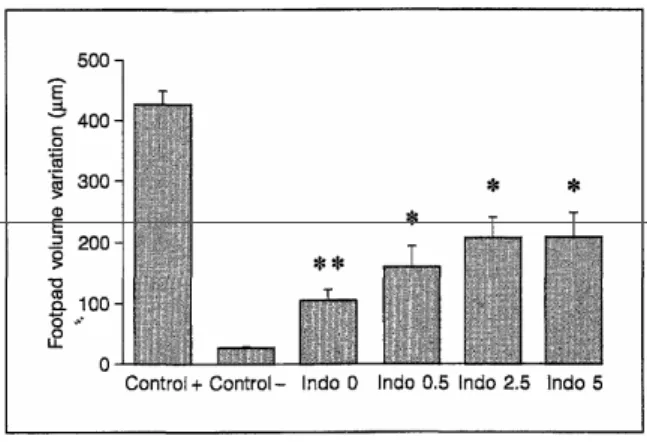
Results are shown in figure 4. There was a significant decrease in footpad swelling after intradermal injection of ovalbumin in ovalbumin-fed mice. This decrease was still significant in indomethacin-treated mice. However, it was less pronounced, and the difference between non-treated mice and mice treated with indomethacin 2.5 or 5 mg/kg was significant.
Fig. 4. DTH to ovalbumin, 10 days after subcutaneous immunization, in the different experimental conditions. There was decrease in the DTH intensity in ovalbumin-fed mice. This decrease was significant both in non-treated (p < 0.0001) and indomethacin-non-treated mice (p < 0.002).
Discussion
This study provides some novel observations of the modifications in intestinal permeability and systemic immune response to fed antigen following oral dosing with indomethacin.
An increased intestinal permeability following oral administration of indomethacin has been shown both in humans and animals using different tests and probes [6-9]. However, permeability to molecules normally present in the digestive tract, which could play a role in the pathophysiology of NSAID enteropathy (e.g. fed antigens, bacterial products), has rarely been studied. Our results show an increased ovalbumin serum level in
ovalbumin-fed mice following oral dosing with indomethacin. This increase was proportional to the dose of indomethacin. It probably reflects an increased passage of undegraded ovalbumin through the digestive epithelium. This increased permeability was associated with histological changes characterized by subepithelial oedema and, for the highest dose of indomethacin, epithelial desquamation.
Systemic immune tolerance to a fed antigen is a well-established phenomenon in mice [10]. Some conditions, such as cyclophosphamide pretreatment [11] or graft-ver-sus-host reaction [12] have been associated with an abrogation of this tolerance. Moreover, abnormalities of the systemic and mucosal immune responses to luminal antigens have been implicated in different human diseases such as coeliac disease and inflammatory bowel diseases [13, 14]. Our results show a breakdown in humoral but only a slight decrease in cellular systemic tolerance to fed ovalbumin in indomethacin-pretreated mice. Such a dissociation between humoral and cellular tolerance induced by fed antigen has already been described in different experimental models [10, 15],
suggesting different mechanisms of induction. Suppressive cells [16], clonal anergy [17] and soluble factors, that is fragments of ovalbumin [18], have been implicated in tolerance induction for cell-mediated immune response. On the other hand, the mechanism of humoral tolerance remains poorly understood [10]. In our study the increased passage of ovalbumin into the blood could allow an increased stimulation of the general immune system not sufficiently suppressed by the mucosal tolerance induction and responsible for the humoral tolerance breakdown. As the epithelial cells may play a role in the induction of tolerance at the mucosal level [19], their dysfunction or destruction induced by indomethacin may also contribute to the defect observed in tolerance induction. Alternatively, indomethacin by itself may have interfered with the induction of systemic tolerance. Indomethacin inhibits the production of several eicosanoids, that have, at physiological concentrations, some immunomodulatory effects [20, 21]. Particularly, in antoher system of systemic tolerance induction in mice, involving injection of de-aggregated human γ-globulin followed by injection of aggregated human γ-globulin, indomethacin has been shown to inhibit the induction of a systemic tolerance for humoral response [22]. It has also been shown to block the suppression of lymph node cells by gut epithelial cells in vitro [23]. Whatever its mechanism, the development of a systemic humoral response to an antigen present in the digestive tract has been shown to induce a mucosal inflammation in an experimental model [24]. Therefore, the humoral tolerance breakdown to a fed antigen in indomethacin-pretreated mice may participate in the development of mucosal inflammation in NSAID enteropathy. Although there was not a breakdown in cellular tolerance, the tolerance level achieved in indomethacin-treated mice was lower than in non-treated mice. This decrease in cellular tolerance was found both in the lymphocyte proliferation test and in the DTH response. An increase in cellular immune responsiveness toward a luminal antigen may also be involved in the development of mucosal lesions [11] and may thus play a role in NSAID enteropathy.
In conclusion, oral administration of indomethacin is followed by an increased passage of fed ovalbumin from the lumen of the digestive tract into the blood, which is probably due to epithelial and subepithelial alteration. This is followed by a decrease in systemic (mainly humoral) tolerance to ovalbumin. Although the precise mechanism of this decrease in tolerance remains to be elucidated, an increased stimulation of the general immune system, a bypass of intestinal epithelial cells or a direct immunomodulating mechanism of indomethacin may be involved. This decrease may participate in the development of inflammatory lesions characteristic of NSAID enteropathy.
References
1 Bjarnason I, Hayllar J, Macpherson AJ, Russel AS: Side effects of nonsteroidal anti-inflammatory drugs on the small and large intestine in humans. Gastroenterology 1993;104:1832-1847.
2 Gargot D, Chaussade S: Nonsteroidal anti-inflammatory drug-induced consequences and disease in the small and large bowel. 1. Experimental data and pathophysiological effects. Gastroenterol Clin Biol 1993;17:485-491.
3 Robert A, Asano T: Resistance of germ free rats to indomethacin-induced intestinal lesions. Prostaglandins 1977;14:333-341. 4 Benoni G, Cuzzolin L, Raimondi MG, Velo GP: Indomethacin-induced intestinal lesions and fecal flora; in Rainsford KD, Velo GP (eds.): Advance in Inflammation Research. New York, Raven Press, 1984, vol 6, pp 103-108.
5 Brune K, Dietzel K, Nurnberg B, Schneider HT: Recent insight into the mechanism of gastrointestinal-tract ulcerations. Scand J Gastroenterol 1987;22(suppl65):8-14.
6 Bjarnason I, Williams P, So A, Zanelli G, Levi J, Gumpel J, et al: Intestinal permeability and inflammation in rheumatoid arthritis: Effect of nonsteroidal anti-inflammatory drugs. Lancet 1984;ii:1171-1175.
7 Bjarnason I, Macpherson A: The changing gastrointestinal side effect profile of nonsteroidal anti-inflammatory drugs: A new approach for the prevention of a new problem. Scand J Gastroenterol 1989;24(supρl 163):56-64.
8 Oman H, Henriksson K, Blomquist L, Johansson SG: Increased intestinal permeability to polysucrose in NSAID-treated patients. Eur J Gastroenterol Hepatol 1992;4:235-240.
9 Mion F, Cuber J-C, Minaire Y, Chayvialle J-A: Short term effects of indomethacin on rat small intestinal permeability: Role of eicosanoids and platelet activating factor. Gut 1994;35:490-495.
10 Mowat AM: The regulation of immune responses to dietary protein antigens. Immunol Today 1987;8:93-98.
11 Mowat AM, Ferguson A: Hypersensitivity in the small intestinal mucosa. V Induction of cell-mediated immunity to a dietary antigen. Clin Exp Immunol 1981;43:574-582.
12 Strobel S, Mowat AM, Ferguson A: Prevention of oral tolerance induction to ovalbumin and enhanced antigen presentation during a graft-versus-host reaction in mice. Immunology 1985; 56:57-64.
13 Cerf M, Cerf-Bensussan N: Le syndrome d'atrophie villositaire. Hepato-gastroentérologie 1994;1:63-70.
14 Fantry GT, James SP: Cellular and molecular immunology and biochemistry of inflammatory bowel disease. Curr Opin Gastroenterol 1993;9: 544-551.
15 Lamont AG, Gordon M, Ferguson A: Oral tolerance in protein-deprived mice. I. Profound antibody tolerance but impaired DTH tolerance after antigen feeding. Immunology 1987;61:333-337.
16 Bland P, Warren LG: Antigen presentation by epithelial cells of the rat small intestine. II. Selective induction of suppressor T cells. Immunology 1986;58:9-14.
17 Whitacre C, Gienapp IE, Orosz CG, Bitar DM: Oral tolerance in experimental autoimmune encephalomyelitis. III. Evidence for clonal energy. J Immunol 1991;147:2155-2163.
18 Bruce MG, Ferguson A: The influence of intestinal processing on the immunogenicity and molecular size of absorbed, circulating ovalbumin in mice. Immunology 1986;59:295-300.
19 Mayer L, Eisenhardt D: Lack of induction of suppressor T cells by intestinal epithelial cells from patients with inflammatory bowel disease. J Clin Invest 1990;86:1255-1260.
20 Webb DR, Osheroff PL: Antigen stimulation of prostaglandin synthesis and control of immune responses. Proc Natl Acad Sci USA 1976;73: 1300-1304.
21 Goodwin JS, Messner P, Peake GT: Prostaglandin suppression of mitogen-stimulated lymphocytes in vitro. J Clin Invest 1978;62:753-760.
22 Scheuer WV, Hobb MV, Weigle WO: Interference with tolerance induction in vivo by inhibitors of prostagladin synthesis. Cell Immunol 1987;104:409-418.
23 Santos LM, Lider O, Audette J, Khoury SJ, Weiner HL: Characterization of immunomodulatory properties and accessory cell function of small intestinal epithelial cells. Cell Immunol 1990;127:26-34.