Université de Montréal
Examining how PSMB11 orchestrates T cell development
par Anca Apavaloaei
Programme de Biologie Moléculaire Faculté de Médecine
Mémoire présenté
en vue de l’obtention du grade de Maître ès Sciences en Biologie Moléculaire
option Générale
Résumé
Les lymphocytes T CD8 jouent un rôle majeur dans l’immunosurveillance de l’organisme contre les pathogènes de même que les cellules pré-cancéreuses. Afin d’induire une réponse cytotoxique efficace contre ces événements anormaux, les lymphocytes T CD8 doivent exprimer à leur surface un récepteur des cellules T (TCR) qualifié et capable de reconnaître les molécules étrangères. Cette qualification s’effectue au sein du thymus, qui représente l’unique organe se spécialisant dans l’éducation des lymphocytes T CD4 et CD8. Dans le cortex thymique, les thymocytes exprimant un TCR fonctionnel sont sélectionnés positivement par les cellules épithéliales corticales thymiques (cTECs). Ceux-ci transitent ensuite vers la médulla thymique où les thymocytes autoréactifs sont éliminés afin de prévenir l’autoimmunité.
PSMB11 est une sous-unité catalytique du thymoprotéasome, qui est exprimé de façon exclusive par les cTECs, des cellules spécialisées dans la production et la présentation des peptides associés au CMH de classe I (MIPs). Ainsi, une déficience en PSMB11 altère sévèrement le développement des thymocytes CD8. Étant donné que les thymoprotéasomes présentent une faible activité chymotrypsique, leur rôle présumé est de produire des peptides ayant une faible affinité pour le CMH de classe I pour ainsi assurer la sélection positive des thymocytes CD8 exprimant un TCR de faible affinité. À ce jour, aucune étude n’a toutefois réussi à élucider le rôle fondamental de PSMB11 dans la sélection positive.
Puisque les protéasomes peuvent être impliqués dans plusieurs processus cellulaires, nous avons vérifié si PSMB11 1) orchestre des activités peptide-indépendantes dans les cTECs, et 2) son impact sur le développement des thymocytes. Nous avons montré que les cTECs de souris Psmb11-/- présentent une expression différentielle de gènes impliqués dans l’adhésion cellule-cellule ainsi que dans la signalisation des cytokines, deux processus étroitement liés à la sélection positive. Nous avons également observé des niveaux élevés de CXCR4 à la surface des thymocytes 8SM des souris Psmb11–/–, suggérant que les thymocytes CD8 sont retenus plus longtemps dans le cortex comparativement aux souris WT. Des gènes pro-inflammatoires sont aussi surexprimés dans les cTECs déficientes en PSMB11, menant à la détection d’une augmentation du stress dans les thymocytes. Enfin, nous avons montré que PSMB11 altère l’expression de Cd83 et Prss16, deux gènes ayant un rôle essentiel dans le développement des
lymphocytes T CD4. En conclusion, notre étude dévoile un tout nouveau rôle de PSMB11 dans le développement des lymphocytes CD4 et CD8, soit l’orchestration de la transcription des gènes au sein des cTECs.
Mots-clés: Thymoprotéasome, PSMB11, cellules épithéliales thymiques, thymocytes, sélection positive.
Abstract
CD8 T cells are central to the body’s immunosurveillance against pathogens and pre-cancerous cells. To achieve a cytotoxic response to abnormal events, CD8 T cells must bear a competent T cell receptor (TCR). The thymus is unique for “educating” developing CD4 and CD8 T cells to recognize foreign material. In the thymic cortex, thymocytes displaying functional TCRs are positively-selected by cortical thymic epithelial cells (cTECs). Upon arrival to the medulla, T cells which recognize self-antigens presented by medullary thymic epithelial cells (mTECs) receive the “death sentence” to prevent autoimmunity.
PSMB11 is a catalytic subunit of the thymoproteasome expressed exclusively in cTECs, which mediates the generation of major histocompatibility complex class I-associated peptides (MIPs). In consequence, PSMB11-deficiency severely impairs the development of CD8 thymocytes. Since thymoproteasomes display a low chymotrypsin activity, it was inferred that they produce peptides with low MHC class I affinity, and by similarity, that they produce low TCR affinity. However, indirect studies failed to elucidate the fundamental role of PSMB11 in positive selection.
Proteasomes regulate essentially all cellular processes, therefore we investigated 1) if PSMB11 orchestrates peptide-independent processes in cTECs, and 2) the impact on thymocyte development. We found that cTECs in Psmb11-/- mice show differential expression of genes
involved in cell-cell adhesion and cytokine signaling, which are at the core of positive selection. Elevated levels of CXCR4 at the cell surface of 8SM thymocytes in Psmb11–/– mice suggest that
CD8 thymocytes have a longer retention time in the cortex compared to the WT. Pro-inflammatory genes were upregulated in cTECs lacking PSMB11, which might underlie the increased stress detected in medullary thymocytes. Finally, we found that PSMB11 alters the expression of Cd83 and Prss16, genes which have an essential role in CD4 T cell development. In conclusion, our study describes novel peptide-independent means of PSMB11 to regulate CD4 and CD8 thymocyte development, by orchestrating the transcriptome in cTECs.
Table of Contents
Résumé ... i Abstract ... iii Table of Contents ... iv List of Tables ... vi List of Figures ... vii List of Acronyms ... viii Acknowledgements ... x Chapter 1 – Introduction ... 1 1.1 The adaptive immune system ... 1 1.1.1 Antigen recognition by immune cells ... 1 1.1.2 Antigen presentation ... 2 1.1.3 MHC class I presentation ... 2 1.1.4 MHC class II presentation ... 4 1.1.5 Human HLA molecules ... 5 1.1.6 Antigen processing ... 5 1.2 T cell development ... 6 1.2.1 The thymus and the thymic journey of lymphocytes ... 6 1.2.2 Antigen presentation in the thymus ... 8 1.2.3 Positive selection ... 8 1.2.4 Negative selection ... 9 1.2.5 Agonist selection and unconventional T cells ... 9 1.3 Research context ... 10 1.3.1 The thymoproteasome ... 10 1.3.2 Objectives ... 11 Chapter 2 – PSMB11 shapes the transcriptome of cTECs and has pervasive effects on both CD4 and CD8 thymocyte population ... 12 2.1 ABSTRACT ... 13 2.2 INTRODUCTION ... 13 2.3 RESULTS ... 152.3.2 Psmb11-/- cTECs resemble mTECs in their transcription factor activity ... 18
2.3.3 Psmb11 deletion affects CD4 and CD8 thymocyte maturation irrespective of age………19
2.3.4 CD8 thymocytes selected in Psmb11-/- mice show increased susceptibility to apoptosis………..21
2.3.5 Lack of thymoproteasomes induces stress in developing thymocytes ... 22
2.3.6 CD8 thymocytes show patterns of agonist selection, whereas CD4 thymocytes upregulate stress-responsive genes ... 24 2.3.7 Data suggest impaired cytokine responsiveness and migration of CD8 thymocytes in Psmb11-/- thymi ... 28 2.3.8 Data suggest a direct impact of PSMB11 on the CD4 lineage ... 31 2.4 DISCUSSION ... 31 2.5 EXPERIMENTAL PROCEDURES ... 33 2.3.9 Mice ... 33 2.3.10 Flow cytometry and cell sorting ... 33 2.3.11 Immunofluorescence analyses ... 34 2.3.12 BrdU administration ... 34 2.3.13 RNA sequencing ... 34 2.3.14 Promoter region analysis ... 35 2.3.15 Statistical test ... 35 2.6 AUTHOR CONTRIBUTIONS ... 35 2.7 CONFLICT OF INTEREST ... 36 2.8 ACKNOWLEDGMENTS ... 36 2.9 REFERENCES ... 37 2.10 Supplementary Figures ... 42 Chapter 3 – Discussion & Perspectives ... 46 Conclusion ... 49 Bibliography ... i Appendix 1 – DEGs in the 8SM subset ... v Appendix 2 – DEGs in the 8M1 subset ... xvii Appendix 3 – DEGs in the 8M2 subset ... xx Appendix 4 – DEGs in the 4SM subset ... xxvi Appendix 5 – DEGs in the 4M1 subset ... xl Appendix 6 – DEGs in the 4M2 subset ... xli
List of Tables
Table 1. DEGs CD8 thymocytes in Psmb11-/- mice show aberrant expression of genes specific to unconventional T cell lineages.. ... 26 Table 2. CD4 thymocytes in Psmb11-/- mice upregulate expression of stress-responsive and rescue genes.. ... 27
List of Figures
Figure 1. MHC class I presentation pathway.. ... 3
Figure 2. MHC class II presentation pathway.. ... 5
Figure 3. Different forms of proteasomes.. ... 6
Figure 4. Thymus morphology in the mouse.. ... 7
Figure 5. PSMB11 is a master regulator of the transcriptome in cTEC. ... 17
Figure 6. Impact of Psmb11-deletion on CD4 and CD8 thymocyte numbers. ... 20
Figure 7. Loss of PSMB11 induces stress onto developing thymocytes. ... 23
Figure 8. Impact of Psmb11-deletion on CD4 and CD8 thymocytes. ... 30
Figure 9. Analysis of TEC populations in WT and Psmb11-/- mice. ... 42
Figure 10. Flow cytometry analysis of the thymocyte maturation subsets. ... 43
Figure 11. Immunofluorescence analysis of HSP70. ... 45
List of Acronyms
7-AAD 7-amino-actinomycin D APC Antigen-presenting cell β2m β2-microglobulin BCR B cell receptor CMJ Cortico-medullary junction cTEC Cortical thymic epithelial cell CTL Cytotoxic T cell DC Dendritic cell DEG Differentially expressed gene DNA Deoxyribonucleic acid DP Double positive ECM Extracellular matrix e.g. for example ER Endoplasmic reticulum ERAD ER-associated degradation FACS Fluorescence activated cell sorting FPKM Fragments per kilobase million GMFI Geometric mean fluorescence intensity GO Gene-ontology HLA Human leukocyte antigen i.e. for instance IFNg Interferon-γ IL-2 Interleukin-2 iNKT Invariant natural killer T cell M1; M2 Mature 1; mature 2 MHC Major histocompatibility complexMIP MHC class I-associated peptide mTEC Medullary thymic epithelial cell PCD Programmed cell death PWM Position weight matrix RNA Ribonucleic acid RNA-Seq RNA-Sequencing SM Semi-mature TAP Transporter associated with antigen processing TCR T cell receptor TEC Thymic epithelial cell TF Transcription factor TGFβ Tfh Tumor growth factor β Follicular T helper Th T helper TNFa Tumor necrosis factor α Treg nTreg Induced regulatory T cell Natural regulatory T cell TSSP Thymus specific serine protease
Acknowledgements
First and foremost, I am grateful to my research director Dr. Claude Perreault for welcoming me to his team with a firm belief in my abilities. It boosted my enthusiasm and encouraged me to pursue this work during the past two years. I admire his wisdom and I especially enjoyed the engaging scientific conversations with him. I feel very fortunate to have had him as a mentor.
Thank you to Sylvie for introducing me to flow cytometry, and to Alex who patiently helped me through my first attempt to analyze RNA-Seq data. I am greatly appreciative for the friendly and scientific discussions with Charles and Marie-Pierre during early-mornings, and also for being ready to answer my countless inexperienced questions. Thank you to the entire Perreault group for sharing ideas, concepts and personal experiences over coffee or lunch. I believe I had the opportunity to work with some of the most talented and passionate people.
At IRIC it has been a pleasure to do research with the personnel at the flow cytometry, microscopy, histology, genomics and bioinformatics platforms. A big thank you to Danièle Gagné for teaching me the intricacies of multicolor flow cytometry and being there for assistance over emails even after her departure from IRIC. Likewise, I thank Gaël Dulude for taking over the many hours of sorting after Danièle’s leave. Special thanks go to Christian Charbonneau and Julie Hinsinger, who guided me through hours of technique optimization, and to Jalila for her invaluable experimental suggestions.
I am grateful for the thought-provoking discussions with Dr. Heather Melichar. Thank you to her and Maggie for joining forces and helping us with cell migration assays. Thank you to Dr. Julie Lessard and Dr. Nathalie Labrecque for their time and effort in revising my master’s thesis.
I would like to acknowledge my family and friends for their constant support from all around the world. Finally, this work would not have been possible without the generous support of the Canadian Institutes of Health Research and NIH.
Chapter 1 – Introduction
1.1 The adaptive immune system
There are two major threats to the immune system, foreign materials (viruses and pathogenic bacteria) and abnormal cellular processes in stressed and neoplastic cells. The immune system has evolved in vertebrates to recognize these threats, fight them and develop long-term memory for antigen re-exposure 1.
1.1.1 Antigen recognition by immune cells
The adaptive immune system is defined by B and T cell lineages, which recognize foreign antigens via surface B cell receptors (BCRs) and T cell receptors (TCRs), respectively. Genetic rearrangements and somatic hypermutations of the DNA regions encoding lymphocyte receptors allow the recognition of a diverse antigen repertoire 2. In the peripheral lymphoid tissues, the binding of an antigen to the BCR on B cells has a two-fold function: it triggers signal transduction in B cells and the internalization of the antigen. The antigen is degraded intracellularly and presented at the cell surface via major histocompatibility complex (MHC) class II molecules (discussed in 1.1.2) for recognition by helper T cells. Helper T cells that recognize the antigenic peptides on B cells and receive co-stimulatory signals become activated. The activation of helper T cells allows the activation of B cells through the interaction between the CD40 ligand and the CD40 protein on B cells, and the production of cytokines which stimulate the proliferation and differentiation of B cells into secreting cells. The release of about 2000 antibodies with the same binding site per second mediates target neutralization 3.
Targets that are not accessible to antibodies, i.e. intracellular pathogens which multiply in the cytoplasm of infected cells or molecules expressed by abnormal cells, can be eliminated solely via proteolytic destruction by the respective antigen-presenting cells (APCs). Subject APCs load peptides resulting from the internal antigen degradation onto MHC class I or class II complexes for surface presentation, which activates a T cell response 4. Recognition of peptide/MHC class I molecules on APCs by the TCR is necessary for a cytotoxic immune response by CD8 T cells. Cytotoxic T cells (CTLs) release granzymes, perforins and cytokines
like interferon-γ (IFNg) into the intercellular space, which induce the programmed-cell death (PCD) of the infected cells. CTLs are also thought to detect quantitative and qualitative antigenic differences at the cell surface. In this regard, cancerous or stressed cells alter their intracellular protein homeostasis and thus the repertoire of peptide/MHC class I at the cell surface, allowing CD8 T cells to scan for cellular alterations and induce apoptosis, a form of PCD 5.
Recognition of surface MHC class II complexes loaded with macromolecular parts of pathogens is the role of CD4 T cells. The initial activation of a naïve CD4 T cell signals its differentiation into effector T helper (Th) cells, including Th1, Th2, Th17, Th9, Tfh (follicular helper T cells) and iTreg (induced regulatory T cells). The task of Th1 cells is to aid CTLs in their immune response by secreting IFNg and tumor necrosis factor α (TNFa), or to stimulate B cells to produce antibodies. Th2 cells are identified by the production of interleukins and mainly priming of B cells to produce immunoglobulin E for the defense against extracellular pathogens
3. In addition to common cytokines produced by more than one Th cell type, e.g. 4 and
IL-13, Th17 and Th9 cells produce the signature cytokines IL-17 and IL-9, respectively. In contrast, Tfh cells are essential for germinal center formation and the development of memory B cells with the highest antigen affinity, whereas iTregs are suppressive cells involved in tolerance that are not generated in the thymus 6.
1.1.2 Antigen presentation
Whereas B cells are capable of recognizing native antigens in their intact form, TCRs recognize antigen-derived peptides in complex with an MHC molecule. Both MHC class I and MHC class II complexes have the purpose of presenting antigen-derived peptides on the cell surface of T cells. What makes the difference between these complexes is the origin of the pathogen. In the classic pathway described below, class I molecules load peptides derived from intracellular sources, whereas class II molecules from extracellular sources. Other non-classical pathways are termed cross-presentation, where the extracellular-derived antigens are presented by class I molecules, and cytosolic antigens by class II molecules via autophagy 7,8.
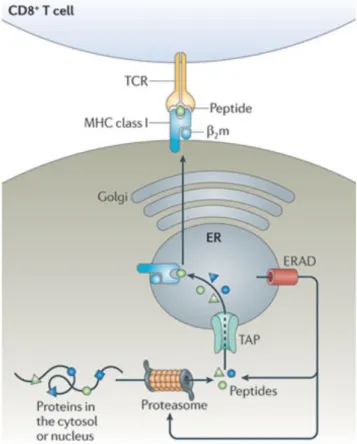
by the transporter associated with antigen presentation (TAP) to the endoplasmic reticulum (ER) where they are loaded onto MHC class I molecules, or further trimmed by the ER-associated degradation (ERAD) machinery to fit the binding groove of the MHC. The trimeric complex formed by the MHC I heavy chain, the β2-microglobulin light chain and the peptide are transported to the cell surface via the Golgi apparatus, where they are accessible for interaction with the TCR on CD8 T cells (Figure 1) 8,9.
Figure 1. MHC class I presentation pathway. Intracellular proteins are degraded into 8-10 amino-acid-long peptides by the proteasome and translocated to the endoplasmic reticulum (ER) via the transporter associated with antigen presentation (TAP) complex. Peptides are loaded onto MHC class I complexes forms by a heavy chain and the light chain β2-microglobulin. Peptides of inappropriate length are further trimmed by the ER-associated degradation (ERAD) machinery to fit the binding groove of the MHC. Peptide/MHC complexes are now ready for transport to the plasma membrane via the Golgi for recognition by CD8 T cells. Figure adapted from 8.
1.1.4 MHC class II presentation
Whereas MHC class I molecules are present on all nucleated cells, MHC class II complexes are only found on professional APCs, i.e. dendritic cells (DCs), B cells and macrophages, or on non-APCs induced with IFNg, which constitutively process protein antigens and present them to the cell surface for immune surveillance in the form of peptide/MHC molecules 8.
The α and β chains of the MHC II complex are assembled in the ER with the Ii chain which is involved in transport and stability, promoting an open conformation of the binding groove. Upon transport to the endosomal compartments, H2-M facilitates the exchange of the CLIP chain, resulted from the proteolytic fragmentation of the Ii chain in the endosome, with the antigenic peptides. The peptides suitable for MHC II presentation are derived from exogenous proteins and cleaved by proteases in early endosomes which fuse with the endosomal compartments in APCs. Stable peptide/MHC class II complexes are translocated to the plasma membrane of APCs and recognized by the TCR on CD4 T cells (Figure 2) 8.
Figure 2. MHC class II presentation pathway. Extracellular proteins are endocytosed and cleaved by proteases in early endosomes that fuse use the endosomal compartments. The α and β chains of the MHC II complex are assembled in the ER lumen and transported to the MHC II compartment (MIIC) via Golgi. With the help from H2-M, the CLIP peptide derived from the surrogate Ii chain is exchanged for an antigenic peptide. Peptide/MHC II complexes are now ready for translocation to the plasma membrane for interactions with CD4 T cells. Figure adapted from 8.
1.1.5 Human HLA molecules
In humans, the MHC molecules are called human leukocyte antigens (HLA), and there are six major types of HLA molecules. Similar to MHC class I in mice, CD8 T cells interact with HLA-A, HLA-B and HLA-C, whereas HLA-DP, -DQ and –DR are recognized by the TCRs on CD4 T cells 10.
1.1.6 Antigen processing
The stability of peptide/MHC complexes is essential for translocation to the cell surface, and is conferred by peptide binding into the cleft of MHC molecules. The peptides resulted from proteolytic degradation have two particularities that allow them to fit into the MHC binding cleft: proper length and anchor residues 9.
The ubiquitously expressed 26S proteasome, also called the constitutive proteasome, degrades cytosolic proteins for MHC class I presentation. Constitutive proteasomes generate peptides with hydrophobic C-termini which serve as anchors to the binding groove of the MHC I molecules. Two alternative proteasomes have been described, the immunoproteasome and the thymoproteasome. Immunoproteasomes are expressed by immune cells, stressed and IFNg-exposed cells, and display an increased activity compared to constitutive proteasomes, thus efficiently cleaving proteins that cannot be handled by constitutive proteasomes. Thymoproteasomes are thymus-specific, and play a role in the development of CD8 T cells 8. The core of the proteasome is composed of two outer rings with 7 α (α1-α7) subunits and two inner rings with 7 β (β1-β7) subunits. The proteolytic subunits of constitutive proteasomes are PSMB6, PSMB7 and PSMB5 (or β1, β2 and β5, respectively), whereas immunoproteasomes assemble PSMB9, PSMB10 and PSMB8 (also known as β1i, β2i and β5i). Lastly, only one catalytic subunit is different between immunoproteasomes and thymoproteasomes: PSMB8 is
replaced by PSMB11 (or β5t) (Figure 3). The assembly of different catalytic subunits alters the protein cleavage preferences and characteristics of the resulting peptides 11.
Figure 3. Different forms of proteasomes. Proteolytic subunits of the constitutive proteasomes PSMB6, PSMB7 and PSMB5 (or β1, β2 and β5, respectively), immunoproteasomes assemble PSMB9, PSMB10 and PSMB8 (also known as β1i, β2i and β5i). Lastly, only one catalytic subunit is different between immunoproteasomes and thymoproteasomes, PSMB8 is replaced by PSMB11 (or β5t). Figure adapted from 11.
Proteolytic cleavage of MHC class II-associated peptides is performed by lysosomal proteases residing in the endosomal compartment. Among them, cathepsin S and cathepsin L are the two known proteases with non-redundant roles in endosomal presentation. Whereas cathepsin S has essential roles in B cells and DCs, cathepsin L is specifically expressed in the thymus 12.
1.2 T cell development
1.2.1 The thymus and the thymic journey of lymphocytes
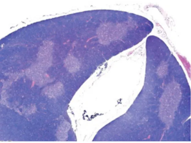
The thymus is unique for its ability to produce immunocompetent T cells 13. Located in the pericardial mediastinum, it is formed by two lobes linked by a connective tissue called isthmus. Each lobe is divided into interconnecting lobules in most species, but not in the mouse. The thymic lobes of mice are subdivided into cortical and medullary areas separated by the corticomedullay junction (CMJ) predominant in blood vessels (Figure 1). Being an epithelial organ, the thymic stroma is formed by cortical thymic epithelial cells (cTECs) in the cortex, medullary thymic epithelial cells (mTECs) in the medulla, endothelial cells forming the
cell development in the thymus are B cells, DCs and macrophages. Lymphoid precursors travel from the bone-marrow (BM) to the thymus as a prerequisite for becoming mature T cells, and emigrate into the periphery as naïve T cells upon completion of the thymic “education” 14.
Lymphoid precursors enter the thymus through the vasculature at the CMJ and progress through 4 different stages of maturation characterized by the surface expression of the CD4 and CD8 coreceptors. Initially, double-negative thymocytes reside in the cortex and receive cues to differentiate into double-positive (DP) cells expressing both CD4 and CD8 coreceptors. DP thymocytes represent around 80% of total thymocytes and express a diverse repertoire of TCR specificities which must be selected for their immunocompetence, i.e. for their ability to bind the limited MHC repertoire to insure instant reaction in the case of pathogenic threats. Each T cell expresses a unique cell surface TCR resulted from the rearrangement of the TCRβ and then TCRα transmembrane chains. Successful engagement of the TCR with MHC class I or class II molecules on professional APCs signals thymocytes to differentiate into CD8 or CD4 single-positive thymocytes, respectively, which migrate to the medulla. Further interactions with the medullary stroma prepare thymocytes for entry into the peripheral circulation. Mature thymocytes can be identified by changes in cell size, proliferation competence and expression of differentiation antigens and interleukin receptors 15,16.
Figure 4. Thymus morphology in the mouse. Hematoxylin and eosin staining of a normal thymus from a 3-month-old B6C3F1 mouse showing the cortex in dark blue and the medulla in light blue. Occasional pink areas inside the organ are stained blood vessels. Figure adapted from 16.
1.2.2 Antigen presentation in the thymus
All thymic epithelial cells, namely cTECs and mTECs, are professional APCs and capable to present both MHC class I and class II molecules loaded with peptides. In the thymus, antigen presentation serves a different purpose than described in 1.1: it prepares T cells for the encounter of a diverse repertoire of antigens in the periphery and prevents an inappropriate recognition of the host. Thus, the cortex is specialized in selecting the T cells with functional TCRs via a process called positive selection, whereas in the medulla the T cells with high affinity to the host are “sentenced to death” through negative selection 17.
1.2.3 Positive selection
The positive selection of DP T cells is mediated by cTECs which exclusively express thymoproteasomes, and the proteases cathepsin L and thymus-specific serine protease (TSSP). cTECs express unique peptide/MHC complexes at the cell surface for TCR engagement, and the TCR signaling strength determines the fate of T cells. A too low interaction results in “death by neglect”, whereas excessive signaling can cause apoptosis. The intermediate affinity of the TCR to the peptide/MHC on cTECs signals the decrease in the CD8 coreceptor expression on DP cells. CD8 thymocytes require a timely (18-53 h) termination of the TCR signaling which allows cytokines to restore surface CD8 levels and induce expression of the transcription factors (TFs) specifying assignment to the CD8 lineage, e.g. Runx3. Since the TCR intensities may vary, the cytokine signaling is used as a “compensatory” mechanism to ensure correct commitment to the CD8 lineage (that is, stronger TCR signals accelerate cytokine responsiveness). If TCR signaling continues, it results in the expression of the TF ThPOK which prevents induction of Runx3 and determines commitment to the CD4 lineage 18. cTECs express
the CD83 receptor on the cell surface, which limits the MHC II turnover and allows longer signaling times of CD4 T cells. Thus CD4 T cells are selected based on stronger TCR signals than CD8 T cells 19.
Whereas the extracellular domains of the CD4 and CD8 coreceptors couple to the MHC II and MHC I molecules respectively, the intracellular tails associate with the Src family protein tyrosine kinase Lck, which is needed for transduction of the TCR signals. CD8 coreceptors
Thus, the strength of TCR signaling determines lineage decision in positive selection, and it is defined by the persistence or cessation of the CD4 and CD8 coreceptor expression 18.
1.2.4 Negative selection
Following positive selection, thymocytes migrate to the medulla. mTECs and medullary DCs that are responsible for the negative selection of self-recognizing TCRs constitutively express immunoproteasomes and constitutive proteasomes. As a safeguard against autoimmunity, mTECs ectopically express a myriad of antigens from peripheral tissues, named tissue-restricted antigens, for presentation to T cells. This phenomenon is controlled by the autoimmune regulator AIRE, and perhaps the transcription factor Fezf2 20. High-affinity TCR interactions with the self-peptide/MHC induces thymocyte apoptosis. Therefore, immunoproteasomes facilitate the cleavage of an otherwise exhausting pool of proteins, but also shape the TCR repertoire of CD8 T cells 21.
Similar to cTECs, mTECs display a high rate of macroautophagy. TECs inefficiently use the classical endocytic pathway for MHC II loading, and instead fuse autophagosomes loaded with intracellular proteins with the endosomal compartments. Cell-surface presentation of self-antigen/MHC II complexes shapes the repertoire of CD4 T cells 21.
1.2.5 Agonist selection and unconventional T cells
Approx. 98% of DP thymocytes die an apoptotic death due to failing positive selection, or, much less often, due to negative selection 4. Nonetheless, in exceptional cases T cells receiving strong TCR signals evade apoptosis and develop into unconventional T cells, i.e. natural regulatory T cells (nTregs), invariant natural killer T (iNKT) cells. This process is called agonist selection 22,23.
iNKT cells are αβ T cells that recognize CD1 non-polymorphic molecules loaded with lipid antigens on the surface of DCs. iNKT cells branch off from the DP stage of thymocyte development in the thymus and lose the CD4 and CD8 coreceptors to become DN cells. iNKT cells originate preferentially from DP cells with high-affinity self-ligands and development of this T cell lineage is dependent on the interaction between the CD28 and B7 costimulatory molecules 23,24.
Finally, nTreg development results from the strong reactivity of CD4thymocytes to self and are identified by expression of Foxp3. The availability of interleukin-2 (IL-2) and transforming growth factor β (TGFβ) in the thymic medulla is favorable and limiting for the commitment to the nTreg fate. Paradoxically, the role of nTreg in the periphery is to promote T cell tolerance to self-antigens and to prevent autoimmune diseases, and it differs from iTregs in that nTregs develop in the thymus, whereas iTregs in the periphery (section 1.1.1) 23.
Another unconventional T cell lineage are γδ T cells, which arise from the DN stage. TCR rearrangements during early T cell development result in αβ or γδ TCR T cells. DN thymocytes rearrange the β, γ and δ TCR genes. The successful in-frame rearrangement of the TCRβ is necessary for the expression of the pre-TCR, a prerequisite to the αβ TCR. This induces the silencing of the TCRγ gene and initiation of the TCRα gene rearrangement. Transition to the DP stage is a hallmark of αβ T cell commitment, whereas γδ T cells with successfully rearranged γ and δ TCRs egress to the periphery as DN cells 22.
1.3 Research context
1.3.1 The thymoproteasome
The proteasome activity is known to be required for MHC class I but not for class II antigen presentation 25. Since the identification of β5t in 2007, scientists have been trying to
understand how its specific activity in cTECs influences the positive selection of CD8 T cells. It has been shown that absence of the thymoproteasome exclusively depletes and affects the repertoire of CD8 T cells (CD4-CD8=TCRβ+ cells) and not the CD4 population (CD4+CD8 -TCRβ+ cells), based on cell numbers and distribution of a set of TCR specificities. CD8 T cells developed in the absence of PSMB11 show impaired allogeneic and antiviral responses, despite normal thymic morphology 26,27. Thymoproteasomes display decreased chymotrypsin activity compared to constitutive proteasomes and immunoproteasomes, theoretically resulting in MHC I-associated peptides (MIPs) with hydrophilic C-termini and low MHC affinity. However, evidence of similar half-life of peptide/MHC complexes between cTECs and mTECs went against this hypothesis 28.
locus with the β5i gene in β5i-deficient mice did not rescue the CD8 thymocyte numbers. The role of PSMB11 is also not to prevent excessive thymocyte deletion during positive selection, since the inactivation of the cell-death mediator Bim did not restore the number of CD8 thymocytes 17.
Since the nature of MIPs cannot be directly assessed with the current mass spectrometry technology which requires 105 times more cells than can be isolated from one mouse (approx. 104 cTECs) 26, researchers examined 100 MIPs on the surface of fibroblasts with transgenic expression of β5t or β5i. This indirect approach unveiled a differential pattern of the amino-acids involved in TCR recognition, and it was inferred that thymoproteasomes generate low TCR-affinity peptides 29. It is known that CD8 thymocytes in PSMB11-deficient thymi receive a stronger TCR signaling compared to cells in WT mice based on two widely-accepted readout molecules, CD5 and Nur77 30. However, 100 MIPs are not enough to draw a strong conclusion and the precise role of thymoproteasomes in positive selection has not been elucidated to date. 1.3.2 Objectives
None of the studies mentioned in 1.3.1 examined a crucial aspect of proteasomes, the regulation of essentially all cellular processes. The cross talk between thymocytes and the cortical microenvironment is critical for the development of T lymphocytes during positive selection (Robey and Fowlkes, 1994). Therefore, we hypothesized that PSMB11 orchestrates cellular functions in cTECs that are peptide-independent, yet essential for thymocyte development. More precisely, we wondered whether a distinct proteolytic activity in PSMB11-deficient cTECs might change the levels of activation and survival-inducing molecules central to thymocyte development. This work had two main objectives:
1) to understand how thymoproteasomes regulate the cTEC biology using a genome-wide RNA-Sequencing (RNA-Seq) approach
2) to evaluate the impact of 1) on thymocyte development. We employed RNA-Seq, flow cytometry and microscopy to examine the cell numbers and the phenotype of CD4 and CD8 thymocytes, including proliferation ability, resistance to apoptosis and cytokine responsiveness.
Chapter 2 – PSMB11 shapes the transcriptome of cTECs
and has pervasive effects on both CD4 and CD8 thymocyte
population
Anca Apavaloaei1,2, Sylvie Brochu1, Alexandre Rouette1,2, Marie-Pierre Hardy1, Geno Villafano3, Claude Perreault1,2.
1Institute for Research in Immunology and Cancer, University of Montreal, Montreal, QC H3C
3J7, Canada.
2Department of Medicine, University of Montreal, Montreal, QC H3C 3J7, Canada.
3Department of Molecular and Cell Biology, University of Connecticut, Connecticut, CT 06269,
USA.
Correspondence: claude.perreault@umontreal.ca
2.1 ABSTRACT
How thymoproteasomes contribute to the positive selection of CD8 T cells is still not completely clear. Indirect evidence led to the dogma that PSMB11 generates a unique peptide repertoire with low T cell receptor (TCR) affinity for major histocompatibility complex (MHC) class I presentation. However, the role of proteasomes in cellular functions has been largely disregarded. Herein, we aimed to investigate if PSMB11 might regulate the cortical thymic epithelial cell (cTEC) biology with an effect on T cell development. We found that thymoproteasomes have an overall repressive activity on the transcriptome of cTECs, principally on the genes encoding extracellular matrix (ECM) components, cell-cell adhesion molecules and tumor-necrosis factor (TNF)-associated chemokines. We show that post-selection T cells of both CD4 and CD8 lineages undergo significant stress in the PSMB11-deficient thymus, likely due to extended interactions with cTECs. CD8 thymocytes appear to be reprogrammed to commit to apoptosis or unconventional T cell lineages, whereas CD4 thymocytes show transcriptional stimulation of recovery and survival genes. Although it remains possible that CD4 thymocytes are affected by the decline in the CD8 cell number, our data suggest a direct effect of PSMB11-deficiency on the selection of CD4 T cells through a decrease in the transcription of Prss16 and Cd83. In summary, our study describes novel means of PSMB11 to orchestrate CD4 and CD8 thymocyte development, by controlling the matricellular and chemokine homeostasis in the thymic cortex.
2.2 INTRODUCTION
Vertebrates express two main types of proteasomes: constitutive proteasomes and immunoproteasomes (Kniepert and Groettrup, 2014). Via proteolysis of cellular proteins, both types of proteasomes regulate many basic cellular processes including cell cycle progression and division, differentiation and development, morphogenesis and response to stress (Glickman and Ciechanover, 2002; de Verteuil et al., 2014). In addition, degradation of proteins by proteasomes generates peptides that are presented by MHC class I proteins (Kincaid et al., 2012; de Verteuil et al., 2010). Quite remarkably, Murata and colleagues reported in 2007 that cTECs
expressed a third type of proteasome that they named the thymoproteasome (Murata et al., 2007). Thymoproteasomes share two catalytic subunits with immunoproteasomes (PSMB9 and PSMB10) and contain a third catalytic chain which is unique to cTECs, PSMB11. Psmb11-/-
mice display defective positive selection of CD8 thymocytes in the thymus, and their CD8 T cells show impaired antigen responsiveness (Nitta et al., 2010; Takada et al., 2015; Xing et al., 2013).
Given that MHC I peptides (MIPs) regulate the positive selection of CD8 thymocytes, it was logical to posit that the key role of thymoproteasomes might be to generate a unique MIP repertoire (Nitta et al., 2010). Indeed, since they are shown to have a lower chymotryptic activity than other types of proteasomes, it was suggested that thymoproteasomes might generate peptides with hydrophilic C-termini and thereby a low MHC binding affinity (Florea et al., 2010). However, indirect approaches failed to support the idea that thymoproteasomes generate unstable peptide-MHC I complexes: the cell surface density and half-life of MHC I-peptide complexes is similar in wild-type (WT) and Psmb11-/- cTECs (Nitta et al., 2010; Xing et al., 2013). To gain some insights into the impact of PSMB11 on the MIP repertoire, Sasaki et al. therefore sequenced about 100 MIPs extracted from mouse embryonic fibroblasts transfected with PSMB8 or PSMB11 and treated with IFN-γ (Sasaki et al., 2015). MIPs from the two cell types showed similar MHC I binding affinity, hydrophobic C-termini and anchor residues. However, analysis of 48 MIPs unique to PSMB8- or PSMB11-transfected cells revealed some discrepancies in residues located in the center of MIPs (P3 and P4). These provocative results suggest that Psmb11 generates MIPs with a relatively low T-cell receptor binding affinity (Sasaki et al., 2015). Nevertheless, they must be considered with some reserve for two reasons: i) it is difficult to estimate whether 100 MIPs extracted from transfected embryonic fibroblasts are representative of the 10,000 MIPs present at the surface of primary TECs (Granados et al., 2015) and ii) mechanistically, it is difficult to envision how PSMB11-mediated cleavage might affect the center rather than the extremities of MIPs (de Verteuil et al., 2010).
including proliferation, survival and differentiation (de Verteuil et al., 2014) and may even enhance organismal lifespan (Pickering et al., 2015). These considerations prompted us to evaluate whether PSMB11 might have some MIP-independent effect on cTEC biology.
Our RNA-Sequencing (RNA-Seq) analyses indicate an overall repressive activity of thymoproteasomes on the transcriptome of cTECs. In the absence of PSMB11, cTECs upregulate expression of extracellular matrix (ECM) organization molecules and cytokines, which potentially extends the contact time with thymocytes and impairs the thymocyte migration patterns. Our data show that lack of PSMB11 has a more pervasive effect on thymocyte development than the changes at the peptide level described in other studies (Nitta et al., 2010; Xing et al., 2013), which appears to distress both helper and cytotoxic T cells developing in the absence of PSMB11. Thus, our study introduces a model of transcriptional regulation by which PSMB11 orchestrates CD4 and CD8 T cell development.
2.3 RESULTS
2.3.1 PSMB11 regulates genes involved in chemokine-mediated signaling and cell adhesion-related processes
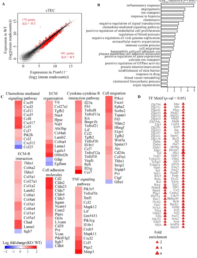
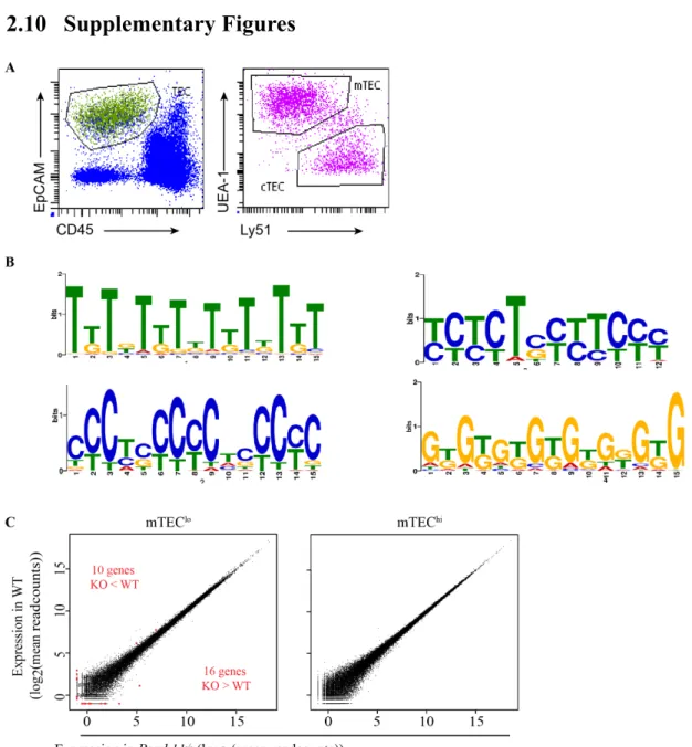
The concept that proteasomes regulate the level of transcriptional regulators by ubiquitin-mediated proteolysis is well established (Geng et al., 2012). Therefore, we performed RNA-Sequencing (RNA-Seq) to assess the overall impact of thymoproteasomes on the transcriptome of cTECs from 10-week-old mice. cTECs were defined as thymic epithelial cells (TECs) (Epcam+CD45-) with a Ly51+UEA1- staining pattern (Supplementary Figure 9A). By comparing cTECs from PSMB11-deficient mice to WT control mice in three replicates, we found that among the genes with FPKM higher than 1, 851 were differentially expressed genes (DEGs) with an adjusted p-value lower than 0.1 and a fold change of at least 1.4. Of these, 681 and 170 DEGs were over- and under-expressed in the Psmb11-/- condition, respectively (Figure 5A).
Gene ontology (GO) term and KEGG-pathway enrichment analyses of the 851 DEGs highlighted processes linked to the interaction between TECs and thymocytes: chemotaxis,
cell-cell adhesion, ECM organization (Figure 5B). The TNF-induced genes Cxcl2, Cxcl9 and Ccl5, which serve as chemoattractants for hematopoietic progenitors to the thymus or for T cell emigration (Bichele et al., 2016) were upregulated in cTECs from Psmb11-/- mice (Figure 5C).
Notably, previous comparative analyses of the transcriptome between cTECs and mTECs have shown that Cxcl2, Cxcl9, Ccl5 and Ccl11 are primarily expressed by mTECs (Bunting et al., 2010; Gotter et al., 2004). In line with this, the transcripts of CCL25 and CXCL12, which are the major chemokines secreted by cTECs (Gameiro et al., 2010), decreased in the absence of PSMB11. These results imply an altered chemokine gradient with potential slow-down of thymocytes in their cortex-to-medulla migration. Notably, Ccl2 was also upregulated in the Psmb11-/- cTECs. Even though CCL2 can recruit peripheral dendritic cells (DCs) to the cortex (Lopes et al., 2015), we found no increase in the abundance of DCs in Psmb11-/- thymi (data not shown).
Positive selection is accompanied by high-speed migration interrupted by brief migratory pauses (Ross et al., 2014). Acting in synergy with chemokines, the ECM components facilitate thymocyte migration and compartmentalization, and also support thymocyte adhesion to TECs, with the thymic medulla containing a denser laminin network than the cortex (Emre et al., 2013; Gameiro et al., 2010; Lannes-Vieira et al., 1991). The integrin subunit alpha m gene (Itgam), and components of the fibronectin (Fndc1, Fndc7) and collagen (16 genes) families were collectively upregulated in cTECs in the absence of PSMB11 (Figure 5C). Additionally, a group of laminin and laminin-associated genes had elevated expression in the Psmb11-/- cTECs, Lama1, Lamb2, Nid1, Ntn4. By similarity to the laminin-related protein Ntn1, the products of these genes may increase thymocyte adhesion in the cortex (Guo et al., 2013). Akin to laminins, the expression of HpsE, Vit, Parvb and Mmp3 was elevated in cTECs lacking thymoproteasomes, which could promote cell-adhesion by degradation of the ECM (Uniprot). Thus, we infer that a denser ECM in the cortex of Psmb11-/- mice can affect the migration pattern of thymocytes and their communication with cTECs.
Figure 5. PSMB11 is a master regulator of the transcriptome in cTEC. (A) Differential gene expression from cTECs between WT and Psmb11-/- mice at 10 weeks of age (mean of 3 replicates per genotype). Differentially expressed genes (DEGs) in red, with
an adjusted p-value < 0.1, FPKM > 1, fold-change between WT and Psmb11-/- > 1.4. (B) GO term analysis of the DEGs in cTECs showing the most significant biological processes identified. (C) Heatmap of the DEGs involved in the biological processes and KEGG pathways associated with cell adhesion, cell migration and cytokine signaling from (B). (D) Transcription factors (TFs) identified as enriched in the DEG dataset for cTECs, based on the most common motifs in the promoter regions. The size of the triangles increases with the fold enrichment. Related to Figure S9.
Lastly, several members of the TNF (Tnfrsf21, Tnfrsf8, Tnfsf10, Lif) and tumor growth factor (TGF) (Tgfb2, Gdf7) families were also among the upregulated DEGs in cTECs (Figure 5C). The cytokine-dependent activation of NF-kB is necessary for thymocyte survival and transition to mature stages (Xing et al., 2016). Thus, although these signaling events occur in the medulla of WT thymi, we found that lack of PSMB11 might influence the secretion of these cytokines in the cortex and therefore affect thymocyte co-stimulation and the timing of maturation. Overall these results indicate that PSMB11 balances the expression of several chemokines and adhesion molecules involved in guiding post-selection T cells through their thymic development.
2.3.2 Psmb11-/- cTECs resemble mTECs in their transcription factor activity
In order to identify possible transcriptional regulators of the DEGs in cTECs, we employed two types of analyses of their promoter regions. Motif enrichment analysis using CIS-BP (Catalog of Inferred Sequence Binding Preferences) (Weirauch et al., 2014) with a 10% false discovery rate (FDR) revealed 118 transcription factor (TF) binding motifs enriched 1.5 to 4 times in the promoters of the downregulated genes, whereas only 19 possible TFs were found for the upregulated genes. In parallel, we performed multiple sequence alignments of the promoter regions of the DEGs using the motif-based sequence analysis tool MEME (Bailey and Elkan, 1994), and examined the top 4 consensus motifs identified (Supplementary Figure 9B). These motifs were similar to the binding sites for 10 TFs in the Tomtom database (meme-suite.org) which were found to be enriched in our dataset, namely RARG, EGR2, FOXJ3, FOXD3, FOXJ2, SP1, IRF3, ELF3, RREB1 and GLIS2 (Figure 5D). The retinoic acid receptor g (RARG) has been shown to preferentially restrict cTEC cellularity, but also to modulate T lymphopoiesis (Joseph et al., 2016; Sitnik et al., 2012). The ubiquitous TF SP1 is implicated in a number of essential cellular processes, including proliferation, apoptosis, DNA damage response,
associated to the Prdm1-mediated autoimmunity in mTECs (Roberts et al., 2017). Moreover, the TF ELF3 is one of the top 50 targets of HDAC3 in the thymic medulla (Goldfarb et al., 2016). In addition, RREB1 activation was also found to be induced by TNF signaling in the thymic stroma, and might be responsible for the upregulation of mTEC-related cytokines in Psmb11-/- thymi (Bichele et al., 2016). Altogether, these data suggest that PSMB11 provides a unique cellular program to cTECs by controlling the homeostasis of TFs, whereas the absence of thymoproteasomes skews the transcriptional activity to an mTEC phenotype.
Of note, the Psmb11 deletion had no impact on the transcriptome of mTEChi, while 26 genes were differentially expressed in mTEClo between WT and Psmb11-/- mice (Supplementary Figure 9C). PSMB11+ cTEC precursors are thought to also give rise to the mTEClo subset which then differentiate into mTEChi31. Thus, our results show that the DEGs identified in cTECs are not spurious differences, but they are induced specifically by lack of PSMB11.
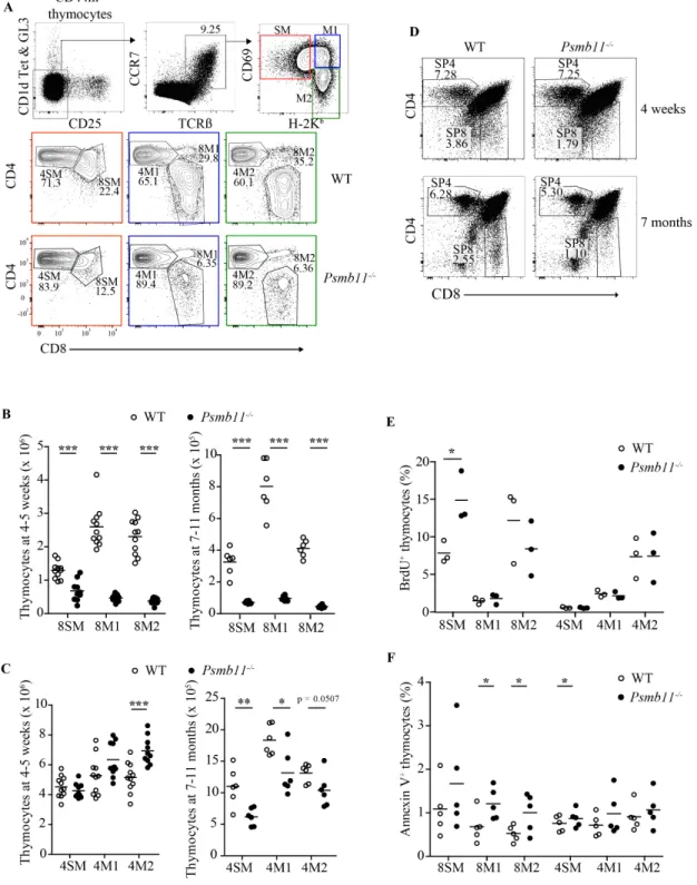
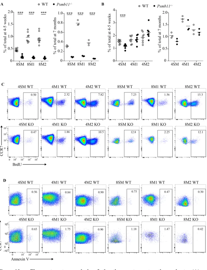
2.3.3 Psmb11 deletion affects CD4 and CD8 thymocyte maturation irrespective of age In order to assess the impact of Psmb11 deletion on αβ T cell development, we examined the cell number of post-selection thymocytes at the semi-mature, mature 1 and mature 2 (SM, M1, M2) developmental subsets defined by Hogquist and colleagues (Hogquist et al., 2015) (Figure 6A). We found that in Psmb11-/- mice, CD8 T cells had a strong decrease in numbers and percentages at all three stages of maturation (Figure 6B and Supplementary Figure 10A). Moreover, the development of CD8 T cells was similarly impaired in 7-month-old Psmb11-/- mice as those at 4-5 weeks of age.
Figure 6. Impact of Psmb11-deletion on CD4 and CD8 thymocyte numbers. (A) Flow cytometry analysis of thymocytes from WT and Psmb11-/- mice. CD1d–α-GalCer tetramers as well as antibodies against CD25 and TCRγδ. Contour plots show the percentages of SM, M1 and M2 elements among post-selection CD4 and CD8 thymocytes (CCR7+TCRβ+).
SM, M1 and M2 subsets in mice at 4-5 weeks and 7 months of age. (C) Absolute numbers of CD4 thymocytes in the SM, M1 and M2 subsets in mice at 4-5 weeks and 7 months of age. (D) Representative gating of total thymocytes in WT vs. Psmb11-/- mice showing the percentages of CD4 and CD8 thymocytes. (E) Flow cytometry analysis of BrdU incorporation into WT and Psmb11-/- CD4 and CD8 thymocytes after 24 h of chase. Each dot represents the proportion of BrdU+ cells in one mouse. (F) Annexin V staining of WT and Psmb11-/- thymocytes. Each dot represents the percentage of AnnexinV+ thymocytes in one mouse. 7-ADD+ thymocytes were excluded from the analysis. Related to Figure S2.
Strikingly, the number of CD4 thymocytes in the thymus of 4-5-week-old mice lacking PSMB11 was significantly increased in the 4M2 subset (Figure 6C). The effect of PSMB11 deletion on the CD4 lineage was masked by population averaging in young mice, yet at 7 months old the total CD4 single-positive T cell population as well as discrete 4SM, 4M1 and 4M2 subsets show a slight decline in the percentage and absolute numbers (Figure 6C and D, and Supplementary Figure 10B). This indicates that absence of PSMB11 affects the commitment of thymocytes to both CD4 and CD8 lineages. Although we observed an age-dependent effect on the CD4 subset, we decided to focus on the 4-5-week-old group for further analysis.
2.3.4 CD8 thymocytes selected in Psmb11-/- mice show increased susceptibility to apoptosis
We considered two non-mutually exclusive explanations for the altered cellularity in the CD4 and CD8 cell subsets in Psmb11-/- mice, proliferation and apoptosis. To examine the
proliferation ability, we performed in vivo 5-bromo-2’-deoxyuridine (BrdU) labeling in young adult mice. BrdU was pulsed twice at 2-hour intervals, and was chased for 24 hours to allow incorporation into newly synthesized DNA. The percentage of BrdU+ 8SM cells was greater in Psmb11-/- mice compared to the WT, suggesting a compensatory reaction to the abnormal decrease in the CD8 cell number (Figure 6E and Supplementary Figure 10C). The proportion of BrdU+ cells in all other CD8 and CD4 subsets was similar to the WT control.
The early apoptosis status was assessed using Annexin V staining. We found that a higher percentage of 8SM, 8M1 and 8M2 thymocytes were early apoptotic in the absence of PSMB11 (Figure 6F and Supplementary Figure 10D). Previous research has shown that splenic CD8 T cells developed in Psmb11-/- mice are functionally capable to survive in lymphopenic
environments and undergo TCR-mediated proliferation (Nitta et al., 2010). Our results indicate that CD8 thymocytes selected in the absence of PSMB11 do not lose their proliferation ability, however these cells are more susceptible to apoptosis. Nonetheless, these experiments did not elucidate the changes identified in the cellularity of the CD4 subsets.
2.3.5 Lack of thymoproteasomes induces stress in developing thymocytes
To gain a global understanding of the commitment of thymocytes to the CD4 and CD8 lineages in the absence of PSMB11, we carried out RNA-Seq on discrete thymocyte subsets and used the DESeq2 package in the R software to extract DEGs between the WT and Psmb11 -/-conditions. Differential gene expression was defined as genes with a fold change expression higher than 2 and an adjusted p-value lower than 0.1. We found a total of 304 DEGs in the CD8 thymocytes and 610 DEGs in the CD4 thymocytes between the WT and Psmb11-/- conditions, with little overlap between the maturation subsets (Figure 11A and Appendix 1-6).
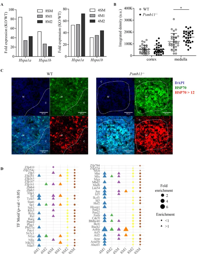
Our RNA-Seq results showed a 20-80-fold upregulation of the Hspa1a and Hspa1b genes, which encode for the stress-responsive HSP70 chaperone, in all CD4 and CD8 thymocyte subsets from Psmb11-/- mice (Figure 7A). This prompted us to perform immunofluorescence staining for HSP70 on thymic slices from 4-week-old mice. By comparing Psmb11-/- vs. WT thymi for the areas of medulla and cortex with an HSP70 intensity at least 3 times higher than the isotype (that is, pixels above a threshold of 12, Supplementary Figure 11A and B), we detected a significant increase in the HSP70 levels in the medulla, but not in the cortex of thymi deficient in PSMB11 (Figure 7B and C). These results demonstrate that the cellular stress was elevated in thymocytes selected in the absence of PSMB11.
Figure 7. Loss of PSMB11 induces stress in developing thymocytes. (A) Bar chart showing the fold-change upregulation of Hspa1a and Hspa1b transcripts expressed as a ratio between the readcounts in Psmb11-/- and WT thymocytes. Data obtained by RNA-Seq. (B) Immunofluorescence quantification of the HSP70 intensity in the cortex and medulla of WT vs. Psmb11-/- mice (n = 2 mice per genotype). The tissue from 4-week-old mice was
snap-frozen and cut into 8 µm slices. Five areas of cortex and medulla were analyzed per tissue section, and three sections were chosen randomly at different Z-planes in the organ. (C) Representative images from immunofluorescence staining for HSP70 on thymic slices from WT vs. Psmb11-/- mice. The cortex and medulla areas were defined based on the nuclei
abundance using DAPI. From left to right, the upper images represent DAPI (blue) and HSP70 (green), and the lower images show the merged channels and HSP70 quantification of pixels > 12 (red). Scale bar 50 µm. (D) Motif comparison on the promoter regions of the DEG in each thymocyte maturation subset, showing transcription factors (TFs) identified as enriched.
2.3.6 CD8 thymocytes show patterns of agonist selection, whereas CD4 thymocytes upregulate stress-responsive genes
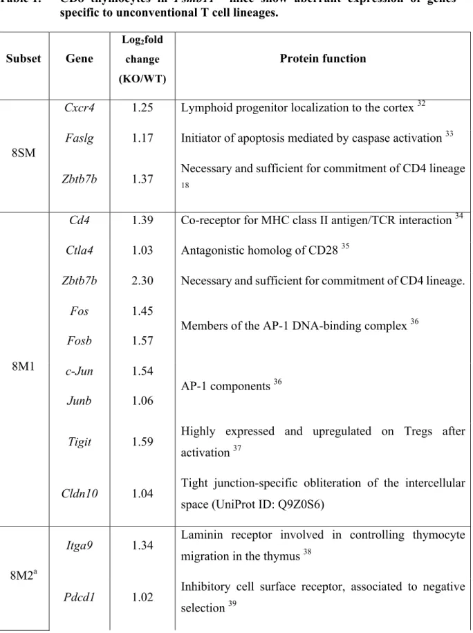
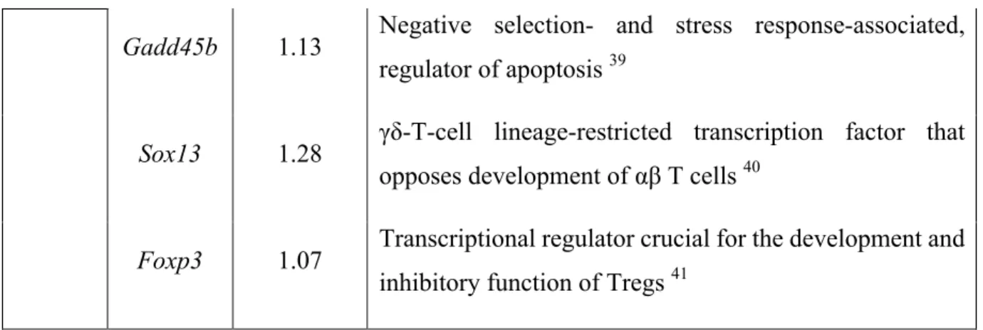
Overall the CD8 cell subsets from Psmb11-/- thymi showed a gene expression pattern with signatures of CD4 and unconventional T cell lineages. To exemplify, 8SM and 8M1 thymocytes displayed a lag in the downregulation of Zbtb7b (Table 1). We found that 8M1 and 8M2 subsets upregulated transcripts restricted to regulatory T cells (Tregs), namely Tigit and Foxp3, and to γδ T cells, namely Sox13. Thus, this transcription pattern is suggestive of an agonist selection in response to stronger TCR signaling (Oh-hora et al., 2013). These data are consistent with the higher expression of negative selection-associated genes in cTECs, including Pvr (CD155, which has high affinity to TIGIT) (Qiu et al., 2010). Correspondingly, the expression level of Ctla4 was also higher in 8M1 thymocytes from mice lacking PSMB11. Moreover, the upregulation of genes involved in apoptosis, Faslg, Pdcd1, Gadd45b, are in line with the increased Annexin V levels in the CD8 maturation subsets from Psmb11-/- mice (Table 1 and Figure 6F). Likewise, the increased expression of Cldn10 and Itga9 genes suggest an induction of negative selection signals which promote cell-cell adhesion (Schmitz et al., 2003). In conclusion, CD8 thymocytes appear to receive stronger TCR signals in the cortex of Psmb11
-/-thymi and skew their expression pattern to unconventional T cell lineages.
Unlike the CD8 lineage, we found that CD4 thymocytes upregulate a group of genes encoding co-chaperones of HSPA1A and HSPA1B, suppressors of apoptosis (Areg, Serpine1, Ppp1r15l) and molecules involved in the shut-off of protein synthesis in response to stress (Ppp1r15a) (Table 2). We reason that the 4SM, 4M1 and 4M2 subsets in Psmb11-/- mice use
stress-responsive mechanisms to rescue their fate from a stress-induced apoptosis and the initial decline in the percentage of 4SM cells (Supplementary Figure 10B).
In an effort to understand what signals trigger the transcriptomic changes in thymocytes, we again searched for enriched motifs in the promoter regions. Interestingly, all thymocyte subsets except 8M2 showed strong enrichment for the binding site of ATF6β (Figure 7D). The processing of ATF6β, an endoplasmic reticulum (ER) transmembrane TF, is activated in response to stress and appears to regulate the strength and duration of the unfolded protein response (UPR) mediated by the homologous protein ATF6α (Thuerauf et al., 2004). Although it is unexpected that lack of PSMB11 in cTECs would cause ER stress in thymocytes and we did not find differential expression of the UPR-specific genes, as stated above, we observed a general response to stressful stimuli in the CD4 subsets marked by the upregulation of HSP70-associated genes (Table 2).
The zinc finger protein Hivep1 was another possible upstream regulator of the genes with increased expression in the 8SM subset (Figure 7D), and its transient expression has been linked to the TCR-induced apoptosis (Schmitz et al., 2003). Similarly, upregulation of apoptosis genes in 8M1 might be induced by the aryl hydrocarbon receptor (AhR), which has been demonstrated to regulate FasL and NF-κB in stromal cells, but also cell motility genes in thymocytes (Camacho et al., 2005). These data suggest that CD8 thymocytes receive longer exposure to TCR signals in Psmb11-/- mice.
The SP1 TF was also identified as a putative regulator of the transcriptional changes in all thymocyte subsets (Figure 7D). However, since SP1 is a versatile partner for many TFs and is implicated in a wide array of cellular functions, it is difficult to clearly characterize its function in our model. In a similar situation is TEF, which is associated with DNA damage repair actions and the circadian clock rhythm (Siddiqui et al., 2017). Other TF identified, but with minimal annotation available were Zkscan1, Zkscan5, Cenpb, Hmbox1, Nfya and Smarcc2.
Table 1. CD8 thymocytes in Psmb11-/- mice show aberrant expression of genes specific to unconventional T cell lineages.
Subset Gene Log2fold change (KO/WT) Protein function 8SM
Cxcr4 1.25 Lymphoid progenitor localization to the cortex 32
Faslg 1.17 Initiator of apoptosis mediated by caspase activation 33
Zbtb7b 1.37 Necessary and sufficient for commitment of CD4 lineage 18
8M1
Cd4 1.39 Co-receptor for MHC class II antigen/TCR interaction 34
Ctla4 1.03 Antagonistic homolog of CD28 35
Zbtb7b 2.30 Necessary and sufficient for commitment of CD4 lineage.
Fos Fosb
1.45 1.57
Members of the AP-1 DNA-binding complex 36
c-Jun Junb
1.54 1.06
AP-1 components 36
Tigit 1.59 Highly expressed and upregulated on Tregs after
activation 37
Cldn10 1.04 Tight junction-specific obliteration of the intercellular space (UniProt ID: Q9Z0S6)
8M2a
Itga9 1.34 Laminin receptor involved in controlling thymocyte
migration in the thymus 38
Pdcd1 1.02 Inhibitory cell surface receptor, associated to negative selection 39
Gadd45b 1.13 Negative selection- and stress response-associated, regulator of apoptosis 39
Sox13 1.28 γδ-T-cell lineage-restricted transcription factor that
opposes development of αβ T cells 40
Foxp3 1.07 Transcriptional regulator crucial for the development and
inhibitory function of Tregs 41
Select DEGs from the 8SM, 8M1 and 8M2 thymocyte subsets showing genes whose expression is associated with unconventional T cells lineage, apoptosis or cell-cell adhesion. a means that all DEGs in 8M1, except Zbtb7b, are also present in the 8M2 subset.
Table 2. CD4 thymocytes in Psmb11-/- mice upregulate expression of stress-responsive and rescue genes.
Subset Gene Log2fold change (KO/WT) Protein function 4SM
Hsph1 1.53 Nucleotide-exchange factor for chaperones HSPA1A and
HSPA1B (UniProt ID: Q61699)
Dnaja1 1.00 Co-chaperone for HSPA1B, protects cells against apoptosis
(UniProt ID: P63037)
Dnajb4 1.34 Stimulates ATP hydrolysis and the folding of unfolded
proteins mediated by HSPA1A/B (UniProt ID: Q9D832)
Dnajb9 1.27 Co-chaperone for HSP70 (UniProt ID: Q9QYI6)
Fzd4 Fzd5 Fzd7 3.48 1.58 1.00
Receptors for Wnt ligands, important for thymus development 42
Fosl1 Fosl2
2.29 1.31
Areg 1.58 Promotes cell proliferation and inhibits apoptosis 43
Serpine1 1.04
Activated in stressed thymic tissue, role in the inhibition of apoptotic external signals and to the monocyte/macrophage chemotaxis 44
4M1
Areg 2.47 Promotes cell proliferation and inhibits cell apoptosis.
Dnajb4 1.17 Stimulates ATP hydrolysis and the folding of unfolded
proteins mediated by HSPA1A/B.
4M2
Hspa1 1.30 Nucleotide-exchange factor for chaperone proteins
HSPA1A and HSPA1B.
Ppp1r15a 1.28
Reverses the shut-off of protein synthesis initiated by stress-inducible kinases and facilitates recovery of cells from stress 45
Ppp1r15l 1.47 Suppresses the activation of apoptosis (UniProt ID:
Q5I1X5)
Select DEGs from the 8SM, 8M1 and 8M2 thymocyte subsets showing genes whose expression is associated with unconventional T cells lineage, apoptosis or cell-cell adhesion.
2.3.7 Data suggest impaired cytokine responsiveness and migration of CD8 thymocytes in Psmb11-/- thymi
A comprehensive analysis of MHC I positive selection by Kimura et al unveiled two phases of surface CD8 expression (Kimura et al., 2016). In phase 1, the TCR signaling intensity determines the extent of CD8 loss. Thus, the higher the TCR signaling intensity, the stronger the decline in surface CD8 at the intermediate maturation stage (CD4+CD69+CCR7+). Phase 2 of positive selection corresponds to cytokine signaling, which fully restores CD8 expression independently of the phase I levels.
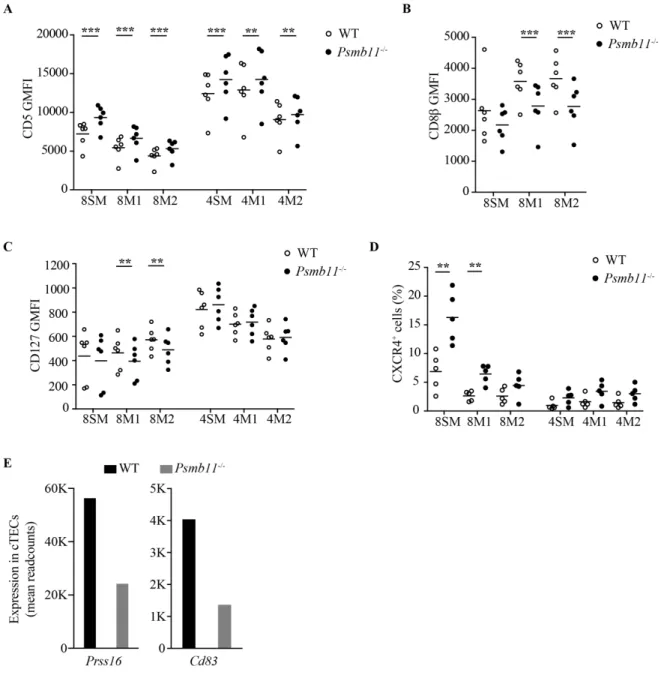
et al., 2013), we checked both CD5 and CD8β levels on post-selection thymocytes. We found that 8SM, 8M1 and 8M2 thymocytes had increased CD5 levels in the absence of PSMB11 (Figure 8A), consistent with bone-marrow (BM) chimera studies on total CD8 T cells (Xing et al., 2013). In addition, the CD8 coreceptor did not reach normal levels on these subsets in Psmb11-/- mice, indicating that phase 2 differentiation was also affected (Figure 8B). Although a longer phase 1 signaling is expected to increase cytokine responsiveness of the MHC I-restricted thymocytes, prolongation of TCR signaling results in lineage errors (Kimura et al., 2016). We found that 8M1 and 8M2 thymocytes, but not CD4 thymocytes, in the Psmb11 -/-thymus showed slightly lower CD127 levels on the cell surface, causing CD8 cells to lose full cytokine response potential and aberrantly express transcripts specific to the CD4 lineage (Figure 8C and Table 1). We suspect that prolonged interactions with cTECs due to a different peptide/class I repertoire and abundance of matricellular molecules in the Psmb11-/- thymus predispose MHC I-restricted thymocytes to lineage errors. Correspondingly, we found that significantly more 8SM and 8M1 thymocytes in the Psmb11-/- condition are positive for CXCR4 relative to the WT, which supports the assumption that CD8 thymocytes have a longer retention in the cortex (Figure 8D).
Figure 8. Impact of Psmb11-deletion on CD4 and CD8 thymocytes. (A) Geometric mean fluorescence intensity (GMFI) of CD5 on the surface of SM, M1 and M2 thymocytes in the CD4 and CD8 lineages from WT and Psmb11-/- mice. (B) GMFI of CD8β on the surface of 8SM, 8M1 and 8M2 thymocytes from WT and Psmb11-/- mice. (C) GMFI of CD127 on the surface of SM, M1 and M2 thymocytes in the CD4 and CD8 lineages from WT and Psmb11-/- mice. (A-C) Statistical analysis was determined using a paired Student’s t-test, with *P < 0.05, ** P < 0.01, ***P < 0.001. (D) Percentages of CXCR4+ thymocytes. Each data point from WT is paired with a data point from the Psmb11-/- condition. (E) Expression level of Prss16 and Cd83 in WT and Psmb11-/- cTECs expressed in mean
2.3.8 Data suggest a direct impact of PSMB11 on the CD4 lineage
A notable characteristic of cTECs is constitutive macroautophagy and the expression of specific enzymes for MHC class II-peptide production, namely cathepsin L and the thymus-specific enzyme protease (TSSP, encoded by Prss16) (Klein et al., 2009; Mizushima et al., 2004). To our surprise, CD5 levels on the 4SM, 4M1 and 4M2 subsets were slightly but reproducibly higher in Psmb11-/- mice (Figure 8A). Together with the significantly elevated cellularity of the 4M2 subset and the extensive amount of DEGs identified at all CD4 maturation stages assessed, it was tempting to infer that an altered pool of endogenous proteins in the Psmb11-/- cTECs become substrates for lysosomal degradation and change the MHC II ligandome. Although this hypothesis remains valid, we found a more than two-fold downregulation in the expression of Prss16 and Cd83 in cTECs (Figure 8E), which have both been shown to control the positive selection of CD4 T cells (Klein et al., 2009; Liu et al., 2016; Rohrscheidt et al., 2016).
We conclude that several factors regulated by PSMB11 in cTECs might be responsible for the changes in the CD4 lineage. These factors include the MHC II levels and ligandome, the cytokine gradient and the ECM composition in the cortex (Luckheeram et al., 2012; Robey, 2016), as suggested by our RNA-Seq data from cTECs.
2.4 DISCUSSION
Our study provides strong evidence that PSMB11 is a master regulator of the transcriptome in cTECs, which is essential for the normal positive selection of both CD4 and CD8 T cells in the thymus. We surmise that population averaging has eluded other researchers from identifying an effect of Psmb11 deletion on the CD4 lineage. Nonetheless, Nitta et al. have reported a significant increase in the number of CD4 T cells in the lymph nodes of Psmb11-/- mice (Nitta et al., 2010). We show here that this increase cannot be a mere consequence of the loss in CD8 T cells and thus an expansion of the CD4 T cell pool. Instead, it is likely a direct effect of lack of PSMB11 in cTECs at the antigen presentation and cell motility levels, and correlated with the greater abundance of the 4M1 and 4M2 subsets detected in the present study. Moreover, FACS analysis of thymocytes from MHCII-/-Psmb11-/- mice confirmed that this expansion was not a result of CD8 thymocytes mistakenly engaging to a CD4 cell-fate (data not shown).
