PCAM: a multi-user facility-based protein crystallization apparatus for
microgravity
Daniel C. Cartera, Brenda Wrighta, Teresa Millerb, Jenny Chapmana, Pam Twigga, Kim Keelinga, Kerry Moodya, Melissa Whitea, James Clicka, John R. Rublea, Joseph X. Hoa, Lawana Adcock-Downeya, Tim Dowlingb, Chong-Hwan Changc, Paul Alac, John Rosed, B.C. Wangd, Jean-Paul Declercqe, Christine Evrarde, John Rosenbergf, Jean-Pierre Weryg, David Clawsong, Mark Wardellh, W. Stallingsi, A. Stevensi
aNew Century Pharmaceuticals, Inc., 895 Martin Road, Huntsυille, AL 35824, USA
b Marshall Space Flight Center, Huntsville, AL 35812, USA
c Experimental Station, DuPont Pharmaceuticals Company, Wilmington, DE 19880, USA
d
Department of Biochemistry and Molecular Biology, University of Georgia, Athens, GA 30602, USA
e Laboratoire de Chemie Physique et de Cristallographie, Université Catholique de Louvain, Belgium f Department of Biological Sciences, University of Pittsburgh, PA 15260, USA
gEli Lilly & Company, Lilly Corporate Center, Indianapolis, IN 46285-0403, USA
hDepartment of Biochemistry and Molecular Biophysics, Washington University, School of Medicine, St. Louis, MO 63110, USA
iMonsanto∕Searle BB4K, 700 Chesterfield Parkway North, St. Louis, MO 63198, USA
Abstract
A facility-based protein crystallization apparatus for microgravity (PCAM) has been constructed and flown on a series of Space Shuttle Missions. The hardware development was undertaken largely because of the many important examples of quality improvements gained from crystal growth in the diffusion-limited environment in space. The concept was based on the adaptation for microgravity of a commonly available crystallization tray to increase sample density, to facilitate co-investigator participation and to improve flight logistics and handling. A co-investigator group representing scientists from industry, academia, and government laboratories has been established. Microgravity applications of the hardware have produced improvements in a number of structure-based crystallographic studies and include examples of enabling research. Additionally, the facility has been used to support fundamental research in protein crystal growth which has delineated factors contributing to the effect of microgravity on the growth and quality of protein crystals.
Keywords: PCAM; Protein; Crystal; Microgravity; Vapor-diffusion; Shuttle
1. INTRODUCTION
Since the inception of protein crystal growth in microgravity research by Littke in 1984 [1], several research groups have developed microgravity hardware and experiments [1-5]. Because of various problems inherent in previous designs which create limitations in sample density, logistics, and handling associated with space flight experiments, a program was established with the aim of design and construction of an improved microgravity protein crystal growth facility. Further motivations for such a facility were based on the many important examples of quality improvements gained from growth in the diffusion-limited environment in space [2-13]. During the course of this investigation hardware design and construction has proceeded rapidly through hand-held versions to complete facility hardware [9]. A large flight co-investigator group has been established and the hardware has been successfully manifested and flown on several shuttle missions. Additionally, in related efforts, a special microdialysis counterdiffussion hardware has been designed, constructed, and is currently active on Mir [10,14].
The program proceeded in two phases. In the first stage a hand-held concept was developed and tested in microgravity to assess the concept which was followed by a second phase involving the design, construction and flight of the facility. Once operational, access to the facility was opened to an international group of scientists from industrial, academic, and government laboratories.
The specific objectives of this research have been to: (1) provide greatly increased experiment and co-investigator capacity in order to improve the odds of obtaining suitable crystals, thereby expanding the overall science return from each mission; (2) investigate the disposable interface concept in development of
microgravity hardware for reduced cost and improvements in logistics and handling; (3) utilize the facility to delineate factors contributing to the effect of microgravity on the growth and quality of protein crystals; and (4)
produce crystals of improved size and quality for application in structural biology.
2. FLIGHT HARDWARE DESIGN AND CONSTRUCTION 2.1. Hand-held hardware (HH-PCAM)
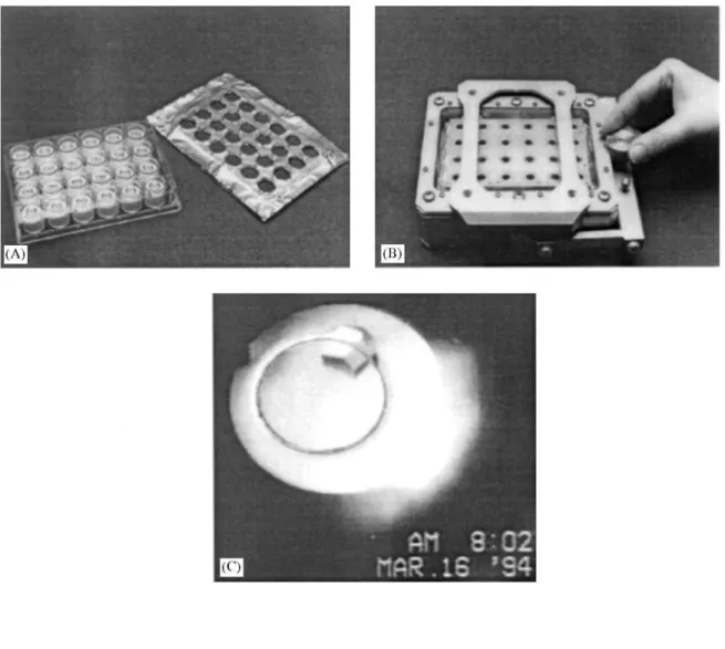
The hand-held protein crystallization apparatus for microgravity (HH-PCAM) consists of two main compartments, a disposable plastic tray (Cryschem™) containing 24 vapor equilibration chambers and a mechanical deactivation mechanism (Fig. 1). The reservoirs are contained within porous high molecular weight polyethylene wicks and the plastic trays are sealed with a specially developed elastomer tape. The tray is then housed in a mechanical device which includes an optical capability to photograph and document the results in microgravity. The experiment is activated or deactivated by rotating the knob (Fig. 1B) in the appropriate direction. The hardware was manufactured and rigorously tested against a variety of performance criteria including, functional, safety, science requirements, and parabolic flights on the KC135 aircraft. Subsequently, four hand-held PCAM were manifested and flown on STS-62 (Fig. 1A and Fig. 1B) as proof-of-concept. For this experiment, a selection of proteins and precipitants were chosen to test the stability of the protein droplets in the pedestal during launch and landing. Additionally, two of the units were stored in the locker with the orientation parallel to the gravity vector (most stable) and two perpendicular. Thus, information could be obtained on the functionality of the hardware/concept in the two likely future payload stowage modes. Astronaut Pierre Thuot photo-graphically documented the flight performance of the hardware on STS-62 which included one of the most spectacular downlinked photos of a growing in situ, protein crystal (hen egg white lysozyme (HEWL)) (Fig. 1C) to date. Analysis of these and other data from HH-PCAM verified the performance of the hardware and crystal growth experiments. Although minor occurrences of small droplets of protein solution on the elastomer were observed, either orientation of the hardware during launch proved satisfactory. In addition to the successful tests of functionality, numerous advantages in logistics and handling were realized with the pro-to-type. Based on these outstanding results, approval to proceed with the full facility configuration of the hardware was granted. The HH-PCAM was considered to be the first flight hardware developed with a disposable interface concept.
2.2. PCAM facility hardware
Through the design and analysis of the HH-PCAM, a set of criteria were established for the facility configuration. First, the number samples per tray were reduced to enable greater co-investigator (Co-I) involvement and to provide the greatest possible flexibility in sample documentation and distribution. The disposable interface concept proved to be extremely advantageous and was retained. Other design factors pertained to the future possibilities of cryogenic storage and handling and glove box interface capabilities. Photographic capabilities within the facility hardware were eliminated in order to increase sample density. However, photographic documentation of individual experiments could, with minor modification, be performed with available microscopes and other facilities in the glovebox.
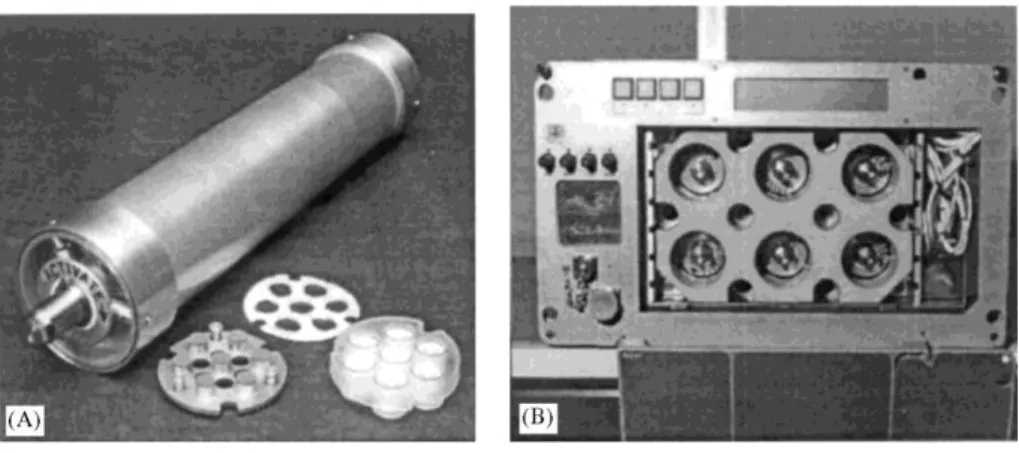
A design meeting all of the requirements was rapidly produced, manufactured (Fig. 2) and tested [9]. The hardware consists of circular injected molded Lexan™ trays containing seven vapor-equilibration chambers (Fig. 2A). The trays are sealed with an improved version of the elastomer. A series of nine trays are disposed within a cylinder (Fig. 3) between interleaving actuator plates. Consequently, each cylinder contains 63 individual experiments for a total of 378 chambers in a standard single locker thermal enclosure system (STES) unit (Fig. 2B) or 504 in a standard middeck locker without active temperature control. This provided a middeck locker experiment capacity expansion several times over previously existing NASA vapor-diffusion hardware [2]. Turning the actuator knob on each cylinder several revolutions to a fixed stop activates or deactivates all of the trays (nine) simultaneously. Some advantages from the new design are synopsized below:
(1) Disposable interface allows for rapid in situ evaluation of results during post flight analysis and post-flight distribution. This is extremely important since a number of co-investigators have noted that their crystals are damaged during post-flight removal from other flight hardware. PCAM allows the individual co-investigator groups to take the crystals undisturbed back to their respective laboratories where familiar equipment and surroundings facilitate the proper handling of the samples for diffraction studies. In the case of extensive launch/ scrub scenarios, the disposable interface allows rapid refurbishment of the hardware and automatic experiment controls.
(2) Cryogenic storage of experiments. Trays were engineered to fit into existing flight dewars available through Johnson Space Flight Center. Previously, it was determined that loaded sample trays could be stored without detriment for over 18 months at -150°C.
(3) Each cylindrical unit contains a hand operated locking and pinning mechanisms to allow for the disassembly in microgravity. This feature, with refinements, could potentially provide for future glovebox interactive experiment opportunities which might become available on Spacelab, Mir, or the future International Space Station.
(4) Facility operation requires minimum crew involvement and skill, and it can be operated as hand-held or easily adapted to automation.
(5) The unprecedented experiment capacity allows for a greater co-investigator group and generous experiment allocation, thus increasing the odds of obtaining suitable crystals and the total science return.
Fig. 1. (A) Cryschem™ tray adapted for microgravity utilizing high molecular weight polyethylene wicks and elastomer sheet. (B) HH-PCAM flight unit which simultaneously activates or deactivates 24 Cells in a standard Cryschem™ tray. (C) Downlinked video image of a lysozyme crystal growing in microgravity during STS-62. Photo taken by astronaut Pierre Thuot.
Fig. 2. (A) A single PCAM cylinder with individual disposable tray with wicks and associated interleaving actuator plate. A total of nine trays,with seven chambers trays, are accommodated in each cylinder, (B) Illustrates the standard flight arrangement of six PCAM units in an STES.
Fig. 3. A schematic illustrating the basic construction of the PCAM and its mechanism. Note trays are loaded in what appears to be upside down configuration, depending on whether the PCAMs are located in the forward or aft lockers.
Table 1 Flight history
Mission Date Hardware # samples # Co-Is # Proteins
STS-62 3-4-94 HH-PCAM 96 1 4 STS-63 2-3-95 6 PCAM 378 7 (5 groups) 9 STS-67 3-2-95 6 PCAM 378 11 (8 groups) 9 STS-73 10-20-95 12 PCAM 756 13 (8 groups) 12 8 Short PCAM 168 1 81 DCAM 81 3 3 STS-76 3-22-96 162 DCAM 162 8 (7 groups) 9 STS-79 9-16-96 162 DCAM 162 9 (7 groups) 11 STS-81 1-12-97 162 DCAM 162 8 (6 groups) 6 STS-83 4-4-97 10 PCAM 630 14 (9 groups) 10 STS-84 5-15-97 162 DCAM 162 7 (6 groups) 6 STS-94 7-1-97 10 PCAM 630 12 (8 groups) 9 STS-85 8-7-97 10 PCAM 630 11 (7 groups) 13 STS-89 1-22-98 162 DCAM 162 10 (7 groups) 9
3. SUMMARY OF PCAM PROTEIN CRYSTAL GROWTH RESULTS
A protocol and procedure was established to coordinate access to the flight resources and facility in collaboration with a diverse guest and co-investigator group. Potential collaborators are requested to submit brief applications which are reviewed through coordination with NASA including internal and external review prior to resource allocation. A co-investigator group was established in 1994 and the PCAM hardware was initially manifested on a series of Shuttle flights during 1995. An outline of the current flight history is presented in Tables 1 and 2, and a selection of the results from these collaborations has been synopsized below and outlined in Table 3.
3.1. Highlights from STS-63, STS-67, and STS-73 (February-October 1995)
Co-investigators: Dr. Chong Hwan Chang and Dr. Paul Ala, DuPont Pharmaceuticals Company HIV protease complex with proprietary inhibitor
Background and objective: to improve the quality of the crystals for increased resolution to aid in the design of new inhibitors against HIV. The structure of the HIV protease complexes with inhibitors has been utilized by several pharmaceutical companies, including Dupont/Merck, to design de novo inhibitors and improve the potency of current inhibitors (structure-based drug design). The most effective AIDS therapeutics have been designed by this approach.
A complete diffraction data set has been collected from selected crystals (Fig. 4a). Overall, diffraction data exhibit superior R-factors in shells of resolution. For example, enhanced I/σ(I) ratios and increased resolution of approximately 0.3 A, thus producing the highest quality crystal of this specific inhibitor/protein complex yet obtained. In addition, it was noted that the crystals did not show significant decay under X-ray exposure which was a marked difference from the terrestrial samples.
Table 2 Guest and co-investigator list
Guest and co-investigators Affiliation
Dr. Mark Wardell Washington University, School of Medicine at St. Louis
Dr. Richard Skinner Cambridge University, UK
Dr. Jean-Paul Declercq Catholic University of Louvain, Belgium Dr. Christine Evrard Catholic University of Louvain, Belgium
Dr. Gerard Bunick Oak Ridge National Laboratory
Mr. Joel Harp Oak Ridge National Laboratory
Dr. Chong-Hwan Chang Dupont/Pharmaceuticals Company
Dr. Paul Ala Dupont/Pharmaceuticals Company
Dr. B.C. Wang University of Georgia
Dr. John Rose University of Georgia
Dr. Jean-Pierre Wery Eli Lilly Corp.
Mr. David Clawson Eli Lilly Corp.
Dr. John Rosenberg University of Pittsburgh
Dr. Bill Stallmgs Monsanto/Searle
Ms. Anna Stevens Monsanto/Searle
Dr. Wolfgang Weber University of Eppendorf, Hamburg, Germany
Dr. Bill Thomas USRA
Dr. Alex Chernov USRA
Dr. Franz Rosenberger University of Alabama at Huntsville
Dr. P. Velkelov University of Alabama at Huntsville
Dr. Glenn Pilkington IntraCel Corporation
Dr. Joseph Ho New Century Pharmaceuticals, Inc.
Dr. John Ruble New Century Pharmaceuticals, Inc.
Dr. Louis Delbaere University of Saskatchewan, Canada
Dr. Dennis Bamford University of Helsinki, Finland
Dr. Udo Heinemann Max-Delbruck Centrum, Berlin, Germany
Dr. Charlotte Foerster Max-Delbruck Centrum, Berlin, Germany Dr. Florian Ruker Institute for Applied Microbiology, Austria Dr. Gottfried Wagner Justus-Liebig University, Germany
Dr. Christian Betzel University of Eppendorf, Hamburg, Germany
Dr. Donald Frazier NASA, Marshall Space Flight Center
Dr. Benjamin Penn NASA, Marshall Space Flight Center
Dr. Roger Kroes NASA, Marshall Space Flight Center
Raf kinase
Background and objective: to determine the three-dimensional structure of the N-terminal regulatory domain of raf kinase. Cancer oncogene product/drug target, raf kinase, is a Ser/Thr kinase involved in the signal
transduction cascade that originates with a receptor at the cell surface, due to binding of a growth hormone. Crystals of the N-terminal regulatory domain of raf kinase were the largest ever produced of this crystal form, approximately an order of magnitude larger in a single dimension (Fig. 4c and Fig. 4d). Previously, crystals were not large enough to pursue the structure. Although the crystals from this experiment did not survive attempts at mounting the crystals for X-ray diffraction studies, this is potentially an extremely important example of "enabling" microgravity research. Since the time of this experiment, these researchers are pursuing new crystal forms of this protein which are more suitable for the structure determination.
Co-investigators: Dr. Jean-Paul Declercq and Dr. Christine Evrard, Laboratoire de Chimie physique et de
L-Alanine dehydrogenase from Bacillus subtilis
Background and objective: to improve the diffraction quality of the crystals in order to aid in structural
determination. L-alanine dehydrogenase catalyses the reversible oxidative deamination of L-alanine to pyruvate and ammonia. This enzyme does not seem to share sequence similarity with other amino acid dehydrogenases, whose three-dimensional structures are known. However, it seems to be structurally related to the
transmembrane proton translocating pump and pyridine nucleotide transhydrogenase. The three-dimensional crystal structure will allow a better understanding of the catalytic reaction and of the enzymologic and structural relationships between these proteins.
Crystals were obtained from nearly all of the 35 experiments, some of which produced the largest crystals of L-alanine dehydrogenase ever grown (Fig. 4e and Fig. 4f). Data have been collected on beam line X31 at EMBL Hamburg [16].
Bacteriophage lambda lysozyme
Background and objective: to improve the diffraction quality of the crystals in order to aid in structural
determination. The bacteriophage lambda lysozyme (λL) is a small protein of 158 amino acids. Like other known lysozymes, it is involved in the lysis of the bacterial peptidoglycan.
This enzyme is remarkable in that its mechanism of action is different from the classical lysozyme mechanism. Moreover, from the point of view of protein evolution, it shows features of lysozymes from different classes. STS-63 and STS-67 PCAM crystals were large enough for data collection for the first time (Fig. 4g and Fig. 4h). A 2.3 A data set was collected at EMBL, Hamburg, Germany [17,18]. This work has enabled the structure determination which represents the recent Ph.D. dissertation of Dr. Christine Evrard [19].
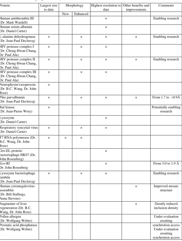
Table 3 PCAM high lights summary
Protein Largest size
to date
Morphology Highest resolution to date
Other benefits and improvements
Comments New Enhanced
Human antithrombin III (Dr. Mark Wardell)
× Enabling research
Human serum albumin (Dr. Daniel Carter)
×
L-alanine dehydrogenase (Dr. Jean-Paul Declercq)
× × × × Enabling research
HIV protease complex I (Dr. Chong-Hwan Chang, Dr. Paul Ala)
× × ×
HIV protease complex II (Dr. Chong-Hwan Chang, Dr. Paul Ala)
× × × × Enabling research
HIV protease complex III (Dr. Chong-Hwan Chang, Dr. Paul Ala) × × × Neurophysin/vasopressin (Dr. B.C. Wang, Dr. John Rose) × Pike parvalbumin (Dr. Jean-Paul Declercq) × × × × From 1.7 to <0.9Ǻ Raf kinase (Dr. Jean-Pierre Wery) × Potentially enabling research Lysozyme (Dr. Daniel Carter) ×
Respiratory syncytial virus (Dr. Daniel Carter) × × × T7 RNA polymerase (Dr. B.C. Wang, Dr. John Rose) × × × Gro-EL protein/ bacteriophage HK97 (Dr. John Rosenberg) × Eco-RI Dr. John Rosenberg × From 3.0 to 1.9 Å Lysozyme bacteriophage lambda (Dr. Jean-Paul Declercq) × × × Enabling research Human cytomegalovirus assemblin (Dr. Bill Stallmgs, Anna Stevens) × Improved mosaic structure Augmenter of liver regeneration (Dr. B.C. Wang, Dr. John Rose)
× Greatly reduced inclusion density Pollen allergen (Dr. Wolfgang Weber) Under evaluation awaiting Prostatic acid phosphatase
(Dr. Wolfgang Weber)
synchrotron access Under evaluation
awaiting synchrotron access
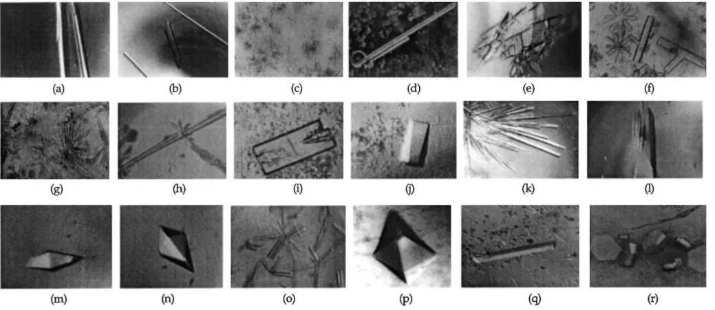
Fig. 4. Comparisons of protein crystals produced in ground-based and flight experiment activities conducted using the PCAM facility on several US Space Shuttle misions. (a) crystals of HIV-I protease inhibitor complex from STS-73; (b) crystals of a drug resistant HIV protease inhitor complex grown on STS-85; terrestrial (c) and flight samples (d) of raf kinase at the same magnification. Photo courtesey of D. Clawson, Eli Lilly; typical terrestrial (e) and flight samples (f) of L alanine dehydrogenase; typical terrestrial crystals (g) and flight (h) of bacteriophage lambda lysozyme; typical examples of terrestrial (I) and flight (j) crystals of augmenter of liver regeneration protein; photos courtesy of J. Rose and B. C. Wang, University of Georgia; flight samples of pike parvalbumin from 83 (k) and an extremely large specimen from the longer STS-94 mission (1); typical terrestrial (m) and flight (USML-2) (n) samples of neurophysin/vasopressin complex; flight crystals of Gro-EL protein/bacteriophage HK97 complex grown on USML2 (o); flight example of the protease assemblin from cytomegalovirus (p); and large flight crystal of a recombinant fab expressed against the respiratory syncytial virus grown during STS-85 (q); flight crystals of Eco RI grown on STS-85 (r).
Human cytomegalovirus assemblin
Background and objective: to increase the crystal size and quality to achieve greater resolution data to aid in drug design. Human cytomegalovirus (HCMV), in addition to other members of the herpesvirus family, encodes a unique serine protease, assemblin, that is necessary for viral replication. Accordingly, specific inhibitors of HCMV assemblin should interfere with the replication of this increasingly important human pathogen. Crystallization and structure determination of assemblin will provide information necessary for the design of antiviral drugs. The assemblin target has represented a high priority structure-based drug design project at Monsanto/Searle.
Two variants of assemblin were grown producing numerous diffraction sized crystals (Fig. 4p). Early reports indicated an improvement in mosaic spread (internal order), but not in resolution. Extended delays in the post-flight analysis were experienced because of X-ray equipment failure at Monsanto. Samples may have
deteriorated by the time analysis was performed.
Co-investigators: Dr. Mark R. Wardell, Department of Haematology, University of Cambridge, MRC. Current
Address: Washington University, School of Medicine, St. Louis, MO.
Human antithrombin III
Background and objective: to improve crystal quality, size, and morphology for obtaining higher resolution data which will aid in the design of antithrombin therapeutics. Antithrombin is a member of the serpin family of serine protease inhibitors which are larger than other inhibitor families and characterized by the remarkable flexibility of their relatively long reactive center loops. Antithrombin has 432 amino acids organized into nine helices and three beta-sheets with three disulfide bonds and four N-linked oligosaccharide chains. Its
physiological function is to control blood coagulation in human plasma which is achieved by forming inhibitory complexes with thrombin and other coagulation proteases in a process greatly accelerated by heparin. Its importance is underscored by the occurrence of severe thrombotic disorders including deep vein thrombosis, pulmonary embolism and cerebral infarction in subjects with antithrombin mutations. This results in either decreased plasma levels or aberrant inhibitory function.
The PCAM crystals grown on STS-67 were superior to any previous grown by any method. The resulting resolution of 2.6 A is the highest resolution example of an active inhibitory serpine to date [11,12]. The newly refined atomic model revealed regions of the structure, previously unseen, including residues involved in the binding of heparin. In addition, it allowed the complete definition of its reactive center loop. Unfortunately, because of the series of long delays this very fragile protein did not crystallize well after the third USML-2 reloading (documented by loading team). This is an added example of "enabling" microgravity research on an extremely important medically significant protein. The resulting structural work contributed significantly to the Ph.D. dissertation of Dr. Richard Skinner [20].
Co-investigator. Dr. John Rosenberg, Department of Crystallography, University of Pittsburgh. E.coli chaperonin Gro EL protein/bacteriophage HK91 capsid protein complex
Background and objective: to improve the diffraction quality of the crystals for aiding in the structure determination. This research is part of a larger effort in collaboration with Roger Hendrix where both
crystallographic and biochemical techniques are used. Chaperonins, including members of the Cpn60 family, are an ubiquitous class of proteins that assist in protein folding in vivo by binding to partially folded proteins. This prevents the aggregation and may unfold "off pathway" intermediates and direct them back into the correct folding pathway. The gp 5 protein is a natural substrate of GroEL. It is the major capsid protein in the icosahedral head of the lambdoid bacteriophage HK97.
Large crystals were grown (Fig. 4o). Crystals from USML-2 produced the highest quality diffraction data yet obtained. Diffraction data were collected at Brookhaven National Synchrotron Light Source.
Co-investigators: Dr. John Rose and Dr. B.C. Wang, Department of Biochemistry and Molecular Biology,
Neurophysin II/vasopressin complex
Background and objective: to improve the diffraction quality of the crystals in order to aid in structural
determination. The hormone vasopressin is synthesized and packaged in the posterior pituitary as a prohormone with its carrier neurophysin. Vasopressin, which has long been associated with cardiovascular function, has recently been shown together with the related hormone oxytocin to help orchestrate social and sexual relationships. Knowledge of this structure could provide information for basic understanding of human relationships, mental illness and endocrinology.
Largest crystal of neurophyisn/vaspressin complex grown to date by any method (Fig. 4m and Fig. 4n). Crystals also appear superior in optical perfection. Although the results have encouraged further studies, no improvement in diffraction resolution was reported for this experiment.
Augmenter of liver regeneration protein
Background and objective: to improve the diffraction quality of the crystals in order to aid in structural
determination. This is a new liver growth factor which promotes liver regeneration after it is damaged or injured. The crystal structure of ALR will provide a road map for other researchers for use in better understanding the biochemical and physiological properties of this new class of growth factors, and thus may open the door for new ways to treat liver diseases or make liver transplants more successful. Large crystals with dramatically improved optical perfection were obtained (Fig. 4i and Fig. 4j).
3.2. Highlights from STS-83, STS-85, and STS-94 (June-October 1997)
Investigators: Dr. D.C. Carter, New Century Pharmaceuticals, Huntsville, AL and Dr. Glenn Pilkington, Intracel,
Issaquah, Washington.
Respiratory syncytial virus (RSV) antibody
Background and objective: to improve crystal size and resolution. RSV is an influenza type virus which produces serious respiratory infections in infants. It is especially dangerous to infants with other health risk/complicating factors. The National Academy of Science Institute of Medicine estimates there are approximately 100000-120000 hospital admissions/year (US) with a mortality rate of 4000 infant deaths per year in the United States. It is considered by many physicians to be the most serious infectious disease affecting infants in the US.
This therapeutic recombinant antibody produced by Intracel Corp. neutralizes all known variants of the respiratory syncytial virus based on isolated strains from the past 40 years. New Century Pharmaceuticals, Inc. (NCP) has recently determined the atomic structure of this antibody which should provide insight into the production of vaccines, as well as the development of small molecule therapeutics which are less expensive to manufacture. This work is part of an ongoing NASA/NCP/Intracel Government/Industry cooperative agreement. Crystals of the antibody grown on STS-85 (PCAM) were the largest to date (Fig. 4q) and diffracted X-rays to significantly higher resolution based on stills (from 2.2 to 1.5 Ǻ) which will contribute to further improvements in the structure and future vaccine development activities.
Co-investigator. Dr. Jean-Paul Declercq, Laboratoire de Chimie physique et de Cristallographie, Université
Pike parvalbumin
Background and objective: to grow crystals of improved size and resolution, and to attempt growing extremely large crystals for application in neutron structure determination.
Crystals of pike parvalbumin (PPA) grown during STS-83 and STS-94 gained improvements which moved the resolution into the ultra-high resolution category diffracting X-rays beyond 0.9 Ǻ at the Hamburg synchrotron. Prior work by the co-investigator group indicated a diffraction limit of 1.7 Ǻ for pike parvalbumin and the Brookhaven Protein Data Bank contains 13 parvalbumin structures of which the highest resolution is 1.5 Ǻ. The data indicate this will result in one of the highest resolution protein structures over 100 amino acids determined. Since the data in highest resolution shell are observed at 97.3%, clearly usable diffraction data exist well below 0.9 Ǻ. Rmerge(/) = 3.6% for 481972 reflections (63 763 unique). Preliminary refinement has revealed the high quality of the resulting structure and answered many questions concerning conformational states in the protein structure and calcium binding (Fig. 4k). Additionally, as a result of the longer duration of STS-94 (MSL-1R), the largest crystal to date was produced of this protein [13] which has provided the first example of microgravity enablement for neutron diffraction (Fig. 41).
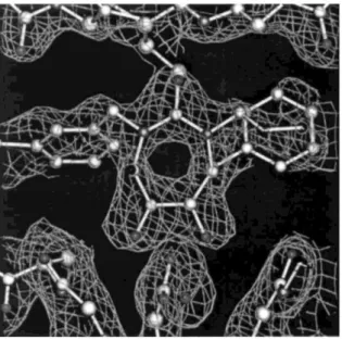
Co-investigator. Dr. Chong Hwan Chang, DuPont Pharmaceuticals Company. Fig. 5. Initial 2Fo-Fc of mutant HIV protease complexed with cyclic area based inhibitor.
HIV protease complex with cyclic area based inhibitor
Background and objective: the objectives of this proposal are to improve the quality of the crystals of selected protease/inhibitor complexes to provide for increased resolution in the design of new therapeutics against HIV. Large crystals of a mutant HIV protease involved in drug resistance complexed with a cyclic area based inhibitor were grown on STS-85 (Fig. 4b). Previous terrestrial crystals were of insufficient size for X-ray analysis. The microgravity crystals were the largest grown to date and provided data to 1.8 Ǻ. A detailed image of the inhibitor bound to the active site was obtained from the microgravity data (Fig. 5).
Eco RI endonuclease-DNA complex
Background and objective: type II restriction enzymes, such as Eco RI endonuclease, present a unique advantage for the study of sequence-specific recognition because they leave a record of where they have been in the form of the cleaved ends of the DNA sites where they had bound. The differential behavior of a sequence-specific protein at sites of differing base sequence is the essence of the sequence-specificity; the core question is how do these proteins discriminate between different DNA sequences especially when the two sequences are very similar.
Prior work by the co-investigator group indicated a diffraction limit of 3.0 Ǻ. STS-85 Eco RI crystals diffracted X-rays to 1.9 Ǻ and contributed to the first high resolution structure of this protein.
4. SUMMARY
In summary, several thousand individual protein crystal growth experiments have been flown using the PCAM facility hardware aboard the US Space Shuttle rivaling or exceeding the total number flown since the inception of the first protein crystal growth activity in 1985. The operation of this facility has benefited an international group of industrial, academic, and government research activities in structural molecular biology. The
accomplishments from both the hardware development and the flight experiment results have been outstanding. The PCAM hardware represents a pioneering development in design and deployment of space flight hardware which are based on disposable interface elements. These disposable interfaces have resulted in reduced costs, improved logistics and numerous additional advantages. Several examples of enabling research in microgravity have been documented and/or published through application of PCAM. These include an ultra high resolution structure and the first example of protein neutron diffraction achieved as a result of protein crystal growth in microgravity [13]. Additionally, using the facility, fundamental differences in protein partitioning in microgravity have been documented which represent the first direct experimental observation of the factors contributing to quality improvements in the growth of protein crystals in microgravity [15].
Acknowledgements
We gratefully acknowledge the enthusiastic support of the crews of Space Shuttle Missions STS-62, STS-63, STS-67, STS-73, STS-83, STS-85, and STS-94 with special appreciation to Pierre Thuot, Katie Coleman, Fred Leslie, Marsha Ivans, Wendy Lawerence, Bill Gregory, Janice Voss, and Roger Crouch. This research was supported through NASA grant NRA 963-23-08 and contract NAS8-97247.
References
[1] W. Littke, C. John, Science 225 (1984) 203.
[2] L.J. DeLucas, F.L. Suddath, R.S. Snyder, R. Naumann, M.B. Broom, M. Pusey, V. Yost, B. Herren, D. Carter, B. Nelson, E.J. Meehan, A. McPherson, C.E. Bugg, J. Crystal Growth 76 (1986) 681.
[3] L.J. DeLucas, C.D. Smith, H.W. Smith, V.K. Senadhi, S.E. Ealick, D.C. Carter, R.S. Snyder, P.C. Weber, F.R. Salemme, D.H. Ohlendorl, H.M. Einspahr, L.L. Clancy, M.A. Navia, B.M. McKeever, T.L. Nagabhushan, G. Nelson, A. McPherson, S. Koszelak, G. Taylor, D. Stammers, K. Powell, G. Darby, C.E. Bugg, Science 246 (1989) 651.
[4] J. Day, A. McPherson, Protein Sci. 1 (1992) 1254. [5] A. McPherson, J. Phys. D 26 (1993) B104.
[6] R.K. Strong, B.L. Stoddard, J. Crystal Growth 119 (1992) 200.
[7] S.B. Larson, J. Day, A. Greenwood, A.J. McPherson, J. Mol. Biol. 277 (1) (1998) 37. [8] W. Littke, C. John, J Crystal Growth 76 (1986) 663.
[9] D.C. Carter, T.E. Dowling, U.S. Patent No. 5 643 540,1997.
[10] D.C. Carter, B. Wright, T. Miller, J. Chapman, P. Twigg, K. Keeling, K. Moody, M. White, J. Click, J. Ruble, J. Ho, L. Adcock-Downey, G. Bunick, J. Harp, J. Crystal Growth 196 (1999) 602.
[11] M.R. Wardell, R. Skinner, D.C. Carter, P.D. Twigg, J.-P. Abrahams, Acta Crystallogr. D 53 (1997) 622.
[12] R. Skinner, J.-P. Abrahams, J.C. Whisstock, A.M. Lesk, R.W. Carrell, M.R. Wardell, J. Mol. Biol. 266 (1997) 601. [13] J.-P. Declercq, C. Evrard, D.C. Carter, B.S. Wright, G. Etienne, J. Parello, J. Crystal Growth 196 (1999) 595. [14] D.C. Carter U.S. Patent No. 5 641 681 (1997).
[15] D.C. Carter, K. Lim, J.X. Ho, B.S. Wright, P.D. Twigg, T.Y. Miller, J. Chapman, K. Keeling, J. Ruble, P.G. Vekilov, B.R. Thomas, F. Rosenberger, A.A. Chernov, J. Crystal Growth 196 (1999) 623.
[16] J.-P. Declercq, D. Deforge, Recent Advances in Macro-molecular Crystallization, International Meeting, Le Bischenberg, France, 1996. [17] C. Evrard, J.-P. Declercq, J. Fatsrez, Acta Crystallogr. D 53 (1997) 217.
[18] C. Evrard, J. Fastrez, J.-P. Declercq, J. Mol. Biol. 276 (1998) 151. [19] C. Evrard, Ph.D. Thesis, University Catholique de Louvam, 1998. [20] R. Skinner, Ph.D. Dissertation, Hughes Hall, Cambridge, May 1996.