HAL Id: dumas-01628550
https://dumas.ccsd.cnrs.fr/dumas-01628550
Submitted on 3 Nov 2017
HAL is a multi-disciplinary open access archive for the deposit and dissemination of sci-entific research documents, whether they are pub-lished or not. The documents may come from teaching and research institutions in France or abroad, or from public or private research centers.
L’archive ouverte pluridisciplinaire HAL, est destinée au dépôt et à la diffusion de documents scientifiques de niveau recherche, publiés ou non, émanant des établissements d’enseignement et de recherche français ou étrangers, des laboratoires publics ou privés.
Uncovered nitinol stenting in airway complications after
lung transplantation
Philippe Junet
To cite this version:
Philippe Junet. Uncovered nitinol stenting in airway complications after lung transplantation. Human health and pathology. 2015. �dumas-01628550�
AVERTISSEMENT
Ce document est le fruit d'un long travail approuvé par le
jury de soutenance et mis à disposition de l'ensemble de la
communauté universitaire élargie.
Il n’a pas été réévalué depuis la date de soutenance.
Il est soumis à la propriété intellectuelle de l'auteur. Ceci
implique une obligation de citation et de référencement
lors de l’utilisation de ce document.
D’autre part, toute contrefaçon, plagiat, reproduction illicite
encourt une poursuite pénale.
Contact au SID de Grenoble :
[email protected]
LIENS
LIENS
Code de la Propriété Intellectuelle. articles L 122. 4
Code de la Propriété Intellectuelle. articles L 335.2- L 335.10
http://www.cfcopies.com/juridique/droit-auteur
1
UNIVERSITE JOSEPH FOURIER FACULTE DE MEDECINE DE GRENOBLE
Année : 2015
POSE DE STENT NON COUVERT EN NITINOL DANS LES COMPLICATIONS DES VOIES AERIENNES
APRES TRANSPLANTATION PULMONAIRE
THESE
PRESENTEE POUR L’OBTENTION DU DOCTORAT EN MEDECINE DIPLÔME D’ETAT
Philippe JUNET
THESE SOUTENUE PUBLIQUEMENT A LA FACULTE DE MEDECINE DE GRENOBLE*
Le : 25 juin 2015
DEVANT LE JURY COMPOSE DE
Président du jury : M. Le Professeur Emile REYT
Membres
M. Le Professeur Christian Adrien RIGHINI, Directeur de thèse
M. Le Professeur Christophe PISON
M. Le Docteur François ARBIB
Me Le Docteur Amandine BRIAULT
*La Faculté de Médecine de Grenoble n’entend donner aucune approbation ni improbation aux opinions émises dans les thèses ; ces opinions sont considérées comme propres à leurs auteurs.
2
UNIVERSITE JOSEPH FOURIER FACULTE DE MEDECINE DE GRENOBLE
Année : 2015
POSE DE STENT NON COUVERT EN NITINOL DANS LES COMPLICATIONS DES VOIES AERIENNES
APRES TRANSPLANTATION PULMONAIRE
THESE
PRESENTEE POUR L’OBTENTION DU DOCTORAT EN MEDECINE DIPLÔME D’ETAT
Philippe JUNET
THESE SOUTENUE PUBLIQUEMENT A LA FACULTE DE MEDECINE DE GRENOBLE*
Le : 25 juin 2015
DEVANT LE JURY COMPOSE DE
Président du jury : M. Le Professeur Emile REYT
Membres
M. Le Professeur Christian Adrien RIGHINI, Directeur de thèse
M. Le Professeur Christophe PISON
M. Le Docteur François ARBIB
Me Le Docteur Amandine BRIAULT
*La Faculté de Médecine de Grenoble n’entend donner aucune approbation ni improbation aux opinions émises dans les thèses ; ces opinions sont considérées comme propres à leurs auteurs.
3
DOMAINE DE LA MERCI
FAX : +33 (0)4 76 63 71 70 ...
...
Affaire suivie par Marie-Lise GALINDO [email protected]
Doyen de la Faculté : M. le Pr. Jean Paul ROMANET
Année 2014-2015
ENSEIGNANTS A L’UFR DE MEDECINE
CORPS NOM-PRENOM Discipline universitaire
PU-PH ALBALADEJO Pierre Anesthésiologie réanimation
PU-PH APTEL Florent Ophtalmologie
PU-PH ARVIEUX-BARTHELEMY Catherine chirurgie générale
PU-PH BALOSSO Jacques Radiothérapie
PU-PH BARRET Luc Médecine légale et droit de la santé
PU-PH BENHAMOU Pierre Yves Endocrinologie, diabète et maladies métaboliques
PU-PH BERGER François Biologie cellulaire
PU-PH BETTEGA Georges Chirurgie maxillo-faciale, stomatologie
MCU-PH BIDART-COUTTON Marie Biologie cellulaire
MCU-PH BOISSET Sandrine Agents infectieux
PU-PH BONAZ Bruno Gastro-entérologie, hépatologie, addictologie
MCU-PH BONNETERRE Vincent Médecine et santé au travail
PU-PH BOSSON Jean-Luc Biostatiques, informatique médicale et technologies de communication
MCU-PH BOTTARI Serge Biologie cellulaire
PU-PH BOUGEROL Thierry Psychiatrie d'adultes
PU-PH BOUILLET Laurence Médecine interne
MCU-PH BOUZAT Pierre Réanimation
PU-PH BRAMBILLA Christian Pneumologie
PU-PH BRAMBILLA Elisabeth Anatomie et cytologie pathologiques
MCU-PH BRENIER-PINCHART Marie Pierre Parasitologie et mycologie
...
UFR de Médecine de Grenoble
38706 LA TRONCHE CEDEX – France TEL : +33 (0)4 76 63 71 44
4
PU-PH BRICAULT Ivan Radiologie et imagerie médicale
PU-PH BRICHON Pierre-Yves Chirurgie thoracique et cardio- vasculaire
MCU-PH BRIOT Raphaël Thérapeutique, médecine d'urgence
PU-PH CAHN Jean-Yves Hématologie
MCU-PH CALLANAN-WILSON Mary Hématologie, transfusion
PU-PH CARPENTIER Françoise Thérapeutique, médecine d'urgence PU-PH CARPENTIER Patrick Chirurgie vasculaire, médecine vasculaire
PU-PH CESBRON Jean-Yves Immunologie
PU-PH CHABARDES Stephan Neurochirurgie
PU-PH CHABRE Olivier Endocrinologie, diabète et maladies métaboliques
PU-PH CHAFFANJON Philippe Anatomie
PU-PH CHAVANON Olivier Chirurgie thoracique et cardio- vasculaire
PU-PH CHIQUET Christophe Ophtalmologie
PU-PH CINQUIN Philippe Biostatiques, informatique médicale et
technologies de communication
PU-PH COHEN Olivier Biostatiques, informatique médicale et
technologies de communication
PU-PH COUTURIER Pascal Gériatrie et biologie du vieillissement
PU-PH CRACOWSKI Jean-Luc Pharmacologie fondamentale, pharmacologie clinique
PU-PH DE GAUDEMARIS Régis Médecine et santé au travail
PU-PH DEBILLON Thierry Pédiatrie
MCU-PH DECAENS Thomas Gastro-entérologie, Hépatologie
PU-PH DEMATTEIS Maurice Addictologie
PU-PH DEMONGEOT Jacques Biostatiques, informatique médicale et
technologies de communication
MCU-PH DERANSART Colin Physiologie
PU-PH DESCOTES Jean-Luc Urologie
MCU-PH DETANTE Olivier Neurologie
MCU-PH DIETERICH Klaus Génétique et procréation
MCU-PH DOUTRELEAU Stéphane Physiologie
MCU-PH DUMESTRE-PERARD Chantal Immunologie
PU-PH EPAULARD Olivier Maladies Infectieuses et Tropicales PU-PH ESTEVE François Biophysique et médecine nucléaire
MCU-PH EYSSERIC Hélène Médecine légale et droit de la santé
PU-PH FAGRET Daniel Biophysique et médecine nucléaire
5
MCU-PH FAURE Julien Biochimie et biologie moléculaire
PU-PH FERRETTI Gilbert Radiologie et imagerie médicale
PU-PH FEUERSTEIN Claude Physiologie
PU-PH FONTAINE Éric Nutrition
PU-PH FRANCOIS Patrice Epidémiologie, économie de la santé et prévention
PU-PH GARBAN Frédéric Hématologie, transfusion
PU-PH GAUDIN Philippe Rhumatologie
PU-PH GAVAZZI Gaétan Gériatrie et biologie du vieillissement
PU-PH GAY Emmanuel Neurochirurgie
MCU-PH GILLOIS Pierre Biostatiques, technologies de communication informatique médicale et
PU-PH GODFRAIND Catherine Anatomie et cytologie pathologiques (type clinique)
MCU-PH GRAND Sylvie Radiologie et imagerie médicale
PU-PH GRIFFET Jacques Chirurgie infantile
MCU-PH GUZUN Rita Endocrinologie, diabétologie, nutrition, éducation thérapeutique
PU-PH HALIMI Serge Nutrition
PU-PH HENNEBICQ Sylviane Génétique et procréation
PU-PH HOFFMANN Pascale Gynécologie obstétrique
PU-PH HOMMEL Marc Neurologie
PU-PH JOUK Pierre-Simon Génétique
PU-PH JUVIN Robert Rhumatologie
PU-PH KAHANE Philippe Physiologie
PU-PH KRACK Paul Neurologie
PU-PH KRAINIK Alexandre Radiologie et imagerie médicale
PU-PH LABARERE José Epidémiologie ; Eco. de la Santé
PU-PH LANTUEJOUL Sylvie Anatomie et cytologie pathologiques
MCU-PH LAPORTE François Biochimie et biologie moléculaire
MCU-PH LARDY Bernard Biochimie et biologie moléculaire
MCU-PH LARRAT Sylvie Bactériologie, virologie
MCU-PH LAUNOIS-ROLLINAT Sandrine Physiologie
PU-PH LECCIA Marie-Thérèse Dermato-vénéréologie
PU-PH LEROUX Dominique Génétique
PU-PH LEROY Vincent Gastro-entérologie, hépatologie, addictologie
6
PU-PH LEVY Patrick Physiologie
MCU-PH LONG Jean-Alexandre Urologie
PU-PH MACHECOURT Jacques Cardiologie
PU-PH MAGNE Jean-Luc Chirurgie vasculaire
MCU-PH MAIGNAN Maxime Thérapeutique, médecine d'urgence
PU-PH MAITRE Anne Médecine et santé au travail
MCU-PH MALLARET Marie-Reine Epidémiologie, économie de la santé et
prévention
MCU-PH MARLU Raphaël Hématologie, transfusion
MCU-PH MAUBON Danièle Parasitologie et mycologie
PU-PH MAURIN Max Bactériologie - virologie
MCU-PH MCLEER Anne Cytologie et histologie
PU-PH MERLOZ Philippe Chirurgie orthopédique et traumatologie
PU-PH MORAND Patrice Bactériologie - virologie
PU-PH MOREAU-GAUDRY Alexandre Biostatiques, technologies de communication informatique médicale et
PU-PH MORO Elena Neurologie
PU-PH MORO-SIBILOT Denis Pneumologie
MCU-PH MOUCHET Patrick Physiologie
PU-PH MOUSSEAU Mireille Cancérologie
PU-PH MOUTET François Chirurgie plastique, reconstructrice et esthétique, brûlogie
MCU-PH PACLET Marie-Hélène Biochimie et biologie moléculaire
PU-PH PALOMBI Olivier Anatomie
PU-PH PARK Sophie Hémato - transfusion
PU-PH PASSAGGIA Jean-Guy Anatomie
PU-PH PAYEN DE LA GARANDERIE Jean-François Anesthésiologie réanimation
MCU-PH PAYSANT François Médecine légale et droit de la santé
MCU-PH PELLETIER Laurent Biologie cellulaire
PU-PH PELLOUX Hervé Parasitologie et mycologie
PU-PH PEPIN Jean-Louis Physiologie
PU-PH PERENNOU Dominique Médecine physique et de réadaptation
PU-PH PERNOD Gilles Médecine vasculaire
PU-PH PIOLAT Christian Chirurgie infantile
PU-PH PISON Christophe Pneumologie
PU-PH PLANTAZ Dominique Pédiatrie
7
PU-PH POLOSAN Mircea Psychiatrie d'adultes
PU-PH PONS Jean-Claude Gynécologie obstétrique
PU-PH RAMBEAUD Jacques Urologie
MCU-PH RAY Pierre Génétique
PU-PH REYT Émile Oto-rhino-laryngologie
MCU-PH RIALLE Vincent Biostatiques, informatique médicale et technologies de communication
PU-PH RIGHINI Christian Oto-rhino-laryngologie
PU-PH ROMANET J. Paul Ophtalmologie
MCU-PH ROUSTIT Matthieu Pharmacologie fondamentale, pharmaco clinique, addictologie
MCU-PH ROUX-BUISSON Nathalie Biochimie, toxicologie et pharmacologie PU-PH SARAGAGLIA Dominique Chirurgie orthopédique et traumatologie
MCU-PH SATRE Véronique Génétique
PU-PH SAUDOU Frédéric Biologie Cellulaire
PU-PH SCHMERBER Sébastien Oto-rhino-laryngologie PU-PH SCHWEBEL-CANALI Carole Réanimation médicale
PU-PH SCOLAN Virginie Médecine légale et droit de la santé
MCU-PH SEIGNEURIN Arnaud Epidémiologie, économie de la santé et prévention
PU-PH STAHL Jean-Paul Maladies infectieuses, maladies tropicales
PU-PH STANKE Françoise Pharmacologie fondamentale
MCU-PH STASIA Marie-José Biochimie et biologie moléculaire
PU-PH TAMISIER Renaud Physiologie
PU-PH TONETTI Jérôme Chirurgie orthopédique et traumatologie
PU-PH TOUSSAINT Bertrand Biochimie et biologie moléculaire
PU-PH VANZETTO Gérald Cardiologie
PU-PH VUILLEZ Jean-Philippe Biophysique et médecine nucléaire
PU-PH WEIL Georges Epidémiologie, économie de la santé et prévention
PU-PH ZAOUI Philippe Néphrologie
PU-PH ZARSKI Jean-Pierre Gastro-entérologie, hépatologie, addictologie
8
A mes maîtres et membres du jury :
Monsieur le Professeur Emile REYT, président du jury
Vous me faites un grand honneur de présider le jury de cette thèse. Je souhaitais vous remercier tout
particulièrement pour votre disponibilité, votre bienveillance et vos conseils toujours avisés tout au
long de ma formation d’interne. J’ai eu la chance de bénéficier de votre enseignement sur la pratique de la chirurgie cervico faciale et endo nasale qui a suscité ma vocation.
Monsieur le Professeur Christian Adrien RIGHINI, directeur de thèse
Vous m’avez fait l’honneur de me confier ce travail. Je vous remercie vivement d’avoir dirigé ce travail de manière claire et précise et de m’avoir tant appris durant toutes ces années d’internat surtout dans le domaine de la cancérologie et de la chirurgie cervico faciale. Votre rigueur est un exemple
que vos élèves s’attachent à suivre.
Monsieur le Professeur Christophe PISON
Vous m’avez fait l’honneur de me proposer ce travail de thèse et de me guider dans la publication de cette étude. Votre enthousiasme et vos précieux conseils ont été indispensables. Veuillez trouver ici
l’expression de ma profonde reconnaissance.
Docteur François ARBIB
Je vous remercie d’avoir accepté d’être membre de ce jury. Veuillez trouver en ce travail l’expression de mes remerciements les plus sincères.
9
Docteur Amandine BRIAULT
Je souhaitais te remercier très sincèrement d’avoir accepté d’être membre de mon jury ainsi que pour ton aide et ta disponibilité lors de la mise en place de ce travail et du recueil de données. Je te souhaite
10
Table des matières
Préambule... 12
1) Introduction ... 16
2) Methods ... 17
a) Study cohort ... 17
b) Clinical care post LT ... 17
c) Endoscopic and stent management ... 17
d) Statistical Analysis ... 18
LT recipient stent insertion predictive factors ... 18
Survival Analysis ... 19
3) Results... 19
a) Patient characteristics ... 19
b) PGD ... 20
c) Infections ... 20
d) Acute cellular rejection (ACR) ... 20
e) Stenting risks factors (Table 2) ... 23
f) Stent insertion procedure and outcomes (Tables 3 and 4) ... 26
g) Survival and CLAD-Free survival analysis (Figure 4) ... 31
4) Discussion ... 33
5) Conclusion and perspectives ... 36
Annexes ... 37 Conclusion de thèse ... 38 References ... 39 SERMENT D’HIPPOCRATE ... 44 Remerciements ... 45 Résumé ... 53
11
Cette thèse a été soumise sous forme d’article pour publication dans la revue Journal of heart and lung transplantation depuis le 27/05/2015
12
Préambule
Depuis la première transplantation pulmonaire (TP) chez l’homme en 1963, les complications
post transplantation des voies respiratoires restent un problème majeur. Leur incidence varie de 1,6 à
33%, avec un taux de mortalité associé de 4% (1). Ces complications sont liées à plusieurs facteurs
(1-7): l’ischémie de la bronche du donneur dans la période post-opératoire immédiate, la technique
chirurgicale utilisée, la longueur des bronches du donneur, les incompatibilités de diamètre entre les
bronches du receveur et celles du donneur, le rejet aigu de greffe, la sur-immunosuppression, un
protocole de préservation d'organe inadaptée, la présence d'agents microbiens avant la transplantation
chez le receveur et le donneur, les infections post-opératoires et l’utilisation de la ventilation
mécanique en post opératoire.
Neuf à 20% des anastomoses bronchiques développeront des complications sévères
nécessitant un traitement (5,8-10). Il existe six types de complications bronchiques anastomotiques
(1) dont les sténoses sont les plus fréquentes, les autres correspondant aux nécroses ou déhiscences,
aux granulomes, aux malacies, aux fistules et aux infections.
L'amélioration des techniques de préservation d’organe, le dépistage de cross match donneur /
receveur et les avancées médicales et chirurgicales ont abouti à une réduction de la fréquence de ces
complications. Des traitements ont cependant dû être développés afin de prendre en charge ces lésions
qui peuvent mettre en jeu le pronostic fonctionnel pulmonaire et le pronostic vital du patient.
Différentes types d’interventions endoscopiques ont été développées et ont permis une gestion efficace des complications obstructives symptomatiques (11), comprenant la dilatation au ballonnet,
la coagulation au laser ou au plasma argon, la curiethérapie ou l’insertion d'une endoprothèse (ou
13
Cette dernière technique est surtout utilisée en présence de bronchomalacie, de sténose anastomotique
ou d’une déhiscence lorsque ces conditions entrainent des symptômes respiratoires, une baisse
significative de la fonction pulmonaire ou des complications infectieuses post-obstructives.
Il existe trois catégories principales de stents utilisables au niveau des voies respiratoires, décrites en
fonction du matériau utilisé pour leur fabrication: en silicone, en métal, et hybride ou biodégradable.
Ces 3 types de stents peuvent être utilisés pour les sténoses anastomotiques. Leur rôle est de rétablir
la perméabilité des voies aériennes de manière mécanique, et peuvent offrir des avantages cliniques
immédiats. Les stents métalliques sont composés d’un alliage de substances inertes, de type nitinol et
peuvent être auto-extensibles ou expansibles par ballonnet, non couverts ou couverts.
Les stents métalliques auto-expansibles en nitinol sont utilisés en raison de leur facilité d'insertion et
leur efficacité rapide sur la symptomatologie respiratoire, mais peuvent conduire à de nombreuses
complications telles que les migrations, les saignements, les obstructions, les infections, et les
sténoses récidivantes (11).
Concernant la procédure précédant la pose de stent et la technique de pose de stent, de nombreux pré
requis doivent être précisés :
Avant l'insertion du stent :
- Une évaluation au fibroscope souple sous anesthésie locale par un pneumologue doit être effectuée.
Les critères d’insertion de stent sont les suivants : lésions bronchiques obstructives avec réduction de
la lumière ≥ 50% associée à des symptômes respiratoires (dyspnée, wheezing, toux ou détresse respiratoire), infections pulmonaires récidivantes et/ou réduction de la capacité fonctionnelle
14
-En accord avec Righini et al. (15), une tomodensitométrie à l’aide d’un scanner multibarette doit être
effectuée avant toute procédure interventionnelle, chez tous les patients nécessitant une insertion de
stent. L’acquisition des données est réalisée avec des coupes fines de 1mm tous les 0,5 mm, de 100 à
120 kV et de 60 à 200 mA. L’ensemble des images des voies respiratoires centrales est acquis entre
3 et 25 secondes, sans injection de produit de contraste veineux. Toutes les mesures ainsi que les
reconstructions 3D ou la bronchoscopie virtuelle sont effectuées afin d’évaluer les conditions
anatomiques locales et de déterminer la taille du ou des stents qui pourront être utilisés. Une « zone
adjacente saine » définie comme de morphologie normale, doit être sans aucune déformation ou
réduction apparente de diamètre et sans aucune formation de fistules. Une longueur de 10 mm est
mesurée et ajoutée à partir des extrémités proximale et distale de l'obstruction afin de mesurer la
longueur du stent à utiliser, pour être sûr qu’il couvrira toute la partie malade et empêcher la
migration. Le positionnement optimal et le nombre de stents potentiels nécessaire sont également
évalués au cours de cet examen, en tenant compte de l'emplacement et de la longueur de l'obstruction.
La procédure d’insertion du stent est effectuée au bloc opératoire par un ORL à l’aide d’un bronchoscope rigide (KARL STORZ, Allemagne) sous anesthésie générale :
- L’anesthésie durant cette intervention représente un véritable défi : avoir accès aux voies aériennes
à la fois pour l’ORL et l'anesthésiste. Le risque majeur de cette procédure est l’hypoventilation dont
les conséquences peuvent être graves, en particulier pour ce type de patient atteint de troubles
ventilatoires prévalents. La technique de jet ventilation représente une approche sécuritaire avec un
maintien de la physiologie respiratoire normale, un minimum de mouvement des cordes vocales, une
15
l’utilisation d’un respirateur particulier de type Monsoon (Acutronic, Zurich). Les paramètres de ventilation utilisés doivent bien calibrés et maitrisés par l’anesthésiste: pression = 3-3,5 bar,
fréquence= 120 cpm, alarme expiratoire 8-10 cm H2O, FiO2 = 40-60%. L'injecteur est le
bronchoscope relié au respirateur par un connecteur de verrouillage Luer et un tube sans perte de
pression (pression> 3 bar). Sous certaines conditions, il est nécessaire d'utiliser une ventilation
manuelle (bypass = 60 litres / min, avec ventilation à 4 mains). Seule une anesthésie par voie
intraveineuse en continu est possible, le propofol et le rémifentanil sont administrés selon le principe
du titrage. L'anesthésie locale de la trachée et des bronches est essentiel et exécutée dès que
l'anesthésie permet la laryngoscopie. A la fin de la procédure sont souvent utilisés des injections
d'adrénaline intratrachéale via le bronchoscope (200 à 500 microgrammes) surtout en présence de
saignements abondants. Les patients ne sont pas intubés avant le réveil.
Si besoin et avant l’insertion du stent, une reperméabilisation des voies respiratoires centrales est réalisée avec l’aide d’un ballon (balloon dilatator, Boston Scientific Corporation).
L’insertion des stents non couvert en nitinol (Ultraflex, Boston Scientific Corporation, Natick, MA) est effectué à l’aide d’un cathéter de « libération » sous contrôle directe de la vision. Une dilatation au ballon est souvent effectuée afin de permettre une expansion complète du stent.
16
Pose de stent non couvert en nitinol dans les
complications des voies aériennes après
transplantation pulmonaire
Uncovered nitinol stenting in airway complications
after lung transplantation
1) Introduction
Central airways complications (CAC) are still a challenging problem after lung transplantation (LT)
(incidence ranging between 1.6 and 33%) with an associated mortality rate of 2 to 4% (1). CAC are
caused by several disputed factors (1–7). Nine to 20 % of bronchial anastomosis develop
complications severe enough to require treatment (5,8–10). There are six types of bronchial
anastomosis complications (1): necrosis and dehiscence, granuloma, malacia, fistulae, infections and,
most common being, bronchial stenosis. In symptomatic obstructive lesions, suggested interventional
techniques include balloon dilatation and desobliteration, laser application, argon plasma coagulation,
brachytherapy and stent insertion (11). Stent placement is required in case of bronchomalacia,
bronchial stenosis, combined stenosis and malacia or bronchial dehiscence when these conditions
induce respiratory symptoms, persistent fall in lung function or post-obstructive complications. Three
main categories of airway stents are used for anastomotic stenosis: silicone, metallic, and hybrid. In
our center, self-expanding metallic stents (SEMS) made from nitinol are used because of their ease
of insertion and immediate relief but they lead to some complications such as migration, bleeding,
atelectasis, infection and restenosis (11), thus their use in benign airway complications is challenged.
17
and monocentric study to evaluate the outcomes after stent insertion in terms of survival and
complications.
2) Methods
a) Study cohort
LT recipients who benefited from a single LT, double LT or heart-LT in Grenoble University Hospital
between January 1, 2002 and November 11, 2013, and survived more than 3 months were eligible for
analysis. Participation in the database was a part of the agreement signed when patients were listed
for LT as a national policy of quality control and clinical research conducted by the Agence de la
BioMédecine, St Denis, France. Data used for these analyses were extracted from the Grenoble University
Hospital database recorded for surveillance of LT recipients. Donor lung procurement and preservation and
the surgical procedure were performed, according to the standard requirements (12,13).
b) Clinical care post LT
Clinical care post-LT, including immunosuppressive regimen, surveillance, and management of
infections in Grenoble University hospital were already described in a previous study(14). Early
infections were defined as infections during the first six months.
Spirometric data were collected before and after stent placement. Best pre-stented forced expiratory
volume at 1 s (FEV1) was taken within 3 months prior to the insertion of the first stent. The best
post-stented FEV1 was taken within 6 months following last stent insertion. Successful treatment was defined by
improved clinical symptoms and/or an FEV1 increase of ≥10% from the pre-treatment level.
c) Endoscopic and stent management
18
or not with respiratory symptoms, recurrent pulmonary infections and/ or FEV1 fall. All patients
underwent multidetector CT measurement for a potential stent insertion before carrying out any
interventional procedure, according to Righini et al. (15). Stent was inserted with rigid bronchoscopy
(KARL STORZ, Germany) under general anesthesia and jet ventilation. Uncovered nitinol stents
(Ultraflex, Boston Scientific Corporation, Natick, MA) with appropriate size were deployed from its
delivery catheter under direct vision and subsequent balloon dilatation to allow full stent expansion.
d) Statistical Analysis
Quantitative data are expressed as mean (standard deviation) or median and 1st and 3rd quartiles [Q1 Q3]
if not normally distributed. Comparison of baseline quantitative data was performed by unpaired
Student's t test. Data exhibiting non-normal distribution were analyzed by nonparametric unpaired
statistical tests (Mann-Whitney U test). Categorical variables are expressed as numbers and proportions
and were compared using the chi-square test. A p value less than 0.05 was considered to be statistically
significant and Stata 13 software, Statacorp, College Station, TX was used. Stented (S) patients could
possibly differ from non-stented (NS) patients for recorded characteristics that might also affect the
outcomes. To make an appropriate comparison because patients were not randomized to stent
insertion, we built a propensity score with variables contemporaneous of the transplantation and which
were available for all patients: donor characteristics (sex, age and ventilation time before harvesting),
recipient characteristics (sex, BMI and age), CMV mismatch, use of non-invasive ventilation, type of
preservation liquid, use of extracorporeal circulation, surgical reoperation and severe primary graft
dysfunction (PGD) at H48.
LT recipient stent insertion predictive factors
To find independent predictors of stenting, we used logistic regression model with stenting as the
19
score: number of transplantations per patient, pre-transplantation data (native lung disease, FEV1 %
predicted, type of colonization), procedure data (type of transplant), post-transplantation data (blood
transfusion, ICU and hospitalization time, ventilation time, early bacterial and fungal infection).
Predictors from the univariate analyses with a p value less than 0.20 entered in the multivariate
analysis and were selected by backward elimination. Variables remaining with p values less than 0.05
at the final step were considered independent predictors of stenting.
Survival Analysis
In observational studies, patients are classified in the ''treatment'' group from the time that they received
the treatment. This means that a patient who died before this moment will always be in the ''NS'' group
and the patients in the ''S'' group are immortal until the first day of treatment, which induces a bias called
''immortal time'' bias (16). To limit this immortal time bias, we used a time-dependent analysis and
divided the follow-up of the "S" group subjects in two parts : time before the first stent date was
classified in the NS group, and in S group thereafter (16,17). Survival and CLAD-free survival were
estimated with Kaplan-Meier curves and compared with the log-rank test.
3) Results
a) Patient characteristics
Of the 133 LT recipients who survived more than 3 months in Grenoble transplantation center
between 2002 and 2013, 34 have been stented corresponding to 21.5 % of the 218
anastomoses-at-risk (Figure 1). Median follow-up time was 2.34 years [0.83-4.75]. Incidence of LT and stent insertion
during the analysis period appears in Figure 2. Both S and NS groups demonstrated similar donor,
recipient and pre-operative characteristics as shown in Table 1. In the S group, ICU and
20
days (p<0.001).
b) PGD
There was no statistical difference between S and NS groups.
c) Infections
Early bacterial and fungal infections were both more frequent in the S group (<6 months after LT),
respectively: 94.1% vs 61.6% (p<0.001) and 35.3% vs 18.2% (p=0.04).
d) Acute cellular rejection (ACR)
The annual incidence of ACR was not significantly different between both groups (p=0.12).
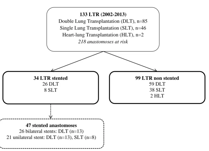
Figure 1: Flow Chart
133 LTR (2002-2013)
Double Lung Transplantation (DLT), n=85 Single Lung Transplantation (SLT), n=46
Heart-lung Transplantation (HLT), n=2 218 anastomoses at risk 99 LTR non stented 59 DLT 38 SLT 2 HLT 34 LTR stented 26 DLT 8 SLT 47 stented anastomoses 26 bilateral stents: DLT (n=13) 21 unilateral stent: DLT (n=13), SLT (n=8)
21
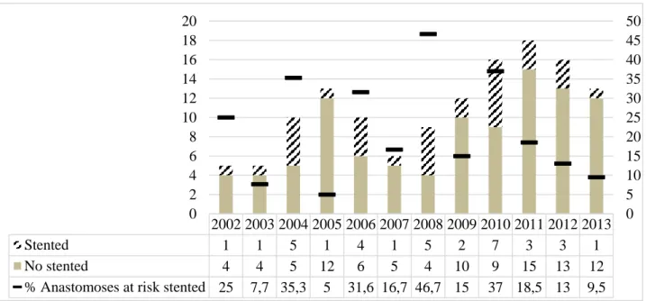
Figure 2: Number of LT and airway complications resulting in stent insertion between 2002 and 2013.
2002 2003 2004 2005 2006 2007 2008 2009 2010 2011 2012 2013
Stented 1 1 5 1 4 1 5 2 7 3 3 1
No stented 4 4 5 12 6 5 4 10 9 15 13 12
% Anastomoses at risk stented 25 7,7 35,3 5 31,6 16,7 46,7 15 37 18,5 13 9,5 0 5 10 15 20 25 30 35 40 45 50 0 2 4 6 8 10 12 14 16 18 20
22 TABLE 1. Characteristics of LT recipients in the non-stented and stented groups
NS group, n=99 S group, n=34 p Donor characteristics
Age, y (SD)a 43 (16.4) 45.8 (14.7) 0.81
Sex ratio, M:F 1.3 1.3 0.95
Ventilation in donor, hb 48 [24:48] 30 [24:48] 0.83
Recipient characteristics pre-LT
Age, y (SD)a 47.8 (16.3) 50.3 (13.2) 0.79
Sex ratio, M:F 1.8 1.0 0.13
BMI, kg/m² (SD)a 21.1 (4.4) 22.4 (5.0) 0.94
Native lung disease, n [%] COPD
Cystic fibrosis
Interstitial pulmonary fibrosis Pulmonary hypertension 45 [45.4] 31 [31.3] 17 [17.1] 5 [5.1] 18 (52.9] 7 (20.6] 6 [17.6] 3 [8.82] 0.69
Unilateral lung carcinoma 1 [1] 0
FEV1, % pred. (SD)a 28.3 (15.7) 31 (19.3) 0.79
Non invasive ventilation, n [%] 59 [59.6] 20 [5.8] 0.94 CMV Status (R / D), n [%] -/- +/- -/+ +/+ 24 [24.2] 19 [19.2] 23 [23.2] 33 [33.3] 7 [20.6] 6 [17.6] 12 [35.3] 9 [26.5] 0.58 Colonization, n [%] Bacterial Fungal
Multibacterial and/or multifungal
49 [51] 20 [20.8] 3 [3.1] 26 [27.1] 13 [38.2] 9 [26.5] 0 4 [11.8] 0.20 Procedures Type of transplant, n [%]
Single lung transplantation 59 [59.6] 26 [76.5]
23 Categorical variables are expressed as number and proportion, and compared with the chi-square test
aResults expressed as mean (SD) are compared with Student’s t test
bResults expressed as median (Q1:Q3) are compared with Mann Whitney U test
SD, standard deviation; LT, lung transplantation; COPD, chronic obstructive pulmonary disease; CMV, cytomegalovirus; FEV1,
forced expiratory volume in one second; PGD, primary graft dysfunction; ACR, acute cellular rejection; ICU, intensive care unit; CLAD, chronic lung allograft dysfunction
*considered only before stenting
e) Stenting risks factors (Table 2)
In univariate logistic model analysis adjusted with propensity score, hospitalization time (OR
+1week=1.18 [1.02-1.36], p=0.03), early bacterial infection (OR=10.64, [2.31 - 49.05], p=0.002) and
Double lung transplantation Heart-lung transplantation 38 [38.4] 2 [2] 8 [23.5] 0 Surgical reoperation [%] 18 [18.2] 3 [8.8] 0.20 Preservation liquid, n [%] Celsior Perfadex 36 [36.4] 63 [63.6] 13 (38.2] 21 [61.8] 0.85 Extracorporeal circulation, n [%] 35 [35.3] 14 [41.2] 0.80 Ventilation, hb 18 [7:7] 24 [10:144] 0.08 Blood transfusion, n [%] 38 [40] 8 [25] 0.19 Ischemia time, hb 343.5 [290:422] 379 [314:405] 0.30 ICU time, db Hospitalization time, db 10 [7:15] 24 [20:30] 14 [8:34] 30 [25:50] 0.007 <0.001 Post-transplantation events during follow-up
Early infections < 6 months*, n [%] Bacterial Fungal Severe PGD h48, n [%] Severe PGD h72, n [%] 61 [61.6] 18 [18.2] 13 [13.1] 11 [11.1] 32 [94.1] 12 [35.3] 6 [17.6] 5 [14.7] <0.001 0.039 0.52 0.58
24
fungal infection (OR=2.95, [1.17 - 7.40], p=0.021) seemed to be predictive factors. In multivariate
logistic model analysis adjusted with propensity score, only hospitalization time (+ 1 week) and early
bacterial infection were found to be independent predictors (respectively OR (+1 week = 1.23 [1.05
25 *considered only before stenting
TABLE 2. Predictive factors of stent insertion for LT recipients (n=133)
Odds ratio p 95% Confidence interval Univariate logistic regression analysis (adjusted with propensity
score)
Transplantation type (ref is double-mono transplantation)
Monopulmonary transplantation 0.349 0.034 0.132 - 0.921 Native lung disease (ref is COPD)
Cystic fibrosis 0.859 0.776 0.301 - 2.450
Interstitial pulmonary fibrosis 0.627 0.437 0.194 - 2.031 Pulmonary hypertension 0.944 0.944 0.186 - 4.777
FEV1 % pred., +1% 1.001 0.957 0.976 - 1.026
Blood Transfusion : yes 0.575 0.243 0.227 - 1.456 Pre LT colonization (ref is no)
monobacterial 1.437 0.480 0.526 - 3.929
multibacterial and/or fungal 0.444 0.190 0.132 - 1.493
UCI time, +1 day 1.020 0.060 0.999 - 1.042
Hospitalization time, +1 week 1.18 0.030 1.02 - 1.36
Ventilation, +1 hr 1.000 0.984 0.999 - 1.001
Post LT early bacterial infection (<6 months)* 10.640 0.002 2.308 - 49.050 Post LT early fungal infection (<6 months)*
Multivariate logistic regression analysis (adjusted with propensity score)
2.948 0.021 1.174 - 7.404
Transplantation type (ref=double-mono transplantation)
Monopulmonary transplantation 0.284 0.032 0.090 - 0.899 Pre LT colonization (ref. is no)
monobacterial
multibacterial and/or fungal
0.877 0.233 0.830 0.041 0.263 - 2.918 0.057 - 0.943 Hospitalization time, + 1 week 1.030 0.011 1.007 - 1.053 Post LT early bacterial infection (<6 months)* 20.98 0.004 2.57- 171.08
26
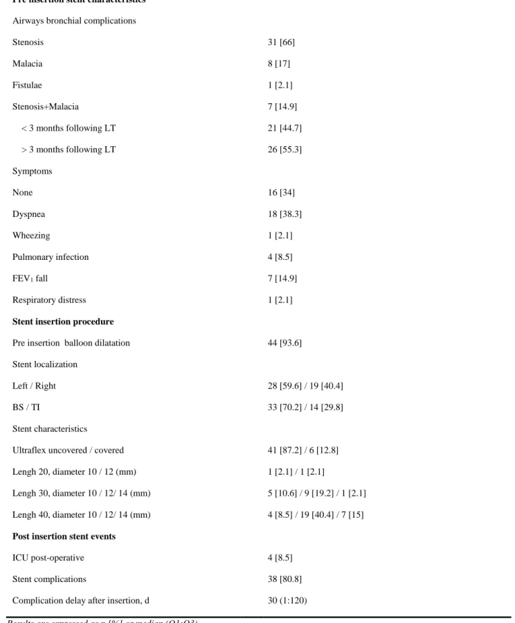
f) Stent insertion procedure and outcomes (Tables 3 and 4)
Prior to stent insertion, patients (n=34) presented obstructive lesions: stenosis (n= 31, 66%), malacia
(n= 8, 17%) and association stenosis-malacia (n=7, 15%). One presented fistulae. Clinical indications
were dyspnea (n=18, 38.3%), FEV1 fall (n=7, 14.9%), pulmonary infection (n=4, 8.5%), wheezing
(n=1, 2.1%) and respiratory distress (n=1, 2.1%). Sixteen patients (34%) had no significant clinical
symptoms.
Median time for first stent insertion was 2.4 months after transplantation [1.6 - 4.2] (n=34) and second
stent insertion, for those treated (n=13), 3.3 months after [2.8 - 6.8]. Twenty-one stents (44.7%) were
inserted during the first 3 months following LT, corresponding with early airways complications.
Median number of stent procedures per patient was 2 [1 - 3] with a maximum of 7. Forty-four (93.6%)
stented anastomoses required a pre insertion balloon dilatation (Table 3). Four (8.5%) stented
procedures required ICU. Thirty-eight (80.8%) stented anastomoses developed in a second time
airway complications. Median time to first complication was 30 days [1 - 120]. Migration and
27
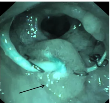
Figure 3: Follow up after nitinol stent insertion (flexible bronchoscopy): presence of a granuloma at the proximal end of the stent in the left main bronchus.
28
In 47.4% cases, there was an error of the diameter stent measurement which required a change of
stent (Table 4). A stent in stent insertion was also performed in 15.8%. Thermocoagulation (13.2%),
balloon dilatation (10.5%), removal (7.9%) and mobilization of the stent (5.3%) were the others
management of post-stent insertion complication.
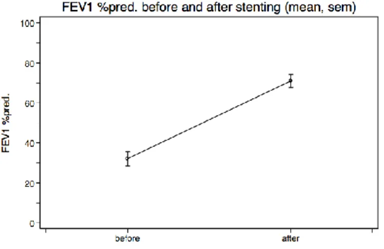
Spirometry with adequate baseline values before and after stent insertion was available for 30 subjects
(88.2%). After stent insertion, functional improvement was measured with a mean FEV1 gain of
38.9% ± 25.8 % but not statistically significant (n=30, p >0.1) (Fig.3).Twenty patient (66.7%) have
a FEV1 increase ≥ 10% corresponding in a successful treatment in the six first months. No data were available concerning improved clinical symptoms.
29 Results are expressed as n [%] or median (Q1:Q3)
TABLE 3. Description of stent indications, procedures and follow-up (stented anastomosis, n=47) Pre insertion stent characteristics
Airways bronchial complications Stenosis Malacia Fistulae Stenosis+Malacia 31 [66] 8 [17] 1 [2.1] 7 [14.9] < 3 months following LT > 3 months following LT Symptoms None Dyspnea Wheezing Pulmonary infection FEV1 fall Respiratory distress 21 [44.7] 26 [55.3] 16 [34] 18 [38.3] 1 [2.1] 4 [8.5] 7 [14.9] 1 [2.1]
Stent insertion procedure
Pre insertion balloon dilatation 44 [93.6] Stent localization Left / Right BS / TI 28 [59.6] / 19 [40.4] 33 [70.2] / 14 [29.8] Stent characteristics
Ultraflex uncovered / covered Lengh 20, diameter 10 / 12 (mm) Lengh 30, diameter 10 / 12/ 14 (mm) Lengh 40, diameter 10 / 12/ 14 (mm) 41 [87.2] / 6 [12.8] 1 [2.1] / 1 [2.1] 5 [10.6] / 9 [19.2] / 1 [2.1] 4 [8.5] / 19 [40.4] / 7 [15]
Post insertion stent events
ICU post-operative 4 [8.5]
Stent complications
Complication delay after insertion, d
38 [80.8] 30 (1:120)
30 TABLE 4. Etiology and treatment of stent related complications, n=38
Stent complications Stent causes Treatment
Early (< 2 days) , n=3 [7.9]
Acute dyspnea (n=1) Migration (n=2)
Default deployment of the prosthesis Default prosthesis diameter (too small)
Removal Change Tardive, n=35 [92.1] Mucus plugging, n=4 [10.5] Stenosis, n=5 [13.16] Granuloma, n=10 [26.32] Migration, n=14 [36.8] Malacia, n=2 [5.26]
Default prosthesis diameter (too small) (n=3) Obstruction (n=1)
Default prosthesis length (too small) (n=1) Default prosthesis diameter (too small) (n=2) None (n=2)
Default prosthesis diameter (too small) (n=2) None (n=7)
Obstruction (n=1)
<30 days
Default prosthesis diameter (too small) (n=4) Default prosthesis length (too small) (n=4)
>30 days
Default prosthesis diameter (too small) (n=4) None (n=2)
Default prosthesis diameter (too small) (n=1) Default prosthesis length (too small) (n=1)
Change Removal Addition Change Dilatation Change Thermocoagulation (n=5) Dilatation (n=2) Removal Change Addition Change Mobilization Change Addition
31
g) Survival and CLAD-Free survival analysis (Figure 4)
Using the Kaplan-Meier estimator without taking into account immortality bias, survival was not
statistically different in S and NS groups (log-rank: p=0.87) (Fig. 5A). In time-dependent analysis
taking into account the immortal time bias, the 75% survival time was 1.28 years in S group and 2.6
years in NS group, and using the Kaplan-Meier estimator, survival was better in NS group (log rank:
p=0.02) (Fig. 5B).
Using the Kaplan-Meier estimator without taking into account immortality bias, CLAD-free survival
was not statistically different in S and NS groups (log-rank: p=0.60) (Fig. 5C). In time-dependent
analysis, the 75% CLAD-free survival time was 3.4 years for S group and 3.78 years months for NS
group, and using the Kaplan-Meier estimator, CLAD free survival was not statistically different in S
32
5: Survival. 5A Kaplan-Meier estimates of survival between stented group and non-stented group. 5B Kaplan-Meier estimates of survival between stented group and non-stented group in a
time-dependent analysis. CLAD-free survival. 5C Kaplan-Meier estimates of CLAD-free survival
between stented group and non-stented group. 5D Kaplan-Meier estimates of CLAD-free survival
between stented group and non-stented group in time-dependent analysis. CLAD: chronic lung
allograft dysfunction.
B A
D C
33
4) Discussion
Results challenge four issues to be discussed: bronchial anastomoses complications are still frequent
after LT with 47 (21.4 %) of the 218 anastomoses-at-risk requiring stent insertion; infections,
considered before stenting were associated with a higher risk of stent insertion; in patients treated
with uncovered nitinol stent, a high rate of complications (80.8%); and a lower survival in cases of stenting
challenges the uncovered nitinol stents use in CAC post LT.
Many risk factors of bronchial anastomosis complications after LT were described: surgical
techniques including minimal dissection “no touch” of recipient main bronchi and location of anastomose (7), mechanical ventilation time (18), recipient age < 45 years old and early graft rejection
(6), post LT colonization and type of transplantation (DLT or lobar transplantation (7,19), organ
preservation solution and postoperative tracheostomy(20). In univariate analysis, post LT fungal
infection seems to be an important risk factor, in agree with literature (6,10,18,20) even if we have
described less post LT fungal infection than others studies (22.6% vs 48.5% -82%) (2,10).Our fungal
prophylaxis was probably started too late, only after ICU period. A lower rate of fungal infection
could be achieved with an immediate post-surgical treatment.
However, multivariable analysis adjusted for a propensity score, we found that only hospitalization
time and early bacterial infections were independent predictor of stent insertion. Our treatment against
Pneumocystis Jiroveci is probably not efficiency against others microorganism like encapsulated
bacteria infections (Pneumococcus, Haemophilus). These antibiotherapy was usually short like
prophylaxis. Indeed, there is no consensus about prophylaxis or antibiotic against bacterial infection
in LTR. For example, Dutau and al (2) used systematic ceftriaxone or individualized antibiotic
34
careful attention of bacterial or fungal colonization before LT with infection identification may clarify
this issue.
Our second significant predictive factor is hospitalization time and it is difficult to know if it is a
cause or a consequence for stent insertion. During this time a lot of events can be described. However,
ICU time, blood transfusion, ventilation time, use of extra corporeal circulation, presence of primary
graft dysfunction are not considered as risk factors. Others data could be analyzed as length of
intubation (19) or all potential medical complications, medical personal history, medical and
paramedical treatment (physiotherapy, respiratory rehabilitation) to allow an appropriate
interpretation of this result.
Twenty one percent of our patients required stent placement, in agreement with literature (9,10).
According with the literature, we had not procedure related deaths (21–25). Only 4 patients required ICU,
one case not due to the stenting procedure, 2 cases due to an immediate stent migration and one case due to
a malfunction during stent placement. Successful treatment corresponding in lung function improvement
after stenting was described but did not reach significance due to a lack of data and limited power of
these analysis.
Post-stent insertion complications were very frequent reaching 80.6%. Most of the previous studies had a
lower rate of complications, 22 and 67% (26,27), except one, from Gottlieb et al (11). This higher rate is
explained by a longer follow up and a larger cohort in our study (854 days) and Gottlieb’s one (777 days), than in others studies (between 263 to 400 days).
Default of pre stent insertion sizing is important in the development of post stenting complications. Stent
migration was described in 42.1% of cases, with 26.3% diagnosed in the first 30 days after the procedure
higher than in the literature, occurring in 0-21% of metal stent (28). A small diameter and/or length
stent result in a disparity between stent size and airway diameter and required a change or an addition
35
inflammation induced by the friction of the prosthesis against the bronchial wall; alternative
explanation could be related to wall ischemia due to stent pressure favoring granulation at stent
extremities. Pre insertion CT SCAN avoid invasive procedures before definitive treatment decisions
are made and give a better evaluation of the sizing and location of stents required (15). In our
experience, disparity in airway measurement and stent sizing are still present. A better analysis and
cooperation among ENT, pulmonologist and radiologist should reduce these purely technical
complication. Restenosis were described in 13.16% according with literature (21,24,25,29,30), unlike
Gottlieb et al. and Chhajed et al. (11,31). Others procedures like balloon dilatation, thermocoagulation
or stent removal could be more used (11). However, they do not seem to be indicated for the treatment
of post stent delivery complications because they also result in short-term complications (32), with
their own side effects like tissue damage for dilatation, deep tissue injury for coagulation or airway
re obstruction for removal. Some centers perform serial bronchoplasty many times followed by stent
insertion and have been able to resolve these complications in 22 (37%) of cases just with balloon
dilation alone (9,20). This way may be necessary to induce a better epithelialization before stent
insertion, which improved anatomic and functional results.
According with Gottlieb and Chaajed (11,31), survival was significatively better in NS group. Our
time-dependent analysis, which takes into account the immortal bias, allows us to have a better
interpretation of this result. Most observational old studies (2,20,33) presented artificially improved
results by increasing event outcome in control groups because they did not consider this bias. This bias
has already been well described in many observational studies with a variety of cohort designs (34,35). We hypothesize that this result seems to due to a higher risk of infections and a lower functional lung
reserve in case of bronchial complications treated with nitinol stent insertion. Temporary stent, like
36
post insertion complications and stent removal achieved in 69.5% without recurrence of stenosis (2).
Concerning the CLAD free survival, there is no significant difference between groups, according with
Shofer et al. (20). It confirms that stent insertion is not associated with CLAD development and that
the mechanisms underlying central and peripheral airways complications are different.
As with any retrospective study design, there are serious limitations that may have introduced bias.
But our report merits discussion about methodological issues in observational studies in the field of
LT and could be viewed as a use-case of how to control biases in cohort studies by using propensity score.
5) Conclusion and perspectives
Use of nitinol stent in the management of anastomotic complications after LT is challenged after our
study. Temporary or biodegradable stent may play a role in the future. We show a strong association
between both early bacterial infection and hospitalization time, and the development of CAC
requiring stent insertion. The improvement of infections prophylaxis and analysis of underlying
37
Annexes
Sténoses bronchiques anastomotiques
Stent en nitinol non couvert en position normale
Stent en nitinol non couvert avec complications locales : granulomes, sténose à l’extrémité distale, obstruction par mucus plugging
38
39
References
1. Santacruz JF, Mehta AC. Airway complications and management after lung transplantation:
ischemia, dehiscence, and stenosis. Proc Am Thorac Soc. 2009;6:79‑93.
2. Dutau H, Cavailles A, Sakr L, et al. A retrospective study of silicone stent placement for
management of anastomotic airway complications in lung transplant recipients: short- and
long-term outcomes. J Heart Lung Transplant Off Publ Int Soc Heart Transplant. 2010;29:658‑64.
3. Groetzner J, Kur F, Spelsberg F, et al. Airway anastomosis complications in de novo lung
transplantation with sirolimus-based immunosuppression. J Heart Lung Transplant Off Publ Int
Soc Heart Transplant. 2004;23:632‑8.
4. Van Berkel V, Guthrie TJ, Puri V, et al. Impact of anastomotic techniques on airway
complications after lung transplant. Ann Thorac Surg. 2011;92:316‑20; discussion 320‑1.
5. Van De Wauwer C, Van Raemdonck D, Verleden GM, et al. Risk factors for airway
complications within the first year after lung transplantation. Eur J Cardio-Thorac Surg Off J Eur
Assoc Cardio-Thorac Surg. 2007;31:703‑10.
6. Thistlethwaite PA, Yung G, Kemp A, et al. Airway stenoses after lung transplantation: incidence,
management, and outcome. J Thorac Cardiovasc Surg. 2008;136:1569‑75.
7. Weder W, Inci I, Korom S, et al. Airway complications after lung transplantation: risk factors,
prevention and outcome. Eur J Cardio-Thorac Surg Off J Eur Assoc Cardio-Thorac Surg.
40
8. Shennib H, Massard G. Airway complications in lung transplantation. Ann Thorac Surg.
1994;57:506‑11.
9. Murthy SC, Blackstone EH, Gildea TR, et al. Impact of anastomotic airway complications after
lung transplantation. Ann Thorac Surg. 2007;84:401‑9, 409.e1‑4.
10. Herrera JM, McNeil KD, Higgins RS, et al. Airway complications after lung transplantation:
treatment and long-term outcome. Ann Thorac Surg. 2001;71:989‑93; discussion 993‑4.
11. Gottlieb J, Fuehner T, Dierich M, Wiesner O, Simon AR, Welte T. Are metallic stents really safe?
A long-term analysis in lung transplant recipients. Eur Respir J. 2009;34:1417‑22.
12. Bisson A, Bonnette P. A new technique for double lung transplantation. « Bilateral single lung »
transplantation. J Thorac Cardiovasc Surg. 1992;103:40‑6.
13. Sundaresan S, Trachiotis GD, Aoe M, Patterson GA, Cooper JD. Donor lung procurement:
assessment and operative technique. Ann Thorac Surg. 1993;56:1409‑13.
14. Claustre J, Quétant S, Camara B, et al. Nonspecific immunoglobulin replacement in lung
transplantation recipients with hypogammaglobulinemia: a cohort study taking into account
propensity score and immortal time bias. Transplantation. 2015;99:444‑50.
15. Righini C, Aniwidyaningsih W, Ferretti G, et al. Computed tomography measurements for airway
stent insertion in malignant airway obstruction. J Bronchol Interv Pulmonol. 2010;17:22‑8.
16. Liu J, Weinhandl ED, Gilbertson DT, Collins AJ, St Peter WL. Issues regarding « immortal time »
41
17. Gail MH. Does cardiac transplantation prolong life? A reassessment. Ann Intern Med.
1972;76:815‑7.
18. Choong CK, Sweet SC, Zoole JB, et al. Bronchial airway anastomotic complications after
pediatric lung transplantation: incidence, cause, management, and outcome. J Thorac Cardiovasc
Surg. 2006;131:198‑203.
19. Moreno P, Alvarez A, Algar FJ, et al. Incidence, management and clinical outcomes of patients
with airway complications following lung transplantation. Eur J Cardio-Thorac Surg Off J Eur
Assoc Cardio-Thorac Surg. 2008;34:1198‑205.
20. Shofer SL, Wahidi MM, Davis WA, et al. Significance of and risk factors for the development of
central airway stenosis after lung transplantation. Am J Transplant Off J Am Soc Transplant Am
Soc Transpl Surg. 2013;13:383‑9.
21. Breitenbücher A, Chhajed PN, Brutsche MH, Mordasini C, Schilter D, Tamm M. Long-term
follow-up and survival after Ultraflex stent insertion in the management of complex malignant
airway stenoses. Respir Int Rev Thorac Dis. 2008;75:443‑9.
22. Hautmann H, Bauer M, Pfeifer KJ, Huber RM. Flexible bronchoscopy: a safe method for metal
stent implantation in bronchial disease. Ann Thorac Surg. 2000;69:398‑401.
23. Husain SA, Finch D, Ahmed M, Morgan A, Hetzel MR. Long-term follow-up of ultraflex metallic
42
24. Madden BP, Park JES, Sheth A. Medium-term follow-up after deployment of ultraflex
expandable metallic stents to manage endobronchial pathology. Ann Thorac Surg.
2004;78:1898‑902.
25. Miyazawa T, Yamakido M, Ikeda S, Furukawa K, Takiguchi Y, Tada H, et al. Implantation of
ultraflex nitinol stents in malignant tracheobronchial stenoses. Chest. 2000;118:959‑65.
26. Carré P, Rousseau H, Lombart L, et al. Balloon dilatation and self-expanding metal Wallstent
insertion. For management of bronchostenosis following lung transplantation. The Toulouse
Lung Transplantation Group. Chest. 1994;105:343‑8.
27. Kapoor BS, May B, Panu N, Kowalik K, Hunter DW. Endobronchial stent placement for the
management of airway complications after lung transplantation. J Vasc Interv Radiol JVIR.
2007;18:629‑32.
28. Mehta AC, Dasgupta A. Airway stents. Clin Chest Med. 1999;20:139‑51.
29. Mughal MM, Gildea TR, Murthy S, Pettersson G, DeCamp M, Mehta AC. Short-term
deployment of self-expanding metallic stents facilitates healing of bronchial dehiscence. Am J
Respir Crit Care Med. 2005;172:768‑71.
30. Burns KEA, Orons PD, Dauber JH, et al. Endobronchial metallic stent placement for airway
complications after lung transplantation: longitudinal results. Ann Thorac Surg.
2002;74:1934‑41.
31. Chhajed PN, Malouf MA, Tamm M, Glanville AR. Ultraflex stents for the management of airway
43
32. Lunn W, Feller-Kopman D, Wahidi M, Ashiku S, Thurer R, Ernst A. Endoscopic removal of
metallic airway stents. Chest. 2005;127:2106‑12.
33. Abdel-Rahman N, Kramer MR, Saute M, Raviv Y, Fruchter O. Metallic stents for airway
complications after lung transplantation: long-term follow-up. Eur J Cardio-Thorac Surg Off J
Eur Assoc Cardio-Thorac Surg. 2014;45:854‑8.
34. Van Walraven C, Davis D, Forster AJ, Wells GA. Time-dependent bias was common in survival
analyses published in leading clinical journals. J Clin Epidemiol. 2004;57:672‑82.
35. Li Y, Gottlieb J, Ma D, Kuehn C, et al. Graft-protective effects of the HMG-CoA reductase
inhibitor pravastatin after lung transplantation--a propensity score analysis with 23 years of
44
SERMENT D’HIPPOCRATE
En présence des Maîtres de cette Faculté, de mes chers condisciples et devant l’effigie d’HIPPOCRATE,
Je promets et je jure d’être fidèle aux lois de l’honneur et de la probité dans l’exercice de la Médecine.
Je donnerai mes soins gratuitement à l’indigent et n’exigerai jamais un salaire au-dessus de mon tr avail. Je ne participerai à aucun partage clandestin d’honoraires.
Admis dans l’intimité des maisons, mes yeux n’y verront pas ce qui s’y passe ; ma langue taira les secrets qui me seront confiés et mon état ne servir a pas à corrompre le s mœurs, ni à favoriser le crime.
Je ne permettrai pas que des considérations de religion, de nation, de race, de parti ou de classe sociale viennent s’inter poser entre mon devoir et mon patient.
Je garderai le respect absolu de la vie humaine.
Même sous l a menace, je n’admettrai pas de faire usage de mes connaissances médicales contre les lois de l’humanité.
Respectueux et reconnaissant envers mes Maîtres, je rendrai à leurs enfants l’instruction que j’ai reçue de leurs pères.
Que les hommes m’accordent le ur estime si je suis fidèle à mes promesses. Que je sois couvert d’opprobre et mépr isé de mes confrères si j’y manque.
45
Remerciements :
A mes maîtres et aux personnes rencontrées lors de mon cursus :
En ORL au CHU de Grenoble
Monsieur le Professeur Sébastien SCHMERBER
Je souhaitais vous remercier très sincèrement pour votre enseignement clinique et chirurgical dans le
domaine de la chirurgie otologique. Votre enthousiasme et votre excellence chirurgicale dont nous
avons bénéficié durant ces années sont un exemple. Vous m’avez fait l’honneur de me confier et de
diriger un travail sur la labyrinthectomie chimique. Soyez assuré de mon profond respect et de ma
reconnaissance.
A ceux qui sont partis depuis tout ce temps :
A Alexandre KARKAS, qui m’a tant appris en otologie et en chirurgie endonasale, tu as laissé un grand vide en nous quittant pour Saint Etienne. Merci pour ta gentillesse et ta disponibilité.
A Romain TOURNIAIRE, mon premier CCA, qui m’a fait découvrir dans la bonne humeur l’ORL lors de ma première année d’internat et qui maintenant s’éclate à Annemasse, en famille qui plus est !
A l’équipe actuelle :
A Alice HITTER, ma première CCA (aussi !), chef des n’enfants et des n’internes. Un grand merci
pour ton écoute, ta disponibilité, tes qualités d’enseignements et humaines et surtout ta bonne humeur
inégalable. C’est toujours un plaisir de venir aux apéros chez toi, été comme hiver, avec Marc et tes 2 petits.
46
labyrinthectomie chimique, je vous en suis sincèrement reconnaissant. Votre enthousiasme pour la
vestibulologie ainsi que pour toutes les facettes de l’ORL nous forcent à l’admiration.
Aux Docteurs Anne RIVRON et Joëlle TROUSSIER, je vous remercie pour votre professionnalisme
et votre gentillesse.
Aux équipes du bloc, de consultation et du service ORL, avec qui cela a été et est toujours un plaisir
de travailler et dont certaines m’ont connu depuis le début ! Le plus des grand des merci à vous toutes, quelques souvenirs me reviennent, soirées de fin de stage ou apéros, qui n’ont pas eu d’égal avec
d’autres services.
A mes co internes, aux anciens : Alexandra (bertogrololo) et Anne (soooo amazing!) des amies avant
tout, Etienne (la grosse Berte ou bonpapy) qui me manque déjà, Elea, Akil (outou), Raphaëlle (Rachel
4), Jennifer, et cin’dyyy. Aux plus jeunes que j’apprends à connaitre: Ashley, Christol et Ludo. Une pensée pour Jean mich mich et Nasser, ces premiers semestres avec vous restent de supers souvenirs !
Merci !
En Chirurgie vasculaire au CHU de Grenoble
Au Professeur Jean Luc MAGNE, je vous remercie pour votre bienveillance et votre enseignement
chirurgical durant ces 6 mois passés dans un service de grande qualité. Aux Docteurs Emmanuel
COCHET, Hélène BLAISE et Caroline DUCOS ainsi qu’au Professeur Carmine SESSA qui dans la
bonne humeur m’ont transmis leurs passions et leurs rigueurs dans l’exercice de la chirurgie.
Au Docteur Albéric de LAMBERT, ou Albé, qui finit sont clinicat actuellement avant de partir sur
Chambéry. Très heureux d’avoir partagé ton dernier semestre d’internat. Ton parcours familial, professionnel et sportif est assez impressionnant et je suis bien content de te retrouver prochainement
sur Chambéry. Bon courage pour tes ultratrails cet été !
A Marine, co interne du moment, dont les siestes mémorables et la répartie tranchante reste de supers
47
En Chirurgie Maxillo faciale et chirurgie plastique au CHU de Grenoble
Au Professeur Georges BETTEGA, j’ai été heureux d’avoir pu bénéficier à votre contact de votre excellence chirurgicale et de votre rigueur. Vous m’avez confié un travail de master 2, conseillé et dirigé, voire même été un « cobaye » à certains moments. Votre disponibilité et votre implication
envers les internes et étudiants est un exemple. Soyez assuré de mon profond respect et de ma
reconnaissance.
Au Docteur Béatrice MORAND, merci pour votre enseignement, votre disponibilité et votre
gentillesse.
A Antoine GROSDIDIER, j’ai été bien heureux de partager avec toi tes derniers mois de CCA, ce fut un plaisir de travailler à tes côtés et de bénéficier de tes conseils. Merci aussi pour les leçons de skis !
Profites bien de ta petite famille.
A Cynthia HAMOU, ton enthousiasme et ta disponibilité ainsi que tes conseils m’ont beaucoup appris durant ces 6 mois. Je te souhaite une belle carrière mais surtout une vie de famille épanouie.
A mes co internes du moment, Jujuliette, Romain Lewandowskichtaïa et Aurélie Vigneron, un très
bon cru !
Au laboratoire HP2 au CHU de Grenoble
Au Professeur Renaud TAMISIER, qui a su me donner l’envie d’effectuer un M2R passionnant, qui m’a conseillé et dirigé pendant cette année, et a même été un « cobaye » (aussi !) pour certaines expériences. Ta grande expertise, ta disponibilité et tes conseils m’ont beaucoup appris. Sois assuré
de mon plus grand respect et de ma sincère reconnaissance. J’espère que nous pourrons continuer à
travailler ensemble.
En ORL, au CHU de Montpellier
48
enseignement de grande qualité ainsi que leur excellence chirurgicale, soyez assuré de mon plus grand
respect.
Aux docteurs César CARTIER, Guillemette PIERRE, des exemples à suivre, j’ai énormément appris à vos côtés autant d’un point de vue chirurgicale qu’humain. Un énorme bonus pour Guillemette qui m’a bluffé par ses conseils avisés, même jusqu’à 5h du matin, parfois, lors d’un Fizz à l’improviste ;) A Vincent TREVILLOT et Camille GALY, les 2 CCA du service. Malgré un rythme toujours
soutenu, l’ambiance était toujours là et les fins de visite n’ont jamais été aussi dures que lorsqu’un
fou rire débutait à la chambre n°1. De très bons souvenirs à vos côtés. Continuez comme ça !
Aux équipes du bloc et du service, tellement nombreux, avec une ambiance comme on voit rarement.
Un grand merci à vous pour votre accueil et votre professionnalisme.
Aux co internes d’ORL A, les champions Yann, Valentin et Thibault sans oublier Noémie qui a dû nous supporter ;) Une superbe entente et de bons moments malgré des journées de folie.
Aux autres que j’ai pu côtoyer pendant ce semestre et avec qui j’ai aussi de bons moments entre 2 plats mixés tièdes sortis du frigo du A ou du B : Lylou (l’exploratrice.. ?!), Agnès (cachée à Palavas),
David (le sauveur ;) ), Fanny (la fan de chocolat), Julie (la chef du B).
A Hubert Roth, pour ton aide précieuse et ta disponibilité lors de l’élaboration et l’analyse statistique de ce travail. Un plaisir de travailler avec toi, sois assuré de ma gratitude.