HAL Id: dumas-02159885
https://dumas.ccsd.cnrs.fr/dumas-02159885
Submitted on 19 Jun 2019HAL is a multi-disciplinary open access archive for the deposit and dissemination of sci-entific research documents, whether they are pub-lished or not. The documents may come from teaching and research institutions in France or abroad, or from public or private research centers.
L’archive ouverte pluridisciplinaire HAL, est destinée au dépôt et à la diffusion de documents scientifiques de niveau recherche, publiés ou non, émanant des établissements d’enseignement et de recherche français ou étrangers, des laboratoires publics ou privés.
Endolymphatic sac decompression versus endolymphatic
duct blockage in patients with saccular and utricular
hydrops, as evidenced by MRI
Marie Soula
To cite this version:
Marie Soula. Endolymphatic sac decompression versus endolymphatic duct blockage in patients with saccular and utricular hydrops, as evidenced by MRI. Human health and pathology. 2019. �dumas-02159885�
AVERTISSEMENT
Ce document est le fruit d'un long travail approuvé par le
jury de soutenance et mis à disposition de l'ensemble de la
communauté universitaire élargie.
Il n’a pas été réévalué depuis la date de soutenance.
Il est soumis à la propriété intellectuelle de l'auteur. Ceci
implique une obligation de citation et de référencement
lors de l’utilisation de ce document.
D’autre part, toute contrefaçon, plagiat, reproduction illicite
encourt une poursuite pénale.
Contact au SID de Grenoble :
bump-theses@univ-grenoble-alpes.fr
LIENS
LIENS
Code de la Propriété Intellectuelle. articles L 122. 4
Code de la Propriété Intellectuelle. articles L 335.2- L 335.10
http://www.cfcopies.com/juridique/droit-auteurUNIVERSITE GRENOBLE ALPES FACULTE DE MEDECINE DE GRENOBLE Année: 2019
ENDOLYMPHATIC SAC DECOMPRESSION VERSUS ENDOLYMPHATIC DUCT BLOCKAGE IN PATIENTS WITH SACCULAR AND UTRICULAR HYDROPS, AS
EVIDENCED BY MRI
THESE PRESENTEE POUR L’OBTENTION DU DOCTORAT EN MEDECINE DIPLÔME D’ETAT
Marie SOULA
THESE SOUTENUE PUBLIQUEMENT A LA FACULTE DE MEDECINE DE GRENOBLE* - Le 14 juin 2019
DEVANT LE JURY COMPOSE DE
Président du jury : Monsieur le Professeur Christian Adrien RIGHINI
Membres : Monsieur le Professeur Sébastien SCHMERBER, directeur de thèse Monsieur le Docteur Arnaud ATTYE
Monsieur le Docteur Georges DUMAS Monsieur le Docteur Alexandre KARKAS
*La Faculté de Médecine de Grenoble n’entend donner aucune approbation ni improbation aux opinions émises dans les thèses ; ces opinions sont considérées comme propres à leurs
R
EMERCIEMENTS
A MES MAITRES ET MEMBRES DU JURY
Monsieur le Professeur Christian Adrien RIGHINI, président du jury
Vous me faites l’honneur de présider la soutenance de cette thèse. Je vous remercie pour votre enseignement tout au long de ces années d’internat. Merci également de m’avoir permis de faire un semestre d’inter CHU.
Monsieur le Professeur Sébastien SCHMERBER, directeur de thèse
Vous me faites l’honneur de diriger ce travail. Je vous remercie pour votre enseignement clinique et chirurgicale. Merci de nous transmettre au jour le jour votre passion pour l’otologie.
Monsieur le Docteur Arnaud ATTYE
Je te remercie d’avoir accepté de juger ce travail. Merci pour ton aide si chère tout au long de ce travail. Nous sommes si admiratifs de tes connaissances en ORL. Je te souhaite un excellent séjour en Australie.
Monsieur le Docteur Alexandre KARKAS
Tu me fais l’honneur de juger ce travail et de venir jusqu’à Grenoble aujourd’hui. Je n’ai pas eu l’honneur de travailler avec toi hélas... Heureusement, j’ai eu la chance de pouvoir bénéficier de tes cours en congrès qui sont passionnants.
Monsieur le Docteur Georges DUMAS
Je vous remercie d’avoir accepté de juger ce travail, si cher à votre cœur. Je vous remercie pour votre bienveillance et votre disponibilité sans failles tous au long de ces années.
AUX MEDECINS RENCONTRES PENDANT MON INTERNAT
En ORL au CH de Chambéry : Aux Docteurs Patrick MANIPOUD et Christophe GUICHARD, j’ai passé un excellent second semestre dans votre équipe, vous m’avez tant appris. Merci également aux Docteurs GUIRAUD, NICOLLET, SAUMUR et FILIDORO. Il me tarde de revenir dans votre équipe dès le mois de novembre prochain.
En ORL au CH d’Annecy : Aux Docteurs Bernard FONLUPT, Frédérique LAGARDE, Guillaume ANGEL, Etienne BERTA et Dominique PAOLETTI. Mon 5ème semestre parmi vous fut passionnant, le tout dans la simplicité et la bonne humeur.
En ORL au CHU de Grenoble : Au Docteur Ihab ATALLAH, tu fais un travail admirable. Merci de nous transmettre ta passion pour la laryngologie. Aux Docteurs Anne RIVRON et Joëlle TROUSSIER, vous êtes si chère à notre cœur, merci pour votre soutien. Au Docteur Alice HITTER, tu nous manques beaucoup…
En ORL au CHIC Centre Hospitalier Intercommunal de Créteil : Au Professeur André COSTE, votre enseignement et vos connaissances cliniques et chirurgicales sont inégalables. Ce fut un honneur de travailler dans votre équipe. Aux Docteurs Lydia BRUGEL, Anne GAUTHIER, Héloise DE KERMADEC, Emilie BEQUIGNON, Philippe BEDBEDER… les 6 mois sont passés tellement vite, j’aurais aimé qu’ils se poursuivent…
En Chirurgie vasculaire et thoracique au CH de Chambéry : Aux Docteurs Hélène BLAISE, Alberic DE LAMBERT et Nathalie DAVID, j’ai beaucoup aimé travailler avec vous et ce n’est que le début !
En CMF et chirurgie plastique au CHU de Grenoble et CH d’Annecy : Au Docteur Béatrice MORAND, j’ai passé un excellent semestre à tes côtés, merci pour ton enseignement et ta rigueur. Au professeur Georges BETTEGA, aux docteurs Dorothée DENEUBOURG, Isé MARZLOFF et Leslie NOYELLES merci pour votre accueil.
AUX EQUIPES RENCONTREES PENDANT MON INTERNAT
Dans le service au CHU : Marion, Sabrina, Margaux, Amélie, Stéphanie(s), Alice, Ayette, Gwen, Guillaume… c’était un bonheur de travailler avec vous et ça continue pour certain(e)s ! A Chambéry : Océane, Tammy… A Créteil : Fanny, Sandra, Séverine, Margaux, c’était tellement agréable de compter sur vous.
En consultation au CHU : Martine, Jennyfer, Laurence et Sabine. A Chambéry : Françoise, Sandrine, Faustine, Pierre et Sophie, j’ai tellement hâte de vous retrouver !
Au bloc au CHU : Céline, Julie, Jacqueline, Karine, Sabiha, Marjorie, Myriam… Aux secrétaires : Christelle (s), Caroline, Ludivine…
A Sophie, la meilleure des cadres de tous les temps !
Aux audioprothésistes : Brieux, Julien, Fabien, Leslie, Adeline, Jonathan et bien d’autres encore.
A MES CO-INTERNES, CCA ET ASSISTANTS
Grenoblois : A Ludo, tu me feras toujours rire et pleurer de joie. Merci pour ton écoute et ta
bienveillance. A mon acolyte Mathieu pour ces fous rires en stage, ta rigueur et ton sérieux, c’est un vrai plaisir de travailler avec toi. A Bagouz, pour ta gentillesse qui se rebêle ! A Akil é bo le lavabo, une perle qui nous manque beaucoup trop, profites bien de Montpellier… A Cindy, el professor ;-), Noémie, Claire D, Claire S, Christol, Jenifer, Alexandra, Ilan, Olivier, Adrien et Mathilde (petit piou piou)… A Pia, Kévin, Charlotte M, Florian et Virginie, pour ces semestres passés à vos côtés.
Parisiens : A Vincent, pour ces délires en stage. A Sophie, Vincent, Guillaume.
A Eléa, tu es mon mentor, tu m’as tellement appris. Je suis triste de ton départ, mais qui sait peut-être qu’un jour nos chemins se retrouveront. A Anne, c’est grâce à toi que j’ai atterri ici. Je n’oublierai jamais nos longs trajets en voiture entre Grenoble et Chambéry. Lalalalalala lalalalala… A Raphaele, je t’admire pour ton assurance et tes connaissances, qui m’aident beaucoup à progresser. Merci ! A David, au surnom le plus pourri de tout mon internat… A Chrystelle, de bons moments passés en ta compagnie… What else ? A philippe, Eric…
A MES AMIS
A mon bichon, je m’en souviens comme si c’était hier…c’est de la que tout a commencé… Sans toi, je n’aurais jamais pu en arriver là. Nos choix de vies nous ont un peu éloigné mais notre amitié n’en demeure pas moins intense.
A mon Choupi, une amie, un rock. Merci, merci, merci. Merci pour ces soirées de l’espace, pour ces craquages et surtout pour tes précieux conseils pour ma vie personnelle et professionnelle. Le semestre au CHIC était Bêêêêêêê !!! Romain, merci de prendre soin d’elle. A Sarah, nos deux ans de vie commune ne m’ont pas laissé indemne. Me voila à l’affut de la meilleure crème de marron et à la recherche de la petite idée à faire germer pour créer des choses de moi-même… Je vous souhaite beaucoup de bonheur et d’aventure avec Julien.
A Fanny, mon petit chat, j’ai vraiment beaucoup de chance de t’avoir à mes côtés. Merci pour ton écoute si précieuse et d’être si fidèle. Je ne serai te remercier pour ton aide pour cette thèse… On va pouvoir commencer à profiter maintenant que nos thèses sont passées !
A Gagathe, toutes les saisons à l’école de voile défilent… Ma partenaire inégalable de voile et de vie… Hey hey hey ou ah, i want to know…
A ma Mathou… on s’est construit ensemble. Merci pour tout…
A Aldric, plus de 15 ans que notre amitié traverse les frontières… J’espère vous voir rapidement emménager dans la région avec Lise et Hélio !
A mon Maxi Nore ! Le seul, l’unique ! Tu fais des milliers de km pour toujours être là pour les grands moments, Merci pour ta fidèle amitié.
A mes parisiens préférés : Ol pour ces révisions D4 magiques, vive les Pingus ! Vous êtes radieux avec Louis L. Maxou, Notre Captain ! Que nos croisières nous emmènent jusqu’au bout du monde avec Marie, Bertcl (tellement ouf que tu partes en Guyane !) et Raphael, Anne et Louis S, Vanille (Profites à fond du Canada !), Marie A et Fx, Clémence…
Aux grenoblois : Yohan, nos dégustations me feront peut-être un jour changer de voie :), Gautier (Un nounours), Dysmas (mon coloc préféré.), Momo (une joie de vivre), Giovanni (tu aimes les P…. ?), Anne sophie (Tellement magique notre rencontre), Bouba (sans toi j’aurais probablement craqué plus d’une fois à Annecy !) et Manon, Davy (toujours le sourire, un régal), Hubert (merci d’avoir décalé ton réveil), Jojo (je te vois avec ton ciré jaune !), Elliot, Wassima, Alison (on se retrouve dans quelques mois à Chambéry), Camille TV, Camille T et Laura, Clément et Laure, Liza, Bibou, Mariette, Marine, Paul, Jean Charles… Notre premier semestre restera à graver dans ma mémoire… On m’avait dit qu’au premier semestre c’était à ce moment-là que tu te faisais des amis pour la vie…
Aux copains chambériens : Benja (garde ta joie de vivre), Victor (ses pas de danses), Florian et Julie, Elo et Nico, j’ai hâte de me rapprocher de vous ! Et surtout merci à Herbert qui a endiablé nos soirées…
A MA FAMILLE
A ma maman, tu es un exemple pour moi. C’est grâce à toi que j’ai trouvé l’énergie toute ces années durant. Tu le sais déjà, je ne sais pas ce que je ferai sans toi…
A mon papa, merci pour tout ce que tu as fait pour moi. Merci de m’avoir guidé à prendre les bonnes décisions. Merci pour ton soutien toutes ces années passées et pour celle à venir… A mon toutoun, la force qui nous lie est indescriptible… Je te souhaite de t’accomplir dans la formation que tu entreprends en septembre. Quelle fierté d’être la tante de Theodore et Dimitri ! N’oublie pas que tu es un papa en OR !
A ma dodie, tu es partie loin mais cela ne change en rien notre complicité et notre amour… Je suis fière que tu aies mené à terme ton projet au Canada, accroche-toi ! Je serai à jamais là pour toi…
A ma marraine, merci d’être présente ce jour. Souvenir de ces 4 mois passés chez toi…
A Coco, Pierre, Laurent, Naynou et Mado merci de m’avoir si bien accueilli dans votre famille…Je me sens bien parmi vous…
Et pour finir en beauté, ma cerise sur le gâteau, ma pépite d’or, mon bijou, c’est toi Nicolas. Tu es simplement tout ce dont je rêvais, la vie est belle, la vie est simple. Notre complicité, nos fous rires, ta tendresse, ta bienveillance me sont si chers. Que nos rêves et nos envies se concrétisent, je sais qu’avec toi j’irai loin...
T
ABLE DES MATIERES
I PREAMBULE ... 13 I.1 HISTORIQUE ... 13 I.2 EPIDEMIOLOGIE ... 13 I.3 PHYSIOPATHOLOGIE ... 14 I.4 DIAGNOSTIQUE ... 15 I.5 TRAITEMENT ... 18 II ARTICLE ... 22 II.1 ABSTRACT ... 23 II.2 INTRODUCTION ... 24 II.3 METHODS ... 26 II.4 RESULTS ... 29 II.5 DISCUSSION ... 32 II.6 CONCLUSION ... 36 II.7 REFERENCES ... 37I PREAMBULE
I.1 HISTORIQUE
Prosper MENIERE fut le premier médecin à décrire en 1861 un trouble fonctionnel associant des vertiges répétés, des acouphènes de nature variable et un trouble de l’audition reflétant une maladie de l’oreille interne.
Ce fut néanmoins Georges Portmann qui en 1921 mis en évidence la relation entre l’hydrops endolymphatique et la maladie de Ménière.
Puis Hallpike et Cairns ont confirmé cette théorie en 1938 par l’analyse post-mortem d’os temporal chez des patients.
Depuis, de nombreuses études ont confirmé la théorie que l'hydrops endolymphatique est une condition pathologique dans laquelle les structures qui délimitent l'espace endolymphatique sont distendues par l'élargissement du volume endolymphatique (I. Y. Liu, Sepahdari, Ishiyama, & Ishiyama, 2016; Salt & Plontke, 2010).
I.2 EPIDEMIOLOGIE
De nos jours, la prévalence de la maladie de Ménière est estimée entre 3,5 et 513 pour 100000 habitants, variant en fonction des pays.
Certaines équipes ont émis l'hypothèse que le stress de la société moderne ou même les changements de régime ont entraîné une augmentation de son apparition au fil du temps (Alexander & Harris, 2010).
Elle atteint préférentiellement les femmes et survient entre 40 et 70 ans.
La maladie de Ménière est un problème majeur de santé publique, sa prévalence est plus élevée que le lupus ou la sclérose en plaque (Alexander & Harris, 2010), de plus, elle touche environ 10 % de l’ensemble des patients consultant en ORL pour un vertige (Chays, Florant, Seidermann, & Ulmer, 2012).
I.3 PHYSIOPATHOLOGIE
Sa cause est encore inconnue alors que, depuis plus de 70 ans, nous savons que l’hydrops endolymphatique constitue son substratum histopathologique.
L’association de symptômes cliniques au cours de la vie et la découverte d'hydrops endolymphatique lors de l'examen post-mortem de l'os temporal ont conduit à penser que la perte auditive et les vertiges du patient atteint de la maladie de Ménière sont associés à une production d’endolymphe anormale et/ou à une résorption anormale. L’hydrops correspond à une dilatation du secteur endolymphatique de l’oreille interne aux dépens de l’espace périlymphatique.
Une fissure dans la membrane de Reissner entraînerait une libération d'endolymphe riche en potassium (140 mEq/l) dans la périlymphe. L'intoxication du compartiment périlymphatique de l'oreille interne par le potassium de l'endolymphe entraînerait le blocage des terminaisons nerveuses, au niveau des organes neurosensoriels de l'audition et de l'appareil vestibulaire (Schuknecht, s. d.) responsable des symptômes.
D’après l’étude de Pender et al., la maladie commencerait toujours à l'apex cochléaire, même dans les cas non symptomatiques, et impliquerait ensuite le saccule, l'utricule, l'ampoule et le système canalaire dans cette chronologie précise au cours de l'évolution de la maladie (Pender, 2014).
I.4 DIAGNOSTIQUE
CLINIQUE
Le rapport des critères AAO-HNS – American Academy Otolaryngology Head and Neck Surgery (L.-E. J. Lopez-Escamez et al., 2015) propose deux classifications en fonction des symptômes de la maladie de Ménière dont le diagnostic est clinique.
Definite :
A. Deux épisodes ou plus de vertiges de survenus spontané, durant entre 20 minutes et 12 heures.
B. Perte auditive neuro-sensorielle de fréquence basse à moyenne documentée par audiométrie dans une oreille, définissant l'oreille affectée au moins une fois avant, pendant ou après l'un des épisodes de vertige.
C. Symptômes auditifs fluctuants (audition, acouphènes ou plénitude) dans l'oreille affectée.
D. Pas mieux expliqué par un autre diagnostic vestibulaire.
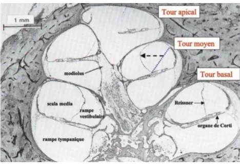
Figure 1: Coupes histologiques longitudinales de cochlée. La distension (flèche en pointillé) de la membrane de Reissner, structure qui sépare le compartiment endolymphatique, la scala média ou canal cochléaire et le compartiment périlymphatique, la rampe vestibulaire, signe l’hydrops endolymphatique. La position normale de la membrane de Reissner est indiquée en trait pointillé au tour moyen de la cochlée (Couloigner, Sterkers, Rask-Andersen, Teixeira, & Ferrary, 2004).
Probable :
A. Deux épisodes de vertiges ou plus, d’une durée de 20 minutes à 24 heures chacun. B. Symptômes auditifs fluctuants (audition, plénitude des acouphènes) dans l'oreille
affectée.
C. Pas mieux expliqué par un autre diagnostic vestibulaire.
L’évolution de la maladie est chronique pouvant conduire à une bilatéralisation en quelques années chez jusqu’à 50% des patients (Frejo et al., 2016).
PARACLINIQUE
L’audiométrie tonale et vocale :
L’atteinte est perceptive, pure et unilatérale prédominant sur les graves, pouvant être fluctuante au début de la maladie.
Les examens vestibulaires :
En crise : Ils peuvent retrouver un syndrome vestibulaire périphérique harmonieux avec un
nystagmus spontané dont l’intensité est supérieure à 5°/s et un déficit vestibulaire au épreuves caloriques. Le signe d’Halmagyi est généralement négatif. Tous les tests peuvent être présent ou absent lors de la crise. A distance des crises : L’examen peut être normal ou pathologique, tout dépend du capital sensoriel de l’oreille atteinte.
Les potentiels évoqués auditifs :
Ils permettent de confirmer l’atteinte périphérique avec l’absence d’allongement des intervalles des latences des ondes I-III et I-V et l’absence de désorganisation du pattern.
Les potentiels évoqués vestibulaires myogéniques cervicaux :
Ils étudient la voie sacculo-spinale. Les réflexes sacculo-colliques sont enregistrés en réponse à une stimulation sonore. Ils peuvent être absents dans la maladie.
L'impédancemétrie multifréquentielle :
Elle évalue l’admittance (l’inverse de l’impédance) à 2kHz, la faculté du système tympano-ossiculaire à se laisser mobiliser lorsqu’il est soumis à un mouvement alternatif. Chez les patients atteints de maladie de Ménière, la largeur entre 2 pics observés sur la courbe de conductance G, composante de l’admittance, à 2kHz est augmentée.
Les otoémissions acoustiques :
Les otoémissions acoustiques correspondent à des sons produits par l’activité contractile des cellules ciliées externes de la cochlée, en réponse à une stimulation sonore. Concernant les produits de distorsion des otoémissions acoustiques, une stimulation acoustique bitonale est appliquée (présentation simultanée de deux sons purs). Chez les patients sains comme ceux atteints d’un hydrops endolymphatique, les changements posturaux sont à l’origine d’une augmentation de la pression intra-cochléaire (exagérée en cas de maladie de Ménière) entraînant un déphasage des produits de distorsion des otoémissions acoustiques.
L'électrocochléographie :
Elle enregistre l’activité électrique de l’organe de Corti et des fibres nerveuses constituant le nerf auditif à l’intérieur de la cochlée, en réponse à une stimulation sonore. Une augmentation du rapport SP (Potentiel de sommation) / AP (Potentiel d’action) est en faveur d’un hydrops endolymphatique.
L’IRM :
Elle présente maintenant deux avantages, réaliser un diagnostic positif d’hydrops endolymphatique et un diagnostic d’élimination. Elle permet d’éliminer des pathologies pouvant être responsable d’un ménière like ; les schwannomes vestibulaires, les méningiomes, les tumeurs du sac endolymphatique, les malformations de l’oreille interne et les malformations d’Arnold-Chiari. Il y a 12 ans, Nakashima (Nakashima et al., 2007) a proposé une méthode d'imagerie originale pour évaluer le compartiment endolymphatique avec acquisition par IRM
quantitatif initial pour évaluer la quantité de liquide endolymphatique (Nakashima et al., 2009) a été progressivement amélioré en distinguant la forme de l’utricule et du saccule (Attyé et al., 2017; Bernaerts et al., 2019) permettant d'évaluer si l'efficacité des procédures chirurgicales pouvait être améliorée par la sélection par l’imagerie des patients atteints de la maladie de Ménière présentant un hydrops sacculaire ou utriculaire.
I.5 TRAITEMENT
Le traitement est avant tout médical. Il peut être chirurgical lorsque la maladie de Ménière est devenue invalidante et paraît d’autant plus intéressant à connaître que l’acte chirurgical permet de guérir définitivement les invalidantes crises de vertiges. Ce caractère invalidant est retrouvé chez 10 % environ des patients souffrant d’une maladie de Ménière.
TRAITEMENT MEDICAL DE LA CRISE :
Le malade doit cesser toute activité dès la perception des prodromes, adopter une position assise ou allongée pour éviter la chute.
L’administration parentérale ou per os d’acéthylleucine (Tanganil), de benzodiazépine tel que le diazépam (Valium), d’antihistaminiques, de métoclopramide (Primpéran) sont utilisés. Le mannitol peut être prescrit tant que dure l’accès vertigineux.
TRAITEMENT MEDICAL DE FOND :
Un soutien psychologique, des règles hygiénodiététiques, des médicaments antivertigineux, des corticoïdes, des « protecteurs cellulaires » telle que la trimétazidine, de l’extrait de gingko biloba semble favoriser la compensation vestibulaire. Les diurétiques, associés à un régime hyposodé le permettent aussi. La labyrinthectomie chimique consiste à détruire le labyrinthe vestibulaire aux moyens d’agents ototoxiques telle que la Gentamycine du fait de son affinité préférentielle pour le vestibule et moindre pour la cochlée.
TRAITEMENT CHIRURGICALE :
Environ 10 % des patients atteints de la maladie de Ménière évoluent en résistant à tout traitement médical et deviennent invalidé du fait de la violence et de la fréquence de survenue des crises de vertige (impossibilité de travailler, de mener des activités extrascolaire). La chirurgie peut alors être indiquée. Les méthodes chirurgicales sont là aussi nombreuses mais peuvent être schématiquement classées en interventions « destructrices » qui suppriment la fonction vestibulaire ou « non destructrices ».
Interventions non destructrices : 1. La pose d’aérateur transtympanique
2. La chirurgie de décompression du sac endolymphatique 3. La chirurgie du blocage du canal endolymphatique 4. La chirurgie d’oblitération des canaux semi-circulaires
La chirurgie de décompression sac endolymphatique consiste à l’aborder, le disséquer, l’ouvrir et établir un « shunt » pour assurer un drainage durable du liquide endolymphatique.
Cette technique chirurgicale est basée sur la physiopathologie de la sécrétion de glycoprotéines osmotiquement actives par le sac endolymphatique (Sood, Lambert, Nguyen, & Meyer, 2014) et la perturbation de l'homéostasie causée par le dysfonctionnement des cotransporters et des aquaporines Na+/K+/2CL (Maekawa et al., 2010).
La nouvelle chirurgicale du blocage du canal endolymphatique consiste à l’aborder, le disséquer procéder au blocage de celui-ci par deux clips en titane.
Elle est basée sur la théorie que les canaux périductals du canal endolymphatique pourraient être impliqués dans l'hydrodynamique de l'endolymphe, contribuant à son absorption (Linthicum, Doherty, Webster, & Makarem, 2014) et que le sac endolymphatique sécrète plus d'endolymphe que ce qu’il absorbe (Saliba, Gabra, Alzahrani, & Berbiche, 2015).
La chirurgie d’oblitération des canaux semi-circulaires consiste à obturer une extrémité de chaque canal semi-circulaire de façon à empêcher les mouvements d’endolymphe et donc les mouvements de la cupule. La fonction vestibulaire est éteinte ou fortement diminuée.
Interventions destructrices : 1. La neurotomie vestibulaire
2. La labyrinthectomie chirurgicale, sacrifice de l’audition. Contrôle similaire à la neurotomie vestibulaire
La neurotomie vestibulaire consiste à sectionner le nerf vestibulaire par voie rétrosigmoidienne, par voie rétrolabyrinthique ou par la voie de la fosse moyenne. Elle préserve l’audition.
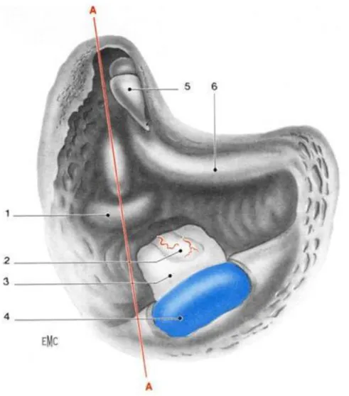
Figure 2: Décompression du sac endolymphatique par voie transmastoïdienne côté droit. 1. Canal semi-circulaire postérieur ; 2. Sac endolymphatique ; 3. Dure-mère fosse cérébelleuse ; 4. Sinus sigmoïde ; 5. Enclume ; 6. Nerf facial. A-A. Ligne de Donaldson (J.Magnan, Chirurgie des vertiges,1994, EMC)
La labyrinthectomie chirurgicale consiste en la destruction du labyrinthe postérieur, par la suppression du capteur vestibulaire. Elle conduit à une cophose et exclut toute possibilité d'implant cochléaire si par malheur l'oreille controlatérale était altérée.
II
ARTICLE
ENDOLYMPHATIC SAC DECOMPRESSION VERSUS ENDOLYMPHATIC DUCT BLOCKAGE IN PATIENTS WITH SACCULAR AND UTRICULAR HYDROPS, AS
EVIDENCED BY MRI
*Marie Soula, ϸArnaud Attyé, ϯAlexandre Karkas, *Georges Dumas, *ϕSébastien Schmerber
*Department of Otolaryngology-Head and Neck Surgery, ϸDepartment of Neuroradiology and MRI, Univ. Hospital Grenoble Alpes, Univ. Grenoble Alpes, UFR de Medicine,
Grenoble, France,
ϯDepartment of Otolaryngology-Head and Neck Surgery, Univ. Hospital of Saint Etienne, Univ. Jean Monnet, School of Medicine, Saint Etienne, France
ϕUniv. Grenoble Alpes, Brain Tech Lab Inserm UMR 1205, F-38000 Grenoble, France
Address correspondence and reprint requests to Marie SOULA, Department of Otolaryngology-Head and Neck surgery, BP 217, Grenoble University Hospital, France.
Email: msoula@chu-grenoble.fr, Tel: +337 61 77 94 62, Fax: +334 76 76 51 20. Conflicts of Interest and Sources of Funding: None
II.1 ABSTRACT
OBJECTIVES: Endolymphatic sac surgery (ESS) is commonly used to shunt the excess of endolymph in the inner ear in Meniere’s disease (MD). Recently, a new surgical technique has been proposed using an endolymphatic duct blockage (EDB). The aim of this work was to evaluate these procedures faced with preoperative and postoperative magnetic resonance imaging (MRI) with delayed acquisition, using the so-called hydrops protocol.
METHODS: In this retrospective study, the 3D-FLAIR sequences (4 hours after contrast media injection) were used before and after surgery in 17 patients with MD. Nine patients who underwent endolymphatic sac decompression (ESD) and 8 who underwent EDB were retrospectively included in our study. Two radiologists independently graded saccular hydrops (SH) and utricular hydrops (UH) before and after surgery based on the latest published imaging classifications.
RESULTS: All patients with MD had pre-operative saccular or utricular hydrops evaluated by MRI. In ESD group, 2 patients showed SH disappearance on postoperative MRI and their vertigo attacks were controlled. In EDB group, 2 patients also showed SH decrease, yet vertigo attacks were controlled in only one patient. In ESD group, 8 patients (89%) no longer experienced vertigo attacks compared to only 3 patients (37.5%) improved in the EDB group. CONCLUSIONS: A careful selection of patients with MD using MRI has allowed ESD to be efficient in almost all cases with pre-operative saccular hydrops, unlike EDB technique. There was no association between the imaging results after surgery and the control of vertigo. A larger cohort would be required to corroborate our conclusions.
KEYWORDS: Inner Ear; Meniere’s Disease; Magnetic Resonance Imaging; Endolymphatic Hydrops; Endolymphatic sac surgery; Endolymphatic duct blockage
II.2 INTRODUCTION
Meniere’s disease (MD) is a clinical entity defined by episodes of spontaneous vertigo usually accompanied by tinnitus, pressure within the ear and fluctuating sensorineural hearing loss (J. A. Lopez-Escamez et al., 2015). The syndrome is a heterogeneous condition and several comorbidities have been consistently associated. The presence of endolymphatic hydrops (EH) is thought to be strongly linked with clinical symptoms (Sajjadi & Paparella, 2008).
Since the first study of Kimura (Kimura, 1982), who investigated an animal model in guinea pigs, the blockage of the endolymphatic sac and duct with obstruction of endolymphatic outflow has been associated with endolymphatic hydrops occurrence. Twelve years ago, Nakashima et al. (Nakashima et al., 2007) were the first to suggest an imaging method to evaluate the endolymphatic compartment with magnetic resonance imaging (MRI) acquisition between 4 and 6 hours after contrast media injection. The initial semi-quantitative grading system to evaluate the amount of endolymphatic fluid (Nakashima et al., 2009) has been progressively improved by distinguishing the utricle from the saccule (Attyé et al., 2017; Bernaerts et al., 2019), allowing to evaluate whether surgical efficiency could be improved by an imaging selection of MD patients with saccular hydrops (SH) and utricular hydrops (UH).
Surgical treatment is usually offered when the medical therapy has failed, and the vertigo becomes incapacitating. Different surgical techniques exist: Pneumatic-Equalisation tube (or Grommet tube) placement, endolymphatic sac surgery (ESS) including endolymphatic sac decompression (ESD) and semicircular canal obliteration surgery are conservative procedures while vestibular neurotomy, surgical labyrinthectomy and chemical labyrinthectomy are more destructive (Sajjadi & Paparella, 2008).
Endolymphatic sac decompression, initially described by Portmann in 1927 (Portmann, 1991), is a surgical treatment of reference for patients with MD because of its low morbidity and preservation of hearing. The improvement of vertigo after surgery has been reported in 75% to
85% of patients, depending on the studies (Bento, Cisneros, & De Oliveira Fonseca, 2017; Huang, Lin, & Chang, 1991; Sood et al., 2014; Telischi & Luxford, 1993).
Recently, a new surgical technique has been proposed, namely the endolymphatic duct blockage (EDB). The inherent pathophysiological concept is different from ESD: by blocking the endolymphatic duct distally near the sac, the objective aims at reducing the accumulation of endolymph in the inner ear without affecting its potential absorption, taking place at the endolymphatic duct, as suggested by Saliba et al. (Saliba et al., 2015).
To evaluate these surgical techniques, recent studies evaluated the postoperative EH variation after ESD, showing a significant volume decrease of the endolymphatic compartment after surgery (F. Liu et al., 2014) associated with control of vertigo/dizziness (Ito et al., 2019). Yet, these studies have not distinguished saccular hydrops and the more recently described utricular protrusion into the lateral semicircular canal, named utricular hydrops (Robert Gürkov et al., 2012; Sugimoto et al., 2018).
The main objective of this clinico-radiological study was to evaluate whether the preoperative selection of patients with endolymphatic hydrops would allow to compare the efficiency of the two surgical techniques in controlling vertigo. The second objective was to describe the potential post-operative decrease of EH in each membranous labyrinthine compartment, depending on the surgical technique.
II.3 METHODS
The study was conducted in accordance with the Declaration of Helsinki and international standards of Good Clinical Practice. It was approved by our institutional review board (IRB 6705/15-CHUG 02). Signed informed consent was obtained from all patients.
PATIENTS
Seventeen patients from our tertiary care center were retrospectively included from March 2013 to March 2019.
The inclusion criteria were:
-A definite or probable clinical diagnosis of MD based on the last consensus statement (J. A. Lopez-Escamez et al., 2015).
-Failure of medical treatment, with patients still presenting with violent and highly frequent vertigo attacks, thus requiring ESD or EDB surgery.
-A pre and post-operative MRI examination including the 3D FLAIR sequence with delayed acquisition showing either an utricular or a saccular hydrops.
The exclusion criteria were: any previous destructive procedure or surgical treatment for inner ear disease, a middle ear disease, an active bilateral MD. Allergic conditions, a history of allergy to gadolinium, pregnancy and children.
The non-inclusion criteria were: a preoperative MRI, which did not show a SH or UH.
POSTOPERATIVE QUESTIONNAIRE AND CLINICAL EXAMINATION
The presence of vertigo and unsteadiness after the surgery was carefully recorded by a control phone call between 1 and 4 months. A clinical examination was performed during the control visit between 1 month to 26 months post operatively.
AUDIOMETRY
The hearing was measured using a pure tone audiometer before the surgery and during the control visit between 1 month to 26 months post operatively. The average hearing threshold was calculated based on the average of 0.5, 1, 2 and 4 kHz. After the surgery, a change of more than 10 dB was considered clinically significant. A positive value beyond +10dB was considered as hearing improvement. Conversely, a negative value below -10dB was considered as hearing loss.
IMAGING
All subjects underwent an MRI with delayed acquisition 4 hours after the injection of contrast medium. Imaging examinations were carried out on a 3T Philips Achieva® or 3T Siemens Skyra®.
Using Philips MR scan, the 3D-FLAIR sequence was performed with the following parameters: TR: 7600 ms, TE: 345 ms, TI: 2300 ms, an isotropic voxel size of 0.8 mm for acquisition and 0.4 mm for reconstructions, and a scan time of 9 mins.
Using Siemens MR scan, we performed the 3D-FLAIR with the following parameters: field of view: 160 × 160 mm, TR: 10000 ms, TE: 323 ms, TI: 2500 ms, and scan time of 7 mins 50 seconds.
Images were evaluated independently by two radiologists, both with a specialist qualification in head and neck radiology, who were blinded to the clinical data. The imaging data of inner ears were analysed with Osirix MD®. The presence of the following structures was checked: the cochlear duct, the saccule and the utricle.
Using the MRI data of the temporal bone, we defined the pre/post-operative presence of saccular hydrops with the two published methods: firstly, patients were evaluated with the SURI score (Saccule to Utricle Ratio Inversion).
SURI was considered positive when the saccule appeared equal or larger than the utricle, in both radiologists’ evaluations. In the most important cases when the saccule was not
individualised from the utricle, we have used the recent classification of Bernaerts et al. to define the presence of a vestibular hydrops (Bernaerts et al., 2019).
SURGICAL PROCEDURES
The procedure of endolymphatic sac decompression employed by the surgeon was previously described by Paparella et al. (Paparella & Hanson, 1976).
A 4 cm postauricular incision was performed. The mastoidectomy consisted in a complete removal of presigmoid, infra-labyrinthine and retro-labyrinthine mastoid air cells. The lateral and posterior semicircular canals were then identified. The bone covering the sigmoid sinus, the posterior cranial fossa and the middle cranial fossa was thinned. The distal portion of the lateral sinus and all accessible dura in Trautman’s triangle, and often an adjacent small portion of dura of the middle cranial fossa was exposed. The sac was identified as a white, dense thickening in the dura, pointing towards the inferior portion of the posterior semicircular canal. A small opening in the sac was performed with a joint knife. Retracting the dura allowes further identification of the sac under the bony edge. The lumen of the sac was identified. A 0.5mm Silastic sheet was pushed in the small opening, thus forming a tubular drain.
The procedure of endolymphatic duct blockage was described by Saliba (Saliba et al., 2015). When the sac was identified, the bone of the vestibular aqueduct operculum and of the posterior fossa was dissected from the retrolabyrinthine bone. The endolymphatic duct was identified in continuity from the endolymphatic sac. Finally, it was blocked with 2 small titanium clips.
Qualitative variables were expressed as n (%) and quantitative variables as median [interquartile range 25%-75%]. We have also tried to make a Yate correction, but due to the small size of our population and the results analysed, a high-performance statistical analysis cannot be used.
II.4 RESULTS
Patient demographic parameters are shown in Table I.
All patients from both groups suffered from vertigo, unsteadiness and tinnitus at the time of surgery.
Before surgery, median PTA was 53.8 dB and 21 dB for EDB and ESD, respectively.
In the EDB group, preoperative MRI showed saccular hydrops in 7/8 ears (87.5%) and utricular hydrops in 1/8 ear (12.5%). In the ESD group, preoperative MRI showed saccular hydrops in 7/9 ears (77.8%) and utricular hydrops in 2/9 ears (22.2%). We have classified utriculo-saccular hydrops (1 ear in each group) in the same group than patients with saccular hydrops. Patients operated in 2013, 2017 and 2018 were decompressed and those operated in 2015 and 2016 were clipped. This was the surgeon's choice at that time.
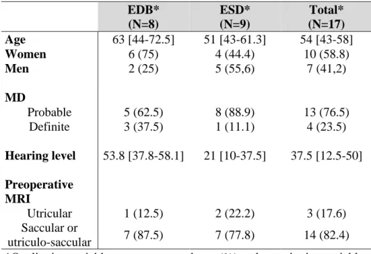
Table I. Demographic parameters
EDB* (N=8) ESD* (N=9) Total* (N=17) Age 63 [44-72.5] 51 [43-61.3] 54 [43-58] Women 6 (75) 4 (44.4) 10 (58.8) Men 2 (25) 5 (55,6) 7 (41,2) MD Probable 5 (62.5) 8 (88.9) 13 (76.5) Definite 3 (37.5) 1 (11.1) 4 (23.5) Hearing level 53.8 [37.8-58.1] 21 [10-37.5] 37.5 [12.5-50] Preoperative MRI Utricular 1 (12.5) 2 (22.2) 3 (17.6) Saccular or utriculo-saccular 7 (87.5) 7 (77.8) 14 (82.4) *Qualitative variables were expressed as n (%) and quantitative variables as median [interquartile range 25%-75%]
EDB: Endolymphatic duct blockage, ESD: Endolymphatic sac decompression, MD: Meniere’s disease, N: Total number of patients, MRI: Magnetic resonance imaging
POSTOPERATIVE MRI
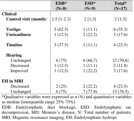
The results are presented in Table II.
In both groups, two patients had a reduced SH after the surgery. UH was reduced in no case after surgery.
In ESD group, 2 patients showed disappearance of their SH on MRI after surgery and their vertigo attacks were controlled. In EDB group, 2 patients also showed SH decrease yet vertigo attacks were controlled in only one patient.
VERTIGO AND UNSTEADINESS
In the ESD group, surgery cured patients from vertigo in all but one case (8/9 patients, 88.9%) and unsteadiness in 7 out of 9 patients (77.8%). In the EDB group, only 3 patients out of 8 (37.5%) had their vertigo attacks improved after surgery and 7 patients out of 8 (87.5%) had her unsteadiness improved.
TINNITUS
In the ESD group, surgery cured patients from vertigo in all but one case (8/9 patients, 88.9%). This patient had vertigo attacks too. In the EDB group, 5 patients out of 8 (62.5%) had their tinnitus improved after surgery.
A
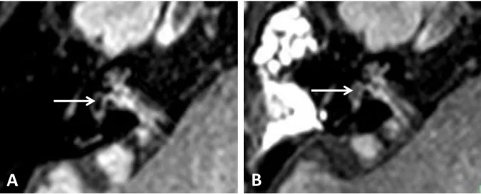
B
Figure 3 : Changes in MR images after endolymphatic sac decompression (ESD). 3D-FLAIR sequences in the axial slice. (A) Image of saccular hydrops (SH) before ESD. (B) Image of the decrease of SH after ESD. White arrow indicates the saccule
AUDIOMETRY
In the EDB group, considering that a change of more than 10 dB was clinically significant, hearing was improved in 1 patient, remained unchanged in 6, and decreased in 1. Likewise, in the ESD group, hearing improved in 2 patients, remained unchanged in 6, and decreased in 1. In the 4 patients in whom SH disappeared on postoperative MRI, the hearing levels were improved in two patients and remained unchanged in the 2 other patients.
Table II. Postoperative clinical and MRI results
EDB* (N=8) ESD* (N=9) Total* (N=17) Clinical
Control visit (month) 1.5 [1-2.3] 2 [1;3] 2 [1;3] Vertigo 5 (62.5) 1 (11.1) 6 (35.3) Unsteadiness 1 (12.5) 2 (22.2) 3 (17.6) Tinnitus 3 (37.5) 1 (11.1) 4 (23.5) Hearing Unchanged 6 (75) 6 (66,7) 12 (70,6) Decreased 1 (12.5) 1 (11.1) 2 (11.8) Improved 1 (12,5) 2 (22,2) 3 (17,6) EH in MRI Decreased 2 (25) 2 (22.2) 4 (23.5) Unchanged 6 (75) 7 (77.8) 13 (76.5) *Qualitative variables were expressed as n (%) and quantitative variables as median [interquartile range 25%-75%]
EDB: Endolymphatic duct blockage, ESD: Endolymphatic sac decompression, MD: Meniere’s disease, N: Total number of patients, MRI: Magnetic resonance imaging, EH: Endolymphatic hydrops
II.5 DISCUSSION
Meniere's disease is a chronic disease which medical or surgical treatment remains difficult due to its uncertain pathophysiology. Different surgical techniques are offered when the medical therapy has failed. The conservative surgical reference is the ESD technique, based on the knowledge of secreting osmotically active glycoproteins by the endolymphatic sac (Sood et al., 2014) and the disturbance of homeostasis caused by the dysfunction of Na+/K+/2Cl cotransporters and aquaporins (Maekawa et al., 2010). The new surgical technique, EDB is based on the theory that the periductal channels of the endolymphatic duct could be involved in the hydrodynamics of the endolymph, contributing to its absorption (Linthicum et al., 2014) and that the endolymphatic sac would secrete more endolymph than it absorbs (Saliba et al., 2015). Since there are different surgical treatments based on different pathophysiologies, we are still looking for the appropriate surgical treatment.
Twelve years ago, Nakashima et al. proposed the first magnetic resonance images to evaluate the endolymphatic compartment with delayed acquisition between 4 and 6 hours after contrast media injection, allowing a better analysis of the disease by clearly identifying the EH. The initial semi-quantitative grading system has been improved by distinguishing the utricle from the saccule (Attyé et al., 2017; Bernaerts et al., 2019).
Regarding the control of vertigo attacks and hearing: in ESD group, 8/9 patients (89%) were cured for their vertigo attacks. This result is higher than the data reported in the literature, which ranges from 75% for Sood et al. to 85.89% for Huang et al. (Huang et al., 1991; Sood et al., 2014). Hearing was preserved or improved in 8/9 patients (89%) compared to 72% in Sood et al. study (Sood et al., 2014). In EDB group, only 3/8 patients were cured from their vertigo attacks, compared to 95% in other studies conducted by Saliba et al. (Asmar & Saliba, 2016; Saliba et al., 2015). As for hearing, we observed the same results, hearing was preserved or improved in 7/8 patients, 87.5% of cases. In this study, the ESD technique seemed to yield better results than the EDB technique in controlling vertigo attacks.
We did not included the cochlear endolymphatic compartment into inner ear analysis because in contrast to saccular dilation, imaging in healthy subjects often revealed cochlear duct dilation (Attyé et al., 2017; Yoshida et al., 2018) and the most recent published classification found no added value from the cochlear endolymph evaluation (Bernaerts et al., 2019). Furthermore, given the hypothetic action of the two surgical techniques studied, only a disappearance of the vestibular was expected. That's why we analysed patients who had a preoperative MRI with a SH or UH.
In ESD group, 2 patients had a SH which disappearance after surgery, their vertigo attacks were controlled, and their hearing improved or was unchanged. In EDB group, 2 patients also presented with SH decrease yet vertigo attacks were controlled for only one patient and hearing were improved or unchanged.
In the study of Zhang et al. vestibular hydrops in 13 patients showed no significant change after sac surgery, but vertigo attacks were controlled in all patients (Zhang et al., 2016). These results corroborate those from the study conducted by Chung et al. on the temporal bone (Chung, Fayad, Linthicum, Ishiyama, & Merchant, 2011) which showed that the endolymphatic sac surgery procedure did not decrease EH in patients. Likewise, Uno et al. reported that only two patients among 7 had a reduction in vestibular hydrops on postoperative MRI, despite a good control of vertigo after ESS procedure (Uno et al., 2013). In 3 cases where vestibular hydrops was unchanged after surgery, vertigo attacks were yet significantly suppressed. These findings support our results that the effects of endolymphatic sac or duct surgery are not limited to hydrops relief. As a matter of fact, even in the cases where hydrops was not modified after surgery, vertigo spells were significantly suppressed (6 patients in ESD group and 2 patients in EDB group), which indicates that the effects of surgery are not limited to hydrops reduction. The mechanism of vertigo attack is not completely understood, or at least not explained by the state of endolymphatic hydrops alone.
On the other hand, two studies showed a significant volume reduction in the endolymphatic compartment volume on MRI after endolymphatic sac surgery and this was correlated with a good control of vertigo/dizziness, in 60% of cases for Liu et al. and 80% for Ito et al. (Ito et al., 2019; F. Liu et al., 2014b). However, those authors have used a semi-quantitative grading system which includes the cochlear endolymph. It could be adapted to follow small changes in
the amount of endolymph yet other research team (Attyé et al., 2017) have reported the
technical impossibility to distinguish controls from patients when the cochlear endolymph is included in the grading system. In our study, the absence of UH modification after the surgical procedure could be further explained by different physiological mechanisms in the utricle and the saccule, requiring new therapies to selectively manage patients with UH. MRI could then be a good postoperative control element in addition to the clinical outcome.
We did not excluded patients who had receiving medical treatment between MRI examinations because it does not appear to cause MRI changes in EH as demonstrated by Fiorino et al; the intravenous administration of furosemide did not reduce EH even though vertigo attacks were disappeared (Fiorino et al., 2016). Furthermore, Gürkov et al. reported on six patients treated with betahistine while no modification in the EH was seen on posttreatment MRI although their symptoms improved significantly (R. Gürkov et al., 2013). Also, Sepahdari et al. reported that only 3 patients among 7 showed a reduced EH on MRI after medical treatment using Acetazolamid (Sepahdari, Vorasubin, Ishiyama, & Ishiyama, 2016).
Our study has some limitations. First, the number of cases is small given that the surgical indication remains rare in MD. Second, the follow-up period of patients may be too short for a chronic disease as MD. Furthermore, there are inherent technical limitations of the MRI technique and limits of image interpretation.
In summary, a careful selection of patients with MD using MRI has allowed ESD to be efficient in almost all patients showing pre-operative saccular hydrops, in contrast with EDB technique. There was no association between the imaging results after surgery and the control of vertigo.
There was no change in hearing in both surgical techniques, thus confirming that these two surgical techniques are safe for the hearing.
II.7 REFERENCES
Alexander, T. H., & Harris, J. P. (2010). Current Epidemiology of Meniere’s Syndrome.
Otolaryngologic Clinics of North America, 43(5), 965‑970.
https://doi.org/10.1016/j.otc.2010.05.001
Asmar, M. H., & Saliba, I. (2016). Endolymphatic Duct Blockage for Refractory Ménière’s Disease: Assessment of Endolymphatic Sac Biopsy on Short-Term Surgical Outcomes.
The Journal of International Advanced Otology, 12(3), 310‑315.
https://doi.org/10.5152/iao.2016.3069
Attyé, A., Eliezer, M., Boudiaf, N., Tropres, I., Chechin, D., Schmerber, S., … Krainik, A. (2017). MRI of endolymphatic hydrops in patients with Meniere’s disease: a case-controlled study with a simplified classification based on saccular morphology.
European Radiology, 27(8), 3138‑3146. https://doi.org/10.1007/s00330-016-4701-z
Bento, R. F., Cisneros, J. C., & De Oliveira Fonseca, A. C. (2017). Endolymphatic sac drainage for the treatment of Ménière’s disease. The Journal of Laryngology & Otology, 131(02), 144‑149. https://doi.org/10.1017/S0022215116009713
Bernaerts, A., Vanspauwen, R., Blaivie, C., van Dinther, J., Zarowski, A., Wuyts, F. L., … De Foer, B. (2019). The value of four stage vestibular hydrops grading and asymmetric perilymphatic enhancement in the diagnosis of Menière’s disease on MRI.
Neuroradiology. https://doi.org/10.1007/s00234-019-02155-7
Chays, A., Florant, A., Seidermann, L., & Ulmer, É. (2012). Les vertiges. Consulté à l’adresse https://nls.ldls.org.uk/welcome.html?ark:/81055/vdc_100052770899.0x000001
Chung, J. W., Fayad, J., Linthicum, F., Ishiyama, A., & Merchant, S. N. (2011). Histopathology After Endolymphatic Sac Surgery for Ménière’s Syndrome: Otology & Neurotology,
32(4), 660‑664. https://doi.org/10.1097/MAO.0b013e31821553ce
Fiorino, F., Mattellini, B., Vento, M., Mazzocchin, L., Bianconi, L., & Pizzini, F. B. (2016). Does the intravenous administration of frusemide reduce endolymphatic hydrops? The
Journal of Laryngology & Otology, 130(03), 242‑247.
https://doi.org/10.1017/S0022215115003527
Frejo, L., Soto-Varela, A., Santos-Perez, S., Aran, I., Batuecas-Caletrio, A., Perez-Guillen, V., … Lopez-Escamez, J. A. (2016). Clinical Subgroups in Bilateral Meniere Disease.
Frontiers in Neurology, 7. https://doi.org/10.3389/fneur.2016.00182
Gürkov, R., Flatz, W., Keeser, D., Strupp, M., Ertl-Wagner, B., & Krause, E. (2013). Effect of standard-dose Betahistine on endolymphatic hydrops: an MRI pilot study. European
Archives of Oto-Rhino-Laryngology, 270(4), 1231‑1235.
https://doi.org/10.1007/s00405-012-2087-3
Gürkov, Robert, Flatz, W., Louza, J., Strupp, M., Ertl-Wagner, B., & Krause, E. (2012). Herniation of the membranous labyrinth into the horizontal semicircular canal is correlated with impaired caloric response in Ménière’s disease. Otology & Neurotology:
Official Publication of the American Otological Society, American Neurotology Society [and] European Academy of Otology and Neurotology, 33(8), 1375‑1379.
https://doi.org/10.1097/MAO.0b013e318268d087
Huang, T. S., Lin, C. C., & Chang, Y. L. (1991). Endolymphatic sac surgery for Meniére’s disease. A cumulative study of twelve years’ experience. Acta Oto-Laryngologica.
Supplementum, 485, 145‑154.
Ito, T., Inui, H., Miyasaka, T., Shiozaki, T., Matsuyama, S., Yamanaka, T., … Kitahara, T. (2019). Three-Dimensional Magnetic Resonance Imaging Reveals the Relationship Between the Control of Vertigo and Decreases in Endolymphatic Hydrops After Endolymphatic Sac Drainage With Steroids for Meniere’s Disease. Frontiers in
Neurology, 10. https://doi.org/10.3389/fneur.2019.00046
Kimura, R. S. (1982). Animal models of endolymphatic hydrops. American Journal of
Linthicum, F. H., Doherty, J., Webster, P., & Makarem, A. (2014). The Periductal Channels of the Endolymphatic Duct, Hydrodynamic Implications. Otolaryngology–Head and Neck
Surgery, 150(3), 441‑447. https://doi.org/10.1177/0194599813516420
Liu, F., Huang, W., Chen, Q., Meng, X., Wang, Z., & He, Y. (2014a). Noninvasive evaluation of the effect of endolymphatic sac decompression in Ménière’s disease using magnetic resonance imaging. Acta Oto-Laryngologica, 134(7), 666‑671.
https://doi.org/10.3109/00016489.2014.885118
Liu, F., Huang, W., Chen, Q., Meng, X., Wang, Z., & He, Y. (2014b). Noninvasive evaluation of the effect of endolymphatic sac decompression in Ménière’s disease using magnetic resonance imaging. Acta Oto-Laryngologica, 134(7), 666‑671.
https://doi.org/10.3109/00016489.2014.885118
Liu, I. Y., Sepahdari, A. R., Ishiyama, G., & Ishiyama, A. (2016). High Resolution MRI Shows Presence of Endolymphatic Hydrops in Patients Still Symptomatic After Endolymphatic Shunt Surgery: Otology & Neurotology, 37(8), 1128‑1130. https://doi.org/10.1097/MAO.0000000000001144
Lopez-Escamez, J. A., Carey, J., Chung, W.-H., Goebel, J. A., Magnusson, M., Mandalà, M., … Korean Balance Society. (2015). Diagnostic criteria for Menière’s disease. Journal
of Vestibular Research: Equilibrium & Orientation, 25(1), 1‑7.
https://doi.org/10.3233/VES-150549
Lopez-Escamez, L.-E. J., John, C., Won-Ho, C., A, G. J., Måns, M., Marco, M., … Alexandre, B. (2015). Diagnostic criteria for Meniere’s disease. Journal of Vestibular
Research, (1), 1–7. https://doi.org/10.3233/VES-150549
Maekawa, C., Kitahara, T., Kizawa, K., Okazaki, S., Kamakura, T., Horii, A., … Kiyama, H. (2010). Expression and Translocation of Aquaporin-2 in the Endolymphatic Sac in Patients with Meniere’s Disease: AQP2 in Meniere’s disease. Journal of
Neuroendocrinology, 22(11), 1157‑1164.
https://doi.org/10.1111/j.1365-2826.2010.02060.x
Nakashima, T., Naganawa, S., Pyykkö, I., Gibson, W. P. R., Sone, M., Nakata, S., & Teranishi, M. (2009). Grading of endolymphatic hydrops using magnetic resonance imaging. Acta
Oto-Laryngologica, 129(sup560), 5‑8. https://doi.org/10.1080/00016480902729827
Nakashima, T., Naganawa, S., Sugiura, M., Teranishi, M., Sone, M., Hayashi, H., … Ishida, I. M. (2007). Visualization of Endolymphatic Hydrops in Patients With Meniere???s Disease: The Laryngoscope, 117(3), 415‑420.
https://doi.org/10.1097/MLG.0b013e31802c300c
Paparella, M. M., & Hanson, D. G. (1976). ENDOLYMPHATIC SAC DRAINAGE FOR INTRACTABLE VERTIGO (METHOD AND EXPERIENCES): The Laryngoscope,
86(5), 697‑703. https://doi.org/10.1288/00005537-197605000-00010
Pender, D. J. (2014). Endolymphatic hydrops and Ménière’s disease: a lesion meta-analysis.
The Journal of Laryngology & Otology, 128(10), 859‑865.
https://doi.org/10.1017/S0022215114001972
Portmann, G. (1991). The saccus endolymphaticus and an operation for draining the same for the relief of vertigo. 1927. The Journal of Laryngology and Otology, 105(12), 1109‑1112.
Sajjadi, H., & Paparella, M. M. (2008). Meniere’s disease. Lancet, 372(9636), 406‑414. https://doi.org/10.1016/S0140-6736(08)61161-7
Saliba, I., Gabra, N., Alzahrani, M., & Berbiche, D. (2015). Endolymphatic Duct Blockage: A Randomized Controlled Trial of a Novel Surgical Technique for Ménière’s Disease Treatment. Otolaryngology-Head and Neck Surgery, 152(1), 122‑129.
Salt, A. N., & Plontke, S. K. (2010). Endolymphatic Hydrops: Pathophysiology and Experimental Models. Otolaryngologic Clinics of North America, 43(5), 971‑983. https://doi.org/10.1016/j.otc.2010.05.007
Schuknecht, H. (s. d.). Schuknecht HF. Pathology of the ear. Cambridge: Harvard University
Press, 1974.
Sepahdari, A. R., Vorasubin, N., Ishiyama, G., & Ishiyama, A. (2016). Endolymphatic Hydrops Reversal following Acetazolamide Therapy: Demonstration with Delayed Intravenous Contrast-Enhanced 3D-FLAIR MRI. American Journal of Neuroradiology, 37(1), 151‑154. https://doi.org/10.3174/ajnr.A4462
Sood, A. J., Lambert, P. R., Nguyen, S. A., & Meyer, T. A. (2014). Endolymphatic Sac Surgery for Ménière’s Disease: A Systematic Review and Meta-analysis. Otology &
Neurotology, 35(6), 1033‑1045. https://doi.org/10.1097/MAO.0000000000000324
Sugimoto, S., Yoshida, T., Teranishi, M., Kobayashi, M., Shimono, M., Naganawa, S., & Sone, M. (2018). Significance of Endolymphatic Hydrops Herniation Into the Semicircular Canals Detected on MRI. Otology & Neurotology: Official Publication of the American
Otological Society, American Neurotology Society [and] European Academy of Otology and Neurotology, 39(10), 1229‑1234.
https://doi.org/10.1097/MAO.0000000000002022
Telischi, F. F., & Luxford, W. M. (1993). Long-Term Efficacy of Endolymphatic Sac Surgery for Vertigo in Meniere’s Disease. Otolaryngology-Head and Neck Surgery, 109(1), 83‑87. https://doi.org/10.1177/019459989310900115
Uno, A., Imai, T., Watanabe, Y., Tanaka, H., Kitahara, T., Horii, A., … Inohara, H. (2013). Changes in endolymphatic hydrops after sac surgery examined by Gd-enhanced MRI.
Acta Oto-Laryngologica, 133(9), 924‑929.
Yoshida, T., Sugimoto, S., Teranishi, M., Otake, H., Yamazaki, M., Naganawa, S., … Sone, M. (2018). Imaging of the endolymphatic space in patients with Ménière’s disease. Auris
Nasus Larynx, 45(1), 33‑38. https://doi.org/10.1016/j.anl.2017.02.002
Zhang, Y., Cui, Y., Hu, Y., Mao, Z., Wang, Q., Pan, C., & Liu, A. (2016). Changes in endolymphatic hydrops visualized by magnetic resonance imaging after sac surgery.
Journal of Huazhong University of Science and Technology [Medical Sciences], 36(5),
R
ESUME
Objectifs : La technique chirurgicale de référence de la maladie de Ménière (MdM) réfractaire au traitement médical est la décompression du sac endolymphatique (DSE). Cette chirurgie est basée sur le drainage de l’excès de liquide du secteur endolymphatique. Récemment, une nouvelle technique chirurgicale a été proposée réalisant le blocage du canal endolymphatique (BCE). Ces deux techniques ont été évaluées par l’analyse de l'imagerie par résonnance magnétique (IRM) pré et postopératoire avec acquisition retardée, en utilisant le protocole hydrops.
Matériels et méthodes : Dans cette étude rétrospective, des séquences 3D-FLAIR (4 heures après injection de produit de contraste) ont été réalisées avant et après l'intervention chirurgicale chez 17 patients atteints de MdM. 9 patients ont subi une DSE et 8 patients un BCE. Les patients ont été inclus rétrospectivement dans cette étude. Deux radiologues ont classé indépendamment l'hydrops sacculaire (HS) et l'hydrops utriculaire (HU) avant et après l'intervention chirurgicale en fonction des dernières classifications d'imagerie publiées. Les patients opérés ont été évalués cliniquement (vertiges et instabilité) entre 1 et 4 mois postopératoires par un appel téléphonique de contrôle. Une audiométrie a été réalisée lors de la visite de contrôle entre 1 et 26 mois. Résultats : Tous les patients avec MdM présentaient un HS ou un HU préopératoire, selon l’IRM. Dans le groupe DSE, 2 patients ont présenté une disparition de l’HS après chirurgie et leurs crises de vertige ont été contrôlées. Dans le groupe BCE, 2 patients ont également présenté une disparition de l’HS mais les crises de vertige ont été contrôlées pour un seul patient. Dans le groupe DSE, 8 patients (89%) n’ont plus éprouvé de crises de vertige contre uniquement 3 patients (37,5%) améliorés dans le groupe BCE. Dans chaque groupe, un patient a aggravé son audition et 6 patients ont maintenu leur audition. Dans le groupe DSE, 2 patients ont amélioré leur audition contre 1 patient dans le groupe BCE.
Conclusion : Une sélection minutieuse des patients atteints de MdM à l'aide de l'IRM a permis à la DSE d'être efficace dans presque tous les cas d'hydrops sacculaire ou utriculaire préopératoire, contrairement à la technique BCE. Il n'y avait pas d’association entre les résultats de l'imagerie après la chirurgie et le contrôle du vertige. Une cohorte plus importante serait nécessaire pour corroborer nos conclusions.
Mots clés : Maladie de Ménière ; Imagerie par Résonance Magnétique ; Hydrops Endolymphatique ; Décompression du Sac Endolymphatique ; Blocage du Canal Endolymphatique