Cutting Balloon Angioplasty for the
Prevention of Restenosis: Results of the
Cutting Balloon Global Randomized Trial
Laura Mauri,
MD,
MSc, Raoul Bonan,
MD, Bonnie H. Weiner,
MD,
Victor Legrand,
MD,
PhD, Jean-Pierre Bassand,
MD, Jeffrey J. Popma,
MD,
Paulette Niemyski,
RN,
BSN, Ross Prpic,
MD, Kalon K. L. Ho,
MD,
MSc,
Manish S. Chauhan,
MD, Donald E. Cutlip,
MD, Olivier F. Bertrand,
MD, and
Richard E. Kuntz,
MD,
MScThe cutting balloon (CB) is a specialized device designed to create discrete longitudinal incisions in the atheroscle-rotic target coronary segment during balloon inflation. Such controlled dilatation theoretically reduces the force needed to dilate an obstructive lesion compared with standard percutaneous transluminal coronary angio-plasty (PTCA). We report a multicenter, randomized trial comparing the incidence of restenosis after CB angio-plasty versus conventional balloon angioangio-plasty in 1,238 patients. Six hundred seventeen patients were random-ized to CB treatment, and 621 to PTCA. The mean ref-erence vessel diameter was 2.86 ⴞ 0.49 mm, mean lesion length 8.9ⴞ 4.3 mm, and prevalence of diabetes mellitus in patients was 13%. The primary end point, the 6-month binary angiographic restenosis rate, was 31.4% for CB and 30.4% for PTCA (p ⴝ 0.75). Acute procedural success, defined as the attainment of<50% diameter stenosis without in-hospital major adverse
car-diac events, was 92.9% for CB and 94.7% for PTCA (pⴝ 0.24). Freedom from target vessel revascularization was slightly higher in the CB arm (88.5% vs 84.6%, log-rank pⴝ 0.04). Five coronary perforations occurred in the CB arm only (0.8% vs 0%, pⴝ 0.03). At 270 days, rates of myocardial infarction, death, and total major adverse cardiac events for CB and PTCA were 4.7% versus 2.4% (p ⴝ 0.03), 1.3% versus 0.3% (p ⴝ 0.06), and 13.6% versus 15.1% (pⴝ 0.34), respectively. In summary, the proposed mechanism of controlled dilatation did not reduce the rate of angiographic restenosis for the CB compared with conventional balloon angioplasty. CB angioplasty should be reserved for difficult lesions in which controlled dilatation is believed to provide a bet-ter acute result compared with balloon angioplasty alone. 䊚2002 by Excerpta Medica, Inc.
(Am J Cardiol 2002;90:1079 –1083)
T
he search for a method to dilate an obstructivecoronary lesion without invoking the proportional injury response observed in already approved devic-es1,2 led to the development of the cutting balloon
(CB) (Boston Scientific Interventional Technologies, Natick, Massachusetts), an angioplasty balloon with 3 to 4 longitudinally bonded microtomes that is de-signed to score atherosclerotic plaque. It was hypoth-esized that the discrete longitudinal incisions created during balloon inflation might improve the success of angioplasty by reducing elastic recoil and minimizing intimal injury, thereby minimizing the neointimal pro-liferative response.3,4 Theoretically, this effect may
allow CB angioplasty to achieve and maintain a larger lumen diameter using lower balloon inflation pres-sures and durations than conventional percutaneous transluminal coronary angioplasty (PTCA). Moreover, by creating controlled incision planes, CB treatment may reduce rates of major dissection associated with angioplasty.5Multiple case series from North
Amer-ica, Europe, and Japan—as well as small randomized studies— have suggested the safety and utility of CB angioplasty4,6 –10 and a reduction in restenosis in
pa-tients treated with CB compared with PTCA.5,11,12We
present a large multicenter randomized clinical trial designed to determine the acute and late-term effects of CB angioplasty on obstructive coronary disease as compared with PTCA.
METHODS
Study design:The primary objective of this study was to evaluate rates of angiographic restenosis after CB angioplasty as compared with PTCA. The trial complied with the provisions of the Declaration of Helsinki, and all investigational sites received ap-proval from their institutional review boards. Written informed consent was obtained from all patients.
Criteria for eligibility:Patients aged 25 to 75 years were eligible for the study if they had evidence of
From the Brigham and Women’s Hospital and Harvard Medical School, Boston, Massachusetts; Montreal Heart Institute, Montreal, Canada; University of Massachusetts, Worcester, Massachusetts; CHU Sart Tilman, Lie`ge, Belgium; University Hospital Jean-Minjoz, Besanc¸on, France; Harvard Clinical Research Institute, Boston, Massachusetts; Beth Israel Deaconess Medical Center, Boston, Massachusetts; Lahey Clinic Medical Center, Burlington, Massachusetts; and Quebec Heart-Lung Institute/Laval Hospital, Quebec, Quebec, Canada. This study was sponsored by Boston Scientific Interventional Technologies, Natick, Massachusetts. Manuscript received March 19, 2002; re-vised manuscript received and accepted June 27, 2002.
Address for reprints: Raoul Bonan, MD, Montreal Heart Institute, 5000 Belanger Street, Montreal, Quebec H1T 1C8, Canada. E-mail: raoul.bonan@mmic.net.
myocardial ischemia attributable to a de novo stenosis
⬍20 mm in length in a native coronary artery ⱖ2 mm
in diameter. The principal clinical criteria for patient exclusion were a history of angioplasty of the target lesion, contraindication to emergency coronary artery bypass surgery, angina at rest or Q-wave myocardial infarction within the preceding 48 hours, and concom-itant congestive heart failure. The angiographic crite-ria for patient exclusion were total occlusion, presence of thrombus, severe calcification, ulceration, vessel angulation of⬎45°, and American College of Cardi-ology/American Heart Association class C lesions.13
Coronary intervention: Eligible patients were ran-domized to a strategy of CB or PTCA. For the CB strategy, a balloon-to-artery (diameter) ratio was lim-ited to between 0.9 and 1.1, and a balloon length (10 or 15 mm) to cover the proximal and distal margins of the lesion was chosen. Tandem inflations were per-formed for lesions 15 to 20 mm in length. At each site, a single CB inflation was performed with a final pressure of 4 to 8 atm for ⱕ90 seconds. If a residual stenosis of⬎40% was present after CB dilation, sub-sequent dilations were performed using a conventional angioplasty balloon. For the PTCA strategy, the final pressure and duration, and number of balloon infla-tions were left to the discretion of the operator. All patients received aspirin and intravenous heparin be-fore angioplasty. Intracoronary nitroglycerin was ad-ministered before baseline and postintervention an-giography.
Collection of data and analyses at the core labora-tory: Case report forms were completed at each site, supervised by independent study monitors, and sub-mitted to the data-coordinating center (Cardiovascular Data Analysis Center/Harvard Clinical Research In-stitute, Boston, Massachusetts). An independent clin-ical events committee that was unaware of each pa-tient’s treatment assignment adjudicated all major ad-verse cardiac events.
Angiograms obtained during the procedure and at the 6-month follow-up were submitted to the angio-graphic core laboratory (Washington Hospital Center Angiographic Core Laboratory, Washington, DC). Pa-tients who developed recurrent symptoms of ischemia before the scheduled 6-month follow-up also under-went angiography. Quantitative angiographic analysis was performed using selected cine frames at baseline and at the end of the procedure, and at 6 months. A minimum of a 7Fr guiding catheter was used for accurate calibration. Reference and minimum lumen diameters were determined with an automated edge-detection algorithm (Cardiovascular Measurement System, Medis Medical Imaging Systems, Nuenen, The Netherlands). The acute gain, late loss, and late-loss index were defined as previously described.2,14
Creatine kinase (CK) and isoenzyme (CK-MB) determinations were performed at 8, 16, and 24 hours following completion of the procedure, and a 12-lead electrocardiogram was performed immediately after the procedure and before hospital discharge. Electro-cardiograms were submitted to a core laboratory for analysis (Duke Clinical Research Institute, Durham,
North Carolina). Any patient with signs or symptoms of ischemia had additional CK and CK-MB determi-nations recorded.
Study end points: The primary end point for the present study was binary angiographic restenosis, de-fined as percent diameter stenosis ⬎50% at 6-month follow-up. The secondary end points included: acute procedural success, defined as a composite of attain-ment of a ⬍50% diameter stenosis in the absence of in-hospital major adverse cardiac events; acute device success, defined as the achievement of⬍50% stenosis of the target lesion without crossover treatment or unplanned coronary stenting; target lesion revascular-ization, defined as clinically driven revascularization of the target lesion; target vessel revascularization, defined as clinically driven revascularization of any lesion in the target vessel; and myocardial infarction. Myocardial infarction was defined as Q wave—the development of new, pathologic Q waves in ⱖ2 con-tiguous leads with postprocedure CK-MB levels above normal, or as non–Q-wave— elevation of post-procedure CK levels to ⬎2 times than normal, with CK-MB levels above normal in the absence of new, pathologic Q waves. The major adverse cardiac event rate, at 30 and at 270 days, was defined as the com-posite of death, Q-wave myocardial infarction, emer-gency bypass surgery, and target lesion revasculariza-tion. The loss index was defined as the slope of the linear regression model of late loss (dependent vari-able) and acute gain (independent varivari-able).1
Statistical analysis:The present study was designed to have a power of 90% to reject the null hypothesis of no difference between the treatment groups with a 5% level of significance in 2-tailed tests. On the basis of an expected 6-month restenosis rate of 40% for the conventional angioplasty strategy, and 30% for the CB strategy (25% treatment effect), and assuming 80% angiographic follow-up, a sample size of 1,244 patients was calculated.
All comparisons were performed in accordance with the intention-to-treat principle. Continuous vari-ables were compared using Student’s t test if data were normally distributed and the Wilcoxon rank-sum test if they were not. Binary variables were compared using Fisher’s exact test. Survival was estimated using the Kaplan-Meier method15 and compared using the
log-rank test. A multivariable model of restenosis was constructed in which the dependent variable was bi-nary angiographic restenosis at 6 months and the independent variables were selected baseline covari-ates. A 2-tailed p value of ⬍0.05 was considered to indicate significance. All analyses were performed using SAS software (version 6.12, SAS Institute, Cary, North Carolina). All data were analyzed accord-ing to the intention-to-treat principle.
RESULTS
Between April 1994 and November 1996, 1,245 patients were enrolled at 34 investigational sites in North America and Europe. A total of 1,238 patients were treated. Six hundred seventeen patients were randomized to CB treatment, and 621 to PTCA. Seven
patients were excluded after randomization but before treatment assignment because of failure to meet eligi-bility criteria.
Baseline characteristics: The baseline clinical and angiographic characteristics were similar in patients of the 2 treatment groups (Tables 1 and 2). Importantly, the 2 groups were well-matched with respect to vari-ables known to increase the risk of restenosis— dia-betes mellitus, Canadian Cardiovascular Society an-gina class III or IV, and location of lesion in the left anterior descending artery.16 –18 The mean reference
vessel diameter before the procedure was 2.84⫾ 0.49
mm for CB and 2.87⫾ 0.49 mm for PTCA, whereas
the mean lesion length was 8.9⫾ 4.2 and 8.9 ⫾ 4.4 mm, respectively. Both groups had a high prevalence of current smokers. Most of the treated lesions were moderately complex (American College of Cardiolo-gy/American Heart Association grade B1 or B2).13
Acute procedural success:Acute procedural success
was 92.9% for CB and 94.7% for PTCA (p⫽ 0.24),
and acute device success was also similar (77.7% vs 77.8%, p⫽ 1.0). The balloon-to-artery ratio employed was similar in the 2 study arms (1.0 ⫾ 0.1 vs 1.0 ⫾
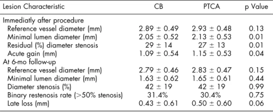
0.1). In addition, there were no significant differences in rates of dissection after the procedure (36% vs 35%, p ⫽ 0.34) or “bail-out” stenting (13% vs 16%, p ⫽ 0.13). However, CB left a smaller immediate minimal lumen diameter after the procedure (2.05 ⫾ 0.52 vs 2.13 ⫾ 0.53 mm, p ⫽ 0.01) and a greater residual percent diameter stenosis (29⫾ 14% vs 27 ⫾ 13%, p
⫽ 0.01) than PTCA (Table 3).
Angiographic follow-up: Repeat angiography was performed at 6 months in 551 (82%) and 559 (80%) of lesions treated with CB and PTCA, respectively. Dur-ing the 6-month angiographic follow-up the primary end point of binary angiographic restenosis was not significantly different between the 2 groups (CB 31.4% vs PTCA 30.4%, p ⫽ 0.75; Table 3), and the follow-up minimal lumen diameter and percent diam-eter stenosis were also similar (Table 3).
A nonsignificant reduction in lower absolute late loss at 6 months in the CB arm (0.43⫾ 0.61 vs 0.50
⫾ 0.60, p ⫽ 0.06) failed to translate to lower
angio-graphic restenosis rates because CB achieved a smaller acute gain than PTCA (1.09⫾ 0.54 vs 1.15 ⫾ 0.53, p⫽ 0.04). In addition, the proportional response to injury, as measured by the late loss index, was not different between CB and PTCA arms (0.49⫾ 0.04 vs 0.44⫾ 0.05, p ⫽ 0.3).
Clinical follow-up: At 270 days, clinical follow-up was available in 580 patients (94%) assigned to CB and 572 patients (92%) assigned to PTCA. The 30-day major adverse cardiac event rate was 3.7% for CB versus 2.7% for PTCA (p ⫽ 0.34), and the 270-day major adverse cardiac event rate was 13.6% versus
15.1%, respectively (p ⫽ 0.47; Table 4). Freedom
from target vessel revascularization, however, was higher in the group treated with CB (88.5%) compared with PTCA (84.6%, log-rank p ⫽ 0.04; Figure 1). In comparison to the PTCA arm, there was a higher incidence of myocardial infarction, largely non–Q-wave, in the CB arm (4.7% for CB vs 2.4% for PTCA, p⫽ 0.03) and higher mortality at 9 months (1.3% vs 0.3%, p ⫽ 0.06; Table 4).
There were 6 cases of emergency bypass surgery in each arm of the study (Table 4). Five coronary perfo-rations occurred in the CB arm, and none occurred in
the conventional angioplasty arm (0.8%, 0%, p ⫽
0.03). Of the 5 perforations, 2 were detected angio-graphically by the appearance of contrast extravasa-tion into the periadventitial space without tamponade; however, 3 perforations were clinically important. One patient underwent successful pericardiocentesis for treatment of tamponade. Two other patients died following perforation, tamponade, and emergency coronary artery bypass graft surgery. Within the CB arm, the balloon-to-artery ratio was higher in the cases of perforation than in the cases without perforation (mean 1.4 ⫾ 0.4, range 1.1 to 1.8 for perforations vs 1.0⫾ 0.1 for nonperforations, p ⫽ 0.05).
Multivariable regression analysis: Despite adjust-ment for differences in baseline characteristics and minimal lumen diameter after the procedure in a mul-tivariable model, treatment strategy (CB vs PTCA) TABLE 1 Baseline Demographics and Clinical Characteristics
Patient Characteristic CB PTCA (n⫽ 617) (n ⫽ 621) Age (yrs) 59⫾ 10 58 ⫾ 11 Men 72% 77% Smoking 67% 69% Diabetes mellitus 13% 12% Dyslipidemia 45% 47% Hypertension 34% 37%
Prior myocardial infarction 37% 39% Prior coronary artery bypass graft surgery 3% 3% CCS class III or IV angina 64% 64%
Values are expressed as percentages (counts/sample size) or mean⫾ SD. CCS⫽ Canadian Cardiovascular Society.
TABLE 2 Baseline Angiographic Characteristics
Lesion Characteristic
CB PTCA (n⫽ 689) (n⫽ 696) Lesion length (mm) 8.9⫾ 4.2 8.9⫾ 4.4 Reference vessel diameter (mm) 2.84⫾ 0.49 2.87 ⫾ 0.49 Minimal lumen diameter (mm) 0.96⫾ 0.33 0.98 ⫾ 0.34 Diameter stenosis (%) 66⫾ 11 66⫾ 11 Target vessel
Left anterior descending artery 42% 42% Left circumflex artery 28% 24% Right coronary artery 31% 34% Calcification (moderate or severe) 14% 15%
Thrombus 4% 3% Eccentric lesion 44% 44% Angulation⬎45° 6% 5% Lesion class* A 14% 15% B1 40% 37% B2 42% 45% C 3% 3%
Values are expressed as percentages (counts/sample size) or mean⫾ SD. *American College of Cardiology/American Heart Association Lesion Class.13
was not a significant independent predictor of 6-month binary angiographic restenosis.
DISCUSSION
The search for a percutaneous coronary interven-tion that would relieve obstructive stenosis and mini-mize the inevitable tissue injury response that leads to restenosis in a sizeable minority of patients has been
ongoing since the advent of balloon angioplasty in 1977.1First proposed
by Schwartz et al,19the proportional
injury model of balloon dilatation in porcine coronary arteries demon-strated that the healing neointimal response was proportional to the de-gree of underlying histologic injury created by the initial balloon dilata-tion. This model has been upheld in clinical trials of coronary devices, in which a consistent proportional rela-tion has been observed between in-jury, measured as acute gain im-parted by the device, and the healing injury response, measured as the 6-month late loss in lumen diame-ter.2,20
Despite a wide variety of me-chanical approaches designed to en-large coronary lumen size beyond balloon dilatation alone (including plaque atherectomy, ablative me-chanical and laser atherectomy, and metal stenting), a common propor-tional vascular injury response ap-pears to transcend the wide array of mechanical interventions. That is, the relation between 6-month late loss and initial acute gain in the tar-get lesion lumen diameter for any given intervention, known as the late-loss index,1 has been
consis-tently observed to be approximately 50%.
We evaluated whether CB angio-plasty could reduce rates of resteno-sis by its proposed mechanism of controlled injury causing minimal injury response. The results from this well-powered multicenter trial of 1,238 patients randomized to the CB versus PTCA demonstrated that the CB was associated with high rates of procedural success. However, there was no difference in the primary end point of binary angio-graphic restenosis between the 2 arms. This primary negative finding is well-supported by the trial design with 90% power to detect a 25% treatment effect.
The observation that the late loss index in the CB arm was no different from that of PTCA, even at the low pressures and durations used for CB inflation, suggests that the CB does not reduce the proportional response to injury. These results and the multivariable model are consistent with the conclusion that a larger minimum lumen diameter after the procedure is the most influential procedural predictor of restenosis, irrespective of the mechanism of plaque displace-ment.2
It had been proposed that by creating controlled linear incisions, CB use could reduce the incidence of flow-limiting dissection after the procedure. In this study, however, rates of dissection were the same in both treatment groups. Perforation is a risk of all TABLE 3 Quantitative Angiographic Analysis
Lesion Characteristic CB PTCA p Value Immediatly after procedure
Reference vessel diameter (mm) 2.89⫾ 0.49 2.93⫾ 0.48 0.13 Minimal lumen diameter (mm) 2.05⫾ 0.52 2.13⫾ 0.53 0.01 Residual (%) diameter stenosis 29⫾ 14 27⫾ 13 0.01 Acute gain (mm) 1.09⫾ 0.54 1.15⫾ 0.53 0.04 At 6-mo follow-up
Reference vessel diameter (mm) 2.79⫾ 0.46 2.83⫾ 0.47 0.15 Minimal lumen diameter (mm) 1.63⫾ 0.62 1.65⫾ 0.61 0.44 Diameter stenosis (%) 42⫾ 19 42⫾ 19 0.99 Binary restenosis rate (⬎50% stenosis) 31.4% 30.4% 0.75 Late loss (mm) 0.43⫾ 0.61 0.50⫾ 0.60 0.06
Values are expressed as percentages (counts/sample size) or mean⫾ SD.
TABLE 4 Early and Late Adverse Events
Event
CB PTCA
(n⫽ 617) (n⫽ 621) p Value Early complications (within 30 days)
Total major adverse cardiac events 23 (3.7%) 17 (2.7%) 0.34
Death 4 (0.6%) 0 0.06
Myocardial infarction 26 (4.2%) 14 (2.3%) 0.05 Q wave 6 (1.0%) 6 (1.0%) 1 Non–Q-wave 20 (3.2%) 8 (1.3%) 0.02 Emergency bypass surgery 6 (1.0%) 6 (1.0%) 1 Target lesion revascularization 16 (2.6%) 16 (2.6%) 1 Subacute closure 8 (1.3%) 10 (1.6%) 0.81 Vessel perforation 5 (0.8%) 0 0.03 Major vascular complications 2 (0.3%) 1 (0.2%) 0.62 Total complications (through 9 mo)
Total major adverse cardiac events 84 (13.6%) 94 (15.1%) 0.47 Death 8 (1.3%) 2 (0.3%) 0.06 Myocardial infarction 29 (4.7%) 15 (2.4%) 0.03 Q wave 9 (1.5%) 7 (1.1%) 0.63 Non–Q-wave 20 (3.2%) 8 (1.3%) 0.02 Emergency bypass surgery 6 (1.0%) 6 (1.0%) 1 Target lesion revascularization 72 (12%) 92 (15%) 0.11
FIGURE 1. Kaplan-Meier estimated freedom from target vessel revascularization (TVR), demonstrating a higher survival for CB. (pⴝ 0.04).
percutaneous coronary interventions. Five coronary perforations occurred after CB treatment (0.8%) and none after conventional angioplasty (p⫽ 0.03). Given the small number of events, it is difficult to predict with certainty what procedural variables predispose to coronary perforation following CB treatment, but high balloon-to-artery ratios (⬎1.1) were associated with this complication.
The role of CB may be for difficult lesions in which controlled dilatation is believed to provide a better acute result. Since the completion of this study, CB angioplasty has been proposed, with success in case series, for the treatment of resistant lesions,10and
also for the treatment of the in-stent restenotic lesions with or without vascular brachytherapy.21
In this multicenter, randomized clinical trial, CB angioplasty did not reduce rates of angiographic re-stenosis but did reduce the rate of target vessel revas-cularization. The CB demonstrated excellent acute procedural results; however, it is an example of an-other new coronary angioplasty device, that despite its novel mechanism of plaque displacement results, in the expected proportional response to injury previ-ously seen with conventional angioplasty, atherec-tomy, and stenting. It is unlikely that different me-chanical solutions will alter the arterial response to injury enough to prevent restenosis. To reduce the injury response, we will instead need to look forward to interventions that address the biology of restenosis, such as the promising drug-coated stents.22
1.Kuntz RE, Baim DS. Defining coronary restenosis. Newer clinical and angio-graphic paradigms. Circulation 1993;88:1310 –1323.
2. Kuntz RE, Gibson CM, Nobuyoshi M, Baim DS. Generalized model of restenosis after conventional balloon angioplasty, stenting and directional atherectomy. J Am Coll Cardiol 1993;21:15–25.
3.Barath P, Fishbein MC, Vari S, Forrester JS. Cutting balloon: a novel approach to percutaneous angioplasty. Am J Cardiol 1991;68:1249 –1252.
4.Popma JJ, Lansky AJ, Purkayastha DD, Hall LR, Bonan R. Angiographic, and clinical outcome after cutting balloon angioplasty. J Invasive Cardiol 1996; 8(suppl A):12A–19A.
5.Ergene O, Seyithanoglu BY, Tastan A, Ergene U, Kozan O, Belgi A, Kaya D. Comparison of angiographic and clinical outcome after cutting balloon and conventional balloon angioplasty in vessels smaller than 3 mm in diameter: a randomized trial. J Invasive Cardiol 1998;10:70 –75.
6.Unterberg C, Buchwald AB, Barath P, Schmidt T, Kreuzer H, Wiegand V.
Cutting balloon coronary angioplasty—initial clinical experience. Clin Cardiol 1993;16:660 –664.
7.Hosokawa H. Cutting balloon angioplasty. A Japanese center’s early experi-ence. J Invasive Cardiol 1996;8(suppl A):20A–24A.
8. Kondo T, Kawaguchi K, Awaji Y, Mochizuki M. Immediate and chronic results of cutting balloon angioplasty: a matched comparison with conventional angioplasty. Clin Cardiol 1997;20:459 –463.
9.Muramatsu T, Tsukahara R, Ho M, Ito S, Inoue T, Akimoto T, Hirano K. Efficacy of cutting balloon angioplasty for lesions at the ostium of the coronary arteries. J Invasive Cardiol 1999;11:201–206.
10.Bertrand OF, Bonan R, Bilodeau L, Tanguay JF, Tardif JC, Rodes J, Joyal M, Crepeau J, Cote G. Management of resistant coronary lesions by the cutting balloon catheter: initial experience. Cathet Cardiovasc Diagn 1997;41:179 –184.
11.Molstad P, Myreng Y, Golf S, Sirnes PA, Kassis E, Abilgaard U, Andersen PE, Thuesen L. The Barath cutting balloon versus conventional angioplasty. A randomized study comparing acute success rate and frequency of late restenosis.
Scand Cardiovasc J 1998;32:79 –85.
12.Izumi M, Tsuchikane E, Funamoto M, Kobayashi T, Sumitsuji S, Otsuji S, Sakurai M, Awata N. Final results of the CAPAS trial. Am Heart J 2001;142: 782–789.
13.Smith SC Jr, Dove JT, Jacobs AK, Kennedy JW, Kereiakes D, Kern MJ, Kuntz RE, Popma JJ, Schaff HV, Williams DO, et al. ACC/AHA guidelines for percutaneous coronary intervention (revision of the 1993 PTCA guidelines)-executive summary: a report of the American College of Cardiology/American Heart Association task force on practice guidelines (Committee to revise the 1993 guidelines for percutaneous transluminal coronary angioplasty) endorsed by the Society for Cardiac Angiography and Interventions. Circulation 2001;103:3019 – 3041.
14.Kuntz RE, Hinohara T, Robertson GC, Safian RD, Simpson JB, Baim DS. Influence of vessel selection on the observed restenosis rate after endoluminal stenting or directional atherectomy. Am J Cardiol 1992;70:1101–1108.
15.Kaplan EL, Meier P. Nonparametric estimation from incomplete observa-tions. J Am Statist Assoc 1958;53:457–481.
16.Kuntz RE, Safian RD, Carrozza JP, Fishman RF, Mansour M, Baim DS. The importance of acute luminal diameter in determining restenosis after coronary atherectomy or stenting. Circulation 1992;86:1827–1835.
17.Kereiakes D, Linnemeier TJ, Baim DS, Kuntz R, O’Shaughnessy C, Her-miller J, Fink S, Lansky A, Nishimura N, Broderick TM, Popma J. Usefulness of stent length in predicting in-stent restenosis (the MULTI-LINK stent trials). Am J
Cardiol 2000;86:336 –341.
18.Bourassa MG, Lesperance J, Eastwood C, Schwartz L, Cote G, Kazim F, Hudon G. Clinical, physiologic, anatomic and procedural factors predictive of restenosis after percutaneous transluminal coronary angioplasty. J Am Coll
Car-diol 1991;18:368 –376.
19.Schwartz RS, Huber KC, Murphy JG, Edwards WD, Camrud AR, Vlietstra RE, Holmes DR. Restenosis and the proportional neointimal response to coronary artery injury: results in a porcine model. J Am Coll Cardiol 1992;19:267–274.
20.Baim DS, Cutlip DE, Sharma SK, Ho KK, Fortuna R, Schreiber TL, Feldman RL, Shani J, Senerchia C, Zhang Y, et al. Final results of the balloon vs optimal atherectomy trial (BOAT). Circulation 1998;97:322–331.
21.Eeckhout E, Roguelov H, De Benedetti E, Coucke P, SilberS, Albiero R, Schiele T, Bonan R, Urban P. Treatment of in-stent restenosis by combining cutting balloon angioplasty and vascular brachytherapy: a report from the RENO Registry. American College of Cardiology Scientific Session. Atlanta, GA, 2002.
J Am Coll Cardial 2002;5(suppl A):4A.
22. Morice MC, Serruys PW, Sousa JE, Fajadet J, Hayashi EB, Perin M, Colombo A, Schuler G, Barragan P, Guagliumi G, Molnar F, Falotico R. A randomized comparison of a sirolimus-eluting stent with a standard stent for coronary revascularization. N Engl J Med 2002;346:1773–1780.