C L I N I C A L
R e v I e w
European Advisory Board on Cat Diseases www.abcdcatsvets.org
www.abcd-vets.org
Corresponding author: Albert Lloret Email: Albert.LLoret@uab.cat
CYTAUXZOONOSIS IN CATS
ABCD guidelines on prevention
and management
Albert Lloret, Diane D Addie, Corine Boucraut-Baralon, Herman Egberink, Tadeusz Frymus, Tim Gruffydd-Jones, Katrin Hartmann, Marian C Horzinek, Margaret J Hosie, Hans Lutz, Fulvio Marsilio, Maria Grazia Pennisi,
Alan D Radford, Etienne Thiry, Uwe Truyen and Karin Möstl
Overview: Cytauxzoon species are apicomplexan haemoparasites, which may cause severe disease in domestic cats, as well as lions and tigers. For many years, cytauxzoonosis in domestic cats was only reported in North and South America, but in recent years the infection has also been seen in Europe (Spain, France and Italy).
Infection: Cytauxzoon felis is the main species; it occurs as numerous different strains or genotypes and is transmitted via ticks. Therefore, the disease shows a seasonal incidence from spring to early autumn and affects primarily cats with outdoor access in areas where tick vectors are prevalent. Domestic cats may experience subclinical infection and may also act as reservoirs.
Clinical signs: Cytauxzoonosis caused by C felis in the USA is an acute or peracute severe febrile disease with non-specific signs. Haemolytic anaemia occurs frequently; in some cats neurological signs may occur in late stages. The Cytauxzoon species identified in Europe differ from C felis that causes disease in the USA and are probably less virulent. The majority of infected cats have been healthy; in some cases anaemia was found, but disease as it occurs in the USA has not been reported to date.
Diagnosis: Diagnosis is usually obtained by Cytauxzoon detection in blood smears and/or fine-needle aspirates from the liver, spleen and lymph nodes. PCR assays are able to detect low levels of parasitaemia and may be used for confirmation.
Treatment: Currently a combination of the antiprotozoal drugs atovaquone and azithromycin is the treatment of choice. Concurrent supportive and critical care treatment is extremely important to improve the prognosis. Cats that survive the infection may become chronic carriers for life.
Prevention: Cats with outdoor access in endemic areas should receive effective tick treatment.
Introduction
Cytauxzoonosis has been documented in wild felids such as bobcats, Florida panthers and Texas cougars. The first cases in domestic cats were documented in 1976.1For many years, cytauxzoonosis in
domes-tic cats was only reported in North America (south eastern and central states and mid-Atlantic regions) and South America, but in recent years the infection has also been documented in Europe.
Agent properties
Cytauxzoon species are apicomplexan haemoparasites (family Theileriidae) of wild and domestic cats, which are transmitted by ticks.
Several species have been identified. Cytauxzoon felis is the main species, with numerous different strains or genotypes2,3producing infection and
severe disease in domestic cats, lions and tigers. Wild cats (bobcats, mountain lions, ocelots, spotted cats and jaguars) in North and South America can act as reservoir or incidental hosts. Recent studies have shown that domestic cats can also harbour subclinical infections and may act as reservoirs.4,5 In some endemic areas, the prevalence of
subclinical infection in cats may be as high as 30%.6 Tick vectors for C felis are Amblyomma americanum and Dermacentor variabilis.7–9
Other species have been identified: Cytauxzoon manul in Pallas cats (Mongolia), Cytauxzoon spp in Iberian lynx and domestic cats in Spain,10and C spp in domestic cats in Italy.11The tick vectors for the
European species are still not known, but most likely are Dermacentor spp or Ixodes ricinus.
Epidemiology
It has been hypothesised that infection in domestic cats involved a species jump from bobcats, in which the prevalence of infection may be high in certain geographic areas.8Disease shows a seasonal incidence
from spring to early autumn,12,13associated with peak activity of the tick
vectors. There is a significant association between infection and both out-door access and feral cats in areas where vector ticks are prevalent.12No
638
JFMSCLINICAL PRACTICEA hyperendemic focus may be found within endemic areas, but is likely due to tick expo-sure of cats rather than cat-to-cat transmis-sion, which has never been proven.14,15 In
some areas of the USA an increase in cytaux-zoonosis diagnoses has been observed in the past decade and it is considered an emerging disease.13
In recent years, the infection has also been documented in Europe. Cases have been described in the Iberian lynx (Figure 1)10,16,17
and in domestic cats18 in the south of Spain,
and in domestic cats in France.19Moreover, a
case series was reported in north-eastern Italy (Trieste) and two cases in central Italy.11,20 In
the Trieste region, samples from domestic and feral cats showed a 23% prevalence of infec-tion, with a higher prevalence in feral cats (30%). Cytauxzoon species in the European cases is different from C felis,
which produces infection and disease in the USA.
Pathogenesis
The life cycle and complex pathogenesis has been well described for this infection.21
Vector ticks ingest merozoite-infected red blood cells from the natural reservoir host (bob-cat, lynx or domestic cat). The parasite initiates a process of sexual replication (gameto -genesis) in the tick gut and
sali-vary glands. This leads to the formation of sporozoites, which are the infective form and can be transmitted if the tick attaches to a domestic cat. Sporozoites infect endothelial-associated mononuclear cells and undergo asexual replication within the macrophages; these, in turn, develop into large structures known as schizonts – large enough to occlude blood vessels, especially in the liver, spleen and lungs. Widespread dissemination of schizonts results in parasitic thrombosis, circulatory impairment, tissue infection and a severe systemic inflammatory response, which can lead to multi-organ dysfunction and failure and death within 3 weeks of infection.22When
schizonts rupture in the circulation, large num-bers of merozoites are released, infecting red blood cells and additional mononuclear cells. This is late-stage disease, with erythropara-sitaemia (piroplasm structures within red blood cells) which can be readily observed in blood smears, and may lead to haemolytic anaemia and erythro phagocytosis.
Recent studies have evaluated systemic and lung immune responses in cats naturally infected with C felis based on serum concen-trations of cytokines (TNFα, IL-1β) and serum
Figure 1 Merozoites within red blood cells in an Iberian lynx from southern Spain.
Courtesy of Professor Josep Pastor, Veterinary School of Medicine, Universitat Autònoma de Barcelona, Spain
proteins, immunohistochemical expression of several inflammatory mediators and PCR assay for CD18.23,24Both studies
demonstrat-ed a markdemonstrat-ed systemic and lung pro-inflamma-tory response that can contribute to the pathogenesis of the disease; the response was even more pronounced in cats that died com-pared with survivors.23,24
Clinical presentation
Cytauxzoonosis (C felis) in the USA is typical-ly an acute or peracute severe febrile disease. Clinical signs are non-specific and consist of depression, anorexia, high fever, icterus, dys-pnoea, tachycardia, generalised pain and vocalisation. Signs of haemolytic anaemia are frequent (pale mucous membranes, pigmenturia, splenomegaly, hepato megaly).
Some cats may present or evolve to late-stage disease with neurological signs (ataxia, seizures, nystagmus), hypother-mia, moribund state and coma. Many cats die within 1 week of the onset of clinical signs.14,25
Veterinarians practising in an endemic area must suspect cytauxzoonosis when faced with any cat with an acute severe disease.
Frequent clinicopathological signs include non-regenerative anaemia, leukopenia with toxic changes, thrombo cytopenia, hyperbilirubinaemia, bilirubinuria and an increase in liver enzymes. These changes are associated with erythrophago cytosis and systemic inflammatory response syndrome (SIRS). Coagulation times are usually pro-longed due to disseminated intravascular coagulation. Other biochemical abnormalities include hypo albuminaemia, hyperglycaemia, pre-renal azotaemia, and electrolyte and acid–base disturbances associated with the SIRS state.14,25
Diagnostic imaging reveals non-specific signs consisting of hepatosplenomegaly on abdominal radiography and/or ultrasound, and a pulmonary interstitial–alveolar pattern on thoracic radiography.
Cytauxzoon species infection reported in
European cats (Italy, Spain, France) is proba-bly less virulent than C felis infection. The majority of infected cats have been healthy, showing only low-level erythroparasitaemia (merozoites within red blood cells) as an incidental finding. In some cats anaemia was described and one cat died after severe dis-ease of a short duration, but no schizont struc-tures were found in tissues, so cytauxzoonosis was not confirmed.
In some
endemic areas,
the prevalence
of subclinical
infection in
cats may be as
high as 30%.
European Advisory Board on Cat DiseasesThe European Advisory Board on Cat Diseases (ABCD) is a body of experts in immunology, vaccinology and clinical feline medicine that issues guidelines on prevention and management of feline infectious diseases in Europe, for the benefit of the health and welfare of cats. The guidelines are based on current scientific knowledge of the diseases and available vaccines concerned.
The latest version of the cytauxzoonosis in cats guidelines is available at
www.abcdcatsvets.org and www.abcd-vets.org
at Universite de Liege on November 8, 2015
jfm.sagepub.com
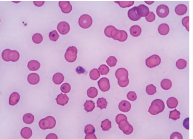
Figure 2Merozoites within red blood cells in a cat from Trieste (Italy). Courtesy of Dr
Erika Carli and Dr Laia Solano-Gallego, Clinica Veterinaria Privata San Marco, Padova, Italy
Cat-to-cat
transmission of
cytauxzoonosis
has never
been proven.
Diagnosis
In clinical practice, diagnosis is usually obtained by identification of C felis in blood smears and/or fine-needle aspirates from the liver, spleen and lymph nodes using rapid Romanowsky-type stains.
Observation of schizont-infected myeloid cells on blood and/or tissue smears is the diag-nostic test of choice because it confirms acute disease. These are seen as very large (50–250 µm diameter) single cells with an eccentric nucleus containing a single prominent nucleolus. The cytoplasm contains variable numbers of basophilic particles (a few to thousands), which are developing merozoites. These cells may be confused with platelet clumps. The sensitivity of blood smears may be low, so fine-needle aspirates and cytology of liver, spleen, lymph nodes and lungs are indicated if
blood smears are not diagnostic in a suspected case.
Observation of merozoites (piroplasms) within red blood cells in thin blood smears pre-pared with Romanowsky-type stains is supportive of a diagno-sis of cytauxzoonodiagno-sis. However, it does not confirm acute disease as merozoites can be an inciden-tal finding in healthy cats, and may also be observed in cats that have survived acute infec-tion or those with clinical signs of another disease. Piroplasms
are usually round to oval structures, 1–2 µm in diameter, with a dark purple eccentric nucleus within a pale blue cytoplasm (signet ring shaped), but in some cases may be more elon-gated with a bipolar nucleus (Figure 2). One to four merozoites may be observed within indi-vidual red blood cells. Sensitivity is not very high, as merozoites appear late in the course of the disease; they are either absent or present in very low numbers in probably more than 50% of cats with acute disease. Blood smears should be performed daily because merozoites can appear over the course of the disease. The dis-tal edges of a blood smear are the best place to look for them.
PCR assays have been developed to confirm the presence of C felis and other Cytauxzoon species,10,11,14but so far they are not useful as a
quick diagnostic tool in practice. It is recom-mended though that samples from suspected cats are submitted to appropriate laboratories to further confirm the infection. Low levels of par-asitaemia can only be detected by PCR assay.5
In one clinical trial, parasitaemia was deter-mined by qPCR and at significantly lower lev-els in surviving cats versus non-surviving cats, so qPCR results might be of prognostic value.26
Treatment
Historically, cytauxzoonosis has been consid-ered a fatal disease, with mortality approach-ing 100%. With the recent advances in treatment and/or differences in strain patho-genicity, this is no longer true, although the prognosis remains guarded in some cats.27,28
Supportive and critical care treatment (intensive fluid and oxygen therapy, anti-thrombotic therapies such as unfractionated heparin 200 U/kg SC q8h, blood products, antibiotics, analgesics) is extremely important to keep the cat alive while the anti protozoal drugs and immune system do their work. Many cats deteriorate during the first days and often die; but, if they survive, a gradual improvement is seen over the ensuing days.26
A variety of antiprotozoal drugs have been used in case reports or experimental studies (diminazene, imidocarb dipropi-onate, thia cetarsamide sodium, tetra cycline, parvaquone, bupar -va quone) but efficacy has not been proven [EBM grade IV].27–29
Imidocarb had been the drug of choice for many years, although it was not known if it provided any advantage over supportive care alone. How -ever, an open-label randomised prospective clinical trial demon-strated better survival rates (60% vs 26%) with the combination of atovaquone (15 mg/kg PO q8h) and azithro -mycin (10 mg/kg PO q 24h) compared with imidocarb (3.5 mg/kg IM once) in 80 cats with acute disease.26 Mortality was high (41/80
cats). Most cats died during the first 3 days after presentation, only three cats dying after the third day of treatment. Supportive treat-ment was the same in all cats, comprising fluid therapy and heparin. This study sug-gests that this antiprotozoal combination plus supportive treatment is the current approach of choice [EBM grade I].26In some cats, a
naso-oesophageal tube may be needed to adminis-ter drugs and enadminis-teral feeding.
Cats surviving the acute infection may become chronic carriers for life, with piro-plasms within the red blood cells. These cats act as reservoirs and may transmit the infec-tion through tick vectors.
A recent study failed to demonstrate effica-cy of diminazene at higher doses (4 mg/kg IM) for 5 consecutive days in eliminating or reducing the parasite burden in chronic carri-er cats. Moreovcarri-er, multiple advcarri-erse effects appeared, so this treatment is not recom-mended [EBM grade III].30
EBM grades
The ranking system for grading the level of evidence of various statements within the treatment and prevention sections of this article is described on page 574 of this Special Issue.
640
JFMSCLINICAL PRACTICEPrevention
There is currently no vaccine against C felis, although preliminary studies are being conducted.31
Prevention is based on living indoors or use of effective tick treatment in cats with outdoor access. Efficacy of an acaricide collar (imida-cloprid 10% plus flumethrin 4.5%) for preven-tion of C felis transmission has been proven in a controlled prospective clinical trial. Two groups of cats (with and without a collar) were exposed to ticks (A americanum) infected with C felis. No cats with a collar, vs 90% of the cats with no collar, were infected [EBM grade II].32
Testing for the presence of Cytauxzoon species in feline blood donors is advised. Although inoculation of merozoites within red blood cells in a blood transfusion does not lead to the development of schizont structures and disease, cats can become chronic carriers and an infection reservoir.
Prognosis
The prognosis for cats with cytauxzoonosis in the USA should be considered guarded to fair, if proper intensive care is provided and ato-vaquone is available. It has been suggested that different C felis strains may vary in pathogenicity, as some cats have survived after not receiving antiprotozoal drugs.2,27,33
It is recommended that cats are treated in well-equipped hospitals where the best sup-portive treatment can be provided.
Cytauxzoon infection in Europe reportedly
has a good prognosis: so far, only cats with sub clinical infection or signs of mild disease (anaemia, diarrhoea), possibly unrelated to the infection, have been documented.11,20
Funding
The authors received no specific grant from any funding agency in the public, commercial or not-for-profit sectors for the preparation of this article. The ABCD is supported by Merial, but is a scientifically independent body and its members receive no stipends from Merial.
Conflict of interest
The authors do not have any potential conflicts of interest to declare.
References
1 Wagner JE. A fatal cytauxzoonosis-like disease in cats. J Am Vet Med Assoc 1976; 168: 585–588. 2 Brown HM, Berghaus RD, Latimer KS, et al.
Genetic variability of Cytauxzoon felis from 88 infected domestic cats in Arkansas and Georgia. J Vet Diagn Invest 2009; 21: 59–63. 3 Shock BC, Birkenheuer AJ, Patton LL, et al.
Variation in the ITS-1 and ITS-2 rRNA genom-ic regions of Cytauxzoon felis from bobcats and pumas in the eastern United States and com-parison with sequences from domestic cats. Vet Parasitol 2012; 190: 29 –35.
4 Haber MD, Tucker MD, Marr HS, et al. The detection of Cytauxzoon felis in apparently
Veterinarians
practising in an
endemic area
must suspect
cytauxzoonosis
when faced
with any cat
with an acute
severe disease.
< Cytauxzoonosis has been reported worldwide, both in domestic cats and wild cat species.
< The parasite is transmitted via ticks, and the prevalence of infection is higher in cats with outdoor access and in feral cats.
< In the USA, cytauxzoonosis is typically an acute or peracute, severe febrile disease. Non-regenerative haemolytic anaemia is often present, as are neurological signs, followed by death in nearly 100% of cases.
< Cats infected with Cytauxzoon spp have been reported in southern Europe, but clinical signs in those cats were mild and possibly unrelated to the infection.
< In practice, diagnosis is often based on blood smears and/or fine-needle aspirates from the liver, spleen and lymph nodes using rapid Romanowsky-type stains.
< PCR assays have been developed to confirm the presence of C felis and Cytauxzoon species, but are not useful for a quick diagnosis in practice.
< Current treatment of choice is a combination of atovaquone (15 mg/kg PO q8h) and azithromycin (10 mg/kg PO q24h), as well as fluids, heparin and supportive care.
< Surviving cats may become chronic carriers.
< Prevention is based on living indoors or use of effective tick treatment in cats with outdoor access.
KEY
pOINTS
at Universite de Liege on November 8, 2015
jfm.sagepub.com
641
healthy free-roaming cats in the USA. VetParasitol 2007; 146: 316 –320.
5 Brown HM, Latimer KS, Erikson LE, et al. Detection of persistent Cytauxzoon felis infec-tion by polymerase chain reacinfec-tion in three asymptomatic domestic cats. J Vet Diagn Invest 2008; 20: 485–488.
6 Brown HM, Lockhart JM, Latimer KS, et al. Identification and genetic characterization of Cytauxzoon felis in asymptomatic domestic cats and bobcats. Vet Parasitol 2010; 172: 311–316. 7 Reichard MV, Edwards AC, Meinkoth JH, et al. Confirmation of Amblyomma americanum (Acari: Ixodidae) as a vector for Cytauxzoon felis (Piroplasmorida: Theileriidae) to domestic cats. J Med Entomol 2010; 47: 890–896.
8 Shock BC, Murphy SM, Patton LL, et al. Distribution and prevalence of Cytauxzoon felis in bobcats (Lynx rufus), the natural reser-voir, and other wild felids in thirteen states. Vet Parasitol 2011; 175: 325–330.
9 Blouin EF, Kocan AA, Glenn BL, et al. Transmission of Cytauxzoon felis Kier, 1979 from bobcats, Felis rufus (Schreber), to domes-tic cats by Dermacentor variabilis (Say). J Wild Dis 1984; 20: 241–242.
10 Millán J, Naranjo V, Rodríguez A, et al. Prevalence of infection and 18S rRNA gene sequence of Cytauxzoon species in Iberian Lynx (Lynx pardi-nus) in Spain. Parasitology 2007; 134: 995–1001. 11 Carli E, Trotta M, Chinelli R, et al. Cytauxzoon
sp infection in the first endemic focus described in domestic cats in Europe. Vet Parasitol 2012; 183: 343–352.
12 Reichard MV, Baum KA, Cadenhead SC, et al. Temporal occurrence and environmental risk factors associated with cytauxzoonosis in domestic cats. Vet Parasitol 2008; 152: 314–320. 13 Miller J and Davis CD. Increasing frequency of
feline cytauxzoonosis cases diagnosed in west-ern Kentucky from 2001 to 2011. Vet Parasitol 2013; 198: 205–208.
14 Birkenheuer AJ, Le JA, Valenzisi AM, et al. Cytauxzoon felis infection in cats in the mid-Atlantic states: 34 cases (1998–2004). J Am Vet Med Assoc 2006; 228: 568–571.
15 Woods JP. Feline cytauxzoonosis. In: Bonagura and Twedt (eds). Kirk’s current veterinary thera-py XV. 15th ed. St Louis, MO: Elsevier Saunders, 2013, pp e405–408.
16 Luaces I, Aguirre E, García-Montijano M, et al. First report of an intraerythrocytic small piro-plasm in wild Iberian lynx (Lynx pardinus). J Wild Dis 2005; 41: 810–815.
17 Millán J, Candela MG, Palomares F, et al. Disease threats to the endangered Iberian lynx (Lynx pardinus). Vet J 2009; 182: 114–124. 18 Criado-Fornelio A, González-del-Rio MA,
Buling-Saraña A, et al. The “expanding universe” of piroplasms. Vet Parasitol 2004; 119: 337–345. 19 Criado-Fornelio A, Buling A, Pingret JL, et al.
Hemoprotozoa of domestic animals in France:
prevalence and molecular characterization. Vet Parasitol 2009; 159: 73–76.
20 Carli E, Trotta M, Bianchi E, et al. Cytauxzoon sp. infection in two free ranging young cats: clinicopathological findings, therapy and follow up. Turkiye Parazitol Derg 2014; 38: 185–189.
21 Kier AB, Wagner JE and Kinden DA. The pathol-ogy of experimental cytauxzoonosis. J Comp Pathol 1987; 97: 415–432.
22 Snider TA, Confer AW and Payton ME. Pulmonary histopathology of Cytauxzoon felis infections in the cat. Vet Pathol 2010; 47: 698–702.
23 Frontera-Acevedo K, Balsone NM, Dugan MA, et al. Systemic immune responses in Cytaux -zoon felis-infected domestic cats. Am J Vet Res 2013; 74: 901 –909.
24 Frontera-Acevedo K and Sakamoto K. Local pulmonary immune responses in domestic cats naturally infected with Cytauxzoon felis. Vet Immunol Immunopathol 2015; 163: 1–7. 25 Hoover JP, Walker DB and Hedges JD.
Cytauxzoonosis in cats: eight cases (1985–1992). J Am Vet Med Assoc 1994; 205: 455–460.
26 Cohn LA, Birkenheuer AJ, Brunker JD, et al. Efficacy of atavaquone and azithromycin or imidocarb dipropionate in cats with acute cytauxzoonosis. J Vet Intern Med 2011; 25: 55–60.
27 Meinkoth J, Kocan AA, Whitworth L, et al. Cats surviving natural infection with Cytauxzoon felis: 18 cases (1997–1998). J Vet Intern Med 2000; 14: 521–525.
28 Greene CE, Latimer K, Hooper E, et al. Administration of diminazene aceturate or imidocarb dipropionate for treatment of cytauxzoonosis in cats. J Am Vet Med Assoc 1999; 215: 497–500.
29 Motzel SL and Wagner JE. Treatment of experi-mentally induced cytauxzoonosis in cats with parvaquone and buparvaquone. Vet Parasitol 1990; 35: 131–138.
30 Lewis KM, Cohn LA, Marr HS, et al. Failure of efficacy and adverse effects associated with dose-intense diminazene diaceturate treatment of chronic Cytauxzoon felis infection in five cats. J Feline Med Surg 2014; 16: 157–163. 31 Tarigo JL, Scholl EH, McK Bird D, et al. A novel
candidate vaccine for cytauxzoonosis inferred from comparative apicomplexan genomics. PLoS One 2013; 8: e71233. DOI: 10.1371/journal. pone.0071233.
32 Reichard MV, Thomas JE, Arther RG, et al. Efficacy of an imidacloprid 10%/flumethrin 4.5% collar (Seresto, Bayer) for preventing the transmission of Cytauxzoon felis to domestic cats by Amblyomma americanum. Parasitol Res 2013; 112: 11–20.
33 Walker DB and Cowell RL. Survival of a domes-tic cat with naturally acquired cytauxzoonosis. J Am Vet Med Assoc 1995; 206: 1363–1365. Available online at jfms.com