HAL Id: hal-02115232
https://hal.archives-ouvertes.fr/hal-02115232
Submitted on 30 Apr 2019
HAL is a multi-disciplinary open access
archive for the deposit and dissemination of
sci-entific research documents, whether they are
pub-lished or not. The documents may come from
teaching and research institutions in France or
abroad, or from public or private research centers.
L’archive ouverte pluridisciplinaire HAL, est
destinée au dépôt et à la diffusion de documents
scientifiques de niveau recherche, publiés ou non,
émanant des établissements d’enseignement et de
recherche français ou étrangers, des laboratoires
publics ou privés.
Linker chemistry dictates the delivery of a phototoxic
organometallic rhenium( i ) complex to human cervical
cancer cells from core crosslinked star polymer
nanoparticles
Sul Hwa Yu, Malay Patra, Stefano Ferrari, Paulina Ramirez Garcia, Nicholas
Veldhuis, Lisa Kaminskas, Bim Graham, John Quinn, Michael Whittaker,
Gilles Gasser, et al.
To cite this version:
Sul Hwa Yu, Malay Patra, Stefano Ferrari, Paulina Ramirez Garcia, Nicholas Veldhuis, et al.. Linker
chemistry dictates the delivery of a phototoxic organometallic rhenium( i ) complex to human cervical
cancer cells from core crosslinked star polymer nanoparticles. Journal of materials chemistry B, Royal
Society of Chemistry, 2018, 6 (47), pp.7805-7810. �10.1039/C8TB02464B�. �hal-02115232�
Linker Chemistry Dictates the Delivery of Phototoxic Organometallic
Rhenium(I) Complex to Human Cervical Cancer Cells from Core Crosslinked
Star Polymer Nanoparticles
Sul Hwa Yu
a†, Malay Patra
b†, Stefano Ferrari
c, Paulina Ramirez Garcia
a, Nicholas A. Veldhuis
a, d, Lisa M. Kaminskas
e, Bim
Graham
a, John F. Quinn
a, Michael R. Whittaker
a*, Gilles Gasser
f* and Thomas P. Davis
a* a. ARC Centre of Excellence in Convergent Bio‐Nano Science and Technology; Monash Institute of Pharmaceutical Sciences, Monash University, Parkville, VIC 3052, Australia. b. Department of Chemistry, University of Zurich, Winterthurerstrasse 190, 8057 Zurich, Switzerland. Current address: Department of Chemical Sciences, Tata Institute of Fundamental Research, 400005 Mumbai, India. c.Institute of Molecular Cancer Research, University of Zurich, Winterthurerstrasse 190, 8057 Zurich, Switzerland. d. Drug Discovery Biology Theme, Monash Institute of Pharmaceutical Sciences, Monash University, Parkville, VIC 3052, Australia. e. School of Biomedical Sciences, University of Queensland, St Lucia QLD, 4072, Australia. f.Chimie ParisTech, PSL University, Laboratory for Inorganic Chemical Biology, F‐75005 Paris, France.*email:thomas.p.davis@monash.edu,gilles.gasser@chimie‐paristech.fr, Michael.whittaker@monash.edu † These authors have contributed equally to the work
Electronic Supplementary Information (ESI) available: [experimental and additional data]. See DOI: 10.1039/x0xx00000x
We have investigated core‐crosslinked star polymer nanoparticles designed with tunable release chemistries as potential nanocarriers for a photoactive Re(I) organometallic complex. The nanoparticles comprised of a brush poly(oligo‐ethylene glycol) methyl ether acrylate(POEGA) corona and a cross‐linked core of non‐biodegradable N,N’‐methylenebis(acrylamide) (MBAA) and either pentafluorophenyl acrylate (PFPA), 3‐vinyl benzaldehyde (VBA) or diacetone acrylamide (DAAM). Each star was modified with an amine functionalized photodynamic agent (i.e. a rhenium(I) organometallic complex) resulting in the formation of either a stable amide bond (POEGA‐Star‐PFPA), or hydrolytically labile aldimine (POEGA‐Star‐VBA) or ketimine bonds (POEGA‐Star‐DAAM). These materials revealed linker dependent photo‐ and cytotoxicity when tested in vitro against non‐cancerous lung fibroblast MRC‐5 cells and HeLa human cervical cancer cells: The toxicity results correlated with final intracellular Re concentrations
Rhenium(I) organometallic compounds show great potential as therapeutic agents, demonstrating cytotoxicity against various cancer cell lines at least equal to cisplatin.1‐12Their photoactive properties can also be exploited as photosensitizers (PS) in photodynamic therapy (PDT), where Re(I) organometallic complexes generate highly reactive oxygen species (ROS), including singlet oxygen (1O
2), upon light irradiation.6, 13‐22 The light‐mediated activation of Re(I) organometallic complexes allows spatial and temporal control of activity, and has been demonstrated to have anti‐cancer activity by inducing oxidative damage of cellular components leading to cell death.7,16,17 However, these complexes lack a selective affinity for cancer cells, thereby limiting therapeutic efficacy. Also, their long systemic half‐lives lead to significant off‐target side‐effects that include long‐term phototoxicity. Consequently, the development of carrier systems has been proposed as a strategy to improve their selectivity and specificity towards the target cancer cells and reduce off‐target adverse effects.20, 23
Multi‐armed structured star polymers have been considered as well‐defined nanoparticles24‐26 that have been widely used for a number of applications, including as nanomedicines, catalysts and emulsification reagents.27‐33 Our group previously reported the synthesis of narrow dispersity core crosslinked star polymer nanoparticles using a reversible addition fragmentation transfer polymerization (RAFT) arm‐first approach,34, 35 in which the arm polymers were chain extended with a suitable cross‐linker. We have previously shown that these polymeric nanoparticles demonstrate size tunable pharmacokinetics as well as passive tumor accumulation properties,32 and cellular association could be manipulated via coronal functionality.36 The incorporation of a functional co‐monomer in the nanoparticle design allowed post‐modification for applications such as biological imaging and drug delivery.37‐40
In this communication we probe the suitability of core cross‐linked star polymers nanoparticles as carriers to support the delivery of a Re(I) organometallic complex, using three different linker chemistries, into target cells in vivo. Three different star polymers were developed comprising poly(ethylene glycol) (PEG)coronas, non‐biodegradable N,N’‐methylenebis(acrylamide) (MBAA) cross‐linkers and three different functional cores; 3‐vinyl benzaldehyde (VBA), diacetone acrylamide (DAAM) or pentafluorophenyl acrylate (PFPA) (Scheme 1). POEGA linear polymer was firstly prepared by RAFT polymerization in the presence of 3‐(benzyl sulfanyl thiocarbonyl sulfanyl)‐propanoic acid, and characterized by 1H nuclear magnetic resonance spectroscopy (1H NMR) and gel permeation chromatography (GPC) (Scheme 1, Figure S1, Table 1). The purified linear polymer was then chain extended separately with individual functional co‐monomers (VBA, DAAM or PFPA) in the presence of MBAA cross‐linker (Scheme 1). The resulting narrow polydispersity core cross‐linked star polymers were purified by precipitation and characterized via 1H NMR, 19F NMR and GPC (Table 1, Figure S2‐S5, S7‐S9). NMR revealed that 20 ‐ 40 mol % (molar composition relative to OEGA monomer) of each functional group was incorporated into the core of stars (23.5/76.5 mol % in VBA/OEGA, 37.4/62.6 mol % in DAAM/OEGA and 31.0/69.0 mol % in PFPA/OEGA, as described in the ESI).
To conjugate the Re(I)‐based photosensitizer into each functional core of the star polymers, a rhenium tricarbonyl complex containing a free amine group (Re‐NH3∙Cl, – Scheme 1) was synthesized as presented in the ESI (Scheme S1).1,2,17 To prepare reversibly linked nanoparticle conjugates, Re‐NH3∙Cl was transformed, in the presence of triethylamine (TEA), via the aldehyde groups of VBA and the ketone groups of DAAM to form Schiff base aldimine/ketimine linkages which are hydrolytically unstable under acidic conditions. The Re‐NH2 conjugated star polymers were then exhaustively dialyzed against water (adjusted to pH= 8‐9 by triethylamine addition) and analyzed by dynamic light scattering (DLS), transmission electron microscopy (TEM) and inductively coupled plasma mass spectrometry (ICP‐MS) (Figure S10, Figure S11, Table 2). The Re‐content (wt %) in Re‐NH2 conjugated POEGA‐ Star‐VBA (Star 1) and Re‐NH2 conjugated POEGA‐Star‐DAAM (Star 2) was quantified by ICP‐MS (Table 2). Additionally, the ICP‐MS values (wt %) measured for each star polymer was in excellent agreement with the maximum theoretical amount of Re (wt %) attached in each star polymer core, indicating that the functional cores (VBA and DAAM) in the star polymers were mostly conjugated with Re‐NH2. Characterization by DLS revealed that the hydrodynamic diameters of Star 1 and Star 2 were 18 and 20 nm, respectively, both with a polydispersity of 0.2 (Table 2, Figure S10).
Scheme 1. Synthesis of Re‐NH2 conjugated star polymers. Star 1 (Re‐NH2 conjugated POEGA‐Star‐VBA); Star 2 (Re‐NH2 conjugated POEGA‐Star‐DAAM); Star 3 (Re‐NH2
Table 1. Molecular weight of polymers, POEGA, POEGA‐Star‐VBA, POEGA‐Star‐DAAM and POEGA‐Star‐PFPA Polymers Mn (g mol‐1)[a] Mn (g mol‐1)[b] Ɖ[b] POEGA 9551 11000 1.10 POEGA-Star-VBA ‐ 85500 1.15 POEGA-Star-DAAM ‐ 79500 1.15 POEGA-Star-PFPA ‐ 76500 1.13
[a] Molecular weight determined by 1H NMR spectroscopy. [b]: Molecular weight determined by gel permeation chromatography. [c]: Dispersity
index, Ɖ, as determined by GPC analysis in DMAc. Table 2 Characterization of Re(I)‐NH2 conjugated star polymers Re(I)‐Stars Theoretical wt % Re [a] Wt % Re[b] Dh (nm)[c] PDI[c] Star 1 7.7 7.4 18 0.2 Star 2 11.0 11.1 20 0.2 Star 3 9.2 9.8 19 0.2 [a]: A = 0.001 g x [(Molar composition (%) of functional core monomer x M.W of functional core monomer)/(Molar composition (%) of functional core monomer x M.W of functional core monomer + molar composition
(%) of OEGA x 480 g)]. B = A x (M.W of Re(I)‐NH2/M.W of functional core monomer). C= B x (M.W of Re(I)/ M.W of Re(I)‐NH2 complex). Re (wt %) =
[C/(0.001 g + B)] x 100. [b]: ICP‐MS measurement. [c]: Size distribution (by number) and polydispersity for each star in water, assessed by dynamic
light scattering.
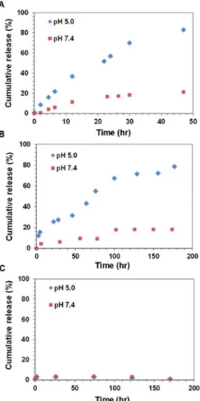
Finally, to prepare a non‐reversibly linked nanoparticle conjugate, the pentafluoro‐activated ester (PFP) of POEGA‐Star‐PFPA was reacted with Re‐NH3∙Cl to form a stable amide bond (Scheme 1). Re‐NH2 conjugated POEGA‐Star‐PFPA (Star 3) was then purified and characterized using 19F NMR, DLS, TEM and ICP‐MS (Table 2, Figure S6, S10). Importantly, 19F NMR analysis confirmed the covalent attachment of the Re‐NH2 complex into the star core by the concomitant release of pentafluorophenol byproduct (Figure S5, S6). Further, ICP‐MS analysis of Star 3 revealed a Re content of 9.78 wt % which is in excellent agreement with the theoretical value of 9.21 wt % (Table 2). The hydrodynamic diameter of Star 3 was determined by DLS to be 19 nm, with a polydispersity of 0.2 (Table 2, Figure S10). The release profile of Re‐NH2 from the star polymers was examined under different biologically relevant pH conditions (pH 7.4 and 5.0), using UV‐visible spectrophotometry (see ESI for detailed procedures, Figure 1). The pH dependent release of the Re‐NH2 via imine linker hydrolysis in Star 1 and 2 was observed (Figure 1, A and B). The acidic pH 5 condition, representing the approximate pH of lyso/endosomes and the lower pH end of the tumour interstitium,41, 42 triggered faster release of Re‐NH2 from both stars, compared to release kinetics at pH 7.4. As expected, Star 2 showed slow hydrolysis of the secondary ketimine bonds at pH 5.0. (Figure 1, B) with approximately 80% of the Re complex released from Star 2 after 180 h, in comparison with 80% released from Star 1 after only 50 h. No Re‐NH2 release was observed from Star 3 under any pH condition used (Figure 1, C). Next, the Re‐NH3∙Cl complex and the nanoparticle conjugates (Stars 1 to 3) were evaluated for their in vitro cytotoxicity against HeLa human cervical cancer cells and non‐cancerous lung fibroblast MRC‐5 cells (Table 3). First, the cells were exposed to increasing
Figure 1: Cumulative release profile (%) of Re(I)‐NH2 complex from the star polymers was examined in different pH conditions (pH 7.4 and 5) over the time period. (A) Re‐NH2 conjuated POEGA‐Star‐VBA (Star 1); (B) Re‐NH2 conjugated POEGA‐Star‐DAAM (Star 2); (C) Re‐NH2 conjugated POEGA‐Star‐PFPA (Star 3).
Figure 2. Re concentration‐response curves of Star 1 (Re‐NH2 conjugated POEGA‐Star‐VBA) and Star 2 (Re‐NH2 conjugated POEGA‐Star‐DAAM) in HeLa cells (4 h incubation with
350 nm light for 10 mins (red line) or without irradiation (blue line).
concentrations of either Re‐NH3∙Cl complex or the nanoparticle conjugates, and incubated for 48 h to evaluate their cytotoxicity under dark conditions. As shown in Table 3, the IC50 values for Re‐NH3∙Cl were found to be >100 µM against both cell lines. Interestingly, our compounds were found to have a slight selectivity for cancer cells over non‐cancerous cells (Table 3).
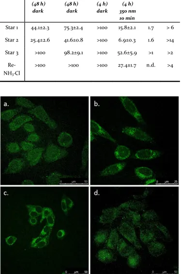
Table 3. IC50 values (µM Re) in the dark (48 h and 4 h assays), and upon light irradiation (350 nm). SI = selectivity index calculated as IC50 (MRC5, 48h, dark)/ IC50 (HeLa, 48h, dark). PI = phototoxic index calculated as IC50 (4h, light)/ IC50 (4h, dark) against HeLa cells. All concentration response curves are given in Figure S12. Figure 3. Fluorescence microscopy images of HeLa cells treated in the dark with a. Re‐NH3∙Cl; and its polymer conjugates: b. Star 1, c. Star 2 and d. Star 3 (50 µM, 9 h). Scale bar, 50 µm. However, the linker chemistry used in the conjugation with polymer stars was found to significantly influence the observed cytotoxicity. The cytotoxicity of the conjugated stars was significantly increased if the linker used was hydrolytically unstable, i.e. Stars 1 and 2, with the order of cytotoxicity determined to be Star 2 > Star 1 > Star 3 under dark conditions. Encouragingly, the most active star polymer conjugate data (Star 1 and Star 2) suggests a potential selectivity for cancer cells as demonstrated by a significant 2 fold higher IC50 values for non‐cancerous cells: the IC50 values of Star 1 and Star 2 against HeLa cancer cells were 44 and 25 µM, whereas the IC50 values against non‐cancerous MRC5 cells were 75 and 41 µM, respectively. The photo‐toxic potential of the Re‐NH3∙Cl complex against HeLa cells was then examined and compared to the nanoparticle conjugates Star 1, Star 2 and Star 3 (Table 3, Figure 2, Figure S12). In this assay, cells were treated with complex for 4 h (followed by replacement of media with fresh media), and then either irradiated with 350 nm UVA (2.58 J/ cm2) HeLa (48 h) dark MRC5 (48 h) dark HeLa (4 h) dark HeLa (4 h) 350 nm 10 min SI PI Star 1 44.1±2.3 75.3±2.4 >100 15.8±2.1 1.7 > 6 Star 2 25.4±2.6 41.6±0.8 >100 6.9±0.3 1.6 >14 Star 3 >100 98.2±9.1 >100 52.6±5.9 >1 >2 Re‐ NH3∙Cl >100 >100 >100 27.4±1.7 n.d. >4
identified when cells were treated with the Re(I) complex and its polymer conjugates were irradiated with 350 nm light. As shown in Table 3 and Figure 1, the phototoxicity followed the order Star 2 > Star 1 > Re‐NH3∙Cl > Star 3. Specifically, the lowest IC50 values were observed with the treatment of Star 1 (15.8) and Star 2 (6.9), and these also showed the highest PI values (>6.3 and >13.5, respectively). This indicates that Star 1 and 2 possess higher phototoxicity against the cells when compared to toxicity in the dark, as well as to other treatments (Re‐NH3∙Cl and Star 3).
To obtain initial insights into the mode of action of the Re‐polymer conjugates, we determined the subcellular localization of the Re‐NH3∙Cl complex and the polymer conjugates Star 1, Star 2 and Star 3 by exploiting the luminescence properties of Re‐ complex.20, 43 The cytosolic accumulation of Re‐NH
3∙Cl seen in Figure 3 (Figure S13) is consistent with previous reports.20, 43 Importantly, similar to the parent complex, Star 1, Star 2 and 3 were also found to accumulate largely in the cytosol of the cells (with some nuclear localization observed). Interestingly those nanoparticles with hydrolytically unstable linkers displayed increased luminescence, indicating greater intracellular Re concentrations. This observation is consistent with the higher observed cytotoxicity and phototoxicity of Star 1 and Star 2 (Table 3, Figure 2).
Owing to the potential for this structural class of Re(I) complexes to undergo cell‐mediated luminescence quenching once internalized into inside cells, fluorescence microscopy may not be a fully convincing method for quantitative evaluation of cellular uptake and quantification.9, 44 Therefore, we employed ICP‐MS to quantify Re in whole cells as well as in isolated cytosolic and nuclear intracellular compartments. As shown in Figure 4A, the total cellular accumulation followed the order Star 2~Star 1 >> Star 3 > Re‐NH3∙Cl, suggesting pH‐responsive Re‐NH2 conjugated star polymers clearly increase intracellular loading of Re‐NH2 complex. These data are consistent with the higher phototoxic properties of Star 1 and Star 2 compared to Star 3 and Re‐NH3∙Cl (Table 3, Figure 2). The observed differences in the cellular accumulation of Re from the nanoparticle reveals the balance of two competing mechanisms: the Re complex release from the internalized nanoparticle into the cell and, the removal of the nanoparticle via exocytosis mechanisms.45 Determination of Re‐content in the cytosol and nuclear fraction confirmed that the Re‐complex, as well as the polymer conjugates, preferentially accumulate in the cytosol of HeLa cells (Figure 4B). The results are consistent with the qualitative interpretation of fluorescent microscopy imaging (Figure 3). The cells exposed to Star 1 and Star 2 exhibited statistically significant higher cytosolic accumulation of Re in comparison with the parent Re(I) complex and that found for the Star 3 treated cells. Interestingly, while accumulation of Re in the cytoplasm of Star 2 exposed cells was not significantly different to that found for Star 1, it did show a statistically significant greater accumulation of Re in the nucleus (Figure 4B). This greater nuclear accumulation of Re may explain the greatest observed photo‐ and cyto‐ toxicity of Star 2 (Table 3).
Figure 4. Determination of final cellular Re concentration in treated HeLa cells: (A) Whole cell uptake of Re‐contents (ng/106 cells) from Re‐NH3∙Cl and its polymer conjugates, Star 1, Star 2 and Star 3 using ICP‐MS. (B) Re content (ng/106 cells) in the nuclear and cytoplasmic compartments of HeLa cells by ICP‐MS after treatment of Re‐NH3∙Cl and its polymer conjugates. *** p < 0.001, ** p < 0.01, * p < 0.05 (see Figure S14 for all analysis), error = SD, n = 3. In conclusion, three star polymers were synthesized with different functional cores: VBA, DAAM or PFPA. Each core was reacted with the amine group of Re‐NH2, resulting in different linker chemistries in the core of the stars. Based on their core linker chemistries, the stars possessed unique release profiles for Re‐NH2 under various pH conditions. In particular, Re‐NH2 conjugated star 2 showed a slower Re‐NH2 release profile compared to Star 1. No Re‐NH2 release was observed from Star 3 under any pH conditions. These unique release characteristics contributed to their dark and photo‐toxicity against HeLa cells and the non‐ cancerous MRC‐5 cells. The order of cytotoxicity of the delivered Re‐complex was determined to be Star 2 > Star 1 > Star 3. These results were also consistent with the cellular uptake of Re‐complex in the order Star 2 ~ Star 1 >> Star 3 > Re‐NH3∙Cl, suggesting that reversible Re‐NH2 conjugation of star polymers clearly facilitated the cellular accumulation of the Re‐NH2. Importantly, while there was not a significant difference in the total cellular Re content between Star 1 and Star 2, the more hydrolytically stable ketamine found in Star 2, led to a statistically significant greater nuclear accumulation of Re when compared with the aldimine linker in Star 1. Thus leading to its greater observed photo‐ and cytotoxicity.
Conflicts of interest
There are no conflicts to declareAcknowledgements
This work was carried out within the Australian Research Council (ARC) Centre of Excellence in Convergent Bio‐Nano Science and Technology (Project No. CE140100036). We gratefully acknowledge financial support from the Swiss National Science Foundation (Professorships No. PP00P2_133568 and PP00P2_157545 to G.G), the ERC (ERC Consolidator Grant PhotoMedMet (GA 681679) to G.G.) and the University of Zurich (G.G). This work has received support under the program «Investissements d’Avenir » launched by the French Government and implemented by the ANR with the reference ANR‐10‐IDEX‐0001‐02 PSL (G.G.). T.P.D. acknowledges the award of an Australian Laureate Fellowship from the ARC. J.F.Q. acknowledges receipt of a Future Fellowship from the ARC (FT170100144). Electron microscopy was performed at the Bio21 Advanced Microscopy Facility, The University of Melbourne.1. N. Viola‐Villegas, A. E. Rabideau, M. Bartholoma, J. Zubieta and R. P. Doyle, J. Med. Chem., 2009, 52, 5253‐5261. 2. N. Viola‐Villegas, A. E. Rabideau, J. Cesnavicious, J. Zubieta and R. P. Doyle, ChemMedChem, 2008, 3, 1387‐1394. 3. K. Yin Zhang, K. Ka‐Shun Tso, M.‐W. Louie, H.‐W. Liu and K. K.‐W. Lo, Organometallics, 2013, 32, 5098‐5102. 4. I. Chakraborty, S. J. Carrington, J. Hauser, S. R. J. Oliver and P. K. Mascharak, Chem. Mater., 2015, 27, 8387‐8397. 5. K. A. Stephenson, L. C. Reid, J. Zubieta, J. W. Babich, M.‐P. Kung, H. F. Kung and J. F. Valliant, Bioconjugate Chem., 2008, 19, 1087‐1094. 6. K. Wähler, A. Ludewig, P. Szabo, K. Harms and E. Meggers, Eur. J. Inorg. Chem. 2014, 2014, 807‐811. 7. A. Leonidova and G. Gasser, ACS Chem. Biol., 2014, 9, 2180‐2193. 8. C. C. Konkankit, S. C. Marker, K. M. Knopf and J. J. Wilson, Dalton Trans., 2018, 47, 9934‐9974. 9. I. Kitanovic, S. Can, H. Alborzinia, A. Kitanovic, V. Pierroz, A. Leonidova, A. Pinto, B. Spingler, S. Ferrari, R. Molteni, A. Steffen, N. Metzler‐Nolte, S. Wölfl and G. Gasser, Chem. Eur. J, 2014, 20, 2496‐2507. 10. K. M. Knopf, B. L. Murphy, S. N. MacMillan, J. M. Baskin, M. P. Barr, E. Boros and J. J. Wilson, J. Am. Chem. Soc., 2017, 139, 14302‐14314. 11. K. Suntharalingam, S. G. Awuah, P. M. Bruno, T. C. Johnstone, F. Wang, W. Lin, Y.‐R. Zheng, J. E. Page, M. T. Hemann and S. J. Lippard, J. Am. Chem. Soc., 2015, 137, 2967‐2974. 12. L. C.‐C. Lee, K.‐K. Leung and K. K.‐W. Lo, Dalton Trans., 2017, 46, 16357‐16380. 13. D. E. Dolmans, D. Fukumura and R. K. Jain, Nat. Rev. Cancer, 2003, 3, 380‐387. 14. J. M. Menter, T. D. Hollins, R. M. Sayre, A. A. Etemadi, I. Willis and S. N. Hughes, Photodermatol. Photoimmunol. Photomed., 1998, 14, 154‐159. 15. A. P. Castano, T. N. Demidova and M. R. Hamblin, Photodiagnosis Photodyn. Ther., 2004, 1, 279‐293. 16. C. Mari, V. Pierroz, S. Ferrari and G. Gasser, Chem Sci, 2015, 6, 2660‐2686. 17. F. S. De Rosa and M. V. Bentley, Pharm. Res., 2000, 17, 1447‐1455. 18. T. A. Debele, S. Peng and H. C. Tsai, Int. J. Mol. Sci., 2015, 16, 22094‐22136.
19. A. Kastl, S. Dieckmann, K. Wahler, T. Volker, L. Kastl, A. L. Merkel, A. Vultur, B. Shannan, K. Harms, M. Ocker, W. J. Parak, M. Herlyn and E. Meggers,
ChemMedChem, 2013, 8, 924‐927. 20. A. Leonidova, V. Pierroz, R. Rubbiani, J. Heier, S. Ferrari and G. Gasser, Dalton Trans., 2014, 43, 4287‐4294. 21. S. C. Marker, S. N. MacMillan, W. R. Zipfel, Z. Li, P. C. Ford and J. J. Wilson, Inorg. Chem., 2018, 57, 1311‐1331. 22. I. Chakraborty, J. Jimenez and P. K. Mascharak, Chem. Commun. (Camb.), 2017, 53, 5519‐5522. 23. M. Hu, J. Zhao, X. Ai, M. Budanovic, J. Mu, R. D. Webster, Q. Cao, Z. Mao and B. Xing, Dalton Trans., 2016, 45, 14101‐14108. 24. M. Sugimoto, T. Koizumi, T. Taniguchi, K. Koyama, K. Saito, D. Nonokawa and T. Morita, J. Polym. Sci. B Polym. Phys, 2009, 47, 2226‐2237. 25. B. M. Erwin, M. Cloitre, M. Gauthier and D. Vlassopoulos, Soft Matter, 2010, 6, 2825‐2833. 26. T. K. Goh, K. D. Coventry, A. Blencowe and G. G. Qiao, Polymer, 2008, 49, 5095‐5104. 27. J. H. Ryu, R. T. Chacko, S. Jiwpanich, S. Bickerton, R. P. Babu and S. Thayumanavan, J. Am. Chem. Soc., 2010, 132, 17227‐17235. 28. Z. An, Q. Qiu and G. Liu, Chem. Commun. (Camb.), 2011, 47, 12424‐12440. 29. W. Cao, J. Zhou, Y. Wang and L. Zhu, Biomacromolecules, 2010, 11, 3680‐3687. 30. Q. Qiu, G. Liu and Z. An, Chem. Commun. (Camb.), 2011, 47, 12685‐12687. 31. V. Rodionov, H. Gao, S. Scroggins, D. A. Unruh, A. J. Avestro and J. M. Frechet, J. Am. Chem. Soc., 2010, 132, 2570‐2572. 32. S. Y. Khor, J. Hu, V. M. McLeod, J. F. Quinn, M. Williamson, C. J. Porter, M. R. Whittaker, L. M. Kaminskas and T. P. Davis, Nanomedicine, 2015, 11, 2099‐ 2108. 33. S. Y. Khor, J. Hu, V. M. McLeod, J. F. Quinn, C. J. Porter, M. R. Whittaker, L. M. Kaminskas and T. P. Davis, J .Pharm. Sci., 2016, 105, 293‐300. 34. J. Ferreira, J. Syrett, M. Whittaker, D. Haddleton, T. P. Davis and C. Boyer, Polym. Chem., 2011, 2, 1671‐1677. 35. J. A. Syrett, D. M. Haddleton, M. R. Whittaker, T. P. Davis and C. Boyer, Chem Commun. (Camb.), 2011, 47, 1449‐1451. 36. J. J. Glass, Y. Li, R. De Rose, A. P. Johnston, E. I. Czuba, S. Y. Khor, J. F. Quinn, M. R. Whittaker, T. P. Davis and S. J. Kent, ACS Appl. Mater. Interfaces, 2017, 9, 12182‐12194. 37. L. Esser, N. A. Lengkeek, B. A. Moffat, M. N. Vu, I. Greguric, J. F. Quinn, T. P. Davis and M. R. Whittaker, Polym. Chem., 2018, 9, 3528‐3535. 38. J. Liu, H. Duong, M. R. Whittaker, T. P. Davis and C. Boyer, Macromol. Rapid Commun., 2012, 33, 760‐766. 39. H. T. Duong, K. Jung, S. K. Kutty, S. Agustina, N. N. Adnan, J. S. Basuki, N. Kumar, T. P. Davis, N. Barraud and C. Boyer, Biomacromolecules, 2014, 15, 2583‐ 2589. 40. Y. Li, H. T. Duong, S. Laurent, A. MacMillan, R. M. Whan, L. V. Elst, R. N. Muller, J. Hu, A. Lowe, C. Boyer and T. P. Davis, Adv. Health. Mater., 2015, 4, 148‐ 156. 41. P. Watson, A. T. Jones and D. J. Stephens, Adv. Drug Deliv. Rev., 2005, 57, 43‐61. 42. J. Kneipp, H. Kneipp, B. Wittig and K. Kneipp, J. Phys. Chem. C, 2010, 114, 7421‐7426. 43. A. Leonidova, V. Pierroz, R. Rubbiani, Y. Lan, A. G. Schmitz, A. Kaech, R. K. O. Sigel, S. Ferrari and G. Gasser, Chem.Sci., 2014, 5, 4044‐4056. 44. G. Gasser, S. Neumann, I. Ott, M. Seitz, R. Heumann and N. Metzler‐Nolte, Eur. J. Inorg. Chem., 2011, 2011, 5471‐5478. 45. R. Sakhtianchi, R. F. Minchin, K. B. Lee, A. M. Alkilany, V. Serpooshan and M. Mahmoudi, Adv. Colloid. Interface Sci., 2013, 201‐202, 18‐29.
![Table 1. Molecular weight of polymers, POEGA, POEGA‐Star‐VBA, POEGA‐Star‐DAAM and POEGA‐Star‐PFPA Polymers M n (g mol ‐1 ) [a] M n (g mol ‐1 ) [b] Ɖ [b] POEGA 9551 11000 1.10 POEGA-Star-VBA ‐ 85500 1.15 POEGA-Star-DAAM ‐ 79500 1.15](https://thumb-eu.123doks.com/thumbv2/123doknet/7762449.255337/4.892.63.700.109.277/table-molecular-weight-polymers-poega-poega-poega-polymers.webp)