HAL Id: tel-01480143
https://tel.archives-ouvertes.fr/tel-01480143
Submitted on 1 Mar 2017
HAL is a multi-disciplinary open access archive for the deposit and dissemination of sci-entific research documents, whether they are pub-lished or not. The documents may come from teaching and research institutions in France or abroad, or from public or private research centers.
L’archive ouverte pluridisciplinaire HAL, est destinée au dépôt et à la diffusion de documents scientifiques de niveau recherche, publiés ou non, émanant des établissements d’enseignement et de recherche français ou étrangers, des laboratoires publics ou privés.
stored as S-nitrosothiols in biological fluids
Abdul Ghani Ismail
To cite this version:
Abdul Ghani Ismail. Miniaturized devices for bioanalysis : case of nitric oxide stored as S-nitrosothiols in biological fluids. Analytical chemistry. Université Pierre et Marie Curie - Paris VI, 2016. English. �NNT : 2016PA066357�. �tel-01480143�
Université Pierre et Marie Curie
Ecole doctorale de Chimie Moléculaire de Paris Centre (ED 406)
Unité de Technologies Chimiques et Biologiques pour la Santé
Equipe "Synthèse, Electrochimie, Imagerie et Systèmes Analytiques pour le Diagnostic"
Miniaturized devices for bioanalysis: case of nitric oxide
stored as S-nitrosothiols in biological fluids
Par Abdul Ghani ISMAIL
Thèse de doctorat de Chimie Analytique
Dirigée par Fethi BEDIOUI et Anne VARENNE
Présentée et soutenue publiquement le 17 octobre 2016 Devant un jury composé de :
Pr Susan LUNTE Rapporteur
Dr Daniel REGIS Rapporteur
Pr Emmanuel MAISONHAUTE Examinateur
Dr Stéphanie DESCROIX Examinateur
Pr Anne VARENNE Directrice de thèse
Preface
NB: This thesis manuscript entiteled “Miniaturized devices for
bioanalysis: case of nitric oxide stored as S-nitrosothiols in biological
fluids” is written in English language except a french summary at the
beginning. It is composed of four chapters in addition to a general
introduction and a conclusion.
This work resulted in five publications:
1. Ismail, A.; D’Orlyé, F.; Griveau, S.; Bedioui, F.; Varenne, A.; Da Silva, J. A. F.; Electrophoresis 2015, 36, 1982-1988
2. Ismail, A.; D’Orlyé, F.; Griveau, S.; Bedioui, F.; Da Silva, J. A. F.; Varenne, A.;Anal. Bioanal. Chem. 2015, 407, 6221-6226.
3. Ismail, A.; D’Orlyé, F.; Griveau, S.; Varenne, A.; Bedioui, F.; Electroanal., 2015, 27, 2857-2863
4. Baldim, V.; Ismail, A.; Taladriz-Blanco, P.; Griveau, S.; de Oliveira, M.; Bedioui, F.; anal. Chem., 2016, 88 (6), 3115-3120
5. Ismail A.; de Oliveira Araújo M.; L. S. Chagas C.; Griveau S.; d’Orlyé F.; Varenne A.; Bedioui F.; K. T. Coltro W.; Analyst, 2016, 141, 6314-6320
Acknowledgment
This thesis becomes a reality with the kind support of several individuals. I take this opportunity to express my gratitude to the people who were essential in the success of this work.
Foremost I am gratefull to my supervisors Fethi BEDIOUI, Anne VARENNE, Sophie GRIVEAU and Fanny D’ORLYE for their support, advices, valuable comments, suggestions and provisions all over these three years. Their generous guidance and encouragement were the reason why i was able to be deeply involved in the subject and to overcome the problems encountered during the thesis. They were always acting professionally but at the same time offering a family atmosphere and this made the work in the laboratory even more interesting. Words can never be enough to thank your kindness.
I am hugely indebted to Alberto FRACASSI, professor at UNICAMP (BRAZIL), who received me the first year of my thesis in Brazil and with who I occasionally worked in the following years. I will always have in mind his kindness and brainstorming techniques. He was an essential part of my success in this thesis. I would like to express my gratitude to Wendell COLTRO, professor at Federal University of Goias (BRAZIL), for receiving me in my second year in Brazil. He was a big source of motivation and ideas. He gave me access to the laboratory and all research facilities. Thank you sir for your kindness and for the three months I spent in your laboratory.
Besides my advisors, I would like to thank the rest of my thesis committee:
-Susan LUNTE, professor at Ralph N. Adams institute for bioanalytical chemistry (KENSAS, USA), for accepting to be in my committee of the thesis and for the deep reviewing and correction. It was a very nice opportunity to meet you professor.
-Regis DANIEL, research director at Evry University (FRANCE), for his comments and deep reviewing of my thesis.
-Emmanuel MAISONHAUTE, professor at UPMC (FRANCE), for his interesting comments and perspectives but also for accepting to be the president of my jury
-Stéphanie DESCROIX, research director at Curie Institute (FRANCE), for her support and interesting comments
I would like to also thank a lot Pr. Richard COLE and Dr. Rodrigue LESCOUZEC (UPMC-FRANCE) for accepting to be in the thesis follow-up committee where they guided and encouraged me all over my thesis.
I am grateful to Rosaria FERRIGNO, professor at Claude Bernard University of Lyon (FRANCE), for enlightening me the first glance of research. I would like to thank also my professors in my doctor of pharmacy diploma at Lebanese University (LEBANON) especially Dr Ziad ZEIDAN and Pr. Edmond CHEBLI who were the first to teach me analytical chemistry and Dr Ahmad YASSIN for his continuous caring.
I thank my fellow labmates for the stimulating discussions and for all the fun we had in the last three years. (Mathieu, FX, Abdelilah, Damaris, Gonzalo, Nidahl, Marcelo, Deborah, Francoise, Ludovic, Laura, Victor, Julien, Camille, Amandine, Nissrine, Huong, Cyrine, Christian, Bic Thuy, Raquel, Gabriella, Vincint, Duc, Samentha, Jérémie, Menel). I would like to thank you for your encouragement and eventual help you gave to me.
I also would like to thank my friends in the laboratory of Pr. Alberto Fracassi especially Grazielle SETTI, Maria LEMOS, Alieth CAVASSA, Aline COELHO and Gabriela ALMEIDA and my friends in the laboratory of Pr. Wendell COLTRO (Marillya DE OLIVEIRA, Gerson DUARTE, Ellen FLAVIA, Kariolanda CRISTINA, Paulo DE TARSO, Wanderson, Thiago MIGUEL, Federico FIGUEREDO, Roger MOREIRA, Karoliny ALMEIDA, Eulicio OLIVEIRA, Lucas DUARTE, Cyro CHAGAS, Fabricio DE SOUZA, Simone LUCAS and Marilia LOPES). I would precise Bruno CONRADO, my friend and my room-mate in Brazil. We enjoyed a lot and we learned from each other. Thank you a lot Bruno.
A good research necessitates a good social life outside the lab. For this, I would like to thank my friends outside the laboratory and at the “Maison du Liban” especially Mona BARAKE, Ghaith ALAYLI, Tamer BADRAN, Gabriella PETHO, Mira SADEK, Rami ALZEIN, Diana OSSEIRAN, Hicham AYYOUBI, Mariane KHATER, Chantal EL KHOURY, Reine RAAD, Janwa ELMAIS, Janah SHAYA, Sarwat MOHYELDINE, Farah HAIDAR, Joseph BASILA, Farah RIFAI, Andre NASR, Dima MORTADA, Fatma KHALILI, Islam ISHRA, Mohammad MAGDY, Elissa NAIM, Omar KARA, Hadi FADLALLAH, Chiara Riccio, Marta ALBERTO, Rim DBAYSI, Céline LETOURIANTE, and Nehme EL HACHEM.
Last but not the least, I would like to thank my family for supporting me spiritually throughout writing this thesis and in my life in general.
Finally I would like to thank a great woman that influenced me all my life. Although the distance, I always felt that she is near me in every step I made in life. I would not have continued without your support. Thank you for every small thing you made to me, for every tear you shed. Simply, thank you MOM.
I
Table of contents
Table of contents ... I List of figures ... VI List of tables ... XII List of Abbreviations ... XIII Glossaire ... XVIII Dictionary ... XIX
Résumé des travaux de thèse ... 1
I. Introduction ... 1
II. Etat de l’art ... 3
A. Biologie de NO et des RSNOs ... 3
B. Décomposition et quantification des RSNOs ... 5
C. Miniaturisation et microfluidique ... 8
III. Résultats ... 13
A. Analyse de la décomposition dans le temps de GSNO par électrophorèse capillaire : cinétique et identification des produits de décomposition ... 13
B. Analyse de la décomposition des RSNOs par le Cu+ en milieu réducteur ou en présence de nanoparticules d’or ... 19
1) Décomposition des RSNOs par Cu+ en milieu réducteur... 19
2) Décomposition des RSNOs en présence de nanoparticules d’or ... 23
C. Miniaturisation ... 25
1) Détection colorimétrique dans un dispositif microfluidique d’analyse à base de papier ... 25
2) Détection électrochimie des RSNOs en microsystème après séparation par électrophorèse de zone. ... 29
IV. Conclusion et perspectives ... 35
General introduction ... 37
Chapter I: State of Art on Nitric oxide and S-nitrosothiols ... 41
I. Chemo and Bio-Properties of Nitric Oxide and Nitrosothiols ... 41
A. Nitric Oxide (NO) ... 41
1) History of NO discovery ... 41
2) NO and NO derivatives characteristics ... 44
3) Biological synthesis of NO ... 44
i. Enzymatic Synthesis of NO by Nitric Oxide Synthase ... 44
ii. Enzymatic synthesis of NO by reduction of nitrite and nitrate [105] ... 47
iii. Non-enzymatic NO synthesis ... 48
4) Biological effects of NO ... 48
i. Role in cardiovascular system ... 48
ii. Role in digestive system ... 50
iii. Role in inflammation ... 50
II
v. Role in central nervous system and neurodegenerative disorders ... 51
vi. Role in diabetes ... 51
vii. Role in immunity ... 52
5) Targets of NO in biological system ... 53
i. Reactions of NO with metals / metalloproteins ... 53
ii. Reactions of NO with low molecular weight chemicals ... 56
iii. Reaction with thiols: ... 57
B. NO-donors drugs ... 58
1) Organic nitrates (RONO2s) ... 58
2) NONOates (Diazeniumdiolates): ... 59
3) C-nitroso compounds: ... 60
4) Iron nitrosyl complexes: ... 60
5) Furoxans ... 61
6) S-nitrosothiols ... 61
7) Other NO-hybrid donors ... 61
C. S-nitrosothiols ... 62
1) Formation of RSNOs in-vivo ... 62
i. Auto-oxidation of NO followed by addition to thiolate ... 63
ii. Oxidative nitrosylation ... 64
iii. Direct nitrosylation ... 65
iv. Transition metal ion / protein nitrosation ... 65
v. Transnitrosation ... 67
vi. Decomposition of low molecular weight DNICs with thiolate ligand ... 67
vii. Nitrite mediated S-nitrosation ... 68
2) RSNOs trans-membrane trafficking ... 68
3) Decomposition of RSNOs ... 69
i. Enzymatic denitrosylation ... 69
ii. Decomposition by metal ions ... 70
iii. Decomposition by ascorbate ... 72
iv. Decomposition by light ... 73
v. Decomposition by heat ... 74
4) RSNOs in health and disease ... 74
i. RSNOs therapeutic effects ... 74
ii. RSNOs as diagnosis indicator ... 76
II. Methods of quantification of RSNOs ... 77
A. Sample pretreatment ... 77
B. Direct vs indirect methods ... 78
Table of Contents
III
i. Phosphines-based detection method ... 79
2) Indirect methods ... 79
i. Colorimetric (Saville reaction) ... 80
ii. Fluorescence detection ... 80
iii. Chemiluminescence ... 82
iv. Biotin Switch Assay (BSA) and derived methods ... 84
v. Electrochemistry ... 86
3) Separation techniques (HPLC, GC, CE) coupled to direct or indirect methods .. 92
III. Miniaturization and microfluidics ... 97
A. Introduction ... 97
B. RSNOs detection using microsystems ... 98
C. Materials for microfluidic devices ... 99
1) Silicon and glass ... 100
2) Polymers ... 100
3) Paper ... 104
4) Comparison ... 105
D. Separation on microfluidic devices ... 106
1) Microchip liquid chromatography ... 106
2) Microchip capillary electrophoresis (MCE) ... 107
i. Injection techniques in MCE ... 108
a) Floating injection ... 111
b) Pinched injection ... 112
c) Gated injection ... 113
ii. Detection techniques ... 114
a) Optical detection methods ... 114
b) Mass spectrometry ... 115
c) Electrochemical detection methods ... 115
Chapter II: Analysis of GSNO decomposition and reactivity by capillary electrophoresis: kinetics and decomposition products identification ... 121
I. EC and MS techniques for the analysis of decomposition products of GSNO at solid state 122 A. Experimental ... 123
1) Chemicals ... 123
2) Sample synthesis ... 123
3) CE apparatus and measurements ... 124
4) MS detection ... 124
B. Results and discussion ... 125
C. Conclusion ... 132
II. EC and C4D for the analysis of the decomposition of GSNO solution under light and heat 132 A. Experimental ... 133
IV
1) Samples, reagents and solutions ... 133
2) Capillary Electrophoresis Instrumentation ... 133
3) Decomposition and transnitrosation protocols ... 134
B. Results and discussion ... 134
1) Characterization of GSNO sample ... 136
2) Decomposition of GSNO using light. ... 139
3) Decomposition by heat ... 141
4) Transnitrosation reaction between GSNO and Cysteine ... 142
C. Concluding remarks ... 143
Chapter III: Decomposition of S-nitrosoglutathione by Cu2+ / GSH and by gold nanoparticles ... 145
I. Quantitation of S-nitrosoglutathione using Saville and electrochemical detection upon its Cu+-catalyzed decomposition ... 146
A. Experimental ... 147
1) Chemicals ... 147
2) Microsensor fabrication and NO detection ... 148
3) Colometric assays ... 149
B. Results and discussion ... 150
C. Conclusion ... 157
II. Quantification of GSNO using gold nanoparticles ... 157
A. Experimental section ... 158
1) Materials. ... 158
2) Preparation of gold nanoparticles. ... 158
3) S-nitrosoglutathione synthesis. ... 159
4) Reconstituted human and mice plasma manipulation. ... 159
5) Preparation of the NO selective Pt ultramicroelectrode (UME). ... 160
6) Amperometric detection of NO. ... 160
B. Results and discussion ... 160
1) Effect of AuNPs on the GSNO quantification. ... 160
2) Effect of plasma thiols on RSNOs quantification. ... 163
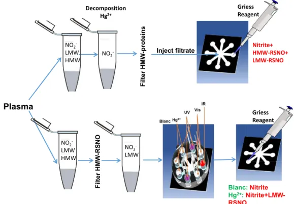
3) Detection of total RSNOs in plasma. ... 166
C. Conclusion of part II ... 167
III. Conclusion of chapter III ... 168
Chapter IV: Miniaturization ... 169
I. Colorimetric analysis of S-nitrosothiols decomposition on paper-based microfluidic devices ... 169
A. Experimental ... 170
1) Chemicals and materials ... 170
2) S-nitrosothiols synthesis ... 170
3) Fabrication of µPADs... 171
4) Fabrication of a 3D printed holder ... 171
Table of Contents
V
6) Plasma RSNOs detection ... 173
B. Results and discussion ... 174
C. Conclusions ... 179
II. Electrochemical detection of RSNOs in electrophoretic micro device: preliminary studies ... 180
A. Experimental ... 180
1) Microchip configuration ... 180
i. PMMA microchip fabrication ... 180
ii. Commercial microchips ... 181
2) Operating conditions for the microchip electrophoresis of GSNO ... 183
i. PMMA and COC microchip ... 183
ii. Commercial glass microchip ... 183
3) Chemicals and GSNO synthesis ... 184
B. Results and discussions ... 184
1) Preliminary study ... 184
i. Employement of wireless potentiostat ... 185
ii. Optimization of the injection mode ... 186
iii. microchip with integrated electrodes ... 187
2) Application to the separation and quantitation of RSNOs ... 189
C. Conclusion ... 192 General conclusion ... 193 Annex ... 198 I. Annex 1 ... 198 II. Annex 2 ... 198 III. Annex 3 ... 200 IV. Annex 4 ... 202 References ... 203
VI
List of figures
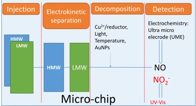
FIGURE 1 : SCHEMA DU MICROSYSTEME ENVISAGE POUR L’ANALYSE DES RSNOS DANS LES FLUIDES BIOLOGIQUES COMPRENANT QUATRE ETAPES : INJECTION, SEPARATION, DECOMPOSITION ET DETECTION. ... 2 FIGURE 2 : MECANISME DE FORMATION DE GSNO SELON LA CONCENTRATION EN GLUTATHION (GSH) ET EN NO. GSSG :
GLUTATHION OXYDEE. ADAPTE DE [14] ... 5 FIGURE 3 : METHODES DIRECTES ET INDIRECTES POUR LA DETECTION DES RSNOS. ... 6 FIGURE 4 : COMPARAISON DES INJECTIONS DITES “FLOATING” (A), “PINCHED” (B), ET “GATED” (C). EN (C) L’IMAGE EN CLAIR
REPRESENTE LE POSITIONNEMENT DES VALVES (A), LES IMAGES EN FLUORESCENCE DE L’INJECTION « GATED » DANS LES ETAPES SUCCESSIVES DE CHARGEMENT (B), D’INJECTION (C), ET DE SEPARATION (D). LA COULEUR BLANCHE MATERIALISE L’ECHANTILLON FLUORESCENT CONTRASTANT AVEC L’ELECTROLYTE SUPPORT INCOLORE. ADAPTE DE [35,36] ... 10 FIGURE 5 : POSITIONNEMENT DES ELECTRODES EN DETECTION AMPEROMETRIQUE. ADAPTE DE [37] ... 12 FIGURE 6 : LES ELECTROPHEROGRAMMES UV ET SM CORRESPONDANT A L’ANALYSE DE GSNO A 4,61 MM DANS L’ELECTROLYTE
SUPPORT APRES STOCKAGE A L’ETAT SOLIDE PENDANT 6 MOIS. ELECTROLYTE SUPPORT : TAMPON CARBONATE D’AMMONIUM (FORCE IONIQUE= 20 MM, PH 8,5). INJECTION : MARQUEUR NEUTRE (30 MBAR, 2S), ELECTROLYTE SUPPORT (50 MBAR, 3S), ECHANTILLON (50 MBAR, 3S), ELECTROLYTE SUPPORT (50 MBAR, 2S). CAPILLAIRE : DIAMETRE INTERNE 75 µM, LONGUEUR TOTALE 80 CM, LONGUEUR JUSQU'A LA FENETRE DE DETECTION UV 22 CM (FIG 6A) ET AU SPECTROMETRE DE MASSE (SM) 80 CM (FIG 6B). TENSION DE SEPARATION 20 KV. SM EN MODE D’IONISATION POSITIVE. ATTRIBUTION DES PICS : A : GSNO, B : GSSG, C : GSO2H, ET D : GSO3H. MARQUEUR NEUTRE (DMF 0.02 %, GLUCOSE 5MM). ... 14
FIGURE 7 : I) STRUCTURE DES PRODUITS IDENTIFIES A, B, C, D. II) VOIE DE DECOMPOSITION DE GSNO. LES NUMEROS ROUGES AU DESSUS DE CHAQUE ATOME DE SOUFRE REPRESENTENT LEUR ETAT D’OXYDATION. ... 15 FIGURE 8 : ELECTROPHEROGRAMME CORRESPONDANT A L’ANALYSE DE SOLUTIONS ETALON DE GSH ET GSSG ET D’UN ECHANTILLON
DE GSNO AGE DE 4 MOIS. TOUS LES ECHANTILLONS ONT ETE PREPARES DANS CHES (C =20 MM, PH 9. INJECTION
HYDRODYNAMIQUE : 3S, 11KPA ; ELECTROLYTE SUPPORT : 20 MM CHES (PH 10) ; TENSION : 27KV, CAPILLAIRE : 47 CM (37 CM EFFECTIF), DIAMETRE INTERNE 75 µM. DETECTION : 600 KHZ, 1,9 VPP. (1) GSSG A 157 µM, (2) GSH A 356 µM, (3)
GSNO ECHANTILLON A 1 MM (PURETE 66 % DETERMINEE PAR DETECTION COLORIMETRIQUE A 336 NM EN UTILISANT Ε=920 M-1. CM-1). ... 16
FIGURE 9 : ETUDE DE LA CINETIQUE DE DECOMPOSITION DE GSNO INDUITE PAR LUMIERE. A) ELECTROPHOREGRAMME CORRESPONDANT A L’ANALYSE DE 1) GSSG A 102 µM, 2) GSH A 77 µM ET (3-9) GSNO A 1 MM (PURETE 66 %) DETERMINE PAR DETECTION COLORIMETRIQUE A 336 NM EN UTILISANT Ε = 920 M-1. CM-1) APRES UN TEMPS DE
DECOMPOSITION DE 3) 0 MIN, 4) 1 MIN, 5) 2 MIN, 6) 4 MIN, 7) 10 MIN, 8) 15 MIN, ET 9) 75 MIN. B) REPRESENTATION DE L’AIRE DES PICS DE GSNO ET GSSG EN FONCTION DU TEMPS D’EXPOSITION. TOUS LES ECHANTILLONS ONT ETE PREPARES DANS CHES (C=20 MM, PH 9). C) REPRESENTATION DU LOGARITHME NEPERIENNE DE L’AIRE DES PICS DE GSNO EN FONCTION DU TEMPS D’EXPOSITION. INJECTION HYDRODYNAMIQUE : 3S, 11KPA ; ELECTROLYTE SUPPORT : CHES (C=20MM, PH 10) ; TENSION DE SEPARATION : 27KV, CAPILLAIRE : 47 CM (37 CM EFFECTIF), DIAMETRE INTERNE 75 µM. DETECTION : 600 KHZ, 1,9 VPP. ... 17
FIGURE 10 : ETUDE DE LA DECOMPOSITION THERMIQUE D’ECHANTILLON DE GSNO (PURETE 66 %). REPRESENTATION DE L’AIRE DES PICS ELECTROPHORETIQUE DE GSNO ET GSSG EN FONCTION DU TEMPS DE CHAUFFAGE. CONDITIONS OPERATOIRES :
VOIR.FIGURE 9 ... 17 FIGURE 11 : ETUDE DE LA TRANSNITROSATION ENTRE LE GSNO ET LA CYSTEINE. ELECTROPHEROGRAMMES CORRESPONDANT A
L’ANALYSE DES SOLUTIONS CONTIENANT 1) GSNO A 331 µM + GSH A 90 µM, 2) CYSTEINE A 495 µM, (3-7) GSNO A 331 µM + CYSTEINE A DES CONCENTRATIONS VARIABLES (76 µM, 152 µM, 305 µM, 381 µM ET 495 µM, DE 3 A 7
RESPECTIVEMENT). CONDITIONS OPERATOIRES : VOIR.FIGURE 9 ... 18 FIGURE 12 : REPRESENTATION DE L’EFFET DE L’AUGMENTATION DE LA CONCENTRATION DE GSH SUR LA DECOMPOSITION NORMALISEE
(CHAQUE COURBE A ETE NORMALISEE PAR RAPPORT A SON MAX D’ABSORBANCE) DE GSNO (39 µM) PAR CU+
(CONCENTRATION VARIABLE DE CUSO4 ENTRE 0 ET 1200 µM) DANS PBS 0.1 M (PH 7,4) + EDTA (450 µM). (N=3) ... 20
FIGURE 13 : REPRESENTATION GRAPHIQUE DE L’EFFET DE LA CONCENTRATION EN GSH SUR LA DECOMPOSITION NORMALISEE (CHAQUE COURBE A ETE NORMALISEE PAR RAPPORT A SON MAXIMUM D’ABSORBANCE) DE GSNO (20, 38, ET 82 µM) PAR CUSO4 A
600 µM OU 1000 µM DANS 0.1 M DE PBS (PH 7,4) CONTENANT DE L’EDTA A 450 µM... 21 FIGURE 14 : A) AMPEROGRAMMES MESURES PAR UNE ULTRA MICROELECTRODE SELECTIVE DE NO A 0,8 V VS AG / AGCL . CUSO4
(1000 µM) EST AJOUTE A DES CONCENTRATIONS DIFFERENTES DE GSNO (DE 4 µM A 290 µM) + PBS (C=0,1M ; PH 7,4) + EDTA (450 µM) + GSH (20 µM). ; B) COURBE DE CALIBRAGE OBTENUE A PARTIR DES DONNEES DE (A)... 23
VII
FIGURE 15 : COURANT OBSERVE APRES L’ADDITION DE GSNO JUSQU’A CONDITION FINALE DE 30 µM SUR DES AUNPS (DISPERSION DE
9 µM)). UNE DEUXIEME ADDITION DE GSNO NE DONNE AUCUN COURANT ADDITIONNEL. ... 24
FIGURE 16 : ILLUSTRATION DES DIFFERENTES ETAPES DE DETECTION DES RSNOS SUR DISPOSITIFS MICROFLUIDIQUES D’ANALYSE A BASE DE PAPIER : INJECTION DES RSNOS AU MILIEU, DECOMPOSITION (LUMIERE ET SEL DE MERCURE), ADDITION DU REACTIF DE GRIESS, SCAN ET ANALYSE PAR LE LOGICIEL COREL PHOTO-PAINT. DES COURBES DE CALIBRAGE PEUVENT ALORS ETRE ETABLIES POUR DETERMINER LES CONCENTRATIONS DANS DES ECHANTILLONS INCONNUS. ... 26
FIGURE 17 : COURBES DOSE-REPONSE CARACTERISTIQUES DE LA DECOMPOSITION DE A) GSNO, B) CYSNO ET C) ALBSNO MESUREE EN DISPOSITIFS MICROFLUIDIQUES D’ANALYSE A BASE DE PAPIER APRES DECOMPOSITION PAR HG2+, LUMIERE UV ET VISIBLE. . 27
FIGURE 18 : SCHEMA DE LA METHODE MISE EN OEUVRE POUR DETECTER LES RSNOS DANS LE PLASMA. ... 28
FIGURE 19 : VISUALISATION AU DESSUS (A) OU DE COTE (B) DE LA CONFIGURATION DU MICROSYSTEME ... 30
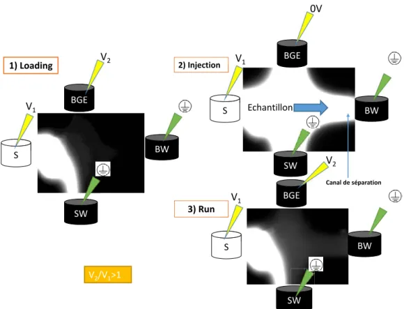
FIGURE 20: PROFILS ELECTROPHORETIQUES CORRESPONDANT A L’INJECTION SEQUENTIELLE (10S A 1KV) DU PARACETAMOL (5,2 MM) DANS UN TAMPON PBS (C=20MM, PH 7,4) DANS LE CANAL D’INJECTION, PUIS SEPARATION SOUS 1 KV DANS LE CANAL DE SEPARATION DE MICROSYSTEME EN PMMA (3 INJECTIONS) ET UNE DETECTION AMPEROMETRIQUE E= 0.8 V VS AG / AGCL. 30 FIGURE 21 : ILLUSTRATION DU MODE D’INJECTION “GATED » EN MICROSCOPIE DE FLUORESCENCE. CONDITIONS OPERATOIRES : PUCE EN COC (SECTION DU CANAL 50 µM X50 µM, LONGUEUR : 87 MM) ; ECHANTILLON COUMARINE 334 (C= 5MG / L DANS BGE / ETOH 99 / 01 (V/V))) IMAGE EN BLANC ; ELECTROLYTE SUPPORT PBS (C 20 MM, PH= 7,4) IMAGE EN NOIR. A) ETAPE DE CHARGEMENT NECESSAIRE POUR QUE LE VOLUME D’ECHANTILLON CIRCULANT ENTRE LE RESERVOIR D’ECHANTILLON (S) ET LE RESERVOIR D’ELECTROLYTE SUPPORT (SW) SOIT DE NATURE HOMOGENE. B) ETAPE D’INJECTION OU L’ECHANTILLON REMPLIT LE VOLUME DE LA CROIX D’INJECTION. C) DEBUT DE L’ETAPE DE SEPARATION OU LE VOLUME D’ECHANTILLON REMPLISSANT LA CROIX D’INJECTION EST ENTRAINE DANS LE CANAL DE SEPARATION. S : RESERVOIR D’ECHANTILLON, SW : RESERVOIR POUBELLE DE L’ECHANTILLON, BGE : RESERVOIR DE L’ELECTROLYTE, DW : RESERVOIR POUBELLE DE L’ELECTROLYTE. ... 32
FIGURE 22 : SCHEMA ET DIMENSIONS DES PUCES MICRUX® COMMERCIALES INTEGRANT UN JEU DE TROIS ELECTRODES DE PT DANS LE PUITS DE SORTIE DU CANAL DE SEPARATION. WE : ELECTRODE DE TRAVAIL, AE : CONTRE ELECTRODE, RE : ELECTRODE DE REFFERENCE. ... 32
FIGURE 23 : ELECTROPHEROGRAMME DE L’INJECTION DE GSNO (3 MM) EN « GATED INJECTION » (-1000, -800 V) POUR 1S. A) LE HG2+ EST DEJA PRESENT DANS LE PUIT DE SORTIE. B) EN T 1 : ARRET DU CHAMP ELECTRIQUE, INJECTION DE HG2+ ET STABILISATION DE L’ELECTRODE FACE A LA NOUVELLE SOLUTION, 16.1 S APRES LE DEBUT DE LA SEPARATION DU GSNO, T2 APPLICATION A NOUVEAU DU CHAMP ELECTRIQUE. ON VOIT LA MEME ALLURE AVEC NITRITE. ... 34
FIGURE 24 : SCHEMA DU MICROSYSTEME PROPOSE OU UN RESERVOIR DU HG2+ EST AJOUTE. SEPARATION VA PROCEDER ENTRE LES RESERVOIRS S, SW, BGE, AND BW. APRES, LE POTENTIEL VA ETRE APPLIQUER ENTRE S, SW, BGE, AND HG2+ ALORS LES RSNOS SE DECOMPOSE EN NITRITE ET ON DETECTE LES NITRITES QUI VIENS DES RSNOS DIFFERENT. ... 35
FIGURE 25: SCHEMATIC REPRESENTATION OF THE OBJECTIVES OF THE PHD PROJECT ... 39
FIGURE 26: ASCANIO SOBRERO (THE DISCOVERER OF NITROGLYCERINE) AND THE THREE NOBLE PRIZE LAUREATE IN 1998 ... 43
FIGURE 27 SCHEMATIC PRESENTATION OF THE SEQUENCE LEADING TO VASORELAXATION WHERE A STIMULATION OF NERVE LEAD TO THE PRODUCTION OF ACETYLCHOLINE THAT BIND TO ITS RECEPTORS ON ENDOTHELIAL CELLS AND LEADS TO THE PRODUCTION OF NO BY NITRIC OXIDE SYNTHASE (NOS) ENZYME. THIS NO DIFFUSES THROUGH THE MEMBRANE OF MUSCULAR CELLS AND BINDS TO GUANYLYL CYCLASE THUS PRODUCING CGMP STARTING FROM GTP. THIS CGMP PROVOKES VASORELAXATION. ... 43
FIGURE 28: STRUCTURAL FORMULA OF NO MOLECULE ... 44
FIGURE 29: REACTION MECHANISM OF TRANSFORMATION L-ARGININE TO L-CITRULLINE (UPSIDE). ELECTRONS (E−) ARE DONATED BY NADPH TO THE REDUCTASE DOMAIN OF THE ENZYME AND PROCEED VIA FAD AND FMN REDOX CARRIERS TO THE OXYGENASE DOMAIN. THERE THEY INTERACT WITH THE HEME IRON AND BH4 AT THE ACTIVE SITE TO CATALYZE THE REACTION OF O2 WITH L-ARGININE, GENERATING CITRULLINE AND NO AS PRODUCTS. ELECTRON FLOW THROUGH THE REDUCTASE DOMAIN REQUIRES THE PRESENCE OF BOUND CA2+/CAM (DOWNSIDE). FAD: FLAVIN ADENINE DINUCLEOTIDE, FMN: FLAVIN MONONUCLEOTIDE, BH 4: TETRAHYDROBIOPTERIN, CAM: CALMODULINE, ADAPTED FROM [109,110]. ... 46
FIGURE 30: EFFECTS OF NO IN CARDIOVASCULAR SYSTEM. ADAPTED FROM [119] ... 49
FIGURE 31: NO BIOLOGICAL ACTIONS CORRELATED WITH ITS CONCENTRATION AND MOLECULAR MECHANISMS. ADAPTED FROM [8] 53 FIGURE 32: CLASSICAL SINGLE SITE MODEL OF SGC ACTIVATION BY NO•. THIS VIEW IS CURRENTLY BEING SUBSTITUTED BY MORE COMPLEX MODELS TO ACCOUNT FOR THE PROPERTIES OF THE PENTACOORDINATED-NO COMPLEX, WHICH IS STRONGLY AffECTED BY THE AVAILABILITY OF ATP, THE GTP SUBSTRATE, OR EXCESS NO• [11]. ... 53
FIGURE 33: VARIATION OF NITROSYLATION BETWEEN SNO:FENO BASED ON THE OXYGENATION LEVEL OF HB [135] ... 54
FIGURE 34: AE 1 TRANSPORT SYSTEM. LEFT PART IS INSIDE RBC AND RIGHT SIDE IN PLASMA. ADAPTED FROM [136] ... 55
FIGURE 35: GENERIC STRUCTURES OF DINITROSYL IRON COMPLEXES (DNIC). DNIC ARE FORMED PRIMARILY FROM THE CHELATABLE IRON POOL (CIP) [11]. ... 55
FIGURE 36: POSSIBLE REACTIONS OF NO+, NO AND NO-. ADAPTED FROM [144] ... 57
FIGURE 37: STRUCTURE OF ORGANIC NITRATES. ... 59
FIGURE 38: GENERAL CONDENSED STRUCTURAL FORMULA OF NONOATE ... 60
List of Figures
VIII
FIGURE 40: STRUCTURE OF FUROXANS ... 61
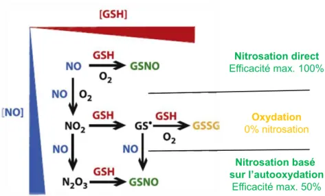
FIGURE 41: ILLUSTRATION OF THE WAY IN WHICH GSH NITROSATION AND OXIDATION ARE EXPECTED TO DEPEND ON THE CONCENTRATIONS OF NO AND GSH. ADAPTED FROM [14] ... 65
FIGURE 42: CYTOCHROME C GSNO SYNTHASE ACTIVITY. RED ARROWS REPRESENT THE FAVORED PATHWAY; GREEN ARROWS REPRESENT HIGH NO CONCENTRATION. SCHEME ADAPTED FROM [187] ... 67
FIGURE 43: THE LEFT GRAPH REPRESENTS TRANSPORT AND METABOLISM OF RSNOS FROM EXTRACELLULAR MEDIUM TO INTRACELLULAR MEDIUM WITH EXISTING TRANSNITROSATION REACTIONS THAT OCCURS OUTSIDE AND INSIDE THE CELL. ONLY CYSNO CAN TRANSVERSE THE MEMBRANE. INSIDE THE CELL GSNO IS METABOLIZED BY MANY ENZYMES (FORMALDEHYDE DEHYDROGENASE IS PRESENTED HERE BUT IT COULD BE OTHERS). THE RIGHT GRAPH REPRESENTS HOW NO TRANSVERSE THE MEMBRANE AND ITS POSSIBLE REACTIONS THAT COULD INCREASE INTRACELLULAR RSNOS CONCENTRATION. SNAP: S-NITROSO-NACETYLPENICILLAMINE. IT IS POSSIBLE SUCH S-NITROSATION OCCURS AT BURIED SITES (BLUE) AS WELL AS EXPOSED SITES (YELLOW) AND THAT THE BURIED RSNOS ARE POORLY REPARABLE BY THE GSH / FDH SYSTEM. ADAPTED FROM [192] 69 FIGURE 44: DECOMPOSITION MECHANISMS OF RSNOS INVOLVING MERCURIC IONS. ADAPTED FROM [27] ... 70
FIGURE 45: (A) STRUCTURE OF SNAP. (B) ABSORBANCE TIME PLOTS FOR THE DECOMPOSITION OF SNAP (5X10⁻⁴ MOL.DM-3), (A) NO ADDED CU2+, (B) [CU2+] 1X10⁻⁵, (C) [CU2+] 5X10⁻⁵, (D) [CU2+] 1X10⁻⁴ AND (E) [CU2+] 5X10⁻⁴ MOL.DM-3. ADAPTED FROM [212] ... 71
FIGURE 46: COPPER DITHIOLATE COMPLEX. ADAPTED FROM [24] ... 72
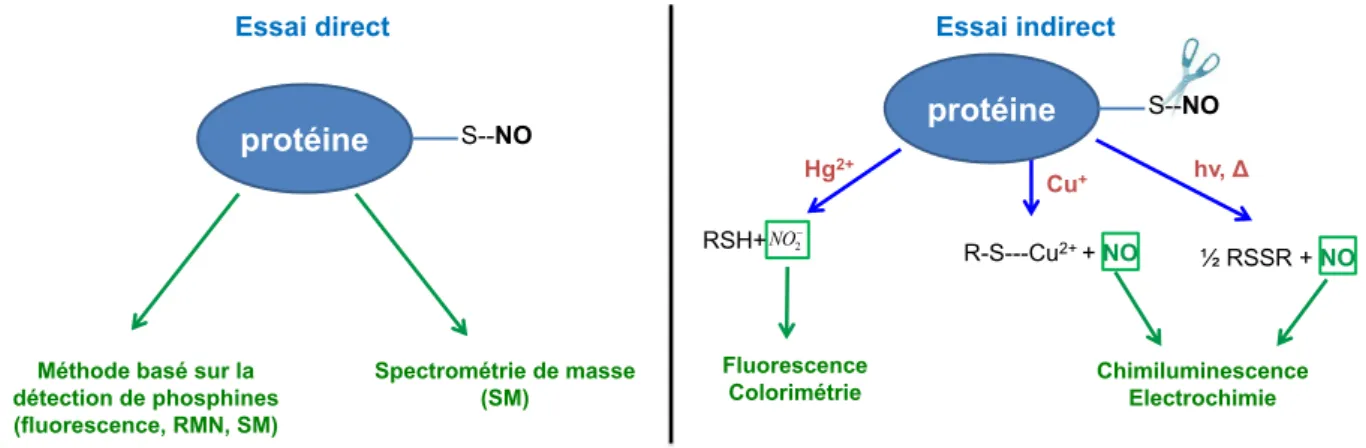
FIGURE 47: STRATEGIES FOR DIRECT AND INDIRECT DETECTION METHODS ... 78
FIGURE 48: STAUDINGER LIGATION PRINCIPLE. ADAPTED FROM [5] ... 79
FIGURE 49: PRICIPLE OF SAVILLE REACTION: DECOMPOSITION OF RSNOS BY HG2+ LEADS TO NO+. THE REACTION BETWEEN NO+ AND SULFANILAMIDE LEADS TO THE FORMATION OF A DIAZONIUM THAT REACTS WITH N(1-NAPHTHYL)ETHYLENEDIAMINE FINALLY FORMING A COLORED AZO-DYE STRONGLY ABSORBING AT 540 NM. AFTER SERIES OF REACTIONS WITH GRIESS REAGENT AT PH 7.4 A COLORED AZO-BYE THAT ABSORBS STRONGLY AT 540 NM. ADAPTED FROM [50] ... 80
FIGURE 50: PRINCIPLE OF FLUORIMETRIC DETECTION OF RSNOS. DAN=DIAMINONAPHTHALÈNE, NAT=NAPHTOTRIAZOLE. ADAPTED FROM [50]. ... 81
FIGURE 51: PRINCIPLE AND SET-UP FOR NO DETECTION USING CHEMILUMINESCENCE. ADAPTED FROM [77] ... 84
FIGURE 52: BIOTIN SWITCH ASSAY. ADAPTED FROM [268] ... 85
FIGURE 53: NORMALIZED STEADY-STATE VOLTAMMOGRAMS (20 MV S-1) OBTAINED FOR THE ELECTROCHEMICAL OXIDATIONS OF H2O2, ONOO-, NOC AND NO2- SOLUTIONS (EACH AT 1 MM IN PBS) AT PLATINIZED CARBON-FIBER MICROELECTRODES. DOTTED LINES DEFINE THE POTENTIALS OFFERING THE BEST SENSITIVITY AND SELECTIVITY OF DETECTION FOR EACH. ADAPTED FROM [275] ... 88
FIGURE 54: GENERAL TYPES OF ELECTROCHEMICAL NO SENSORS. ADAPTED FROM [283]. ... 90
FIGURE 55: PRINCIPLE OF GC-MS ANALYSIS OF RSNOS. ADAPTED FROM [299] ... 93
FIGURE 56: A TYPICAL MICROSCALE ELECTROPHORESIS RUN BEGINS BY ELECTROKINETICALLY INTRODUCING A SAMPLE INTO THE DEVICE, AFTER WHICH THE VOLTAGE IS SWITCHED SO THAT A NARROW BAND IS INJECTED INTO THE SEPARATION CHANNEL. DIFFERENT SPECIES MIGRATE WITH DIFFERENT MOBILITIES AND SEPARATE INTO DISTINCT ZONES THAT ARE DETECTED DOWNSTREAM. ADAPTED FROM [322] ... 99
FIGURE 57: SEM OF A POLYIMIDE MICROCHANNEL WITH TRAPEZOIDAL CROSS SECTION PACKED WITH 5 ΜM C18 PARTICLES (UPPER PANEL); SEVERAL SMALLER CHANNELS CONSTITUTE A FRIT-LIKE STRUCTURE TO CONTAIN THE PACKED PARTICLES (LOWER PANEL). ADAPTED FROM [343] ... 107
FIGURE 58: A) SCHEMATIC REPRESENTATION OF MICROCHIP ELECTROPHORESIS, B) MIGRATION ORDER OF IONS IONIC ANALYTES BASED ON THEIR CHARGE AND MASS. EOF: ELECTROSOSMOTIC FLOW. ADAPTED FROM [353] ... 108
FIGURE 59: SIMULATION OF STEPS OF FLOATING INJECTION ON A CROSS INJECTION SYSTEM. A) INJECTION OF SAMPLE (IN BLACK), B) AND C) RUNNING OF BUFFER. ADAPTED FROM [357] ... 109
FIGURE 60: SIMULATION OF LOADING STEP WITH DIFFERENT FOCUSING RATIOS IN CROSS FORM INJECTION SYSTEM. ADAPTED FROM [357] ... 110
FIGURE 61: SIMULATION OF STEPS OF PULLBACK FLOATING INJECTION ON A CROSS INJECTION SYSTEM. A) SAMPLE (IN BLACK) INJECTION, B) AND C) RUNNING OF BUFFER. ADAPTED FROM [357] ... 110
FIGURE 62: DIFFERENT INJECTION FORMS. S: SAMPLE RESERVOIR, B: BUFFER RESERVOIR, BW: BUFFER WASTE, SW: SAMPLE WASTE. ... 111
FIGURE 63: FLOATING INJECTION. SAMPLE INTRODUCTION LOADING STEP (A) AND DISPENSING STEP WITH (B) OR WITHOUT (C) SAMPLE PULLBACK. ADAPTED FROM [354] ... 112
FIGURE 64: COMPARISON OF FLOATING (A) AND PINCHED INJECTIONS (B) AT 1) INJECTION AND 2)SEPARATION STEPS. THE WHITE COLORATION IS THE SAMPLE AND THE COLORLESS IS THE BACK GROUND ELECTROLYTE. NO SAMPLE DIFFUSION OCCURS IN PINCHED ONE. S: SAMPLE RESERVOIR, B: BUFFER RESERVOIR, SW: SAMPLE WASTE RESERVOIR, BW: BUFFER WASTE RESERVOIR. ADAPTED FROM [35] ... 112
IX
FIGURE 65: ILLUSTRATION OF GATED INJECTION. WHITE IMAGE OF MICROCHIP VALVES (A), AND FLUORESCENT IMAGE OF GATED INJECTION LOADING STEP (B), INJECTION STEP (C) AND RUNNING STEP (D). ADAPTED FROM [36]. ... 113 FIGURE 66: HYDRODYNAMIC VOLTAMMOGRAMS RECORDED FOR CATECHOL USING IN-CHANNEL (♦) AND END-CHANNEL (•) EC
DETECTION. CONDITIONS: 20 MM BORIC ACID BUFFER, PH 9.2, ESEP= 300 V / CM, POTENTIALS VS AG / AGCL REFERENCE.
ADAPTED FROM [373]. ... 118 FIGURE 67: ELECTROPHEROGRAMS FOR CATECHOL USING (A) IN-CHANNEL EC DETECTION AT +2.2 V AND (B) END-CHANNEL. EC
DETECTION AT +1.0 V. CONDITIONS SAME AS IN FIGURE 66. ... 118 FIGURE 68: MODES OF ALIGNMENT OF ELECTRODES IN AMPEROMETRIC DETECTION. ADAPTER FROM [37] ... 119 FIGURE 69: MICROCHIP ELECTROPHORESIS COUPLED TO CONTACTLESS CONDUCTIVITY DETECTION. THE ELECTRODES WERE DRAWN
WITH PENCIL. A, B, C AND D REPRESENT THE DRAWING AND THE ASSOCIATION OF THE PAPER ELECTRODES WITH THE PMMA MICROCHIP. THE LABELS 1, 2, 3 AND 4 (IN (D)) INDICATE THE BUFFER, SAMPLE, SAMPLE WASTE AND BUFFER WASTE
RESERVOIRS, RESPECTIVELY. ADAPTED FROM [375] ... 120 FIGURE 70: GSNO WITH 92 %CONVERSION OF 250 µM GSH TO GSNO (170 µM) AND TO TWO ADDITIONAL UNIDENTIFIED PEAKS
(PEAKS A AND B), ONE OF WHICH IS LIKELY GSSG. SAMPLE BUFFER 0.01 M SODIUM PHOSPHATE, 0.01 M HCL PH 2.3, POSITIVE POLARITY AT 11 KV; ABSORBANCE 200 NM. ADAPTED FROM [89,92,388]. ... 125 FIGURE 71: UV AND MASS SPECTRUM ELECTROPHEROGRAMS OF 4.61 MM GSNO STORED IN SOLID STATE FOR 6 MONTHS, THEN
DISSOLVED IN BGE JUST PRIOR TO ANALYSIS. BGE: AMMONIUM CARBONATE BUFFER (20 MM, PH 8.5). INJECTION: NEUTRAL MARKERS (30 MBAR, 2 S), BGE (50 MBAR, 3 S), SAMPLE (50 MBAR, 3 S), BGE (50 MBAR, 2 S). CAPILLARY: 75 µM ID, TOTAL LENGTH 80 CM, LENGTH TO UV DETECTOR 22 CM (FIG 1A) AND TO MS DETECTOR 80 CM (FIG 1B). APPLIED VOLTAGE: 20KV. MS IN POSITIVE MODE: SEE EXPERIMENTAL PART FOR DETAILS. PEAK ASSIGNMENT: A. GSNO, B. GSSG, C. GSO2H AND D.
GSO3H. NEUTRAL MARKERS (DMF 0.02%, GLUCOSE 5MM). ... 126
FIGURE 72: MASS SPECTRUM EXTRACTED FROM THE FOUR PEAKS IDENTIFIED ON FIGURE 71 BY TOTAL ION CURRENT (TIC). THE LETTERS A, B, C AND D REPRESENT THE SUCCESSIVELY OBTAINED PEAKS (SEE FIGURE 71) ... 129 FIGURE 73: I) STRUCTURES OF THE FOUR IDENTIFIED COMPOUNDS A, B, C, D. II) DECOMPOSITION PATHWAYS OF GSNO, ACCORDING
TO THE CE-MS CHARACTERIZATION OF THIS STUDY. RED NUMBERS ABOVE THE SULFUR ATOMS REPRESENT THE OXIDATION STATE OF SULFUR, S-NITROSOGLUTATHIONE (GSNO), OXIDIZED GLUTATHIONE (GSSG), GLUTATHIONE SULFINIC ACID (GSO2H)
AND GLUTATHIONE SULFONIC ACID (GSO3H). ... 131
FIGURE 74: STRUCTURAL FORMULAS OF REDUCED GLUTATHIONE (GSH), S-NITROSOGLUTATHIONE (GSNO), OXIDIZED GLUTATHIONE (GSSG), GLUTATHIONE SULFINIC ACID (GSO2H), GLUTATHIONE SULFONIC ACID (GSO3H). STRUCTURAL FORMULA OF REDUCED
GLUTATHIONE GSH SHOWS THE AMINO ACID COMPOSITION OF THIS TRIPEPTIDE WITH THE SYMBOLS OF N- AND C-TERMINALS AND CYSTEINE SH. (SEE TABLE 16 FOR PKA VALUES). ... 135 FIGURE 75: ELECTROPHEROGRAMS OF GSSG, GSH STANDARD SOLUTIONS AND A 4 MONTHS OLD GSNO SAMPLE SOLUTION. ALL
SAMPLES WERE PREPARED IN CHES (20 MM, ADJUSTED TO PH 9.0 WITH NAOH). HYDRODYNAMIC INJECTION: 3 S, 11 KPA; BGE: 20 MM CHES (ADJUSTED TO PH 10.0 WITH NAOH); SEPARATION VOLTAGE: +27 KV, CAPILLARY: 47 CM (37 CM EFFECTIVE), ID 75 ΜM. DETECTION: 600 KHZ, 1.9 VPP. (1) GSSG 157 ΜM, (2) GSH 356 ΜM, (3) GSNO SAMPLE 1 MM
(PURITY 66 % DETERMINED BY COLORIMETRIC DETECTION AT 336 NM USING Ɛ=920 M-1 CM-1). AVERAGE CAPILLARY ELECTRIC
CURRENT WAS 36 µA. A. GSNO, B. GSSG, C. GSO2H AND D. GSO3H. ... 137
FIGURE 76; CALIBRATION CURVES OF A) GSNO, B) GSH AND C) GSSG. CONDITIONS SAME AS FIGURE 75 ... 138 FIGURE 77: ELECTROPHEROGRAMS OF GSNO (PURITY 66 %), DIFFERENT STANDARD SOLUTIONS OF NITRITE, NITRATE AND CHLORIDE
AND THEIR MIXTURES. SAMPLES WERE PREPARED IN CHES (20 MM, ADJUSTED TO PH 9.0 WITH NAOH) + DDAB 116 ΜM. BGE: CHES (20 MM, ADJUSTED TO PH 10.0 WITH NAOH) + DDAB 116 ΜM. HYDRODYNAMIC INJECTION: 3 S, 11 KPA; SEPARATION VOLTAGE: -27 KV; CAPILLARY: 50 CM (40 CM EFFECTIVE), ID 75 ΜM. DETECTION: 600 KHZ, 1.9 VPP . (1)
BLANK, (2) NITRITE 125 ΜM, (3) NITRATE 93 ΜM, (4) GSNO SAMPLE 0.9 MM, (5) GSNO SAMPLE 0.9 MM + CHLORIDE 120 ΜM, (6) GSNO SAMPLE 0.9 MM + NITRITE 125 ΜM, (7) GSNO SAMPLE 0.9 MM + NITRATE 93 ΜM. THE EOF TIME WAS 2.7 MIN. AVERAGE CAPILLARY ELECTRIC CURRENT WAS 33 µA. ... 139 FIGURE 78: STUDY OF DECOMPOSITION OF A GSNO SAMPLE INDUCED BY LIGHT. A) ELECTROPHEROGRAMS OF (1) 102 ΜM GSSG,
(2) 77 ΜM GSH, (3-9) 1 MM GSNO (PURITY 66 % DETERMINED BY COLORIMETRIC DETECTION AT 336 NM USING Ɛ=920 M-1 CM-1) AFTER VISIBLE LIGHT DECOMPOSITION FOR (3) 0 MIN, (4) 1 MIN, (5) 2 MIN, (6) 4 MIN, (7) 10 MIN, (8) 15 MIN
AND (9) 75 MIN. B) GSNO AND GSSG PEAK AREAS AS FUNCTION OF TIME OF EXPOSITION. ALL SAMPLES WERE PREPARED IN CHES (20 MM, ADJUSTED TO PH 9.0 WITH NAOH). C) NATURAL LOGARITHM OF PEAK AREAS OF GSNO AS FUNCTION OF TIME OF EXPOSITION. HYDRODYNAMIC INJECTION: 3S, 11 KPA; BGE: 20 MM CHES (ADJUSTED TO PH 10.0 WITH NAOH); SEPARATION VOLTAGE: +27 KV, CAPILLARY: 47 CM (37 CM EFFECTIVE), ID 75 ΜM. DETECTION: 600 KHZ, 1.9 VPP. AVERAGE
CAPILLARY ELECTRIC CURRENT WAS 37 µA. ... 140 FIGURE 79: STUDY OF HEAT DECOMPOSITION OF A GSNO SAMPLE (PURITY 66 %). GSNO AND GSSG PEAK AREAS AS FUNCTION OF
TIME OF HEATING. ALL SAMPLES WERE PREPARED IN CHES (20 MM, PH 9). OTHER CONDITIONS AS IN FIGURE 2 ... 141 FIGURE 80: STUDY OF TRANSNISTROSATION BETWEEN GSNO AND CYSTEINE. ELECTROPHEROGRAMS OF SOLUTIONS CONTAINING: (1)
List of Figures
X
CONCENTRATIONS (3) 76 ΜM, (4) 152 ΜM, (5) 305 ΜM, (6) 381 ΜM AND (7) 495 ΜM. ALL SAMPLES WERE PREPARED IN CHES (20 MM, ADJUSTED TO PH 9.0 WITH NAOH). OTHER CONDITIONS AS IN FIGURE 75. THE EOF TIME WAS 1.2 MIN. A. GSNO, B. GSSG, C. GSO2H AND D. GSO3H. ... 143
FIGURE 81: AMPEROGRAM OF DEPOSITION OF EUGENOL LAYER ON 25 µM PT-UME AT E = 150 MV VS AG / AGCL FOR 15 MIN IN A SOLUTION OF EUGENOL (10 MM) + SODIUM HYDROXIDE (0.1 M). ... 148 FIGURE 82: ADDITION OF NITRITE (100 µM) IN 0.1 M OF PBS (PH 7.4) ON A 25 µM PT-UME COATED WITH
POLYEUGENOL-POLYPHENOL A) DIRECTLY AFTER PREPARATION AND B) AT THE END OF WORKING DAY WHEN IT STARTS LOSING ITS SELECTIVITY. ... 149 FIGURE 83: CALIBRATION OF SAVILLE (A) AND GRIESS (B) METHODS. (A) ABSORBANCE MEASUREMENT AT 540 NM OF 500µL OF PBS
(0.1 M, PH 7.4) CONTAINING DIFFERENT GSNO CONCENTRATIONS + HGCL2 (537 µM) + EDTA (450 µM) + 500 µL GRIESS
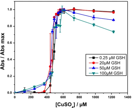
REAGENT. (N=3); (B) ABSORBANCE MEASUREMENT AT 540 NM OF 500 µL OF PBS (0.1 M, PH 7.4) CONTAINING DIFFERENT NITRITE CONCENTRATIONS + EDTA (450 µM) + 500 µL GRIESS REAGENT. THE SLOPE OF GRIESS METHOD IS RELATED TO A A = F([NITRITE]) GRAPH AND THE SLOPES OF SAVILLE METHODS ARE RELATED TO A = F([GSNO]0) GRAPHS... 151 FIGURE 84: NORMALIZED ABSORBANCES OF CU-CATALYZED DECOMPOSITION OF GSNO SOLUTION TREATED WITH GRIESS REAGENT
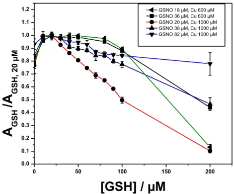
USING COLORIMETRIC DETECTION AT 540 NM (EACH CURVE WAS NORMALIZED WITH RESPECT TO ITS MAXIMUM ABSORBANCE). EFFECT OF THE ADDITION OF INCREASING CONCENTRATIONS OF CUSO4 TO A SOLUTION OF GSNO (39 µM) + EDTA (450 µM) IN PBS (0.1M; PH 7.4) CONTAINING DIFFERENT AMOUNTS OF GSH. (N=3) ... 152 FIGURE 85: EFFECT OF INCREASING GSH CONCENTRATION ON THE NORMALIZED (WITH RESPECT TO THE MAXIMUM OF EACH CURVE AT
EACH DIFFERENT GSNO CONCENTRATION) CU2+-CATALYZED DECOMPOSITION OF GSNO SOLUTION TREATED WITH GRIESS
REAGENT USING COLORIMETRIC DETECTION AT 540 NM. 20, 38, AND 82 µM OF GSNO IN PBS (0.1 M; PH 7.4) + EDTA (450 µM) + CUSO4 (1000 µM) OR 18 AND 36 µM OF GSNO IN PBS (0.1 M; PH 7.4) + EDTA (450 µM) + CUSO4 (600
µM) WERE USED (N=3). THE FIRST POINTS START AT GSH PRESENT AS IMPURITY IN GSNO SOLUTIONS (0.6%) ... 153 FIGURE 86: A) EVOLUTION OF THE AMPEROGRAMS MEASURED BY NO-SENSOR UPON ADDITION OF GSNO TO PBS (0.1M ; PH 7.4) +
EDTA (450 µM) + CUSO4 (1000 µM) + GSH (20 µM). MEASUREMENT MADE AT 0.8 V VS AG / AGCL; B) CALIBRATION
CURVE OBTAINED WITH DATA FROM FIGURE 86A. ... 155 FIGURE 87: (A,C) CALIBRATION CURVES OBTAINED FROM MEASURING THE MAXIMUM CURRENT INTENSITIES OBTAINED BY DIFFERENT
ADDITIONS OF DEA-NONOATE, AS A FUNCTION OF THE ESTIMATED CONCENTRATION OF NO RELEASED BY DEA-NONOATE AT THE MAXIMUM OF THE PEAK (PBS 0.1 M; PH = 7.4; E = 0.8 V VS. AG / AGCL) (B, D) CALIBRATION CURVE OBTAINED FROM AMPEROMETRIC MEASUREMENT OF NO RELEASED FROM COPPER-CATALYZED DECOMPOSITION OF GSNO IN PBS (0.1 M; PH 7.4) + EDTA (450 µM) + CUSO4 (3 MM) + GSH (20 µM). THE NO-SENSOR WAS A PT ULTRAMICROELECTRODE (25 µM DIAMETER) COATED BY (A, B) POLYEUGENOL-POLYPHENOL MEMBRANE OR BY (C, D) POLYEUGENOL AND POLARIZED AT 0.8 V VS AG / AGCL. ... 156 FIGURE 88: REPRESENTATIVE UV-VIS SPECTRUM OF A DISPERSION OF AUNPS OBTAINED BY THE TURKEVITCH METHOD. INSET (RIGHT): TEM OF THE AUNPS SHOWING AVERAGE DIAMETER. INSET (LEFT): SAMPLE OF GOLD NANOPARTICLES DISPERSION. ... 159 FIGURE 89: ELECTROCHEMICAL CURRENT CHANGE AFTER A GSNO INJECTION IN A WELL OF A 24-WELL CELL PLATE CONTAINING A
DISPERSION OF AUNPS. MAXIMUM IS REACHED IN C.A. 3 S. ... 161 FIGURE 90. CURRENT CHANGE OBSERVED AFTER A GSNO ADDITION TO A FINAL CONCENTRATION OF 30 µM ON 9 NM AUNPS
DISPERSION. A SECOND ADDITION OF THE SAME GSNO AMOUNT DID NOT LEAD TO ANY CHANGE IN THE CURRENT. ... 162 FIGURE 91. LINEAR DEPENDENCE OF THE ELECTROCHEMICAL CURRENT WITH THE AUNPS CONCENTRATION (0 TO 5.8 NM) MEASURED WITH A NO SELECTIVE AMPEROMETRIC SENSOR AFTER ADDITIONS OF GSNO SOLUTION TO A FINAL CONCENTRATION OF 30 µM. ... 162 FIGURE 92: CURRENT CHANGES OBSERVED AFTER GSNO ADDITIONS TO FINAL CONCENTRATIONS OF 1.0 µM EACH ON A 9 NM AUNPS
DISPERSION. ... 163 FIGURE 93. CALIBRATION CURVE SHOWING THE CURRENT MAXIMA OBTAINED IN THE ADDITION OF GSNO (IN WATER / EDTA 0.5MM / IAA 10MM) TO DIFFERENT FINAL CONCENTRATIONS ON A 10.9 NM AUNPS DISPERSION WHICH CORRESPONDS TO AN EXCESS OF AU SURFACE RELATIVE TO THE GSNO AMOUNT NECESSARY FOR THE COMPLETE COATING OF THE AU SURFACE. ... 164 FIGURE 94. CURRENT MAXIMA MEASURED AFTER THE ADDITION OF GSNO TO A FINAL CONCENTRATION OF 30 µM ON AUNPS
DISPERSIONS WITH DIFFERENT [ALBUMIN] / [AUNP] (BLACK DOTS) AND [GSH] / [AUNP] (ORANGE DOTS) RATIOS. ... 165 FIGURE 95. CURRENT MAXIMA AFTER ADDITION OF GSNO (FINAL CONCENTRATION OF 30 µM) IN 2 ML OF A 7.5 NM OF AUNPS
DISPERSIONS PREVIOUSLY REACTED WITH DIFFERENT VOLUMES OF RSNOS-FREE PLASMA. ... 166 FIGURE 96. CURRENT CHANGES AFTER INJECTION OF 40 µL OF SULFHYDRYL BLOCKED HUMAN PLASMA TO 2 ML OF A 7.5 NM OF
AUNPS DISPERSION. AFTER 15 MIN, AN ADDITION OF GSNO (FINAL CONCENTRATION OF 25 µM) LED TO A SECOND CHANGE IN THE MEASURED CURRENT. ... 167 FIGURE 97. CURRENT MAXIMA AFTER INJECTION OF DIFFERENT VOLUMES OF SULFHYDRYL BLOCKED HUMAN PLASMA TO 2 ML OF A
10.9 NM AUNPS DISPERSION. ... 168 FIGURE 98: PRESENTATION OF (A) PAPER MICROFLUIDIC DEVICE LAYOUT CONTAINING EIGHT ZONES (1-8), (B) COUPLING OF A 3D
XI
COLORIMETRIC ASSAYS THROUGH THE DECOMPOSITION WITH HG2+, UV, VIS AND IR LIGHT. IN (C), THE LABEL CZ MEANS
CONTROL ZONE. ... 172 FIGURE 99: IMAGE OF THE DEVICE CONNECTED TO LAPTOP BY A USB DRIVE WHICH ASSURES LIGHTENING. A CAMERA COULD BE
CONNECTED ALSO TO DIRECTLY ANALYZE THE RESULTS. ... 172 FIGURE 100: A REAL FIGURE OF PAPER MICROFLUIDIC DEVICE CONTAINING HUMAN SERUM SHOWING COLORED ZONES AFTER
COLORIMETRIC ASSAYS THROUGH THE DECOMPOSITION WITH HG2+, UV, VIS AND IR LIGHT. THE LABEL CZ MEANS CONTROL ZONE. ... 173 FIGURE 101: CALIBRATION CURVE FOR NITRITE DISSOLVED IN 0.1 M PBS (PH 7.4) CONTAINING 0.5 MM EDTA. ... 174 FIGURE 102: GRAPHICAL REPRESENTATION OF THE PERCENTAGE OF GSNO DECOMPOSITION BY UV LIGHT USING TEST TUBES.
PERCENTAGE CALCULATION WERE BASED ON GRIESS AND SAVILLE REACTIONS. Λ=540NM ... 175 FIGURE 103: CALIBRATION CURVES FOR GSNO (A), CYSNO (B), AND ALBSNO (C) DISSOLVED IN 0.1 M PBS (PH 7.4) CONTAINING
0.5 MM EDTA. COLORIMETRIC MEASUREMENTS WERE RECORDED AFTER DECOMPOSITION PROMOTED BY 10 MM HG2+
(BLACK SQUARES), UV (BLUE SQUARES), AND VISIBLE LIGHTS (RED SQUARES). FOR MERCURIC DECOMPOSITION, GRIESS REAGENT WAS MIXED WITH HG2+ AT THE DECOMPOSITION TIME. FOR LIGHT-MEDIATED DECOMPOSITION, GRIESS REAGENT WAS
ADDED AFTER THE DECOMPOSITION REACTION OCCURRED. ... 176 FIGURE 104: CALIBRATION CURVES FOR GSNO DISSOLVED IN 0.1 M PBS (PH 7,4) CONTAINING 0.5 MM EDTA DECOMPOSED BY IR LIGHT DURING 25 MIN AND ... 177 FIGURE 105: (A) SCHEMATIC REPRESENTATION OF MICROFABRICATED PMMA CHIP. TOP (B) AND FRONT (C) VIEW OF THE PMMA
MICROCHIP WITH WORKING AND REFERENCE ELECTRODES SITUATED IN OFF-CHIP END-CHANNEL CONFIGURATION. ... 181 FIGURE 106: SCHEMATIC REPRESENTATION OF A COMMERCIAL COC MICROCHIP FROM MICROFLUIDIC CHIPSHOP (JENA, GERMANY)
... 182 FIGURE 107: SCHEMATIC REPRESENTATION OF A COMMERCIAL GLASS / SU-8 FROM MICRUX TECHNOLOGIES (OVIEDO, SPAIN) .... 182 FIGURE 108: ELECTROPHEROGRAM CORRESPONDING TO THE SUCCESSIVE FLOATING INJECTIONS (1KV FOR 10 S) OF A PARACETAMOL
SOLUTION (5.23 MM) IN A PMMA MICROFLUIDIC SYSTEM. OPERATING CONDITIONS: SEE EXPERIMENTAL PART. ... 185 FIGURE 109: GATED INJECTION ON COC MICRO-CHIP. THE FLUORESCENT MOLECULE COUMARINE 334 (5 MG / L PREPARED IN BGE / ETOH 99 / 01 (V / V)) APPEARS IN WHITE. THE BGE (PBS (C= 20MM, PH 7.4)) APPEARS IN BLACK. S: SAMPLE RESERVOIR, SW: SAMPLE WASTE, BGE: BACK GROUND ELECTROLYTE, BW: BUFFER WASTE. ... 187 FIGURE 110: ELECTROPHEROGRAM CORRESPONDING TO THE ELECTROPHORETIC PROFIL OF 1 MM PARACETAMOL IN GLASS / SU-8
MICROCHIP. GATED INJECTION V1 = 1000 V, V2 = 1200 V, INJECTION TIME 0.5 S, SUCCESSIVES INJECTIONS: 120S, DETECTION
1V VS PT. BGE: 20 MM ARGININE SOLUTION ADJUSTED AT PH 5.6 WITH ACETIC ACID. ... 188 FIGURE 111: ELECTROPHEROGRAM CORRESPONDING TO THE ELECTROPHORETIC PROFILE OF A 50 µM NITRITE IN GLASS / SU-8
MICROCHIP. GATED INJECTION V1 = -800 V, V2 = -1000 V, INJECTION TIME 3 S, SUCCESSIVES INJECTIONS: 70S, DETECTION 1V
VS PT. BGE: 20 MM ARGININ SOLUTION ADJUSTED AT PH 5.6 WITH ACETIC ACID. ... 189 FIGURE 112: ELECCTROPHORETIC PROFILE OF 3 MM GSNO.INJECTED IN « GATED MODE» (-1000, -800 V) FOR 1S. A) HG2+ IS
ALREADY PUT IN THE RESERVOIR. B) T1: STOPPING THE ELECTRIC FIELD, INJECTION OF HG2+, AND STABILIZATION OF THE
ELECTRODE OF DETECTION 16.1 S AFTER THE BEGINNING OF THE SEPARATION OF GSNO. T2: APPLICATION OF A NEW ELECTRIC
FIELD. ... 191 FIGURE 113: SCHEMA OF THE PROPOSED MICROCHIP WHERE AN HG2+ RESERVOIR IS ADDED. SEPARATION OCCURS BETWEEN S, SW,
BGE, AND BW RESERVOIRS. THEN, POTENTIAL IS APPLIED BETWEEN S, SW, BGE, AND HG2+ SO THE RSNOS DECOMPOSE TO
NITRITE AND WE DETECTED THE DIFFERENT NITRITES COMING FROM DIFFERENT RSNOS. ... 192 FIGURE 114: SCHEMATICS OF A SIMPLE MASS SPECTROMETER WITH SECTOR TYPE MASS ANALYZER. ADAPTED FROM [463] ... 199 FIGURE 115: SHEATH FLOW CE-MS ANALYZER. ADAPTED FROM [464,465] ... 200 FIGURE 116: C4D DETECTION PRINCIPLE.A) THE TRANSMITTER ELECTRODE SEND A HIGH FREQUENCY AND LARGE AMPLITUDE AC THE
RECEIVER ELECTRODE RECEIVE THE ATTENUATED SIGNAL, B) THE AC RECEIVED SIGNAL IS AFFECTED BY THE CONDUCTIVITY OF THE SAMPLE, C) DC ANALOG VOLTAGE SIGNAL... 201 FIGURE 117: EQUIVALENT CIRCUIT OF C4D WITH STRAY CAPACITANCE. CW STANDS FOR THE TOTAL CAPACITANCE DUE TO THE SILICA
WALL. RC STANDS FOR THE SOLUTION RESISTANCE BETWEEN THE ELECTRODES. C0 STANDS FOR THE TOTAL STRAY CAPACITANCE BETWEEN BOTH ELECTRODES. ADAPTED FROM [409] ... 201 FIGURE 118: PHOTO OF THE HOME-MADE CE-C4D SHOWING A CAPILLARY CONNECTING TWO VIALS (INLET AND OUTLET). THE INLET,
WHERE THE ELECTROKINETIC PROCESS STARTS, IS CONNECTED TO A PUMP THAT MAKES HYDRODYNAMIC INJECTION. IN ADDITION TO THIS INLET AND OUTLET ARE CONNECTED TO A HIGH VOLTAGE POWER SUPPLY BY PLATINIZED ELECTRODES. THE C4D
DETECTOR ENCAPSULATE THE CAPILLARY JUST BEFORE THE OUTLET VIAL. THE ENTIRE CE-C4D IS PUT IN A HOME-MADE PMMA
XII
List of tables
TABLE 1: DIFFERENT PRODUCTS THAT CAN BE FORMED FROM NO WITH THEIR PRINCIPLE TARGETS AND PULMONARY BIOACTIVITY.
ADAPTED FROM [13,108] ... 45
TABLE 2: PROPERTIES OF NOS1, 2 AND 3. [7,105,107,112] ... 47
TABLE 3: CLASSIFICATION OF DIFFERENT RSNOS ... 63
TABLE 4: NITROGEN OXIDES IN RESPIRATORY BIOLOGY ADAPTED FROM [13] ... 75
TABLE 5: ELEVATED AND DEPRESSED LEVELS OF RSNOS IN HUMAN DISEASES. ADAPTED FROM [1] ... 77
TABLE 6: DIFFERENT METHODS OF DECOMPOSITION BY COPPER TO DETECT RSNOS. ADAPTED FROM [3] ... 91
TABLE 7: DIFFERENT HPLC AND GC CONDITIONS FOR DETECTION OF RSNOS ... 95
TABLE 8: RSNO QUANTIFICATION USING DIFFERENT TECHNIQUES. ADAPTED FROM [3,26,30] ... 96
TABLE 9: SUMMARY OF PHYSICAL PROPERTIES FOR COMMON MICROFLUIDIC THERMOPLASTICS. ADAPTED FROM [330] ... 102
TABLE 10: SUMMARY OF THE PHYSICAL PROPERTIES OF MATERIALS USED IN MICROCHIP ELECTROPHORESIS. LTCC: LOW-TEMPERATURE COFIRED CERAMIC, TPE: THERMOSET POLYESTER, PFPE: POLYFLUOROPOLYETHER, PEGDA: POLY(ETHYLENE GLYCOL)DIACRYLATE, PEO: POLY(ETHYLENE OXIDE), FEP: FLUORINATED ETHYLENE-PROPYLENE, PFA: PERFLUOROALKOXY POLYMER ADAPTED FROM [325] ... 103
TABLE 11: OVERVIEW OF THERMOPLASTIC BONDING TECHNIQUE FOR MICROFLUIDIC DEVICE. ADAPTED FROM[330] ... 104
TABLE 12: COMPARISON OF DIFFERENT MATERIALS USED FOR MICROFLUIDIC DEVICES. ADAPTED FROM [33] ... 105
TABLE 13: COMPARISON OF DIFFERENT MATERIAL COMPATIBILITY WITH CAPILLARY ELECTROPHORESIS AND ELECTROCHEMICAL TECHNIQUES. ADAPTED FROM [54,55,323] ... 106
TABLE 14: ADVANTAGES AND LIMITATIONS OF DIFFERENT TYPES OF INJECTIONS. ... 114
TABLE 15: MASS SPECTRUM ASSIGNMENT OF IONS WITH THEIR % PEAK ABUNDANCE IDENTIFIED BY TOTAL ION CURRENT (TIC). THE DATA IS RELATED TO FIGURE 72. X REPRESENTS A, B, C, OR D ACCORDING TO ITS POSITION IN TABLE ... 127
TABLE 16: PKA VALUES OF THE DIFFERENT CARBOXYLIC AND AMINE MOIETIES OF REDUCED (GSH) AND OXIDIZED (GSSG) GLUTATHIONE. ... 136
TABLE 17. SLOPES OF CALIBRATION CURVES OF GRIESS (USING NITRITE STANDARD SOLUTIONS), SAVILLE (DECOMPOSING GSNO BY HGCL2 TO NITRITE), AND SAVILLE-LIKE (DECOMPOSING GSNO BY CUSO4 AT 600 AND 1000µM WITH GSH 20 µM TO NO WHICH TRANSFORMS TO NITRITE AT THE END). THE SLOPE OF GRIESS METHOD IS RELATED TO A A=F([NITRITE]) GRAPH AND THE SLOPES OF SAVILLE METHODS ARE RELATED TO A=F([GSNO]) GRAPHS. ABSORBANCE MEASUREMENT AT 540 NM AFTER MIXING 500 µL OF GRIESS REACTIVE WITH 500 µL OF INCREASING CONCENTRATIONS OF NITRITE OR GSNO WITH HGCL2 (537 µM) + EDTA (450 µM) IN PBS (0.1M, PH 7.4). R2 WAS ALWAYS > 0.99. (N=3) ... 154
TABLE 18: CONCENTRATIONS OF NITRITE, LMW- AND HMW-RSNOS IN HUMAN PLASMA SAMPLES (N=5)... 178
XIII
List of Abbreviations
µPAD Microfluidic Paper-based Analytical Device
AE1 Anion exchange protein 1
AlbSNO S-nitrosoalbumin
AuNP Gold nanoparticle
Aβ Amyloid β
BBB Blood brain barrier
BGE Back ground electrolyte
BH4 Tetrahydrobiopterine
BSA Bovine serum albumine
BW Buffer waste reservoir
C4D Capacitively coupled contactless conductivity detection
CE Capillary electrophoresis
CGMP Cyclic guanosine monophosphate
COC Cyclic-olefin copolymer
CVD Cardiovascular diseases
CySNO S-nitrosocysteine
CySSyc Cysteine glutathione disulfide
DAF 4,5-diaminofluorescein
DAF-FM 4-amino-5-methylamino-2′,7′-difluorofluorescein
DAN 2,3 diaminonaphthalene
DARPA Defense advanced research projects agency
DDAB Dihexadecyl dimethyl ammonium bromide
XIV
DG Drying gas
DIGE Difference gel electrophoresis
DLS Dynamic light scattering
DMF Dimethylformamide
DNIC Dinitrosyl iron complexes
DTNB 5,5-dithio-bis-(2-nitrobenzoic acid) DTPA Diethylenetriaminepentaacetic acid
Dyneon THV Polymer of tetrafluoroethylene, hexafluoropropylene and vinylidene fluoride
EC Electrochromatography
ECD Electrochemical detection
EDRF Endothelial derived relaxing factor
EDTA Ethylenediaminetetraacetic acid
eNOS Endothelial NO synthase
EOF Electro osmotic flow
ESI Electro spray ionization
GC Gas chromatography
GE Gel electrophoresis
GGT Γ-glutamyl transferase
GSH Reduced glutathione
GSNO S-nitrosoglutathione
GSNOH Aminoxyl radical
GSO2H Glutathione sulfinic acid
GSO3H Glutathione sulfonic acid
GSOH Glutathione sulfenic acid
GSSG Oxidized glutathione
List of Abbreviations
XV
GTP Guanosine-5'-triphosphate
HbFe(II Deoxygenated hemoglobin
HbFe(II)O2 Oxygenated hemoglobin
HbS Sickel-cell hemoglobin
HbSNO S-nitrosohemoglobin
HcySNO S-nitrosohomocysteine
HETP Height equivalent to a theoretical plate HMWSNO High molecular weight S-nitrosothiols
HONO Nitrous acid
HPDP N-[6-biotinamido hexyl]-3’-(2’pyridyldithio)propionamide
HPLC High performance liquid chromatography
Hsp Heat shock protein
HT-29 Human colon adenocarcinoma cells
IAA Iodoacetic acid
IEF Isoelectric focusing
iNOS Inducible NO synthase
IR Infra-red
ITP Isotachophoresis
LIF Laser induced fluorescence
LMWSNO Low molecular weight S-nitrosothiols
LOD Limit of detection
MALDI Matrix-assisted laser desorption/ionization MCE Microchip capillary electrophoresis
MEEKC Microemulsion electrokinetic chromatography MEKC Micellar electrokinetic chromatography
MMTS Methyl methanethiosulfonate
XVI
MS Mass spectrometry
N2O3 Dinitrogen trioxide
NACysNO S-nitroso-N-acetyl-L-cysteine
NADPH Nicotinamide adenine dinucleotide phosphate
NEM N-ethylmaleimide
NG Nebulising gas
nNOS Neuronal NO synthase
NO Nitric oxide
NO+ Nitrosonium ions
NO2 Nitrogen dioxide
NONOates Diazeniumdiolates
NOS Nitric oxide synthase
O3 Ozone OPA O-phtaldehyde o-PD O-phenylenediamine PC Polycarbonate PDMS Polydimethylsiloxane pI Isoelectric point
PLA Poly(latic acid)
PMMA Poly(methyl methacrylate
PPIs Proton pump inhibitors
PR3 Phosphines
PS Polystyrene
PSNO Protein nitrosothiols
PTFE Polytetrafluoroethylene
PTM Post-translational modification
List of Abbreviations
XVII
RAC Resin assisted capture
RBCs Red blood cells
RNS Reactive nitrogen species
ROS Reactive oxygen species
RS* Thiyil radical
RSH Thiol
RS-N=PR3 S-substituted aza-ylides
RSNO S-nitrosothiols
RSSR Thiol disulfide
SDS m-CGE Sodium dodecyl sulphate micro-capillary gel electrophoresis
sGC Guanylyl cyslase
SIM Single ion monitoring
SNAP S-nitroso-N-Acetyl-D,L-penicillamine
SW Sample waste reservoir
Tg Glass transition temperature
TNB 2-nitro-5-thiobenzoic acid
UME Ultramicroelectrodes
XVIII
Glossaire
AuNPs Nanoparticules d’or
BPM Bas poids moléculaires
CG Chromatographie en phase gazeuse
CHES Acide N-Cyclohexyl-2-aminoethanesulfonique
CLHP Chromatographie liquide à haute performance
DEA NONOate Diethylammonium (Z)-1-(N,N-diethylamino)diazen-1-ium-1,2-diolate
EC Electrophorèse capillaire
EOF Flux électro-osmotique
GS* Radicale thiyl
HPM Haut poids moléculaires
PMMA Poly (méthyl méthacrylate)
RSNO S-nitrosothiols
XIX
Dictionary
17-β estradiol The most potent naturally occurring ovarian and placental estrogen in mammals; it prepares the uterus for implantation of the fertilized ovum and promotes the maturation of and maintenance of the female accessory reproductive organs and secondary sex characters.
Acetylcholine A substance that is released at the junction between neurons and skeletal muscle fibers, at the nerve endings of the parasympathetic nervous system, and across synapses in the central nervous system, where it acts as a neurotransmitter
Achalasia A condition in which the muscles of the lower part of the
oesophagus fail to relax, preventing food from passing into the stomach.
Alveolar collapse A condition where the alveoli are deflated down to little or no volume resulting in reduced or absent gas exchange Amyotrophic lateral sclerosis A nervous system (neurological) disease that causes muscle
weakness and impacts physical function.
Anesthetic A substance that induces insensitivity to pain.
Angina pectoris Chest pain due to an inadequate supply of oxygen to the heart muscle. The pain is typically severe and crushing, and it is characterized by a feeling of pressure and suffocation just behind the breastbone. Angina can accompany or be a precursor of a heart attack.
Angiogenesis The growth of blood vessels from the existing vasculature. It occurs throughout life in both health and disease, beginning in utero and continuing on through old age. Anti-oxidant A substance that inhibits oxidation or reactions promoted
by oxygen, peroxides, or free radicals
Arthritis Inflammation of one or more of your joints. The main symptoms of arthritis are joint pain and stiffness, which typically worsen with age. The most common types of arthritis are osteoarthritis and rheumatoid arthritis.
Autacoids Biological factors which act like local hormones, have a
XX
Autocrine Form of cell signaling in which a cell secretes a hormone
or chemical messenger (called the autocrine agent) that binds to autocrine receptors on that same cell, leading to changes in the cell.
Behavioral activity The observable response a person makes to any situation Bradykinin A kinin that is formed locally in injured tissue, acts in
vasodilation of small arterioles, is considered to play a part in inflammatory processes, and is composed of a chain of nine amino acid residues
Calmoduline (CaM) Calcium-modulated protein is a multifunctional
intermediate calcium-binding messenger protein expressed in all eukaryotic cells. It is an intracellular target of the secondary messenger Ca2+, and the binding of Ca2+ is
required for the activation of Calmodulin. Once bound to Ca2+, Calmodulin acts as part of a calcium signal
transduction pathway by modifying its interactions with various target proteins such as kinases or phosphatases Catalase An enzyme found in most living cells that catalyzes the
decomposition of hydrogen peroxide to water and oxygen. They are formed from 4 peptidic chaineseach one composed of more than 500 amino acids. They containe iron atoms inside heme which constitute the active site of protein.
Catecholamine Any of various amines (as epinephrine, norepinephrine, and dopamine) that contain a dihydroxy benzene ring, that are derived from tyrosine, and that function as hormones or neurotransmitters or both.
Cell signaling Part of a complex system of communication that governs basic cellular activities and coordinates cell actions
Chagas disease Also known as American trypanosomiasis, is a potentially life-threatening illness caused by the protozoan parasite Trypanosoma cruzi
Constitutive enzyme Produced constitutively by the cell under all physiological conditions. Therefore, they are not controlled by induction or repression
Dictionary
XXI Cystic fibrosis An inherited disorder that causes severe damage to the
lungs and digestive system. It affects the cells that produce mucus, sweat and digestive juices. These secreted fluids are normally thin and slippery. But in people with cystic fibrosis, a defective gene causes the secretions to become thick and sticky. Instead of acting as a lubricant, the secretions plug up tubes, ducts and passageways, especially in the lungs and pancreas.
Cytochrome oxidase One of a superfamily of proteins which act as the terminal enzymes of respiratory chains. The two main classes are cytochrome c oxidases, and quinol oxidases. The common features are: There are two catalytic subunits, I and II. Subunit I contains two heme centers.
Cytochrome P450 A group of enzymes involved in drug metabolism and found in high levels in the liver. These enzymes change many drugs, including anticancer drugs, into less toxic forms that are easier for the body to excrete
Cytokines Any of a number of substances, such as interferon,
interleukin, and growth factors, which are secreted by certain cells of the immune system and have an effect on other cells.
Dinitrosyl iron complexes
(DNICs) They have been recognized as storage and transport agents of nitric oxide capable of selectively modifying crucial biological targets via its distinct redox forms (NO+, NO• and NO–) to initiate the signaling transduction pathways associated with versatile physiological and pathological responses
Endothelial cells Cells that form the lining of the blood vessels
Gastrointestinal reflux Digestive disorder that affects the lower esophageal sphincter (LES), the ring of muscle between the esophagus and stomach
Gastroparesis Disorder affecting people with both type 1 and type 2 diabetes in which the stomach takes too long to empty its contents (delayed gastric emptying). The vagus nerve controls the movement of food through the digestive tract Hemoglobin Red protein responsible for transporting oxygen in the
blood of vertebrates. Its molecule comprises four subunits, each containing an iron atom bound to a haem group
XXII Hirschsprung's
(HIRSH-sproongz) disease Condition that affects the large intestine (colon) and causes problems with passing stool. Hirschsprung's disease is present when a baby is born (congenital) and results from missing nerve cells in the muscles of part or all of the baby's colon
Histamine A compound which is released by cells in response to injury and in allergic and inflammatory reactions, causing contraction of smooth muscle and dilation of capillaries
Hormone The chemical messengers of the endocrine system and are
transported by blood to distal target cells
Huntington’s disease Progressive brain disorder that causes uncontrolled movements, emotional problems, and loss of thinking ability (cognition)
Hyperaemia The process by which the body adjusts blood flow to meet
the metabolic needs of its different tissues in health and disease
Hypoxia Deficiency in the amount of oxygen reaching the tissues
Inducible enzyme An enzyme that is expressed only under conditions in which it is clear of adaptive value, as opposed to a constitutive enzyme which is produced all the time
Inflammatory bowel disease
(IBD) Involves chronic inflammation of all or part of your digestive tract. IBD primarily includes ulcerative colitis and Crohn's disease. Both usually involve severe diarrhea, pain, fatigue and weight loss. IBD can be debilitating and sometimes leads to life-threatening complications
Ischemic heart disease Disease characterized by reduced blood supply to the heart
Joint The point where two or more bones meet. There are three
main types of joints; Fibrous (immoveable), Cartilaginous (partially moveable) and the Synovial (freely moveable) joint
Lipid peroxidation The oxidative degradation of lipids. It is the process in which free radicals "steal" electrons from the lipids in cell membranes, resulting in cell damage. This process proceeds by a free radical chain reaction mechanism
Dictionary
XXIII
Macrophages Important cells of the immune system that are formed in
response to an infection or accumulating damaged or dead cells. Macrophages are large, specialized cells that recognize, engulf and destroy target cells
Metalloprotein A protein molecule bound to a metal ion
Methemoglobin Stable oxidized form of haemoglobin which is unable to release oxygen to the tissues, produced in some inherited abnormalities and by oxidizing drugs
Multiple sclerosis or MS A long-lasting disease that can affect your brain, spinal cord, and the optic nerves in your eyes. It can cause problems with vision, balance, muscle control, and other basic body functions. MS happens when your immune system attacks a fatty material called myelin, which wraps around your nerve fibers to protect them. Without this outer shell, your nerves become damaged. Scar tissue may form Myoglobin A red protein containing haem, which carries and stores
oxygen in muscle cells. It is structurally similar to a subunit of haemoglobin.
Neurodegenerative disease An umbrella term for a range of conditions which primarily affect the neurons in the human brain. any neurodegenerative diseases including amyotrophic lateral sclerosis, Parkinson's, Alzheimer's, and Huntington's occur as a result of neurodegenerative processes.
Neurotransmitter The chemical messengers found in the nervous system that specifically do the transmission across the synaptic cleft and act on a postsynaptic membrane
Neutrophil Type of white blood cell, a granulocyte that is filled with microscopic granules, little sacs containing enzymes that digest microorganisms
Nitrile hydratase Mononuclear iron or non-corrinoid cobalt enzymes that catalyse the hydration of diverse nitriles to their corresponding amides.
NMDA receptor A type of glutamate receptor that participates in excitatory neurotransmission and also binds N-methyl-D-aspartate; may be particularly involved in the cell damage observed in individuals with Huntington diseas
Nociceptive Relating to or denoting pain arising from the stimulation of nerve cells (often as distinct from that arising from damage or disease in the nerves themselves)
XXIV
Paracrine A form of cell-cell communication in which a cell produces
a signal to induce changes in nearby cells, altering the behavior or differentiation of those cells.
Penile erectile The state of the penis when it is filled with blood and becomes rigid. The penis contains two chambers called the corpora cavernosa, which run the length of the organ, are filled with spongy tissue, and are surrounded by a membrane called the tunica albuginea.
Phosphorylation The addition of a phosphoryl group (PO32−) to a molecule. Phosphorylation and its counterpart dephosphorylation, turn many protein enzymes on and off, thereby altering their function and activity. Protein phosphorylation is one type of post-translational modification.
Pneumonia Swelling (inflammation) of the tissue in one or both of your lungs. It's usually caused by an infection.
Portal hypertension An increase in the blood pressure within a system of veins called the portal venous system. Veins coming from the stomach, intestine, spleen, and pancreas merge into the portal vein, which then branches into smaller vessels and travels through the liver.
Post-translational
modification Refers to the covalent and generally enzymatic modification of proteins during or after protein biosynthesis. Proteins are synthesized by ribosomes translating mRNA into polypeptide chains, which may then undergo PTM to form the mature protein product. PTMs are important components in cell signaling.
Pre-eclampsia A condition in pregnancy characterized by high blood pressure, sometimes with fluid retention and proteinuria. Pulmonary hypertension A type of high blood pressure that affects the arteries in
your lungs and the right side of your heart.
Pyloric stenosis Uncommon condition in infants that blocks food from entering the small intestine.
Sepsis Potentially life-threatening complication of an infection. Sepsis occurs when chemicals released into the bloodstream to fight the infection trigger inflammatory responses throughout the body. This inflammation can trigger a cascade of changes that can damage multiple organ systems, causing them to fail.
Dictionary
XXV Septic shock Serious medical condition that occurs when sepsis, which
is organ injury or damage in response to infection, leads to dangerously low blood pressure and abnormalities in cellular metabolism.
Serotonin An organic compound formed from tryptophan and found
in animal and human tissue, especially the brain, blood serum, and gastric mucous membranes, and active in vasoconstriction, stimulation of the smooth muscles, transmission of impulses between nerve cells, and regulation of cyclic body processes
Stroke Is a "brain attack". It can happen to anyone at any time. It occurs when blood flow to an area of brain is cut off. When this happens, brain cells are deprived of oxygen and begin to die. When brain cells die during a stroke, abilities controlled by that area of the brain such as memory and muscle control are lost.
Substance P A neuropeptide that consists of 11 amino acid residues, that is widely distributed in the brain, spinal cord, and peripheral nervous system, and that acts across nerve synapses to produce prolonged postsynaptic excitation. It involved in regulation of the pain threshold.
Synaptic plasticity The biological process by which specific patterns of synaptic activity result in changes in synaptic strength and are thought to contribute to learning and memory. Both pre-synaptic and post-pre-synaptic mechanisms can contribute to the expression of synaptic plasticity.
Template bleeding time A bedside test to determine the presence of abnormal delays in blood clotting, in which a small cut is made in the skin, and the time it takes for bleeding to stop is measured. Tuberculosis (TB) A potentially serious infectious disease that mainly affects
lungs. The bacteria that cause tuberculosis are spread from one person to another through tiny droplets released into the air via coughs and sneezes.
XXVI Vasopressin A hormone secreted by cells of the hypothalamic nuclei and
stored in the posterior pituitary for release as necessary; it stimulates contraction of the muscular tissues of the capillaries and arterioles, raising the blood pressure, and increases peristalsis, exerts some influence on the uterus, and influences resorption of water by the kidney tubules, resulting in concentration of urine. Its rate of secretion is regulated chiefly by the osmolarity of the plasma. Also prepared synthetically or obtained from the posterior pituitary of domestic animals; used as an antidiuretic. Called also antidiuretic hormone.
![Figure 33: Variation of nitrosylation between SNO:FeNO based on the oxygenation level of Hb [135]](https://thumb-eu.123doks.com/thumbv2/123doknet/2336010.32700/91.892.268.602.717.1023/figure-variation-nitrosylation-sno-feno-based-oxygenation-level.webp)

