Université de Montréal
Role of the 17-beta-hydroxysteroid dehydrogenase type 12
(HSD17B12) in hepatitis C and related flaviviruses replication.
par
Bassim Mohamed
Département de médicine Faculté de Médecine
Thèses présentée
en vue de l’obtention du grade de Philosophiae doctor (Ph.D.) en biologie moléculaire
option générale
August, 2019
Résumé
Dans le monde entier, les infections virales causent des problèmes de santé majeurs et récurrents, engendrant de sérieux problèmes socio-économiques. Notamment, les virus de la famille Flaviviridae qui représentent un fardeau considérable sur la santé mondiale et font partie des domaines prioritaires de la virologie médicale selon le rapport 2016 du ‘Global Virus Network’. Bien que le traitement actuel contre le virus de l’hépatite C (VHC) ait un taux de guérison dépassant 98%, d’autres comme le virus de la dengue (DENV) et le virus zika (ZIKV) n’ont pas encore de traitement spécifique autorisé.
En prenant avantage de la grande expertise de notre laboratoire dans l’étude du VHC, nous avons utilisé des données d’une étude de biologie des systèmes visant à identifier l’interactome des différentes protéines virales. Les techniques utilisées ont combiné l’immunoprécipitation des protéines virales suivie de l’identification des protéines interacteurs humaines par spectrométrie de masse. Des études de génomique fonctionnelle par ARN interférent (ARNi) ont permis d’étudier l’effet de la diminution de l’expression des protéines identifiées sur la réplication du VHC. Cette étude a conduit à la découverte de l’interactant spécifique 17-bêta-hydroxystéroïde déshydrogénase de type 12 (HSD17B12 ou DHB12) de la protéine virale Core comme facteur cellulaire requis à la réplication du VHC. HSD17B12 est une enzyme cellulaire dont l’activité catalytique est requise pour l’élongation des acides gras à très longue chaîne (VLCFA) lors de la deuxième des quatre réactions du cycle d’élongation.
Dans cette étude, nous avons déterminé que les cycles de réplication du VHC, ZIKV et DENV dépendent de l’expression et de l’activité métabolique du facteur cellulaire HSD17B12. Ainsi, nous avons étudié les effets de l’inhibition de l’expression génique par ARNi et de façon pharmacologique sur la réplication de plusieurs flavivirus dans une approche antivirale à large spectre. Nous avons démontré que le silençage de HSD17B12 diminue significativement la réplication virale, l’expression des protéines virales et la production de particules infectieuses de cellules Huh7.5 infectées par la souche JFH1 du VHC. L'analyse de la localisation cellulaire de HSD17B12 dans des
cellules infectées suggère une colocalisation avec l'ARN double brin (ARNdb) aux sites de réplication virale, ainsi qu’avec la protéine Core (et les gouttelettes lipidiques) aux des sites d’assemblage du virus. Nous avons également observé que le silençage de HSD17B12 réduit considérablement le nombre et la taille des gouttelettes lipidiques. En accord avec ces données, la diminution de l’expression de HSD17B12 par ARNi réduit significativement l’acide oléique et les espèces lipidiques telles que triglycérides et phosphatidyl-éthanolamine dans l'extrait cellulaire total. Ces travaux suggèrent une contribution de la capacité métabolique de HSD17B12 lors de la réplication du VHC.
De même, nous avons démontré que le silençage de HSD17B12 réduit significativement les particules infectieuses de cellules infectées par DENV et ZIKV. Ces études supportent le rôle de HSD17B12 dans l’efficacité des processus de la réplication de l'ARN viral et de l’assemblage de particules virales. De plus, l'inhibiteur spécifique de HSD17B12, INH-12, réduit la réplication du VHC à des concentrations pour lesquelles aucune cytotoxicité notable n'est observée. Le traitement avec 20 µM d'INH-12 réduit jusqu'à 1,000 fois les particules infectieuses produite par des cellules Huh-7.5 infectées par DENV et ZIKV lors de plusieurs cycles de réplication, et bloque complètement l'expression des protéines virales.
En conclusion, ces travaux ont conduit à une meilleure compréhension du rôle de HSD17B12 lors de la synthèse de VLCFA et de lipides requise à la réplication du VHC, permettant d’explorer l’inhibition de HSD17B12 et de l’élongation d’acides gras à très longue chaîne comme nouvelle approche thérapeutique pour le traitement à large spectre des infections par les virus de la famille Flaviviridae.
Mots clé: Médecine Moléculaire, Découverte de médicament, Cible antivirale, Agents Antiviraux à large spectre, Flaviviridae, VHC, DENV, ZIKV, 17-bêta-hydroxystéroïde déshydrogénase de type 12, HSD17B12, DHB12, KAR, acide gras à longue chaîne, 3-oxoacyl-CoA réductase à très longue chaîne, inhibiteur de HSD17B12.
Abstract
Infections with viruses are major recurrent socio-economical and health problems
worldwide. These include infections by viruses of the Flaviviridae family, which present
a substantial global health burden and are among the priority areas of medical virology
according to the Global Virus Network 2016 report. While the current treatment regimens
for hepatitis C virus (HCV) infection have cure rates of more than 98%, other important
members of Flaviviridae like dengue virus (DENV) and zika virus (ZIKV) have no
specific licensed treatments.
By taking advantage of the most-studied HCV, which our lab has developed a
vast expertise in the last 20 years, we used proteomics data of an HCV interactome study,
combining viral protein immunoprecipitation (IP) coupled to tandem mass spectrometry
identification (IP-MS/MS) and functional genomics RNAi screening. The study
uncovered the 17-beta-hydroxysteroid dehydrogenase type 12 (HSD17B12, also named
DHB12), as a specific host interactor of core that promotes HCV replication. HSD17B12
catalytic activity is involved in the synthesis of very-long-chain fatty acids (VLCFA)
upon the second step of the elongation cycle.
In this study, taking HCV as a virus model, we elucidated the dependency of
HCV, dengue virus (DENV) and zika virus (ZIKV) replication on expression and
metabolic capacity of the host factor HSD17B12. We investigated the effects of the
inhibition of gene expression by RNAi and of its pharmacological enzymatic inhibition
on flavivirus replication in a broad-spectrum antiviral approach. We showed that
infectious particle production of the JFH1 strain of HCV in Huh7.5 cells. The cellular
localization analysis of HSD17B12 showed a co-staining with double-stranded RNA
(dsRNA) at viral replication sites and with core protein (and lipid droplets) at virus
assembly sites. Furthermore, HSD17B12 gene silencing drastically reduced the number
and size of lipid droplets. In association, the reduced expression of HSD17B12 by RNAi
decreases oleic acid levels and lipids such as triglycerides (TG) and
phosphatidylethanolamine (PE) in whole-cell extract. The data suggested the requirement
of the metabolic capacity of HSD17B12 for HCV replication.
Similarly, we provide evidence that HSD17B12 silencing significantly reduces
DENV and ZIKV infectious particles. The studies support a role of HSD17B12 for
effective viral RNA replication and particle assembly processes. Moreover, the specific
HSD17B12 inhibitor, INH-12, reduces HCV replication at concentrations for which no
appreciable cytotoxicity is observed. The treatment of DENV- and ZIKV-infected
Huh-7.5 cells with 20 µM of INH-12 dramatically reduces production of infectious particles
by up to 3-log10 in infection assays, and completely block viral protein expression.
In conclusion, these studies extends our understanding of the role of HSD17B12
in VLCFA synthesis required for the replication of HCV, allowing to explore the
inhibition of HSD17B12 and elongation of VLCFA as a novel therapeutic approach for
the treatment of a broad-spectrum of viruses of the Flaviviridae family.
Keywords: Molecular Medicine, Drug Discovery, Antiviral host target,
Broad-spectrum antiviral, Flaviviridae, HCV, DENV, ZIKV, 17-beta-hydroxysteroiddehydrogenase type 12, HSD17B12, DHB12, KAR, long-chain fatty acids,
Table of Contents
Résumé ... i
Abstract ... iii
List of Abbreviations & Acronyms ... xi
Acknowledgments ... xvi
Chapter 1. Introduction ... 1
1.1. Flaviviridae family ... 2
1.2. General aspects of HCV ... 4
1.2.1. Discovery of HCV ... 4
1.2.2. HCV infection: disease and fate ... 6
1.2.3. HCV transmission ... 6
1.2.4. Virus composition ... 7
1.2.5. HCV genome ... 7
1.2.6. Genetic variance, genotypic distribution, and origins ... 10
1.3. Molecular biology of HCV ... 11
1.3.1. HCV proteins ... 11
1.3.2. HCV life cycle ... 17
1.3.2.1. HCV entry ... 18
1.3.2.2. HCV translation and replication ... 22
1.3.2.3. HCV assembly ... 28
1.4. In vitro models to study HCV ... 32
1.4.1. Huh7 cells ... 32
1.4.1.1. Subgenomic replicon system ... 32
1.4.1.2. JFH-‐1 System ... 34
1.4.1.3. Primary Human Hepatocytes (PPHs) ... 36
1.4.1.4. HCV pseudo-‐particles ... 36
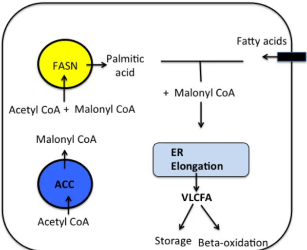
1.5. Fatty acid metabolism is an underexplored area during HCV and flaviviruses infection ... 37
1.6. HCV and the requirement for fatty acid metabolism ... 40
1.6.1. Biogenesis of fatty acids up to 16 carbon (palmitic acid) ... 40
1.6.1.1. ACC and FASN requirement in HCV replication ... 42
1.6.2. Desaturation and activation of fatty acid acyl chains and HCV ... 43
1.6.3. Fatty acid β-‐oxidation and HCV ... 45
1.7. Lipid droplet as an important organelle for HCV and flaviviruses ... 48
1.8. De novo synthesis of very-long-chain fatty acids (VLCFAs) ... 53
1.8.1. ELOVLs ... 53
1.8.2. HSD17B12 ... 58
1.9. Project rationale ... 64
1.10.1. Hypothesis ... 66
1.10.2. Objectives ... 66
1.10.3. Aims ... 66
Chapter 2 ... 67
2.0. Very-long-chain fatty acid metabolic capacity of 17-beta-hydroxysteroid dehydrogenase type 12 (HSD17B12) promotes replication of hepatitis C virus and related flaviviruses. ... 69
2.1. ABSTRACT ... 70
2.2. INTRODUCTION ... 71
2.3. RESULTS ... 74
2.3.2. HSD17B12 promotes HCV RNA replication and infectious particle production75 2.3.3. HSD17B12 contributes to a lipid metabolic environment favoring particle assembly ... 77
2.3.4. Depletion of HSD17B12 disrupts HCV-‐induced MW structures ... 79
2.3.5. Oleate supplementation rescues HCV replication in HSD17B12 KD cells ... 80
2.3.6. HSD17B12 is required for ZIKV and DENV replication ... 80
2.4. DISCUSSION ... 81 2.5. METHODS ... 85 2.6. ACKNOWLEDGEMENTS ... 92 2.7. ADDITIONAL INFORMATION ... 92 2.8. SUPPLEMENTAL INFORMATION ... 92 Chapter 3 ... 107
3.0. Requirement of VLCFA metabolic pathway for HCV replication ... 109
3.1. ABSTRACT ... 110
3.2. INTRODUCTION ... 111
3.3. RESULTS ... 112
3.3.1. Role of de novo VLCFA synthesis in HCV replication ... 112
3.3.2. Role of ELOVLs in HCV replication ... 113
3.3.3. Role of the synthesis of neutral lipids for HCV replication ... 113
3.3.4. Role of de novo synthesis of sphingolipids for HCV replication ... 114
3.3.5. Role of VLCFA (< C22) beta-‐oxidation ... 115
3.3.6. Role of oleic acid synthesis ... 116
3.3.7. Rescue of HCV replication by addition of oleic acid to HSD17B12 KD cells ... 116
3.4. DISCUSSION ... 117 3.5. METHODS ... 122 3.6. ACKNOWLEDGEMENTS ... 125 3.7. ADDITIONAL INFORMATION ... 125 3.8. FIGURES ... 126 Chapter 4. Discussion ... 134
4.1. Elucidating the contribution of HSD17B12 and VLCFA in Flaviviridae virus replication .. 135
4.2. A role of HSD17B12 in the transition of HCV RNA genome from replication to assembly sites ... 136
4.2.1. HSD17B12 contributes to HCV-‐induced membranous replication sites ... 137
4.3. Mode of action of HSD17B12 for the replication of flaviviruses ... 143
4.4. Broad-Spectrum Antiviral activity of specific HSD17B12 inhibitors ... 145
4.4.1. The pharmacological inhibition of HSD17B12 impedes replication of HCV and flaviviruses ... 145
4.4.2. HSD17B12 and FASN inhibition has comparable antiviral effects ... 146
4.5. HSD17B12 targeting as a broad-spectrum antiviral approach ... 148
4.6. Future perspective ... 149
4.7. Potential limitations ... 152
4.8. Conclusion ... 155
List of Figures
Chapter 1. Introduction
Figure 1.1. Schematic illustration for HCV first identification………... 5
Figure 1.2. Genomic organization of HCV virus……….. 9
Figure 1.3. HCV proteins attached to ER membranes………. 12
Figure 1.4. Schematic representation of the HCV life cycle………. 17
Figure 1.5. Schematic representation of hepatocyte HCV entry factors……….. 19
Figure 1.6. Schematic representation of HCV protein interactions necessary for virus particle assembly………... 29
Figure 1.7. An example of an HCV subgenomic replicon………... 33
Figure 1.8. Schematic representation of a) JFH-‐1 genome and b) hybrid JC1 genome. 35 Figure 1.9. Schematic representation of VLCFA de novo synthesis enzymes as hub proteins for several families of viruses……… 39
Figure 1.10. Different sources of the intracellular pool of fatty acids………... 41
Figure 1.11. LDs are the platforms for HCV assembly………. 50
Figure 1.12. Importance of LDs for DENV assembly………... 52
Figure 1.13. Very-‐long-‐chain fatty acid (VLCFA) elongation cycle at ER………. 54
Figure 1.14. ELOVL family substrates and products……… 57
Figure 1.15. Schematic representation of the Genomic Structure of HSD17B3 and HSD17B12……….. 59
Figure 1.16. Comparison between amino acid sequences of HSD17B3 and HSD17B12... 60
Figure 1.17. ELOVLs and HSD17B12-‐catalyzed reactions in the elongation of VLCFA……… 61
Figure 1.18. Schematic representation of the modeled HSD17B12 active site…………. 62
Chapter 2. Figure 2.1. HSD17B12 overlaps replication and core-‐associated assembly sites of HCV infection………..…….…….93
Figure 2.2. HSD17B12 KD decreases HCV replication and infectious viral particle production………...……..…….………94
Figure 2.3. HSD17B12 KD reduces the number of cytoplasmic LDs……….………...96
Figure 2.4. HSD17B12 KD induces alteration in cellular lipid metabolism………...……..97
Figure 2.5. HSD17B12 KD induces dispersion and disruption of HCV replication site………...98
Figure 2.6. HSD17B12 KD decreases DENV and ZIKV replication and infectious viral particle production……….……….……..99
Figure 2.7. HSD17B12 inhibitor INH-‐12 blocks HCV, ZIKV and DENV replication………..100
Figure 2.S1. HSD17B12 overlaps with HCV assembly sites..………...………..………100
Figure 2.S2. HSD17B12 KD has minor effects on protein translation, cell survival and no effect on HCV IRES-‐mediated translation………...………...……….…...….101
Figure 2.S3. HSD17B12 KD increases intracellular HCV RNA levels of HepG2-‐infected cells………...……….……….……102 Figure 2.S4. HSD17B12 KD decreases extracellular HCV RNA levels of HepG2 infected cells………...………...……….…. 103 Figure 2.S5. HSD17B12 KD increases expression of lipolysis genes……….……....104 Figure 2.S6. Oleic acid supplementation restores HCV RNA replication sites and rescues infectious particle production of HSD17B12 KD cells………...………..….105 Figure 2.S7. HSD17B12 inhibitor INH-‐12 has minor effect on cell viability…...106
Chapter 3.
Figure 3.1. Effects of FASN inhibitor and HSD17B12 KD on HCV Con1B replication…... 126 Figure 3.2.Effects of depleting different ELOVLs on HCV RNA replication..……….….127 Figure 3.3. Effects of ASCLs inhibition on HCV Con1B replication……….… 129 Figure 3.4. Effects of CPT1 inhibition on HCV Con1B replication.Effects of 3-‐
ketodihydrosphingosine………..….. 130 Figure 3.5. Inhibition on HCV Con1B replication……….…...131 Figure 3.6. Effects of SCD1 inhibition on HCV Con1B genome replication…………...….…….132 Figure 3.7. Effects of oleic acid treatment on HSD17B12 KD-‐mediated decrease of HCV Con1B replication……….……….….. 133
Chapter 4. Discussion.
Figure 4.1. FASN pathway provides HSD17B12 catalysis with palmitic acid for
elongation………...147 Figure 4.2. A modular RNA-‐guided genome regulation……….150
List of Abbreviations & Acronyms
AA Arachidonic Acid
ACC Acetyl-CoA Carboxylase
ACSL long chain acyl-CoA synthetase
ACP Acyl Carrier Protein of fatty acid synthase
AMPK AMP-activated protein Kinase
AP2M1 clathrin Assembly Protein complex 2 Medium chain
ApoB Apolipoprotein B
ApoE Apolipoprotein E
ARFP Alternative Reading Frame Protein
CD81 Cluster of Differentiation 81
cDNA Complementary DNA
CE Cholesterol Ester
CRISPR Clustered Regularly Interspaced Short Palindromic Repeats
CK IIα Casein Kinase IIα
cLD cytosolic Lipid Droplet
CLDN1 Claudin 1
CoA Co enzyme A
CPT1 Carnitine Palmitoyl Transferase-1
CypA Cyclophilins A
DCI Dodecenoyl Coenzyme A delta isomerase
DH Dehydratase of fatty acid synthase
DG Diglycerides
DGAT Diglyceride acyltransferase
DMV Double membranous vesicles
DRM Detergent resistance membrane
ER Enoyl Reductase of fatty acid synthase
ER Endoplasmic reticulum
FASN Fatty Acid SyNthase
FA (CX, Y) Fatty acid (X is the number of carbons in its acyl chain, while Y is the number of double bonds)
GAGs GlycosAminoGlycans
GTP Guanidine Tri-Phosphate
HACD 3-Hydroxy Acyl Dehydratase
HAV Hepatitis A Virus
HBV Hepatitis B Virus
HCC Hepatocellular Carcinoma
HCV Hepatitis C Virus
HCVpp Hepatitis C Virus pseudo-particles
HIV Human Immunodeficiency virus
HMG-CoA 3-Hydroxy-3-Methyl-Glutaryl Coenzyme A HSD17B12 17 Beta-Hydroxy Steroid Dehydrogenase type 12 HSPG Heparan Sulfate Proteo Glycans
HTS High Throughput Screening
HUH7 Type of human Hepatoma cell line
IP-MS/MS Imunno-precipitation coupled to mass spectroscopy
IRES Internal ribosome entry site
IV Intravenous
JC1 Chimeric genome HCV clone
JFH-1 Japanese Fulminant Hepatitis-1 infectious strain
kDa kilo Dalton
KR Keto-Reductse of fatty acid synthase
LDL Low Density Lipoprotein
LDL-R Low Density Lipoprotein-receptor
let767 Caenorhabditis elegans HSD17B12 analogue
mAbs monoclonal Antibodies
MAPK Mitogen Activated Protein Kinase
mir-122 micro RNA 122
MUFAs Mono Unsaturated Fatty Acids
NADPH Nicotinamide Adenine Dinucleotide Phosphate Hydrogen
NANBH Non-A, Non-B Hepatitis
NPC1L1 Niemann-Pick C1-Like 1
OCLN Occludin
OCT1 Organic Cation Transporter 1
ORF Open Reading Frame
OSBP Oxysterol-Binding Protein
PC Phosphatidyl Cholin
PE Phosphatidylethanoleamine
PH Pleckstrin Homology
PHHs Primary Human Hepatocytes
PI (4,5)P2 Phosphatidylinositol 4,5-bisphosphate
PI4KIII Phosphatidylinositol-4-kinase III
PI(4)P Phosphatidylinositol-4-phosohate
PLA2 Phospholipase A2
PPAR Peroxisome Proliferator-Activated Receptor
PRR Pathogen Recognition Receptor
PPT Phosphopantetheine Transferase
RdRp RNA dependant RNA polymerase
RF Replication Factory
RNA Ribo Nucleic Acid
RTK Receptor Tyrosine Kinase
RXR Retinoid X Receptor
SCD Stearoyl-CoA Dehydrogenase
siRNA Silencing RNA
SR-BI Scavenger Receptor class B type I
SREBP1c Sterol Regulatory Element-Binding Protein 1c
SL- Stem-Loop
TECR Trans-2,3,-Enoyl-CoA Reductase
TE Thioestrase domain
TM Transmembrane segments
TMDs Transmembrane Domain
UTRs Untranslated Regions
VAP-A Vesicle-Associated membrane Protein-associated protein-A
VLCFA Very-Long-Chain Fatty Acids
VLDL Very Low Density Lipoprotein
vRNA viral RNA
WNV West Nile Virus
Alhamdulillah
You think that you are a small luminary (in the sky)
While inside you, the whole universe is hidden!
Acknowledgments
I would first like to thank Dr. Daniel Lamarre for giving me the opportunity to pursue my doctoral degree in a world-class laboratory, filled with innovative research projects and inspiring colleagues. Dr. Lamarre direct supervision paved the foundation for the solid understanding I now have of my field of study. His support and patience was the key in completing my research project and writing my thesis, and I cannot thank him enough for the time he spent in making me a better scientist.
Dr. Martin Baril, the previous research associate in the lab, I deeply thank you since day one in the lab, you taught me everything I needed to know to do good research. I will remember your patience and support as a mentor and your enthusiasm as a researcher. You left a deep impact on my scientific career; I wish I could make you proud.
To Salwa Es-Saad, Nicolas Tremblay, Bridget Gagne, Michael Meloche, and Alex Park my lab colleagues, your kindness and wisdom were always great support for me. I will always remember that you made the work in the lab a great fun. Also, your focus, dedication, and commitment to doing good research were my best motivation.
Marie-Anne Germain, the previous lab member, your dedicated work on HCV was the base of my fruitful work. Dr. Naglaa Shoukry, you were always there for support. Dr. Laurent Chatel-Chaix and Clement Mazeaud, thank you for your sincere help in executing the Flavivirus experiments at Institute Armand Frappier (University of Quebec). Dr. Vladmir Titorenko and Younes Medkour, many thanks for the precious assistance in lipidomic measurements at the University of Concordia. Dr. Donald Poirier (University of Laval), thank you for providing the exclusive HSD17B12 inhibitor. Dr. Arnost Cepica (University of Prince Edward Island), thank you very much for your wholehearted support. I would also like to appreciate the guidance of my doctoral committee members; Dr. Marc Prentki and Dr. Petronela Ancuta.
My Egyptian professors; Dr. Ahmed Abdel-Tawab, Dr. Nemat Yassin, Dr. Wafaa El-Eraqy, Dr. Sawsan Aboul-Alfotouh and Dr. Olfat Hassan, without your sincere support and dedicated mentorship, I could not have the readiness to proceed through my PhD at the University of Montreal.
Last, but not least, I would not have gotten this far without the constant support of my late father, my mother, my brothers, my sister, my father-in-law and my mother-in-law, my wife Dr. Ghada Ebead and my kids Baraa, Khadija and Salman. You are the light of my life. Thank you all from the bottom of my heart.
Chapter 1. Introduction
1.1. Flaviviridae family
Viruses of the Flaviviridae family are enveloped single-strand positive-sense RNA
viruses, with the nucleocapsids surrounded by two or more types of envelope
glycoproteins and a lipid layer [1]). It is composed of different genera, including
Hepacivirus [e.g.,hepatitis C virus (HCV)], Flavivirus [e.g., Zika Virus (ZIKV), Dengue
Virus (DENV)], Pegivirus and Pestivirus [2]. Multiple members within the Flavivirus and
Hepacivirus genera are significant human pathogens.
HCV is a major human liver-specific pathogen that causes chronic infection, and is
the primary cause of liver dysfunction worldwide with more than 70 million people at
risk of progression to liver cirrhosis and hepatocellular carcinoma (HCC) [3]. Medically
relevant flaviviruses, including ZIKV, DENV, West Nile Virus (WNV), Japanese
Encephalitis Virus (JEV) and Yellow Fever Virus (YFV), are usually arboviruses
(transmitted by arthropods, mainly mosquitoes and ticks) that are responsible for severe
mortality in humans and animals worldwide. DENV and YFV infections can cause severe
disease in infected patients, including vascular leakage and haemorrhage [4-6]. DENV
causes 60 million symptomatic infections annually, leading to approximately 10,000
deaths per year [3]. On the other hand, JEV and WNV infections tend to cause
neurological diseases [7, 8]. ZIKV infection is associated with microcephaly (a serious
birth defect) and other neurological disorders [9]. In the last decade, targeting HCV by
direct-acting antiviral drugs achieved unprecedented cure rates reaching about 98% of the
treated patients; however, there is still an urgent need for medications and vaccines
considerable threat to public health due to the increase in their outbreaks and
geographical spread [10].
On the level of molecular virology, viruses within the hepacivirus and flavivirus
genera are similar in their general genome organization and the general principle of the
replication cycles [2]. The similarities in the ways of host cell manipulation by these
viruses may offer the opportunity to learn from the data related to HCV. We can focus on
the importance of the virus-host lipid interaction, in the context of antiviral therapy, to
1.2. General aspects of HCV
1.2.1. Discovery of HCV
The problem of parentally transmitted hepatitis appeared following the
development of serological tests to detect hepatitis A virus (HAV) and hepatitis B virus
(HBV) infections in the 1970s [11]. Most cases of unknown origin surprisingly
represented hepatitis. The condition was termed non-a, non-b hepatitis (NANBH).
Further, it has been demonstrated that NANBH is progressing gradually and has led to
liver cirrhosis in 20% of infected patients [12]. In order to solve this problem, early
studies on viral hepatitis used chimpanzees as reliable models for infection transfer from
human biological materials [13, 14], further proving the existence of NANBH-causing
agents. The chimpanzees’ hepatocytes showed tubule-like structures in their cytoplasm
[15]. Tubule forming agent (TFA) is described as a lipid-enveloped agent that can be
filtered and inactivated by organic solvents [16]. After many efforts to identify the
causative agent of NANBH, a team lead by Dr. Michael Houghton at Chiron Corporation
identified, for the first time, an agent termed HCV by using bacterial expression cDNA
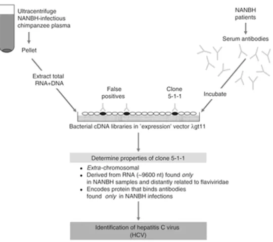
libraries to immune-screen NANBH patient sera [17] (Fig. 1.1). Upon discovery, the
same group produced the first enzyme specifically for NANBH-specific antibodies. The
use of this testing tool demonstrated that the major cause of parentally transmitted
Figure 1.1. Schematic illustration for HCV first identification.
Figure 1.1 shows the use of bacterial expression cDNA libraries to immune-screen NANBH patient sera. This led to discovery of clone 5-1-1 (HCV genome). The figure is adapted with permission from (Houghton, M., Discovery of the hepatitis C virus. Liver Int, 2009. 29 Suppl 1: p. 82-8) [19].
1.2.2. HCV infection: disease and fate
HCV is a significant chronic disease, as 70% of infections progress to persistent
infections [20]. During the first week of infection, HCV titers increase exponentially as
the virus is able to generate up to 1012 virions daily [21, 22]. The patients are mainly
asymptomatic; this leads to the spread of the infection. Further, the patients become
clinically symptomatic late after the liver disease is established, and its symptoms
become apparent [23].
HCV infection is the disease most causative of liver transplantation [24]. The
liver damage occurs despite HCV not being a cytopathic virus. Several studies showed
that the liver pathology leading to HCC is due to direct cytotoxic T-lymphocyte-mediated
killing of hepatocytes [25].
1.2.3. HCV transmission
In developing countries, the primary cause of HCV transmission is iatrogenic,
stemming from the use of blood-contaminated medical instruments. For example, Egypt
has one of the highest populations of HCV infection. This high prevalence was before the
introduction of treatment campaigns with direct-acting anti-HCV drugs. The HCV surge
among the Egyptian population was due to anti-schistosomiasis campaigns of mass
intravenous (IV) injections using glass syringes in the 1950s [26]. In developed countries,
the primary source of transmission is the recreational injection drug use. In Canada, for
1.2.4. Virus composition
HCV has an outer envelope containing two proteins: E1 and E2. Underneath the
membrane is a layer of the viral core protein, which binds to the viral genome forming
the nucleocapsid where the RNA is located [28].
HCV particles purified from sera of humans and chimpanzees exhibit a wide
range of low densities between 1.03 g/cm3 and 1.20 g/cm3 [29, 30]. Further
characterization showed that the particles of lower density have higher infectivity [31].
The virus from patient sera was described for the first time as a lipo-viro-particle when it
was found to be associated with a very low-density lipoprotein (VLDL) or low-density
lipoprotein (LDL). Virus particles obtained from cell culture confirmed the association
with lipoprotein and apolipoproteins ApoE and ApoB. However, they had slightly higher
density and a lower specific infectivity [32, 33]. HCV has a unique lipid profile among
the Flaviviridae family. HCV particles are composed mainly of neutral lipids like
cholesterol esters and triglycerides. In contrast, the lipidomic characterization of bovine
viral diarrhea virus (BVDV), from the same family, did not reveal the presence of those
neutral lipids [34].
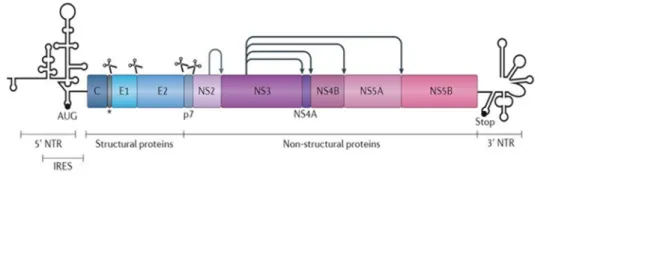
1.2.5. HCV genome
The HCV genome is about 9.6 kb. The genome is represented by a
non-interrupted open reading frame flanked by two untranslated regions (UTRs). The 5'UTR
contains the internal ribosomal entry site (IRES) where the virus genome translation
starts. The 3'UTR has cis-acting RNA elements that stimulate translation. The viral open
reading frame codes for about 3000 amino acids. The HCV polyprotein is processed by a
structural proteins [35] - capsid protein and (E1 and E2) envelope proteins – and seven
Figure 1.2. Genomic organization of HCV virus.
The figure shows the HCV genome and the protein code. Scissors indicate cleavage by cellular proteases, whereas the arrows depict cleavage by the viral proteins. The figure is adapted with permission from (Neufeldt, C.J., et al., Rewiring cellular networks by members of the Flaviviridae family. Nat Rev Microbiol, 2018. 16(3): p. 125-142.) [2].
1.2.6. Genetic variance, genotypic distribution, and origins
HCV is very diverse genetically and based on phylogenetic and sequence analysis
of the viral genome it is classified into seven genotypes (1-7) [19]. Genotyping is based
on genome areas which are highly conserved, namely E1, core, NS5B, and 5'UTR [36].
The different genotypes diverge at about 30-35% of the nucleotide sequence. The strains
in different genotypes have less than 15% difference in nucleotide sequence [37].
HCV genotype 1, as opposed to 2 and 3, is the most distributed genotype in the
developed Western world. The distribution includes North America and Northern and
Western Europe. Regarding genotypes 4, 5, and 6, they are more confined to some
regions of the world. For example, genotype 4 is found mainly in the Middle East [38].
Genotype 5 is found in South Africa [39], while genotype 6 is predominantly found in
1.3. Molecular biology of HCV
1.3.1. HCV proteins
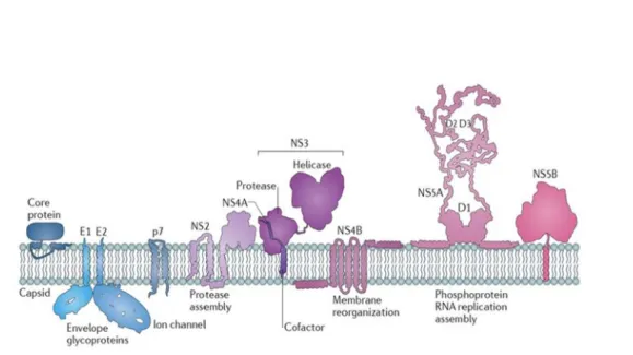
HCV Core
The first structural protein encoded by the HCV open reading frame (ORF) is the
core protein (Fig.1.3), which forms the viral nucleocapsid [42]. The premature core (191
amino acids) is constituted by three domains. Recruitment of the premature core to the
endoplasmic reticulum (ER) depends on the carboxyl terminal (C-terminal) of domain III
[43]. However, the association and targeting lipid droplets (LDs) are dependent on the
proteolytic cleavage of the distal C-terminal (domain III) by the signal peptide peptidase
enzyme, and generation of the mature core (177 amino acids) [44]. The mature core has
two domains. The amino terminal (N-terminal) of domain I is rich in positively charged
amino acids and is mostly hydrophilic. This characteristic contributes to RNA-binding
ability of the protein [45, 46]. Moreover, the same domain catalyzes the
homo-oligomerization of the core and its interaction with glycoprotein E1 [47, 48]. On the other
hand, the hydrophobic C-terminal of domain II is responsible for membrane association,
for instance, to the membrane surrounding LD [47]. Alternative reading frame protein
(ARFP) is a result of a ribosomal frame-shift near codon 11 in core sequence. The protein
is composed of 160 amino acids and is expressed in HCV-infected patients. The function
of this protein is still to be elucidated [49]. HCV core, particularly of genotype 3, induces
the expression of lipid de novo synthesis genes and contributes to the development of
Figure 1.3. HCV proteins attached to ER membranes.
Schematic representation of HCV proteins shows three structural proteins; (core) capsid protein, (E1 and E2) envelope proteins, and seven nonstructural proteins (P7, NS2, NS3, NS4A, NS4B, NS5A, and NS5B) [35]. The figure is adapted with permission from (Neufeldt, C.J., et al., Rewiring cellular networks by members of the Flaviviridae family. Nat Rev Microbiol, 2018. 16(3): p. 125-142.)[2].
The envelope glycoproteins
HCV has two envelope glycoproteins namely, E1 of 35kDa and E2 of 70kDa. The
two proteins are heavily glycosylated. They have a small C-terminal transmembrane
domain that contains the retention signal in the ER, and a large N-terminal ectodomain
facing the ER lumen [51]. The two envelope proteins are heterodimerized through the
transmembrane domain [52]. E1 and E2 are responsible for the early stage of viral entry
through the interaction with permissive cell receptors [53].
P7
P7 is a protein with the ability to form pores in artificial membranes. It also has
ion channel activity. In virtue of these characteristics, it belongs to the family of
viroporins [54]. P7 resides in the ER and has two hydrophobic transmembrane passages
linked together through a short hydrophilic segment [55]. P7 has a significant role in
HCV particle infectivity through its ion channel activity and interaction with other viral
proteins. However, the P7 protein is dispensable in HCV RNA replication and virus entry
[56].
NS5A
NS5A plays an essential role in viral replication, particle production, and
modulation of cellular environment [57]. No enzymatic activity has been reported for
NS5A. NS5A has three domains: one highly structured domain (DI) and two unstructured
domains (DII and DIII). DI integrity is essential for RNA replication, while deletion of
large parts of DII and DIII could be done without affecting it. A large deletion of DIII
replication-competent replicon [58]. However, the NS5A mutants are impaired in particle
production due to the loss of the interaction with core protein [59]. NS5A has two
different phosphorylated forms. They are named according to their molecular mass: the
basal p56, and the hyper-phosphorylated p58. The p56/p58 ratio was reported to alter
HCV RNA replication efficiency [60]. The NS5A N-terminal alpha-helix anchors the
cytosolic leaflet of the ER, which is critical for HCV RNA replication [61]. NS5A
interacts with ER vesicle-associated membrane protein A (VAP-A), which could play an
essential role in the recruitment of effector enzymes such as oxysterol binding protein
(OSBP), which is capable of binding VAP proteins [62]. NS5A importantly interacts with
phosphatidylinositol-4-kinase III alpha (PI4KIIIA). This interaction stimulates the
enzyme. Further, the stimulation of the enzyme leads to the accumulation of
phosphatidylinositol-4-phosphate (PI(4)P) which is essential to the recruitment of other
effector enzymes that have pleckstrin homology domains (PH domains) [63, 64]. Indeed,
NS5A plays a vital role in the preparation of HCV RNA for encapsidation. It is proposed
that NS5A and possibly NS2 [65] deliver the virus RNA genome from sites of RNA
replication to the LD at which core protein processes the encapsidation [66]. Further,
NS5A Domain I was shown to be critical for the induction of double-membrane vesicles
associated with hepatitis C virus replication [67].
NS2
NS2 encodes a cysteine protease that catalyzes the cleavage of the HCV
polyprotein precursor at the NS2/NS3 junction. NS2’s cysteine protease function is
enhanced by the N-terminal one third of NS3 [68, 69]. NS2 is not directly required for
The role involves complex interactions with structural and non-structural viral proteins
[70]. NS2 is proposed to act as a scaffold bringing the replicase machinery (NS3, NS5A)
and the envelope glycoproteins to assembly sites. Mutations in NS2 abrogate the
interaction with NS3, NS5A, P7, and E2, consequently impairing HCV particle
production [65, 71, 72].
NS3
NS3 has multiple enzymatic activities, including protease and helicase activities.
The protease activity is stimulated when the enzyme binds to its co-factor NS4A and
mediates the processing of the polyprotein in the nonstructural protein region [73]. The
C-terminus carry the helicase activity. This activity mediates ATP hydrolysis together
with unwinding HCV RNA [74, 75]. NS3 is critical for HCV replication and particle
assembly [76]. NS3, together with its co-factor NS4A, is localized to the ER membrane
and the contact sites between the ER and the mitochondrial membrane. This localization
promotes its ability to hinder the induction of innate cellular immunity. Indeed, NS3/4A
can cleave mitochondrial antiviral signalling protein MAVS, which is the adaptor of the
retinoic acid-inducible gene (RIG-I) [77, 78].
NS4A
The central part of the NS4A protein acts as an NS3 co-factor, while the
N-terminus plays a role in tethering NS3 to cellular membranes [79]. However, the function
of the C-terminus part is not clear. Mutagenesis studies suggest that through interaction
with other nonstructural proteins, it may play a role in HCV RNA replication and particle
NS4B
NS4B is an integral membrane protein composed of a central core of four
transmembrane domains, and C- and N- terminuses facing the cytoplasm [81]. NS4B can
induce the rearrangement of ER membranes and generation of vesicle-like structures,
which act as HCV replication complex sites [82, 83]. NS4B has shown to bind viral
RNA and to interact with HCV nonstructural proteins [84]. Further, NS4B was shown to
play a role in viral assembly [85]. Moreover, it was demonstrated to harbour NTPase
activity [86], and, similar to the other HCV nonstructural proteins, NS4B has been
reported to form oligomers [87]. Interestingly, it was demonstrated that NS4B protein
oligomerization is critical for membranous web formation and HCV RNA replication,
and hence is required for the assembly of a replication complex, possibly through the
induction of membrane curvature and vesicle formation [70].
NS5B
HCV replication proceeds via synthesis of a complementary negative-strand RNA
using the genome as a template and the subsequent synthesis of genomic positive-strand
RNA from this negative-strand RNA template. The enzyme responsible for this process is
NS5B RNA-dependent RNA polymerase (RdRp), which is the core of the replication
machinery responsible for the amplification of the HCV genome. The NS5B protein is
composed of two domains: a hydrophobic transmembrane domain, and the C-terminal
catalytic domain to which the former is linked [58, 88]. The active site of the polymerase
includes a GDD motif that is involved, along with the contribution of Mg2+ ions, in the
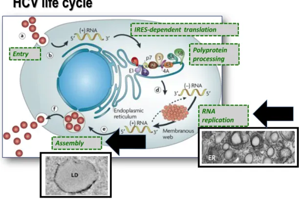
1.3.2. HCV life cycle
The HCV life cycle includes three major steps: the viral entry, the translation and
replication of the virus genome, and the assembly of the viral particles [89]. The HCV
life cycle is represented in (Fig. 1.4).
Figure 1.4. Schematic representation of the HCV life cycle.
Virus binding and internalization (a); cytoplasmic release and uncoating (b); IRES-mediated translation and polyprotein processing (c); RNA replication (d); packaging and assembly (e); virion maturation and release (f). The steps are illustrated as separate steps for simplicity. The bold black arrows refer to the replication and assembly compartments (representative electron microscope images are shown). The steps occur in a tightly coupled fashion. The figure is adapted with permission from (Moradpour, D., F. Penin, and C.M. Rice, Replication of hepatitis C virus. Nat Rev Microbiol, 2007. 5(6): p. 453-63.) [89].
1.3.2.1. HCV entry
HCV, during circulation in the blood, is directly in contact with the basolateral
membrane of hepatocytes. The contact allows it to bind to the receptors on the surface of
these cells and trigger its entry, as reviewed in Lindenbach et al. 2013 [72]. LDL receptor
(LDL-R), glycosaminoglycans (GAGs), and heparan sulfate proteoglycans (HSPGs)
mediate low-affinity attachment of HCV to hepatocytes’ surfaces. This attachment occurs
by virtue of the HCV particle apolipoprotein E (ApoE) content [72]. In addition to this,
there are five hepatocyte surface molecules necessary for HCV particle entry. These are:
CD81, scavenger receptor class B type I (SR-BI), claudin 1 (CLDN1), occludin (OCLN)
and Niemann-Pick C1-like 1 (NPC1L1). As well, receptor tyrosine kinase (RTK) and
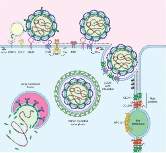
tight junction cadherin are regulatory molecules essential for HCV entry [72]. (Fig. 1.5)
(i) CD81
Human CD81 is widely expressed in many cell types. The molecule is a
tetraspanin adaptor cell surface protein [90]. CD81 promotes a conformational change in
the HCV E1/E2 glycoproteins. The conformational change is important for
low-pH-dependent fusion and viral endocytosis [91]. CD81, through its large extracellular loop,
binds HCV envelope glycoprotein E2 to facilitate HCV entry [53, 92]. The sequences of
the extracellular loops of CD81 are conserved between humans and chimpanzees.
However, cells from different species with different sequences can support HCV entry in
Figure 1.5. Schematic representation of hepatocyte HCV entry factors.
HCV lipo-viro-particle attaches to the cell membrane through interaction with HSPG, LDLR, and SR-BI. SR-BI may produce conformational changes in E2, leading to the exposure of the CD81 binding site. Interaction of E2 with CD81 leads to the activation of the signal transduction through EGFR, Ras, and Rho (Ras homology) GTPases. These signalling events enhance the lateral movement of CD81-HCV complexes to the cell-cell contact sites. Then, CD81 interacts with CLDN1 and promotes HCV internalization via clathrin-mediated endocytosis. The low pH of the endosomes leads to HCV fusion. The figure is adapted with permission from( Lindenbach, B.D. and C.M. Rice, The ins and outs of hepatitis C virus entry and assembly. Nat Rev Microbiol, 2013. 11(10): p. 688-‐700.) [72].
(ii) Scavenger receptor class B type I (SR-BI)
SR-BI is a 509-amino acid polypeptide, and it regulates lipid metabolism through
its function as a primary receptor for high-density lipoproteins (HDLs), supporting its
selective uptake into hepatocytes [94, 95]. SR-B1 was initially identified as a putative
receptor for HCV because it binds soluble E2 (sE2). This binding was suggested to occur
through the interaction with E2 hypervariable region 1 (HVR1) [96]. SR-BI mediates
primary attachment of HCV particles of intermediate density to cells. These initial
interactions involve apolipoproteins, such as ApoE, which is present on the surface of
HCV particles [97]. This suggests that the lipoprotein components in the virion act as
host-derived ligands for important entry factors such as SR-BI. It has been shown that
HCV entry relies on the lipid transfer activity of SR-BI [97]. Indeed, SR-BI antagonist
was demonstrated to have additive and synergistic potency when used in combination
with other antiviral agents [98].
(iii), (iv) Claudin 1 and Occludin
CLDN1 and OCLN are two tight junction proteins. The two proteins were
identified as factors able to render non-human cells supportive of HCV entry [99, 100]. It
was suggested that CLDN1 and OCLN support the later phase of HCV entry, after SRB1
and CD81. Interestingly, CLDN1 and OCLN do not directly interact with the HCV
envelope proteins. However, CLDN1 and CD81 interact to form a part of the HCV
receptor complex [101, 102]. It was also shown that HCV envelope glycoproteins
stimulate endocytosis of both CD81 and CLDN1 and support their fusion with the early
endosome, supporting a model wherein HCV stimulates receptor trafficking to promote
(v) Niemann-Pick C1-like 1
NPC1L1, a 13 transmembrane cell surface cholesterol-sensing receptor, is
expressed in the gastrointestinal tract on the apical surface of intestinal enterocytes, in
addition to the human hepatocytes like Huh7 cells. Further, NPLC1L1 is responsible for
the regulation of cellular cholesterol absorption and controls body cholesterol
homeostasis [104].
Silencing or antibody-mediated blocking of NPC1L1 impairs cell culture-derived HCV
(HCVcc), demonstrating that NPC1L1 expression is required for HCV infection initiation
[105]. The requirement was shown to be dependent on virion cholesterol content, and to
occur before the virion-cell membrane fusion step [105]. Further, ezetimibe, the clinically
available FDA-approved NPC1L1 antagonist, potently blocks HCV uptake in vitro.
[105].
Regulatory molecules
(i) Receptor tyrosine kinases
Using kinase RNAi screens, two receptor tyrosine kinases RTKs, including the
epidermal growth factor receptor (EGFR) and ephrin type-A receptor 2 (EphA2), were
identified as HCV entry cofactors [106]. The two receptors regulate CD81-claudin-1
co-receptor associations and viral glycoprotein-dependent membrane fusion [106]. GTPase
HRas, (Harvey Rat Sarcoma oncogene homolog) activation downstream of EGFR
signalling was shown as a critical host signal transducer for EGFR-mediated HCV entry.
(ii) E-cadherin
E-cadherin is a transmembrane glycoprotein, which acts as a significant adherent
junction protein. It plays an essential role in maintaining cell-cell adhesion. Further, it
plays a vital role in maintaining the epithelial architecture and cell polarity and
differentiation [108, 109]. Li and co-workers showed that E-cadherin regulates HCV
entry [110]. Their functional studies demonstrated that E-cadherin is closely associated
with CLDN1 and OCLN on the cell membrane. It was also demonstrated that the
depletion of E-cadherin severely diminished the cell-surface distribution of these two
tight junction proteins. The phenotype indicates that E-cadherin plays a vital role in the
regulation of CLDN1/OCLN localization on the cell surface [110].
1.3.2.2. HCV translation and replication
HCV has highly structured 5’- and 3’-untranslated regions (UTRs) flanking a
single open reading frame instead of a 5’-terminal cap and a 3’-terminal poly (A) tract
structure characterizing the host cell mRNA [111]. HCV translation initiation occurs by a
cap-independent mechanism mediated by the type III internal ribosomal entry site (IRES)
in the 5’ UTR [112]. The IRES encompasses 5’ UTR stem-loop II-IV [111]. The IRES
includes two stem-loop structures located in the core-coding region. Beside the IRES,
several cis-acting RNA elements in the 3’ UTR stimulate the translation of HCV RNA.
Recent reports suggest the circularization of the HCV genome occurs as a result of the
interaction between stem-loop structures residing in the NS5B coding region and motifs
Further, it has been shown that possible circularization of the HCV genome is
likely facilitated by cellular and viral proteins, for instance, PCBP2 (poly(rc)-binding
protein 2), ILF3 (interleukin enhancer-binding factor 3) and IGF2BP1 (insulin-like
growth factor II mRNA-binding protein 1) [114, 115]. The circularization of the HCV
genome facilitates the translation (5’ to 3’ direction) and the viral replicase-mediated
RNA synthesis (3’ to 5’ direction). Furthermore, liver-specific microRNA-122 (miR-122)
[116] binds to two sites in the 5’ UTR of the HCV RNA genome, leading to stimulation
of IRES-mediated translation, and protecting viral RNA from degradation viarecruitment
of Argonaute 2 [111, 117].
HCV RNA translation leads to a single polyprotein, which is subsequently
processed by viral and host encoded proteases into 10 mature proteins (discussed in
section 1.3.1). Host signal peptidases and signal peptide peptidases process the structural
proteins of HCV by mediating the cleavage at the core/E1, E1/E2, E2/p7 and p7/NS2
junctions. Further, the NS2 cysteine protease, whose activity is enhanced by the
N-terminus of NS3, mediates the cleavage of the NS2/NS3 junction releasing the NS3/4A
serine protease. NS3/4A processes most of the non-structural (NS) proteins by mediating
the cleavage of the NS4A/4B, NS4B/5A, and NS5A/5B junctions while also cleaving
itself and other host factors [70].
On the other hand, viral and cellular proteins orchestrate the process of HCV
RNA replication in the multi-step process [118]. The first step of RNA synthesis
generates a negative-strand genome, which serves as a template for progeny
It is unclear how RNA replication and translation are co-regulated. As described
before, the circularization of the RNA genome might be one mechanism. Another
possibility is the formation of alternative RNA structures. For instance, it was shown that
the 5’ UTR of the positive-strand RNA genome and its complementary sequence – i.e.,
the 3’ UTR of negative-strand RNA – have very different secondary structures [119].
Moreover, RNA sequences in domain II of the IRES are essential for RNA replication
[120]. These characteristics could present multiple signals that might be involved in the
coordination between RNA translations and replication.
HCV, like the other positive-strand RNA viruses, remodels the intracellular
membranes, leading to the formation of organelle-like membranous structures named as
replication factories (RFs) or membranous web (MW). The MW plays many essential
roles, which include (i) compartmental concentration of factors required for efficient viral
RNA replication, (ii) spatial coordination of (RNA translation, replication, assembly) in
the viral replication cycle, and (iii) protecting viral proteins and RNA from intracellular
innate immunity [120].
Positive-strand RNA virus-induced membranous rearrangements could be
included in two morphological types: the double-membrane vesicle (DMV) type and the
invaginated vesicle/spherule type [89, 121]. Interestingly, picornavirus and coronavirus
replication sites belong to the DMV type, though they have only a very distant
evolutionary relationship with HCV, whereas the more closely HCV-related flavivirus
such as DENV and West Nile virus (WNV) have replication sites in the form of
Electron microscopy (EM) studies of HCV-infected cells show the formation of
DMVs of ∼150 nm diameter [122]. The DMVs aggregate to form the structure defined as
the membranous web. The DMVs originate as a protrusion from the ER to the cytoplasm,
by the action of HCV viral proteins NS4B and NS5A [67, 82]. Importantly, the outer
membrane is often linked to the ER membrane. The membranous structures also contain
Single Membrane Vesicles (SMVs) and Multi-membrane Vesicles (MMVs) [123].
Biochemical characterizations have demonstrated that HCV RNA and replicase activity
reside within nuclease-resistant and protease-resistant environments [121]. This suggests
that the HCV replication occurs inside the lumen of DMVs. Only 10% of DMVs have
pores like a connection to the cytoplasm [124]. Hence, it may be only a minor fraction of
the DMVs that support active replication at a given time. Otherwise, DMVs have nuclear
pore complex-like structures, which might enable the traffic in and out of a closed
membrane compartment [125].
Host factors, besides HCV proteins, critically contribute to RF formation. For
instance, cyclophilin (CypA), by acting on NS5A, is suggested to contribute to the
formation of HCV RFs [126]. Another example is PSTPIP2 (proline-serine-threonine
phosphatase interacting protein 2), which belongs to the BAR (Bin-Amphiphysin-Rvs)
domain-containing protein family that acts as a sensor or inducer of positive membrane
curvature [127]. A study by Chao et al. [127] showed that PSTPIP2 is required for
HCV-induced membrane alterations and, thus, RNA replication. Both NS4B and NS5A interact
with PSTPIP2, and recruit it to HCV-remodeled membranes. Further, depletion of this
HCV uses a sterol regulatory element-binding protein-1 (SREBP1) pathway to
induce de novo lipid and membrane biosynthesis [128]. The induction has been shown to
cause distinct changes in the lipidomic profile of HCV-infected cells [129]. The SREBP1
pathway was also activated in the core- and NS4B-overexpressing cells [128, 130].
Further, the overexpression stimulates the transcription of lipogenic genes such as fatty
acid synthase (FASN) and 3-hydroxy-3-methylglutaryl coenzyme-A reductase
(HMG-CoA), the rate-limiting enzyme of the cholesterol biosynthesis of the mevalonate
pathway. Further, viral replication requires the metabolic intermediate geranylgeranyl
phosphate in protein prenylation [131]. NS5B has been shown to also interact with FASN
to stimulate its RdRp activity [132].
HCV is strongly dependent on the lipid kinase PI4KIIIα and its product,
phosphatidylinositol-4-phosphate (PI(4)P) [120]. PI(4)P is predominantly found in Golgi
membranes and the inner leaflet of the plasma membrane of non-infected cells. However,
in HCV infected cells, the PI(4)P is highly enriched in DMVs and RF [63, 133]. PI4KIIIα
knockdown impairs HCV replication and causes the aggregation of DMVs of
significantly reduced diameter [63]. The same phenotype was shown when a PI4KIIIα
pharmacological inhibitor was used [62]. The phenotype shows that PI(4)P is critically
required for DMV functionality. Notably, DMV induction by the distantly related
picornaviruses also requires PI(4)P, suggesting an evolutionarily conserved mechanism
[134].
One pathway linked to PI(4)P which involves non-vesicular cholesterol transport
is the oxysterol binding protein (OSBP)-mediated cholesterol transport [62 352]. OSBP
Importantly, rhinoviruses manipulate this cellular lipid transport system in a strikingly
similar way [135], demonstrating an evolutionarily conserved mechanism of DMV
biogenesis. In HCV-infected cells, it has been shown that DMV diameters were reduced
both by blocking OSBP-mediated cholesterol transport [130] and by depletion of
cholesterol from purified DMVs [121], showing that cholesterol is an essential structural
component of HCV-remodeled membranes. The knockdown of PI4KIIIα or OSBP results
in very similar DMV morphologies. However, PI4KIIIα knockdown blocks HCV RNA
replication potently [120]. In the same context, HCV infection induces the synthesis of
specific sphingolipids that enhance NS5B-mediated RNA replication [136].
Sphingolipids, in addition to cholesterol, play a critical role in detergent-resistant
membranes (DRM) [structural part of RF composed of condensation of phospholipids,
sphingolipids, and cholesterol [120]].
Autophagy is another cellular pathway eventually used by HCV and other
positive-strand RNA viruses to establish RFs. One obvious link is the similarity of the
autophagosomes and DMV-type RFs morphologies. Moreover, HCV infection stimulates
a key event in autophagosome formation, LC3 lipidation [137]. It was demonstrated that
lipidated LC3 associates HCV proteins on viral replication-specialized cell membranes
[138]. However, it is still not well defined which steps of the HCV replication cycle are
modulated by autophagy. For instance, autophagy was reported to stimulate the
translation of incoming HCV RNA without affecting RNA replication [137]. Others
suggest that autophagosomes serve as platforms for HCV RNA synthesis [139]. Further,
multifaceted impact on HCV replication. However, to what extent the autophagy
machinery plays a role in viral DMV biogenesis remains to be investigated.
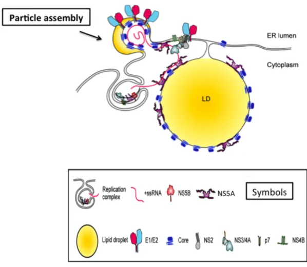
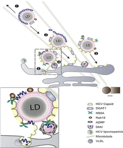
1.3.2.3. HCV assembly
The core protein is the maestro protein of viral assembly. (The HCV core protein
interaction with other viral proteins in the assembly process is represented in (Fig. 1.6))
The mature core with a positively charged N-terminal binds viral genome RNA, whereas
the C-terminal domain facilitates membrane binding via palmitoylated cysteine residues
[47, 141]. The core protein is synthesized on ER ribosomes and then homodimerizes and
traffics to cytosolic LDs. Mutations that prevent core protein from trafficking and binding
to LD strongly inhibit virus assembly and lead to core protein degradation [142, 143].
Cellular factors were demonstrated for core-cLD (cytosolic LD) association. For instance,
diacylglycerol-O- acyltransferase 1 (DGAT1), an enzyme required for the synthesis of
triglycerides that are stored in cytosolic LDs [144], and the activity of cytosolic
phospholipase which is regulated by mitogen-activated protein kinase (MAPK), were
reported to be involved in core trafficking to LDs [145]. Further, core protein processes a
conserved motif YXX* in which X is any amino acid and * corresponds to a hydrophobic
amino acid. This motif interacts with clathrin assembly protein complex 2 medium-chain
(AP2M1) and is involved in transporting core from cLD to ER [146]. Inhibition of RTKs
that regulate AP2M1 activation is reported to strongly inhibit HCV assembly (i.e.,
anti-cancer compounds erlotinib and sunitinib) [147].
Figure 1.6. Schematic representation of HCV protein interactions necessary for virus particle assembly.
NS2, NS3, P7, and phosphorylated NS5A interact with core and envelope proteins to promote particle assembly. The figure is adapted with permission from (Zayas, M., et al., Coordination of Hepatitis C Virus Assembly by Distinct Regulatory Regions in Nonstructural Protein 5A. PLoS Pathog, 2016. 12(1): p. e1005376.) [66]
Recently, imaging studies were conducted on virus-producing cells to obtain
further insight into the trafficking of core protein during virus assembly and release
[148]. It has been demonstrated that the core protein is rapidly trafficked to the surface of
cLDs, and then recruited from the surface of cLD into motile puncta that move in
correspondence with cell microtubules. The movement with microtubule architecture
represents the virus particle within the secretory pathways [148].
While the trafficking of a core protein to LD is critical for the assembly, the core
interaction with other viral factors at the assembly sites is crucial. For example, NS5A
interaction with the LDs bound core protein is a crucial step in HCV assembly [59]. The
C-terminal unfolded domain of NS5A has an essential role in HCV assembly. In
particular, a key event for the virus assembly is the phosphorylation of specific serine in
this domain by casein kinase IIα (CK IIα) [149]. Also, NS5A interactions with ApoE and
annexin A2 were shown to enhance the assembly process [150, 151]. Further, NS2 and
P7 play a pivotal role in organizing the formation of the virus assembly complex. NS2
processes the three transmembrane domains (TMDs). Moreover, the C-terminal cysteine
protease domain of NS2 induces the protein homodimerization. The two parts of the
protein – TMDs and the cysteine protease – are required for the production of the virus
particle [152, 153]. Additionally, NS2 interacts with membrane-bound P7, and the
interaction is essential to localize the proteins to the LD bound core [154].
Moreover, significant defects in HCV assembly were reported when a mutation in
the NS3 helicase domain was created. The demonstration of this suggests the possibility
that NS3 helicase activity serves a purpose in packaging viral RNA during nucleocapsid