Université de Montréal
Contribution of the subthalamic nucleus to visually guided
locomotion
par Nabiha Yahiaoui
Département de neurosciences Faculté de médecine
Mémoire présenté en vue de l’obtention du grade de maîtrise en sciences neurologiques
Décembre 2017
Résumé
Les ganglions de la base (GB) jouent un rôle important dans le contrôle locomoteur. Ceci est illustré par les troubles locomoteurs dont souffrent les patients atteints de maladies dégénératives qui affectent les GB, telles que la maladie de Parkinson, caractérisées par de petits pas lents et traînants, ainsi qu’un gel de la marche (freezing of gait). Une structure centrale dans les GB est le noyau sous-thalamique (NST), de par son rôle de structure d’entrée et ses projections vers le globus pallidus. Cependant, la nature de la contribution du NST au contrôle de la locomotion, ainsi que les caractéristiques de son activité cellulaire durant la marche, sont peu connues.
Afin de mieux comprendre cette contribution, nous avons examiné les propriétés de l’activité neuronale du NST lors de la locomotion non-obstruée et celle sous guidage visuel. Ainsi, nous avons effectué des enregistrements neuronaux chez un chat intact, entraîné à marcher régulièrement sur un tapis roulant et à franchir des obstacles se déplaçant à la même vitesse. Nous avons enregistré 40 cellules montrant une activité reliée au movement du membre antérieur, dont 30 ont montré une activité phasique au cours de la locomotion non obstruée liée aux différentes phases du cycle de la marche, principalement la phase de balancement. Au cours de la modification volontaire de la marche, un groupe de 37/40 cellules, incluant certaines qui étaient modulées pendant la locomotion non-obstruée, ont changé leur fréquence de décharge par rapport à l’obstacle. Ces changement étaient principalement des augmentations de fréquence, mais parfois des diminutions ou une diminution suivie d’une augmentation. Ces modifications se produisaient soit avant l’enjambement de l’obstacle (step-advanced), soit lors de l’enjambement de l’obstacle (step-related). L’activité des cellules step-advanced était indépendante des membres
independent), tandis que celle des cellules step-related était spécifique aux membres (limb-dependent).
Cette étude est la première à examiner les caractéristiques de décharge du NST lors de la marche et montre que cette structure contribue au contrôle de la locomotion non obstruée ainsi que la modification volontaire de la marche, en jouant un rôle dans la planification et l’exécution de cette dernière.
Mots-clés : Ganglions de la base, noyaux sous thalamiques, locomotion, modification volontaire de la marche, locomotion sous guidage visuel.
Abstract
The Basal ganglia (BG) plays an important role in locomotor control. This is emphasized by the impaired walking of patients with neurodegenerative disorders that affect the BG such as Parkinson’s disease. One important structure in the BG is the subthalamic nucleus (STN), which acts as an input structure for the BG and projects to its output structures. Although the STN has been shown to display movement-related activity during reaching, the nature of its contribution to the control of locomotion, together with the characteristics of its neural activity during locomotion, is poorly known.
In order to better understand this contribution, we examined the properties of the neural activity in the STN during unobstructed and visually guided locomotion. To do so, we recorded single neurons in an intact cat trained to walk steadily on a treadmill and to step over obstacles attached to the treadmill belt and moving at the same speed. We recorded 40 neurons which activity was related to the movement of the forelimb during the task. We found that during unobstructed locomotion, many of these cells (30/40) showed phasic step-by-step modulation of their activity pattern, mostly during the swing phase. Most of these swing-related cells discharged throughout the swing phase with no relationship to changes in the pattern of different muscle groups. During voluntary modifications of gait, 37/40 cells, including both cells that were and were not modulated during unobstructed locomotion, changed their firing rate in relationship to the step over the obstacle. The changes observed were mostly increases of activity, but a few cells showed decreases of activity and some showed a decrease followed by an increase of activity. These changes occurred either before the modified step and were classified as step-advanced activity, or they occurred during the modified step and were classified as step-related activity. Step advanced cells mostly showed independent activity, while step related cells showed limb-specific activity.
This is the first detailed account of the contribution of the STN to the control of locomotion and our results indicate that the STN is involved in the control of both unobstructed and visually guided locomotion. The results suggest that during unobstructed locomotion, the STN contributes to the general control of the limb trajectory and to both the planning and execution of voluntary changes of gait.
Keywords: Basal ganglia, subthalamic nucleus, locomotion, voluntary gait modification, visually guided locomotion.
Table des matières
Résumé ... i
Abstract ... ii
Table des matières... iii
Liste des tableaux ... v
Liste des figures ... vi
Liste des abréviations ... viii
Remerciements ... xi
General introduction ... 1
I. Literature review ... 2
1. Neural control of locomotion ... 2
a. Basic locomotion ... 2
b. Supraspinal control of locomotion ... 9
c. Voluntary gait modification ... 12
2. The Basal Ganglia ... 16
a. Structure and pathways of the basal ganglia ... 16
b. Basal ganglia pathological activity ... 18
3. The Subthalamic Nucleus ... 20
a. Anatomical connections ... 21
b. Clinical implications of subthalamic dysfunction... 24
c. Relationship of subthalamic activity to movement ... 26
d. Relationship of subthalamic activity to locomotion ... 28
4. Objectives ... 28
II. Methods... 29
III. Results ... 35
Conclusion ... 58 References ... 60
Liste des tableaux
Liste des figures
Figure 1. (Kandel 2012): The basal ganglia and surrounding structures in the human
brain...……….………..…………. 18
Figure 2. (Volkmann et al 2010): The basal ganglia form anatomically and functionally
segregated neuronal circuits with thalamic nuclei and frontal cortical areas. ... 19
Figure 3. (Redgrave et al 2010): Organization of intrinsic connections within the basal ganglia………21
Figure 4. (Modified from Nambu 2011): Somatotopy of the subthalamic nucleus (STN) in the primate.. ... 23
Figure 5. (Shink et al 1996): Anatomical connections between the STN and the pallidum. ………... 25
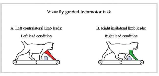
Figure 6. Visually guided locomotor task.. ... 30
Figure 7. Schematic representation of histological reconstruction of recording tracks in the STN……... ... 38
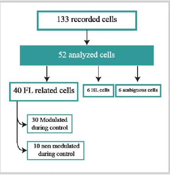
Figure 8. Summary of database.. ... 39
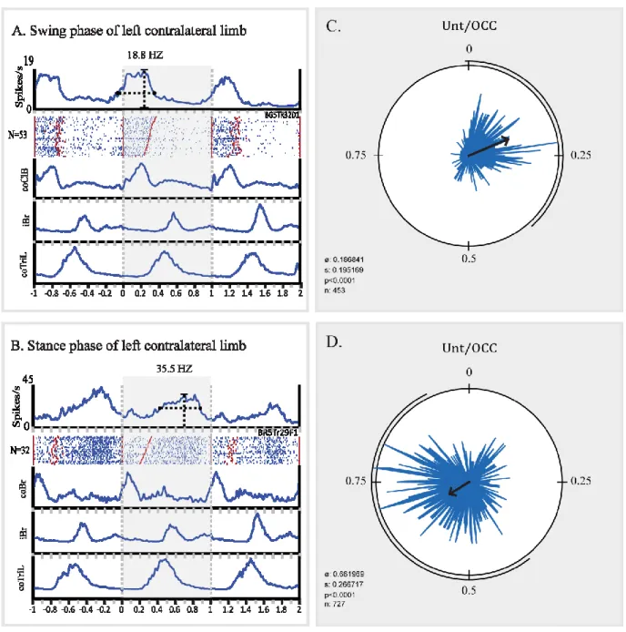
Figure 9. Neuronal activity recorded in the STN during unobstructed locomotion. ... 41
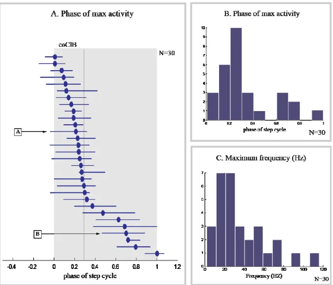
Figure 10. Summary of neural activity during unobstructed locomotion. ... 43
Figure 11. Step-related, limb-dependent activity recorded from 2 cells in the STN during gait modification. ... ... 45
Figure 12. Phase of maximum modification of activity in step-related cells.. ... 46
Figure 13. Maximum change of frequency discharge of step related cells during gait modification… ... 47
Figure 14. Step-advanced, limb-independent activity recorded from 2 cells in the STN during gait modification.. ... 50
Figure 15. Phase of maximum modification of activity in step advanced cells.. ... 51
Figure 16. Maximum change of frequency discharge of step advanced cells during gait modification… ... 52
Liste des abréviations
BG: Basal Ganglia
CMA: Cingulate motor areas co: Contralateral
CPG: Central Pattern Generator EMG: Electromyographic EN: Entopeduncular FOG: Freezing of gait
FRA: Flexor Reflex afferents GABA: γ-Aminobutyric acid GP: Globus pallidus
GPe: Globus pallidus pars externa GPi: Globus pallidus pars interna GTO: Golgi tendon organ
i: Ipsilateral i.v: Intravenous
LVST: lateral vestibulospinal tract MI: Primary motor cortex
MLR: Mesencephalic locomotor region
MPTP: 1-methyl-4-phenyl-1, 2, 3, 6-tetrahydropyridine MRF: Medullary reticular formation
MSN: Medium spiny neurons MT: Movement time
MVST: Medial vestibulospinal tract PD: Parkinson’s disease
PEH: Peri-event histogram PMd: Dorsal premotor area
PMRF: Pontomedullary Reticular Formation PMv: Ventral premotor area
PPC: Posterior parietal cortex PPN: pedunculopontine nucleus RN: Red nucleus
RST: Reticulospinal tract
SMA: Supplementary motor cortex SNpc: Substantia nigra pars compacta SNpr: Substantia nigra pars reticulata STN: Subthalamic nucleus
Remerciements
First I would like to express my deepest gratitude to my supervisor, Dr. Trevor Drew, for his mentorship throughout this master. Thank you for your guidance, patience and support, as well as the inspiration and levity that made this journey, not only possible, but a unique experience of growth.
I would like to thank Toshi Nakajima for consistently setting an example with his stellar work ethic and team spirit. His unfailing support of his colleagues and good-naturedness were both invaluable and humbling.
I would like to thank Dr. Serge Rossignol, Dr. Arlette Kolta and Dr. John Kalaska for being on my thesis committee and for the evaluation of this thesis.
I would like to thank France Lebel and Natasha De Sylva for their technical assistance, their unwavering support is the pillar on which our work relies. I would like to thank Yannick Mullié with whom I collected all the data of this project and Nicolas Fortier-Lebel who has been somewhat of a comrade throughout the years I’ve worked alongside him.
I would like to thank all the friends who have supported me throughout this challenging journey, celebrating the highs and getting me through the lows. Last but certainly not least, I would like to thank my sister Narjes for her unflinching support.
General introduction
Survival critically relies on an animal’s ability to navigate its environment in order to search for food, escape predators and reproduce. Since the 1900s, extensive studies have been conducted to better understand the control of locomotion by investigating the contribution of various parts of the nervous system to the production of stepping. Sherrington was the first to recognize the importance of the spinal cord in producing a locomotor rhythm through what he described as spinal reflexes. From his findings, multiple lines of research have contributed to the understanding of the neural networks behind the basic control of locomotion, which stem from the spinal cord and of brainstem nuclei. However, these structures alone cannot produce visually guided and voluntary gait modifications of locomotion. Such skilled motor behaviors are controlled by a complex network of supraspinal structures that has yet to be fully understood. Most of the major advances relative to this network have concentrated on the contribution of different cortical areas, particularly the primary motor cortex and more recently the posterior parietal cortex. Although clinical evidence also strongly suggests that the basal ganglia play an important role in controlling locomotion, there is little information concerning this contribution at the single neuron level. It is therefore in an effort to better understand the role of the basal ganglia, and more specifically the subthalamic nucleus in the control of visually guided locomotion, that this research project was conducted.
I. Literature review
1.
Neural control of locomotion
Although locomotion may seem like a simple and stereotypical motor task, it requires a complex and dynamic neural control that involves multiple structures of the central nervous system, each controlling a specific aspect of the gait. In order to produce the basic pattern of stepping, a complex signal is produced by central pattern generators at the spinal level, which then activates the appropriate muscles. When external perturbations occur, reflex adjustments and postural adjustments triggered by proprioceptive and cutaneous inputs are superimposed on the centrally generated pattern to maintain balance when walking. When negotiating obstacles on a path, visual information is essential and is processed through complex cortical and subcortical networks to guide the gait modifications needed. In this section, we will be discussing all of these processes and the neural structures involved in the generation of the basic locomotor pattern as well as voluntary gait modifications in response to environmental cues.
a.
Basic locomotion
i. Half centre theory
The first studies suggesting that the generation of locomotion was intrinsic to the spinal cord were conducted by Sherrington (1910a, 1910b). He observed that after a brief period of spinal shock following a spinal cord transection, the hindlimbs were capable of executing ‘reflex stepping’ which strongly resembled a natural step. In decerebrate acute spinal preparations, stepping movements were induced when the animal was lifted and hindlimbs were left suspended. He also showed that stepping could be evoked by continuous, non-rhythmical stimulation of the skin or of the spinal cord. With his results, Sherrington showed that the rhythmic movements elicited by stimulation were intrinsic to the spinal cord. He described the basic motor pattern for walking as a “chain of reflexes” native to the spinal cord, where the activation of one group of muscles would trigger the contraction of another muscle group until the end of the step cycle. Furthermore, this “chain of reflexes” would be evoked by proprioceptors and result in an alternation of flexion and extension movements.
This work was complemented by Graham Brown, who described independently the potential existence of a spinal neuronal network for locomotion. He challenged the idea of reflexes as a source of stepping when, under some conditions, he found that alternating activation of flexor and extensor muscles could be evoked after deafferentation of the studied limb. In one of his experiments, spinal cats with a complete transection of all lumbosacral dorsal roots showed alternating rhythmic contraction of flexor and extensor muscles (Brown 1911, Brown 1914). In addition to that, his study of locomotor movements in fetal cats showed their ability to produce rhythmic progression, both alternating and in-phase (Brown 1915). Brown concluded from his results that rhythmic progression could occur in the absence of any external rhythmic stimulation.
Overall, he concluded from his studies that reflexes rising from sensory afferents were not necessary to the production of the locomotor rhythm and suggested that the spinal cord possessed an intrinsic mechanism that could produce the alternating activation of flexor and extensor muscles in stepping. He further showed that this intrinsic mechanism was innate rather than a learned capability of the central nervous system. He then proposed a ‘half-center’ model comprised of two groups of spinal neurons, reciprocally organized and mutually inhibiting each other, capable of producing the basic rhythm for locomotion.
This idea received little study until it was revitalized by Lundberg and his associates who provided the first direct evidence of Graham Brown's model by using extracellular recordings of flexor and extensor interneurons (Jankowska et al 1967a, Jankowska et al 1967b, Lundberg, 1965). They identified segmental pathways, composed of interneurons located in the lumbar segments of the cord (specifically in the lamina VII) activated by high-threshold afferents (flexor reflex afferents, FRA), that were organized in accordance with Brown’s half-center hypothesis. These FRA pathways, normally inactive in spinal cats, became active following intravenous (i.v) injection of monoaminoxidase inhibitors and L-3,4-dihydroxyphenylalanine (L-DOPA). In this preparation, they observed that single trains of FRA stimulation could evoke rhythmic activity in flexor and extensor nerves (Lundberg 1979) and proposed that the spinal pathways activated by these FRA responses represented the rhythm generator for spinal locomotion (Lundberg 1979). Lundberg and Engberg (1962, 1969) also analyzed the step cycle in unrestrained walking cats and observed that extensor muscle
activity started before the foot touched the ground, concluding that the generation of the alternating pattern of activity in contralateral flexor and extensor muscles was not due to reflexes, but rather centrally programmed in the spinal cord.
Following these findings, Lundberg and Engberg (1969) further suggested that intrinsic limb reflexes “sculpted” the basic rhythmic activity produced by the spinal cord, as the half-center hypothesis could not account for the complexity of the observed locomotor pattern. Their studies on the pattern of afferents coming from muscle spindles to motor nuclei led them to conclude that proprioceptive reflexes play a role in “assisting stepping “as proprioceptive reflexes modify a simple centrally generated program to result in the detailed locomotor pattern observed in intact cats (Engberg & Lundberg 1969, Lundberg 1969).
ii. Central pattern generators
Although the experiments of Graham Brown showed that locomotor movements could occur after dorsal root transection, the degree of normality of the pattern was open to debate, since no objective inspection of the coupling between muscles had been conducted. This issue was addressed by Grillner and Zangger (1975) who examined the locomotor pattern in chronic and acute spinal cats after injection of a noradrenergic precursor (DOPA) or agonist (Clonidine). They showed that, after transection of all dorsal roots in such preparations, a complex pattern of rhythmic alternating movements was obtained during (bilateral or sometimes unilateral) stimulation (5-100 Hz) of cut dorsal roots (Grillner & Zangger 1974, Grillner & Zangger 1975). According to their experiments, the activity in flexors and extensors was clearly reciprocal (Grillner & Zangger 1974). The normality of the locomotor activity was demonstrated in this deafferented preparation by objective inspection of the locomotion using EMG recordings from individual muscles. Such activity could also be evoked after curarization, which excludes the possibility that any phasic sensory activity reaches the spinal cord (Grillner & Zangger 1974, Viala & Buser 1971). Further, the persistence of complex activity patterns following bilateral deafferentation of the hindlimbs in decerebrate cats performing locomotor movements showed that the isolated spinal cord deprived of descending control and of phasic sensory input contains sufficient neuronal elements to produce a complex motor output (Grillner & Zangger 1974, Grillner & Zangger
1975, Grillner & Zangger 1979). These neural elements, likely organized in groups of premotor interneurons, were termed central pattern generators (CPGs) (Grillner 1981, Grillner & Wallen 1985). Further evidence of centrally generated locomotor patterns was presented by Pearson and Rossignol (1991) who recorded the fictive locomotor pattern in cats previously subjected to a thoracic transection (T13).
iii. Interlimb coordination
In order to generate quadrupedal locomotion, the movement of the four limbs must be coordinated in order to maintain equilibrium. The question of whether this coordination is achieved by one CPG controlling four limbs, or by separate CPGs controlling each limb and synchronized through connections, has been thoroughly investigated. Results of multiple studies have suggested that each limb is controlled by its own CPG, which can generate the appropriate motor pattern for its limb (Grillner 1981). In cats walking on a split-belt treadmill with different speeds in the left and right sides, the stance and swing phases of the “fast” leg and the “slow” leg are adjusted separately according to the speed of the treadmill (Forssberg et al 1980b, Frigon et al 2013, Halbertsma 1983). More direct evidence of the existence of separate CPGs for each limb was provided by a study in the spinal rabbit, in which central locomotor activities were recorded from nerves of the fore- and hindlimbs (Viala & Vidal 1978). The locomotor pattern of the limbs was studied in cervical transected preparation (C1), then after a second low thoracic transaction (Th12). The study found that the rhythm of the hindlimbs stayed unchanged after the thoracic transection, but that the burst frequency of forelimb flexors was significantly reduced. Viala and Vidal concluded that both the cervical and the lumbosacral spinal cord have distinct locomotor pattern generators, but that hindlimb CPGs might be driving the rhythm of forelimb GPGs.
Analyzing more complex patterns of locomotor activity such as backward walking and climbing led to the suggestion that a CPG could exist for each joint of each limb (Grillner 1981). Activity from these “units” would be tightly coupled during normal walking but individually controlled by supraspinal input to produce different types of motor patterns. In turns, the locomotor system could be conceived as a group of unit burst generators that,
through connections, could control the limbs to produce a variety of locomotor patterns such as walking forward, backward, as well as different types of gait such as trotting and galloping.
In conclusion, animals with spinal transection can generate the essential and basic features of the step cycle as well as coordinating the limbs and shift from one type of gait to another. However, although the spinal cord plays a major role in the production of basic locomotion, some aspects of the locomotor control are lacking (Grillner 1975). These aspects will be discussed in the next sections.
iv. Adaptive capacities
The central pattern generator is regarded as a central framework for the production of stereotyped locomotor movements. These movements, however, must also be adapted to the environment and compensate for various types of external perturbations or other stimuli. Such adjustments are primarily the result of reflex responses, and therefore are intrinsic to the spinal cord. They arise from somatosensory inputs that produce a range of behaviourally appropriate adjustments to the movements of locomotion. In considering the role of somatosensory input in stepping regulation, Sherrington made a distinction between proprioceptors, located in muscles and joints, and externoceptors, located in the skin (Sherrington 1910).
Proprioceptors regulate timing of stepping
The implication of proprioceptors in the entrainment of locomotor rhythm was first proposed by Sherrington (1910a) when he saw that stepping movement could be induced simply by lifting the thorax of a spinal cat or dog. Since the removal of the skin did not abolish the response, he concluded that this was due to proprioceptors located in the limbs. Later studies showed that spinal cats walking on a treadmill can adapt their locomotor rhythm to the treadmill speed (Budakova 1973, Forssberg & Grillner 1973, Forssberg et al 1980a, Forssberg et al 1980b). One explanation for these phenomena was that the hip joint angle, signaled by afferents from the hip, was an important factor for swing initiation (Andersson et al 1978, Grillner & Rossignol 1978). These afferents were activated as a result of the periodic stretch of muscles around the hip (Andersson & Grillner 1983, Kriellaars et al 1994) and were identified as being produced by Ia afferents of flexor muscles located around the same joint (Hiebert et al 1996). Other muscle spindles were also shown to modulate the locomotor
rhythm based on studies in decerebrate cats that revealed that phasic sensory signals from specific muscle spindle afferents such as Ia from ankle extensor (Guertin et al 1995) and II from flexors (Perreault et al 1995) can entrain and reset the rhythm to extension. These studies support the idea that Ia afferents from primary spindle endings of flexor muscles around the hip as well as Ia from ankle extensor and II from flexors contribute to regulating the stance-to-swing transition (Guertin et al 1995, Perreault et al 1995). A recent study examined the locomotor capabilities of genetically modified mice lacking group Ia/II afferents and found that the absence of proprioceptive feedback from muscle spindles affected the locomotor pattern due to the change of timing of muscle offset during the swing phase (Akay et al 2014). Overall, these studies show that afferent group Ia/II from muscle spindles contribute to regulating the stance-to-swing transition.
Although Grillner and Rossignol showed that a necessary condition for the swing phase to be initiated was that the hip be extended beyond a critical angle, Duysens and Pearson (1980) noticed that it wasn’t a sufficient condition for swing initiation. Indeed, they found that stepping could be inhibited by holding the ankle in a flexed position, even when the hip was extended beyond the angle at which swing would normally be initiated. They suggested that proprioceptors signaling increased load of the extensor muscles could inhibit the central generation of swing. When group I afferents are stimulated during the extensor burst in decerebrate anesthetized cats with a spinal transection at Th12, extensor activity was prolonged while initiation of the following flexor burst was delayed (Conway et al 1987). This effect was mediated at pre-motoneuronal levels resulting from inhibition of centers generating flexor bursts and excitation of centers generating extensor bursts. Conway and colleagues then proceeded to activate Ia and Ib afferents in isolation and found that the main input to rhythm generators arose from Golgi Tendon Organs (GTO) Ib afferents. In the same study mentioned previously (Akay et al 2014), feedback from muscle spindles played a more critical role in pattern generation when feedback from GTO Ib afferent was absent, indicating that both afferents provide essential and distinct functions in the patterning of locomotor output.
Overall, proprioceptive inputs are important for the stance to swing transition. During stance, group Ia and II inputs are activated by extensor muscle stretch, group Ib inputs are activated by load signals coming from the GTO, and afferents signaling hip joint angle all
maintain the stance phase by activating extensor muscles. At the end of the stance phase, when the hip is at its optimal angle and the ankle starts to be unloaded, flexor activity is facilitated and the swing phase is initiated.
Cutaneous stimulation causes phase dependent responses
The neural control of locomotion in animals incorporates mechanisms for coping efficiently with obstacles or painful stimuli occurring at any moment of the locomotor cycle. These mechanisms generate patterns of responses that must be adapted to the ongoing phase of locomotion. Evidence that such mechanisms exist within the spinal cord was first provided by Forssberg (1979) who showed that spinal animals can execute what he termed a “stumble-corrective reaction” during stepping. In the spinal cat, tactile stimulation of the dorsum of the hindlimb paw during the swing phase induced short latency responses in flexor muscles, while the same stimulation during the stance phase induced a marked extensor activity (Forssberg 1979). These responses were abolished after applying a local anesthetic (Xylocaine) to the dorsum of the foot, ruling out activation of muscle or joint receptors as a source of the response (Forssberg et al 1977). Similar reflexes were recorded in the hindlimb of conscious cats, which were abolished after anesthesia of the dorsum of the foot (Prochazka et al 1978). These studies showed that light cutaneous stimulation applied to the dorsum of the paw during the swing or the stance phase yields two opposed responses which occur in synergy with the ongoing locomotor phase.
This switch between extensor and flexor responses was also observed in forelimb muscles when electrical stimulation was applied to the superficial radial nerve of unrestrained cats walking on a treadmill (Drew & Rossignol 1985, Drew & Rossignol 1987). These experiments revealed that the observed phase dependent responses did not necessarily always match the period of locomotor activity, indicating a central modulation of cutaneous reflex responsiveness.
These results show that the “stumbling corrective reaction” does not simply result from the activation of motoneurons that are discharging when the stimulus is applied. Instead, cutaneous inputs during locomotion evoke complex organized responses that are well integrated into the step cycle and recruit flexors and extensors in a very precise sequence, determined by CPG responsiveness to cutaneous reflexes. This emphasizes the importance of
cutaneous afferents in contributing to the control of motor responses to unexpected perturbations.
Cutaneous information in highly demanding motor tasks
It has been shown that cutaneous denervation does not prevent the expression of the locomotor rhythm in undemanding situations such as level walking on a treadmill although cutaneous denervation does cause temporary and minor deficits such as foot drag during swing, and a higher lift during stepping (Bouyer & Rossignol 2003, Sherrington 1910a). However, the deficits become more apparent in more demanding locomotor situations such as walking across a horizontal ladder (Bouyer & Rossignol 2003). These results suggest that cutaneous afferents from the paw are used for fine control of foot placement in locomotor conditions demanding a precise positioning of the foot. Moreover, cats that underwent full cutaneous denervation quickly recovered their locomotor pattern. However, when spinalized, the same cats showed persistent deficits in foot placement and weight bearing of the hindlimbs (Bouyer & Rossignol 2003).This indicates that cutaneous feedback is essential for the expression of spinal locomotion.
b.
Supraspinal control of locomotion
Besides the basic locomotor pattern produced at the spinal level, well-controlled locomotion requires three principal functions (Grillner 1976); 1) initiation of locomotion 2) modification of locomotor posture to adapt to different requirements, and 3) adaptation of each step to the environment. The main structures controlling initiation and posture will be discussed in this section.
i. Initiation
To initiate locomotion, a signal has to be produced somewhere in the nervous system to activate the appropriate structures. Acutely prepared decerebrate animals for example (precollicular-post- mammillary section), do not walk spontaneously, suggesting that the brainstem rostral to the lesion contains a structure essential for locomotor initiation (Wetzel & Stuart 1976). In order to find structures responsible for locomotor initiation, a research group which included Shik, Arshavsky, and Orlovsky, analyzed the brainstem contribution to the initiation of locomotion by systematically applying electrical stimulation throughout the
mesencephalic, pontine and medullary regions of the brainstem (Arshavsky & Orlovsky 1986). An important finding of this group was that tonic stimulation of a region just below the inferior colliculus in supported decerebrate cats triggered controlled quadrupedal locomotion (Shik & Orlovsky 1976). Increasing the strength of the stimulus increased the speed and eventually produced a transition from trot to gallop (Shik 1966). This effect was also seen in the absence of afferent feedback during fictive locomotion (Jordan et al 1979), showing that stimulation of this region is sufficient to produce rhythmic activity in motoneurons in the lumbar spinal cord without any need for afferent inputs. They termed this region the mesencephalic locomotor region (MLR) which was later identified as the cuneiform nucleus and the pedunculopontine nucleus (PPN) (Grillner et al 1997, Jordan et al 1979, Takakusaki et al 2004).
However, the MLR doesn’t directly activate the CPG, as it does not project to the spinal cord. Rather, it is thought that the action of the MLR is exerted through the medullary reticular formation (MRF) (Bayev et al 1988, Steeves & Jordan 1984). Anatomical evidence for this connection was presented by Garcia-Rill and his colleagues in a study using anterograde tracing, showing that the MLR projected to the reticular formation (Garcia-Rill et al 1983). Other studies have shown that cooling of the MRF blocks MLR induced locomotion (Shefchyk et al 1984) and electrical stimulation of MRF in combination with treadmill movements can induce locomotion (Garcia-Rill & Skinner 1987, Mori 1987). The locomotor initiating signal is then transmitted to the spinal cord through the reticulospinal tract that descends in the ventrolateral region of the spinal cord as demonstrated by the fact that destruction of this part of the spinal cord can prevent MLR induced locomotion (Mori et al 1992, Steeves & Jordan 1980).
ii. Posture
Postural control is an essential element of locomotor control. On the one hand, animals need to produce sufficient muscle tonus in antigravity muscles to maintain an adequate weight support, and on the other, they need to be able to maintain equilibrium in the face of perturbations.
Although the neural mechanisms controlling these postural adjustments are not fully understood, studies have shown that lesions in low thoracic level (T11 or T13), restricted to the ventral and the ventrolateral part of the spinal cord (containing the reticulospinal tracts (RST) and the vestibulospinal tract (VST) cause major postural deficits and in some cases rendered cats unable to support their weight or walk on their hindlimbs (Brustein & Rossignol 1998). These finding indicated that the specific brainstem structures from which the RST and the VST originated play an important role in postural control.
The vestibular system is important for maintaining weight support and equilibrium primarily by modifying the excitability of extensor and antigravity muscles, but also through inhibition of reciprocal flexor muscles that constitute mainly axial and proximal limbs muscles. This modulation of muscular tonus is controlled by medial and lateral (Deiters') nuclei which receive vestibular information and projects to muscles in the neck, trunk, and limbs through the lateral vestibulospinal tract (LVST) and the medial vestibulospinal tract (MVST). The LVST mainly controls extensor muscles and axial muscles, but also ipsilateral and contralateral flexor muscles through its projections in the ipsilateral cervical and lumbar spinal cord, allowing the body to maintain an upright posture. The MVST mainly innervate neck extensor muscles and lateral neck motoneurons. It activates neck muscles to counter head movement caused by acceleration, allowing the head to stay stable (Markham 1987).
The Pontomedullary-Reticular-Formation (PMRF), which projects to the spinal cord through the RST, is the other principal structure responsible for postural control during movement (Mori 1987, Mori et al 1992). Postural control is facilitated by reticulospinal neurons (RSN) that project to both cervical and lower levels of the spinal cord through the RST and establish connections with spinal interneurons and motoneurons activating both flexor and extensor muscles (Peterson et al 1975, Peterson et al 1979). During locomotion, stimulation of the PMRF in decerebrate (Drew & Rossignol 1984, Orlovsky 1972a) and chronic cats (Drew 1991, Drew et al 1986) evokes EMG responses in flexor and extensor muscles that are reciprocal, phase dependent and adapted to the locomotor rhythm (Drew & Rossignol 1984). In addition, because the PMRF receives strong projections from the primary motor area of the cat, it was suggested that cortico-reticulospinal projections to the RS neurons are part of the neural pathway that integrates motor commands and postural adjustments
(Drew et al 2004, Matsuyama & Drew 1997, Matsuyama et al 2004, Rho et al 1997). The PMRF is also thought to play a role in the control postural adjustments accompanying voluntary movement, such as voluntary gait modification to step over an obstacle (Prentice & Drew 2001).
c.
Voluntary gait modification
When navigating uneven terrain, animals frequently have to avoid or step over obstacles. For this voluntary adaptation to happen, visual information about the object is used to produce an appropriate motor program that constitutes planning and executing the appropriate gait modification. In the next section, we will discuss some important structures that are involved in each of these two processes.
i. Execution
Motor cortex
It is when anticipatory locomotor adaptations dependent on visual information are needed that the cortex plays an essential role in locomotor control. Indeed, inactivation of the motor cortex (Beloozerova & Sirota 1988, Eidelberg & Yu 1981) or transaction of its efferent pathway, the pyramidal tract (Liddell & Phillips 1944) or of the corticospinal tract (Jiang & Drew 1996) causes very limited motor deficits during unobstructed treadmill. Nonetheless, cells in the motor cortex are rhythmically active and were suggested to contribute to the step-by-step control of locomotion, particularly in regulating the transition period between stance and swing during unobstructed locomotion (Armstrong & Drew 1984a, Armstrong & Drew 1984b, Armstrong & Drew 1985, Beloozerova & Sirota 1988, Orlovsky 1972c, Rho et al 1999).
However, lesions to the motor cortex have much more serious consequences when animals are required to negotiate obstacles in their path. For example, Magoun and Ranson (1938) reported that large, bilateral lesions of the cortex rostral to the ansate sulcus resulted in an inability of cats to “scramble over the rungs of a low stool in their path.” Similarly, Liddell and Phillips (1944) emphasized that a unilateral pyramidotomy left the cats unable to walk along a narrow beam or a horizontal ladder because they were placing their foot too far
forwards, a motor symptom referred to as hypermetria. These motor symptoms persisted in these cats even when they resumed their ability to walk on flat ground. Similar deficits were described by Beloozerova and Sirota (1993) who reported that cats knocked over obstacles placed in their path following cortical ablation. Based on these observations, it seems that for what might be called 'skilled' locomotion, in which the feet must be guided towards specific points in the environment, the integrity of the motor cortex and the corticospinal tract is essential.
Furthermore, studies in a variety of challenging locomotor tasks including walking on circular or horizontal ladders, or stepping over barriers or obstacles, have shown that motor cortical neurons, including those neurons that project towards the spinal cord via the pyramidal tract, significantly change the phase and/or magnitude of their discharge activity when gait has to be modified (Amos et al 1990, Beloozerova & Sirota 1993, Drew 1993, Drew et al 2008a, Drew et al 1996).
The motor cortex, therefore, plays a role in the execution of gait modifications and is involved in specifying limb trajectory and paw placement (Drew et al 2008a, Drew et al 2008b, Krouchev & Drew 2013, Krouchev et al 2006). Changes in limb trajectory are likely the result of the modification of the activity of groups of synergistic muscles active at different times during the gait cycle (Drew & Marigold 2015).
Red Nucleus
The red nucleus (RN) gives rise to the rubrospinal tract (RST) which descends to provide excitatory synaptic terminals at all segmental levels of the cat cord. In general terms, the pathway is flexor-facilitatory (Shapovalov 1975) and microstimulation of the RN in mesencephalic and thalamic cats as well as in intact cats causes responses preferentially in physiological flexor muscles during the swing phase (Orlovsky 1972c, Rho et al 1999).
Lesion studies in the RN have shown that destruction of this structure, especially in the caudal magnocellular part, causes minor deficits such as a slight unsteadiness in gait as well as dysmetria and ataxia (Evans & Ingram 1939, Ingram et al 1934, Ingram et al 1932, Mussen 1927) and therefore does not play a major role in the in generating the basic locomotor synergy. However, Ingram and Ranson (1932) reported stronger deficits when the cats walked
in a cluttered environment, indicating an increased loss of control in a more challenging context. This comment suggests that the RN, like the motor cortex (see previous section, motor cortex), might contribute more strongly to the regulation of locomotion when the cat has to adapt its gait to the environment.
Recording studies have shown that rubrospinal neurons are modulated phasically during unobstructed locomotion, mainly at the end of stance and during the swing phase (Orlovsky 1972b), and increase their discharge frequency during voluntary gait modifications (Lavoie & Drew 2002) in a similar manner to cells in the motor cortex (Drew 1993). From their results, Lavoie and Drew suggested that both structures contribute to the modifications of the pattern of EMG activity that are required to produce the change in limb trajectory needed to step over an obstacle. However, the multiple activations of some neurons time-locked to both contralateral and ipsilateral swing suggest an additional role for the RN in regulating intra- and interlimb coordination. Indeed, while cells in the motor cortex are correlated almost exclusively to the patterns of muscle activity in the contralateral limbs, cells in the RN show correlations with both contralateral and ipsilateral muscles. Moreover, unit recording and microstimulation studies indicate that the RN may also influence the activity of extensor muscles at the end of the swing and during stance phase (Lavoie & Drew 2002, Rho et al 1999). Taken together, these studies strongly suggest that the motor cortex and the RN may play a complementary role in controlling the execution of voluntary gait modifications during visually guided tasks.
ii. Planning
Animal behavior is expressed through two types of movements; reflexive movements generated in response to external or internal stimuli, and voluntary movements that are a “centrally generated intention to act“(Rizzolatti et al 2014). This intention to act could be induced by external stimuli and the goal to interact with it or avoid it. In order for this goal to be achieved, the animal has to plan a series of voluntary movements (Rizzolatti et al 2014). The precise and complex planning of these movements requires the contribution of multiple cortical and subcortical structures including the premotor cortex (PMC), the basal ganglia (BG) and the posterior parietal cortex (PPC). The contribution of only one of these structures (the PPC) has been studied with respect to the planning of visually-guided locomotion.
Posterior parietal cortex
The posterior parietal cortex (PPC), which includes area 5 and 7 is located within and caudal to the ansate sulcus in the cat and projects strongly to all regions of the motor cortex (Andujar & Drew 2005, Andujar & Drew 2007). Moreover, functionally the PPC is located between the primary visual areas and the motor cortex and has been shown to integrate motor and visual information (Beloozerova & Sirota 2003). These projections provide the anatomical basis through which the PPC could directly modulate visually guided motor activity.
Bilateral lesion of the anterior and middle suprasylvian cortex (that includes area 5) in cats trained to press on a moving lever resulted in significant deficits in the ability of the cat to perform the task, although its ability to press on an immobile lever was unaffected (Fabre & Buser 1981) which suggests a role in the spatial adjustment of visually guided arm movements. During visually guided locomotion, unilateral lesion of the PPC in freely moving awake cats resulted in errors in the accuracy of paw placement reflecting errors in motor planning (Lajoie & Drew 2007).
Moreover, neural recording studies (Andujar et al 2010, Beloozerova & Sirota 2003) have shown that cells in the PPC discharge before and during gait modification. In many of these cells, the increase of discharge was best related to the first limb stepping over the obstacle, regardless of whether the limb was ipsilateral or contralateral to the recording site. Because the increase occurred several steps after the obstacle became visible to the animal, this limb independent activity is thought to represent a motor plan that would be later executed when the cat steps over an obstacle. In order to better understand the nature of this motor plan, neural activity in the PPC was recorded when visual input during the approach of the obstacle was briefly interrupted (Marigold & Drew 2011). They found that the largest proportion of recorded cells did not change their discharge following occlusion and concluded that the PPC is not simply involved in processing visual information, but is rather involved in the “visuomotor transformations necessary to plan gait modifications” (Marigold & Drew 2011). Later they suggested that this signal could contribute to the estimation of the position of the limbs and body relative to the location of the obstacle (Marigold & Drew). These studies show that the PPC contributes to the planning of locomotion by providing an estimate of the position of an animal with respect to objects in its path.
2.
The Basal Ganglia
As presented in the previous section, locomotor control requires the contribution of multiple structures that project directly to the spinal cord to modulate the pattern produced by central pattern generators. However, the cerebellum and the basal ganglia (BG) are known to exert their effect indirectly. The former has received extensive study over the years (Armstrong & Marple-Horvat 1996, Armstrong 1988) while the contribution of the latter is little studied, despite the major deficits in locomotion observed in patients with Parkinson’s disease (see section 2.b). The present study forms part of a program to better understand the contribution of the basal ganglia to the control of locomotion in the intact cat. In the following sections, I detail the organization of the basal ganglia as well as information concerning its contribution to motor control. The emphasis is placed on the subthalamic nucleus which is the subject of the current report.
a. Structure and pathways of the basal ganglia
The basal ganglia (BG) are a group of sub-cortical nuclei spread across the telencephalon, diencephalon, and midbrain (Lanciego et al 2012). In primates, they are composed of the striatum (divided into the caudate nucleus and the putamen), the globus pallidus (GP, composed of the external and internal segment; GPe and GPi), the substantia nigra (SN, composed of pars reticulata and pars compacta; SNpr and SNpc) and the subthalamic nucleus (STN) (Fig.1). In the cat, the GPi is referred to as the entopeduncular nucleus (EN) and the GPe as the globus pallidus (or pallidum). In the text that follows, the terminology of the primates will be used.
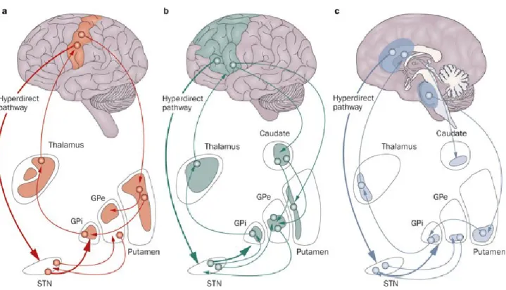
The striatum, pallidum, STN, and SN are strongly interconnected through intrinsic BG pathways that transmit information from the afferent to the efferent targets of the BG. Afferent projections to the BG originate from various cortical areas that have different functions. Information from each type of area is transmitted through BG pathways to the BG output structures and is sent back to the same cortical areas. This creates what is referred to as cortico-basal ganglia functional loops. Three main functional loops have been identified (Alexander & Crutcher 1990, Alexander et al 1986, Middleton & Strick 2000): the associative (oculomotor and prefrontal), limbic, and motor loops (Fig.2).
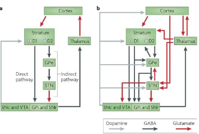
The motor loop starts with cortical areas, mainly from the frontal cortex that project onto the two main BG input structures from which the three major BG pathways originate; the direct, the indirect and the hyperdirect pathways. The first and main input structure is the striatum which gives rise to the direct and indirect pathways. It receives cortical projections mainly onto its GABAergic medium spiny neurons (MSN). These neurons can express one of two different types of dopamine receptors, D1 or D2 (albeit with a small degree of overlap) through which the SNpc dopaminergic projections modulate the striatum. Expression of these neurons corresponds to their implication in the direct and indirect pathways. Indeed, in the direct pathway, MSNs with D1 receptors project monosynaptically to the GPi and the SNpr while the indirect pathway constitutes MSNs with D2 receptors that project polysynaptically to the GPi/SNpr via the GPe (GABAergic projections) and the STN (glutamatergic projections). The GPi and the SNpr, therefore, receive GABAergic input from both the striatum, as part of the direct pathway, as well as a glutamatergic input from the STN as part of the indirect pathway (Fig.3A). More recent studies have found that the GPi also receives projections from the GPe (Fig.3B) (Smith et al 1998).
Figure 1. (Kandel 2012): The basal ganglia and surrounding structures in the human brain. A: The image in the top left shows a 3D representation of the striatum,
composed of the caudate nucleus and the putamen. B: Coronal section of a human brain showing the nuclei of the basal ganglia, identified on the right of the section.
The second input structure of the BG is the STN which receives strong glutamatergic projections from the cortex and projects directly to the BG output structures through what is referred to as the hyperdirect pathway (Nambu et al 2002).
The BG output structures project first to the motor thalamus (Alexander et al 1986), to eventually project back to the cortex. This motor loop is not completely closed since the BG also projects to the pedunculopontine nucleus in the brainstem through the GPi (Redgrave & Coizet 2007). These pallidal projections to the thalamus and the pedunculopontine nucleus are not completely segregated, as a proportion of GPi neurons project to both structures through collaterals (Parent & Hazrati 1995a). Amongst the different BG loops, the motor loop has received a lot of attention mainly because of the various motor disturbances that result in pathological BG motor output. This link between BG dysfunction and motor disorders has resulted in numerous studies investigating the contribution of the BG activity in the production of motor symptoms. In the following text, we will briefly introduce some of the best-known motor disorders that have been linked to BG pathological activity.
b. Basal ganglia pathological activity
Dysfunction of the BG motor circuit that involves the thalamus and the cortex causes a spectrum of motor disturbances that range from hypokinetic to hyperkinetic motor disorders (DeLong 1990).
Hypokinetic disorders are characterized by akinesia (impaired movement initiation and poverty of movement), bradykinesia (slowness of movement), muscular rigidity, and tremor at rest. One of the best-known examples of hypokinetic disorders is Parkinson’s disease (PD), which is caused by a relatively specific dopaminergic loss in the SNpc, which results in a deregulation of striatal activity and by extension, in the direct and indirect pathway. Because
striatal neurons involved in these two pathways express two types of dopamine receptors (D1 and D2) that polarize MSN differently when bound to dopamine, dopaminergic loss causes an imbalance of activity between the two pathways which ultimately results in increased GPi activity (this will be discussed in more detail in the STN section). This increased motor output from the BG results in an increased inhibition of the motor thalamus which in turns causes reduced activity in the thalamo-cortical projections ultimately resulting in hypokinesia (DeLong 1990).
Figure 2. (Volkmann et al 2010): The basal ganglia form anatomically and functionally segregated neuronal circuits with thalamic nuclei and frontal cortical areas. a: The motor circuit involves the motor and supplementary motor cortices, the
posterolateral part of the putamen, the posterolateral globus pallidus pars externa (GPe) and pars interna (GPi), the dorsolateral subthalamic nucleus (STN), and the ventrolateral thalamus. b: The associative loop (that includes the oculomotor and the prefrontal loops) and c: the limbic loop connects the prefrontal and cingulate cortices with distinct regions within the basal ganglia and thalamus. In the STN, a functional gradient is found, with a motor representation in the dorsolateral aspect of the nucleus, cognitive–associative
functions in the intermediate zone, and limbic functions in the ventromedial region. Via a 'hyperdirect' pathway, the STN receives direct projections from the motor, prefrontal and anterior cingulate cortices that can detect and integrate response conflicts (Nambu et al 2002, Frank 2004). This pathway is a powerful contact to influence basal ganglia outflow. (the substantia nigra is not shown).
On the other hand, hyperkinetic disorders are characterized by dyskinesia, defined by excessive motricity expressed through involuntary movements. One example of a hyperkinetic syndrome is hemiballismus, which involves proximal musculature of the limbs (Buruma & Lakke 1986, Lakke 1981, Sellal et al 1992, Whittier 1947) and has been linked to STN lesions in humans (Whittier 1947). These lesions were found to cause hypoactivity in the GPi that equates reduced BG motor output (Hamada & DeLong 1992b) resulting in a hyperactivity in the thalamo-cortical pathway, thought to account for the observed dyskinesia (DeLong 1990). These two examples emphasize that strong clinical manifestations arise when one or multiple parts of the BG show pathological neural activity, causing an imbalanced activity within the BG pathways. In the next section, we will summarize the contribution of one of these nuclei, the STN, to the maintenance of an appropriate motor output, and how deregulation of subthalamic activity contributes to hemiballismus and PD.
3.
The Subthalamic Nucleus
The subthalamic nucleus is a small oval-shaped structure located ventral to the thalamus. It can be divided into three functionally distinct subparts which have been categorized based on afferent cortical projections; a sensorimotor area in the dorsal part of the nucleus, an associative area located ventrally, and a smaller limbic area in the medioventral part of the nucleus (Fig.4) (Nambu 2011). The sensorimotor STN holds a central anatomical position in the motor circuit of the BG considering its role as an input structure as well as its strong excitatory projections to the GPi, the main motor output structure of the BG. The functional importance of these connections is supported by clinical studies linking various motor symptoms to dysfunctional subthalamic activity and by electrophysiological studies in primates. In the following section, we will present the most important connections of the STN as well as its role in motor disturbances and its contribution to motor control.
a. Anatomical connections
i. Afferent projections
The subthalamic nucleus receives strong excitatory projections from the cerebral cortex and a dense inhibitory projection from the GPe (Carpenter et al 1968, Smith et al 1990a). Other less prominent inputs include projections from the excitatory centromedian-parafascicular complex of the thalamus have been described in the rat and the cat (Groenewegen & Berendse 1990, Sugimoto et al 1983) and the pedunculopontine tegmental nucleus and the raphe nucleus in primates (Rinvik et al 1979). Since projections from the cortex and the GPe are the main sources of input to the STN, they will be the focus of this section.
Figure 3. (Redgrave et al): Organization of intrinsic connections within the basal ganglia. A: Model based on the influential proposal by Albin and colleagues (Albin et al
1989), according to which the output of the basal ganglia is determined by the balance between the direct pathway, which involves direct striatopallidal inhibitory connections to the GPi and the SNr, and the indirect pathway, which involves relays in the external
globus pallidus (GPe) and subthalamic nucleus (STN) that eventually project to the GPi and the SNr. The balance between these two projections is thought to be regulated by afferent dopaminergic signals from the substantia nigra pars compacta (SNc) and the ventral tegmental area (VTA) acting on differentially distributed D1 and D2 dopamine receptors. B: Recent anatomical investigations have revealed a more complex organization in which the transformations that are applied to the inputs to generate outputs are less easy to predict. Grey arrows indicate dopaminergic projections, blue arrows GABAergic projections, and red arrows glutamatergic projections. SNr, substantia nigra pars reticulata.
Cortical projections
The STN receives inputs primarily from the M1, the supplementary motor cortex (SMA) and the premotor cortex (Bankiewicz et al 1986). The largest of these inputs is from the M1, which projects mainly to the dorsolateral part of the ipsilateral STN (Monakow et al 1978). In primates, these projections are somatotopically organized into face, forelimb and hindlimb areas on the mediolateral axis (Nambu et al 1996). On the other hand, the dorsomedial part of the STN receives cortical inputs from the SMA and the dorsal and ventral premotor areas (PMd and PMv) (Nambu et al 1996, Nambu et al 1997). The SMA projections to the STN have a somatotopical organization that is a mirror image of the one found in the M1 region (Fig.4) (Nambu et al 1996). The face, forelimb and hindlimb regions of the M1 and the SMA also project in their respective territories to the corresponding body representations, creating a certain level of overlap. Projections from the forelimb representations in the PMv and PMd were found to overlap with each other and with the SMA projections.
Other less prominent cortical inputs to the STN originate from the cingulate motor area (CMA) and the pre-SMA. The caudal cingulate motor areas (CMAc) projects to the lateral STN while the rostral CMA (CMAr) and the pre-SMA project to the medial STN (Inase et al 1999, Takada et al 2001). These anatomical connections show that the STN funnels information from a wide range of cortical areas through the hyperdirect pathway to the BG motor output.
Pallidosubthalamic projections
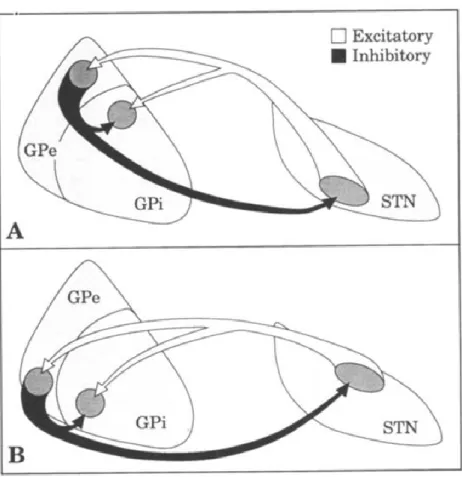
Although connections between the STN and the pallidum have been the subject of many studies, the topography of these connections is still not clear. According to one study,
the ventral GPe projects onto the dorsolateral STN which in turns projects to the ventromedial sensorimotor part of the GPi (Fig.5) (Shink et al 1996). This study is in line with the idea of functional segregation of the intrinsic BG connections where functional parts of each nucleus only project to areas of the same function. On the other hand, studies from Parent and Hazarti have suggested a complete segregation of pallidosubthalamic projections and subthalamopallidal projections (Parent & Hazrati 1995a, Parent & Hazrati 1995b). These results refute the existence of the GPe-STN-GPi connections, known as the indirect pathway (see next section).
Figure 4. (Modified from Nambu 2011): Somatotopy of the subthalamic nucleus (STN) in the primate. A: Somatotopy of the STN is shown in a frontal section. In the
dorsal part of the STN, the lateral part receives somatotopic inputs from MI (solid blue arrow), and the medial part from the SMA (solid red arrow). MI also projects partly to the medial part (dotted blue arrow), and the SMA to the lateral part of the STN (dotted red arrow). Ventral to the motor territory, there exist the oculomotor and prefrontal territories. The most medial part is occupied by the limbic territory. B: Input from motor cortices to the STN is schematically shown in a horizontal section.
ii. Efferent projections
Subthalamopallidal projections
According to a primate study, the majority of efferent subthalamic neurons have axon collaterals that project to both the GPi and the GPe (Sato et al 2000). These projections are thought to originate from the dorsal STN and project onto the sensorimotor part of the GPi located in its ventrolateral two thirds, as well as to the ventral GPe (Fig.5) (Shink et al 1996). On the other hand, the ventromedial part of the STN projects to the non-motor parts of the GPi and the GPe (Shink et al 1996).
Subthalamonigral projections
The SNpr is one of the biggest recipients of STN efferent projections, second to the pallidum (Nauta & Cole 1978). These projections originate mainly from the ventromedial third of the STN (Parent et al 1984, Parent & Smith 1987, Smith et al 1990b). They enter the SNpr from its dorsal or ventral parts and terminate in a patchy manner (Carpenter et al 1981). Although the majority of the STN projections to the SN terminate within the SNpr, some fibers ascend along the dopaminergic cell columns of the SNpc that go through the SNpr (Smith et al 1990b). In both the SNpr and the SNpc, the STN neurons seem to preferentially project onto non-dopaminergic neurons (Nauta & Cole 1978).
b. Clinical implications of subthalamic dysfunction
In order for the BG to produce an appropriate motor output, balanced activity between all the nuclei is essential. Because of the high connectivity between the STN and other important nuclei such as the GPi, an insults to the STN can strongly affect activity in the GPi which results in an inappropriate motor output to the motor thalamus and impedes proper motor control. In this section, we will present how pathological subthalamic activity contributes to BG-related motor disorders.
In the case of hemiballismus, lesion of the STN in primates has been shown to cause motor symptoms closely resembling human hemiballismus (Carpenter et al 1950, Hamada & DeLong 1992b), further supporting the link between reduced STN activity and dyskinesia.
This effect seems to be conveyed by the GPi since lesions to the STN also causes a reduction of GPi activity (Hamada & DeLong 1992a) and a subsequent lesion of the GPi abolishes STN-induced dyskinesia (Carpenter 1961, Carpenter et al 1950). These results show that the STN plays a role in suppressing involuntary movements through the subthalamopallidal pathway. They also suggest that the STN is an important modulator of the BG motor since its hypoactivity is related to a reduced BG motor output that is significant enough to result in dyskinesia.
Figure 5. (Shink et al 1996): Anatomical connections between the STN and the pallidum. Schematic diagram to summarize the relationships between the two pallidal
segments (GPe and GPi) and the STN. Small groups of interconnected neurons in the associative (A) and sensorimotor (B) territories of GPe and STN innervate, via axon collaterals, a common functionally-related region in the GPi.
In the case of PD, activity in the STN was found to be increased. Indeed, loss of SNpr inhibitory dopaminergic projections to striatal D2 neurons (involved in the indirect pathway) increases their GABAergic projections to the GPe. This causes reduced activity in the GPe which releases the STN from its tonic inhibition. This, in turn, causes an increased activity in the STN which sends stronger excitatory glutamatergic projections to the GPi (Bergman et al 1994, Miller & DeLong 1987). Furthermore, when this hyperactivity is countered with STN lesions in parkinsonian 1-methyl-4-phenyl-1,2,3,6-tetrahydropyridine (MPTP) primates, there is a reduction of all of the major motor disturbances in the contralateral limbs, including akinesia, rigidity, and tremor (Aziz et al 1992, Aziz et al 1991, Benabid et al 1993, Benazzouz et al 1993, Bergman et al 1990, Guridi et al 1993). This improvement of motor disturbances that follows STN lesion can be explained by its effect on the GPi. Indeed, one study showed that lesion of the STN in MPTP primates reduces the hyperactivity of the GPi (Wichmann et al 1994b).
These studies show that increased subthalamic activity results in increased inhibition of thalamocortical neurons by GPi output which eventually results in the hypokinetic motor disturbances that characterize parkinsonian symptoms. These studies in primates provided sufficient evidence for clinical studies to use deep brain stimulation on human patients which allows reversible lesioning of the STN or the GPi (Rodriguez-Oroz et al 2005, Samuel et al 1998). This has been successful in reducing pathological BG motor output and in turn, reduces motor symptoms in patients (Wichmann & DeLong 2003).
These clinical studies emphasize the importance of the sensorimotor STN in the control of movement. In the next section, we will present studies that have investigated the role of the STN in motor control.
c. Relationship of subthalamic activity to movement
Neural activity of the STN in relationship to movement has been mainly studied in monkeys, with a few studies in cats, and was found to correlate to different movement parameters. In primates executing a reaction time task, the relationship between the discharge frequency and each movement parameter was tested during both the reaction time (RT) and movement time (MT) (Georgopoulos et al 1983). The results showed statistically significant