Supporting Information
Bimetallic Salen-based compounds and their potential applications
Alba Finelli,
†Sarah-Luise Abram,
†Nelly Hérault,
†Aurélien Crochet,
‡Katharina M.
Fromm,
*,††
Department of Chemistry, University of Fribourg, Ch. du Musée 9, 1700 Fribourg, Switzerland
‡ FriMat, Department of Chemistry, University of Fribourg, Ch. du Musée 9, 1700 Fribourg,
Switzerland.
Table of Contents
General... 2 Synthesis ... 2 TGA analysis ... 3 Crystallography ... 5 Additional structures ... 6 XPRD of complexes 2-6... 10General
All measurements were performed at 250 K or 200 K, with Stoe IPDS-II or IPDS-II T diffractometers equipped with Oxford Cryosystem open-flow cryostats. Single crystals were picked under the microscope and placed in inert oil. All crystals were mounted on loops and all geometric and intensity data were taken from one single crystal. The absorption corrections were partially integrated in the data reduction procedure. The structures were solved and refined u sing full-matrix least-squares on F2 with the SHELX-2014 package. All atoms (except hydrogen atoms)
were refined anisotropically. Hydrogen atoms were refined where possible, and otherwise added using the riding model position parameters. Powder X-ray spectra were collected on a Stoe STADIP using Cu-Kα1 radiation (1.5406 Å) using a Mythen detector. TGA were recorded on a Mettler Toledo TGA7SDTA851. The structures were solved and refined using full-matrix least-squares on F2 with the SHELX-2014 package. All atoms (except hydrogen atoms) were refined anisotropically. Hydrogen atoms were refined when possible, and otherwise added using the riding model position parameters. Crystallographic data can be found in the Supporting Information (see Table S1). CIF files can be obtained from the Cambridge Crystallographic Data Centre, CCDC-1860979 (1), CCDC-1861009 (2), CCDC-1860982 (3), CCDC-1860981 (4), CCDC-1860983 (5), CCDC-1860980 (6), CCDC-1861005 (“LCuMn Cl2”), CCDC-1861004
(“LCuMn ClO4”), and CCDC- 861006 (“LCuMn OAc”). Copies of these data can be obtained
free of charge from the Cambridge Crystallographic Data Centre via www.ccdc.cam.ac.uk or e -mail, [email protected]. Powder X-ray spectra were collected on a Stoe STADIP using Cu-Kα1 radiation (1.5406 Å) using a Mythen detector.
Synthesis
Synthesis H2L. The compound H2L was synthesized as described in the literature. Ethane-1,2-
diamine (0.395 g, 0.875 ml, 6.57 mmol) was added to a solution of o-vanillin (2g, 13.15 mmol) in methanol (1000 ml). After stirring the mixture for 2h at room temperature, the yellow precipitate was filtrated, washed with methanol and, diethyl ether to be dried under vacuum to produce the ligand H2L (4.02 g, 94%). 1H NMR (400 MHz, DMSO): δ 3.76 (s, 6H), 3.92 (s, 4H), 6.77-6.81 (t,
J= 7.9 Hz, 2H), 6.98-7.2 (dd, J=71.7, 1.4Hz, 4H), 8.56 (s, 2H), 13.52 (s, 2H). ESI-MS (m/z): 329.1 [M+H]+ .
Synthesis of [LCu(H2O)] “LCu”. The complex LCu was synthesized as described in the
literature. A solution of Cu(OAc)2·H2O (152 mg, 0.761 mol) was added to a solution of H2L (250
mg, 0.761 mol) in methanol (30 ml). The mixture turned dark green immediately. After the mixture was stirred for 3h at room temperature, the precipitate was collected by filtration and dried under vacuum to produce the complex LCu (278 mg, 90%). Green lozenge -like single crystals suitable for X-ray crystallographic analysis were obtained by recrystallization in DMF. ESI -MS (m/z): 408.0 [M+H]+ .
TGA analysis
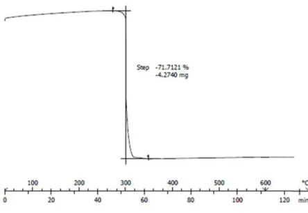
Figure S1: TGA analysis of compound 3 “LCuMn”.
Table S1: Results (phases and crystallite sizes) upon the different thermal decompositions of
LCuMn samples and of the mixture of Cu(NO3)2 and Mn(NO3)2.
Temperature Phase (%) Crystallite size [nm] LCuMn 300°C Cu1.5Mn1.5O4 (100%) 13.5 400°C Cu1.5Mn1.5O4 (100%) 20 500°C Cu1.5Mn1.5O4 (100%) 48 600°C Cu1.5Mn1.5O4 (100%) 125
Cu(NO3)2/Mn(NO3)2 600°C CuMn2O4 (80%)
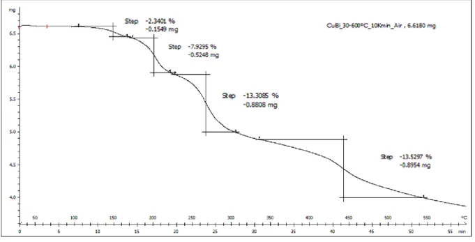
Figure S2: TGA analysis of compound 6 “LCuBi” between 30 and 600°C in air,
The total combustion of the LCuBi complex to CuBi
2O
4should follow the suggested
equation:
2(C18H18BiCuN4O10) + 41.5 O2 → CuBi2O4 + CuO + 36 CO2 + 18 H2O + 8 NO
Table S2: Results (phases and crystallite size) upon the different thermal decompositions of
LCuBi samples and of the mixture of Cu(NO3)2 and Bi(NO3)3.
Temperature Phase (%) Crystallite size [nm] LCuBi 300°C Bi2CuO4 (53%) Bi2O3 (42%) CuO (5%) 90 400°C Bi2CuO4 (55%) Bi2O3 (43%) CuO(2%) 90 500°C Bi2CuO4 (69%) Bi2O3 (26%) CuO (5%) 250 600°C Bi2CuO4 (96%) CuO (4%) 430 Cu(NO3)2/Bi(NO3)3 600°C Bi2CuO4 (70%) B2O3 (20%) B2O3 (sphaerobismoite) (10%) 90
Crystallography
Table S3: Bonds lengths in [
Å]of all crystal structures obtained for 1-6.
LCuBi (6) LCuCu (4) LCuZn (5) LCuAg (1)
Length
[
Å] O2 ̶ M1 1.912(14) 1.884(3) 1.882(2) 1.901(3) O3 ̶ M1 1.926(13) 1.909(3) 1.879(1) 1.905(3) N1 ̶ M1 1.927(19) 1.904(3) 1.908(2) 1.937(3) N2 ̶ M1 1.926(18) 1.917(3) 1.906(2) 1.917(3) O1 ̶ M2 2.605(14) 2.957(3) 2.542(2) 2.713(3) O2 ̶ M2 2.359(13) 2.280(3) 2.070(1) 2.372(3) O3 ̶ M2 2.351(13) 1.996(3) 2.073(2) 2.374(3) O4 ̶ M2 2.738(14) 2.506(4) 2.539(2) 2.729(3) O5 ̶ M2 2.460(18) 2.001(3) 1.92(2) 2.283(3) O6 ̶ M2 2.622(19) 2.052(3) 2.062(5) 2.783(3) O8 ̶ M2 2.744(19) 1.933(8)/1.942(9) 2.010(2) O9 ̶ M2 2.582(16) M1 ̶ M2 3.414(3) 3.115(8) 3.089(5) 3.350(6) BVS M1 1.90 1.93 1.98 1.97 M2 2.46 1.92(50%)/1.91(50%) 1.9(70%)/2.13(30%) 1.04 LCuMn (3) Length[
Å] O2 ̶ M1 1.889(7) O4 ̶ M1’ 1.885(7) N1 ̶ M1 1.914(9) N2 ̶ M1’ 1.913(9) O1 ̶ M2 2.481(7) O2 ̶ M2 2.177(7) O3 ̶ M2’ 2.475(6) O4 ̶ M2’ 2.182(7) O5 ̶ M2 2.290(9) O6 ̶ M2 2.292(10) O8 ̶ M2’ 2.330(10) O9 ̶ M2’ 2.307(10) M1 ̶ M2 3.227(7)/3.226(7) BVS M1 1.93/1.95 M2 2.04/1.96Additional structures
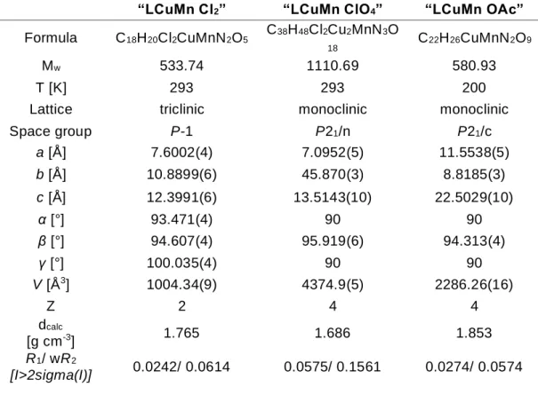
Table S4: Corresponding crystallographic data.
“LCuMn Cl2” “LCuMn ClO4” “LCuMn OAc”
Formula C18H20Cl2CuMnN2O5 C38H48Cl2Cu2MnN3O
18 C22H26CuMnN2O9
Mw 533.74 1110.69 580.93
T [K] 293 293 200
Lattice triclinic monoclinic monoclinic
Space group P-1 P21/n P21/c a [Å] 7.6002(4) 7.0952(5) 11.5538(5) b [Å] 10.8899(6) 45.870(3) 8.8185(3) c [Å] 12.3991(6) 13.5143(10) 22.5029(10) α [°] 93.471(4) 90 90 β [°] 94.607(4) 95.919(6) 94.313(4) γ [°] 100.035(4) 90 90 V [Å3] 1004.34(9) 4374.9(5) 2286.26(16) Z 2 4 4 dcalc [g cm-3] 1.765 1.686 1.853 R1/ wR2 [I>2sigma(I)] 0.0242/ 0.0614 0.0575/ 0.1561 0.0274/ 0.0574
“LCuMn Cl2”
Figure S3: Asymmetric unit of the complex [{LCuMn(Cl)2}(H2O)] abbreviated as “LCuMn Cl”.
Brown needle-like single crystals of compound “LCuMn Cl” were obtained from the mother liquor of the reaction by slow evaporation of the solvent (ACN/EtOH). In this complex, Mn(II) is coordinated by all four O-atoms belonging to the phenoxy and the methoxy groups of the ligand, occupying perfectly the O2O2 cavity. The manganese ion completes its coordination
sphere with two chlorine atoms (Cl1, Cl2). The presence of one non-coordinative molecule of water creates H-Bonds with chlorine atoms allowing an expansion along a-axis.
“LCuMn ClO4”
Figure S4: Asymmetric unit of the complex [{(LCu)(LCuMn(H2O)(µ-H2O))} (ClO4)2] abbreviated
as “LCuMn ClO4”.
Brown rhombohedral-shape single crystals of compound “LCuMn ClO4” were obtained from
the mother liquor of the reaction by slow evaporation of the solvent (ACN/EtOH). In this complex, Mn(II) is coordinated by all four O -atoms belonging to the phenoxy and the methoxy groups of the ligand, occupying perfectly the O2O2 cavity. The manganese ion completes its
coordination sphere with one N-atom (N5) of an acetonitrile molecule solvent and two O-atoms (O9, O10) from two molecules of water which one of them (O10) creates fours H-bonds with a neighboring LCu metalloligand producing a dimer-like structure. In this structure, two non-coordinated perchlorate ions allow an expansion via H -Bonds generated by O9 along a-axis.
“LCuMn OAc”
Figure S5: Asymmetric unit of the complex [LCuMn(OAc)2
(H
2O)
] abbreviated as “LCuMn OAc”.Purple needle-like single crystals of compound “LCuMn OAc” were obtained from the mother liquor of the reaction by slow vapor diffusion of diethyl ether in ACN/EtOH. In this complex, Mn(II) is coordinated by all four O-atoms belonging to the phenoxy and the methoxy groups of the ligand, occupying perfectly the O2O2 compartment. The manganese ion completes its
coordination sphere with one O-atom (O9) from a molecule of water and two O-atoms (O5, O7) from two acetate anions.
XPRD of complexes 2-6
Figure S6: XPRD diffractogram of the “LNiAg” (2) compound obtained (top) and the crystal
structure simulation 2 (below).
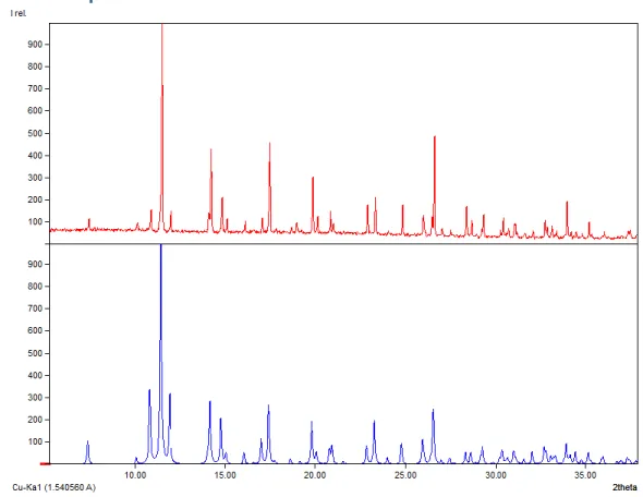
Figure S7: XPRD diffractogram of the “LCuMn” (3) compound obtained (top) and the crystal
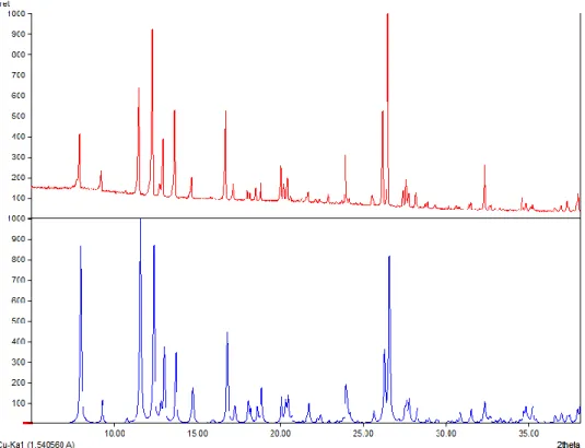
Figure S8: XPRD diffractogram of the “LCuCu” (4) compound obtained (top) and the crystal
structure simulation 4 (below).
Figure S9: XPRD diffractogram of the “LCuZn” (5) compound obtained (top) and the crystal
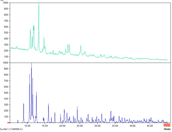
Figure S10: XPRD diffractogram of the “LCuBi” (6) compound obtained (top) and the crystal
![Table S3: Bonds lengths in [ Å] of all crystal structures obtained for 1-6.](https://thumb-eu.123doks.com/thumbv2/123doknet/14486763.525185/5.1262.441.1157.308.735/table-s-bonds-lengths-å-crystal-structures-obtained.webp)
![Figure S3: Asymmetric unit of the complex [{LCuMn(Cl) 2 }(H 2 O)] abbreviated as “LCuMn Cl”](https://thumb-eu.123doks.com/thumbv2/123doknet/14486763.525185/7.893.188.700.140.860/figure-asymmetric-unit-complex-lcumn-cl-abbreviated-lcumn.webp)
![Figure S4: Asymmetric unit of the complex [{(LCu)(LCuMn(H 2 O)(µ-H 2 O))} (ClO 4 ) 2 ] abbreviated as “LCuMn ClO 4 ”](https://thumb-eu.123doks.com/thumbv2/123doknet/14486763.525185/8.893.272.637.163.539/figure-asymmetric-unit-complex-lcu-lcumn-abbreviated-lcumn.webp)
![Figure S5: Asymmetric unit of the complex [LCuMn(OAc) 2 (H 2 O) ] abbreviated as “LCuMn OAc”](https://thumb-eu.123doks.com/thumbv2/123doknet/14486763.525185/9.893.211.688.137.775/figure-asymmetric-unit-complex-lcumn-oac-abbreviated-lcumn.webp)