NOTE TO USERS
This reproduction is the best copy available.
Universite de Sherbrooke
Structural definition of substrate recognition by model RNA capping enzymes and the identification of a novel class of viral RNA capping
enzymes
By
Issur Moheshwarnath Departement de biochimie
Thesis submitted to the Faculte de medecine et des sciences de la sante In partial fulfillment of the requirements for the obtention of the degree
Philosophiae doctor (Ph.D.) in Biochemistry
Library and Archives Canada Published Héritage Branch 395 Weiiington Street Ottawa ON K1A 0N4 Canada Bibiiothèque et Archives Canada Direction du Patrimoine de i'édition 395, rue Weiiington Ottawa ON K1A 0N4 Canada
Yourfile Votre référence ISBN: 978-0-494-83281-3 Our file Notre référence ISBN: 978-0-494-83281-3
NOTICE:
The author has granted a non-exciusive license allowing Library and Archives Canada to reproduce, publish, archive, preserve, conserve, communicate to the public by
télécommunication or on the Intemet,
ioan, distrbute and sell theses
Worldwide, for commercial or
non-commercial purposes, in microform, paper, eiectronic and/or any other formats.
AVIS:
L'auteur a accordé une licence non exclusive permettant à la Bibiiothèque et Archives Canada de reproduire, publier, archiver, sauvegarder, conserver, transmettre au public
par télécommunication ou par l'Intemet, prêter, distribuer et vendre des thèses partout dans le
monde, à des fins commerciales ou autres, sur
support microforme, papier, électronique et/ou autres formats.
The author retains copyright ownership and moral rights in this thesis. Neither the thesis nor
substantiel extracts from it may be
printed or otherwise reproduced without the author's permission.
L'auteur conserve la propriété du droit d'auteur et des droits moraux qui protégé cette thèse. Ni la thèse ni des extraits substantiels de celle-ci ne doivent être imprimés ou autrement reproduits sans son autorisation.
In compliance with the Canadien Privacy Act some supporting forms may have been removed from this thesis.
While these forms may be included in the document page count, their removal does not represent any loss of content from the thesis.
Conformément à la loi canadienne sur la protection de la vie privée, quelques formulaires secondaires ont été enlevés de cette thèse.
Bien que ces formulaires aient inclus dans la pagination, il n'y aura aucun contenu manquant.
Members of the evaluating commitee Dr. Martin Bisaillon
Dr. Eric Marsault
Dr. Lea Brakier-Gingras
Resume (en francais)
La structure coiffe est une unite protectrice situee a l'extremite 5' des ARNm eucaryotes. Cette structure est essentielle pour le transport et la traduction des ARNm. La coiffe est synthetisee co-transcriptionellement par trois activites enzymatiques consecutives : (1) Une ARN triphosphatase (RTase) qui hydrolyse le phosphate terminal de l'extremite 5' de TARN; (2) Une ARN guanylyltransferase (GTase) transfere un groupement GMP a cette extremite diphosphorylee et; (3) une ARN (guanine-N7) methyltransferase (MTase) qui ajoute un groupement methyle a la position N7 de la coiffe.
La cristallographie a permis d'elucider la structure de plusieurs enzymes impliquees dans la synthese de la structure coiffe. En depit du fait que ces structures ont permis de mieux comprendre plusieurs aspects mecanistiques du fonctionnement de ces enzymes, certains points nebuleux persistent, notamment, sur les interactions enzymes-substrats. Parmi les premieres etudes cristallographiques des enzymes de la synthese de la coiffe, la structure de la proteine Cetl, la RTase de Saccharomyces cerevisiae, fut elucidee en complexe avec une molecule de sulfate. La difficulte a elucider la structure du complexe enzyme-ligands est probablement liee a l'instabilite thermodynamique de ce complexe. Afin d'avoir une meilleure comprehension de l'activite RTase de S. cerevisiae une analyse en profondeur du site actif de cette enzyme en complexe avec un substrat approprie reste a etre etablie.
Les nombreuses etudes cristallographiques sur la GTase du virus Paramecium bursaria
chlorella virus-\ (PBCV-1) ont permis d'elucider le chemin reactionnel macroscopique de
cette famille d'enzyme. Cette proteine a ete cristallisee en presence de plusieurs ligands et sous differentes conformations; ce qui a permis de visualiser certaines etapes de la reaction de la synthese de la coiffe. Malgre tout, en depit du fait que le substrat naturel, le GTP, a ete co-cristallise avec la proteine, d'autres etudes permettant la comprehension de la specificite de la GTase envers la guanine doivent etre realisees. De plus, un mecanisme moleculaire de la catalyse de la reaction GTase manque toujours a nos connaissances. L'importance de la structure coiffe pour le processus de traduction est incontestable. La relation entre la coiffe et la machinerie traductionnelle a ete etudiee indirectement par 1'etude des proteines qui lient la coiffe. La plupart des etudes qui investiguaient directement la structure coiffe etaient restreintes a evaluer 1'inhibition de la traduction par des analogues de la coiffe. Des etudes sur les effets des analogues de la coiffe en 5' n'ont debute que depuis peu, et de plus, la fonction principale de la methylation en N7 de la coiffe n'a pas encore ete adressee.
Cette these vise done a fournir un apercu de la dynamique structurale des interactions enzyme-ligand de la RTase de S. cerevisiae et de la GTase de PBCV-1. Nous montrons que des analogues de purines peuvent etre un outil utile pour l'etude de plusieurs processus cellulaires, tels que la traduction. Au cours de mes etudes, nous avons decouvert une nouvelle classe de GTase virale dans la famille des flavivirus, foumissant ainsi un apercu plus succinct du complexe de replication de ce virus.
Key words: Analogues, ARNm, flavivirus, Proteines virales, Structure coiffe, Traduction,
SUMMARY:
The RNA cap structure is a fundamental feature of most known eukaryotic mRNAs and some viral RNAs. It is important for the stability, transport and translation of mRNAs. It is co-transcriptionally synthesized via the action of 3 consecutive enzymatic reactions: (1) A RNA triphosphatase which cleaves off the 5' terminal phosphate of nascent RNAs; (2) A RNA guanylyltransferase which transfers a GMP moiety onto the acceptor RNA; (3) A RNA (guanine-N7) methyltransferase which methylates the cap guanine at the N7 position. Through the end of the 1990's until now, the crystal structures of several capping enzymes have been solved. However, these structures, although very insightful in themselves, failed to provide any instructive information on several key issues regarding enzyme-substrate interactions. For instance, one of the first breakthrough crystallographic studies in RNA capping chemistry led to the elucidation of the yeast RNA triphosphatase structure (the Cetl protein). However, in the crystal structure, the Cetl protein was bound to a sulphate molecule, which was hypothesised to be mimicking the product of the RNA triphosphatase reaction- a phosphate molecule. The inability to capture the RNA triphosphatase in complex with its ligands is probably on account of the inherent thermodynamic instability of this protein when bound to RNA or a nucleotide. A structural definition of the active site of the yeast RNA triphosphatase in complex with an appropriate substrate is still lacking. In addition, the elucidation of the structure of the RNA guanylyltransferase of the
Paramecium bursaria chlorella virus-\ (PBCV-1) in several different conformations has
been a landmark study which greatly contributed towards the understanding of the catalytic pathway of this model enzyme. On the other hand, despite the presence of the natural substrate-GTP, within the active site of the enzyme, the rationale behind the GTP specificity of RNA guanylyltransferase remains poorly understood. Moreover, a molecular mechanism for the RNA guanylyltransferase reaction is still missing. Finally, the importance of the RNA cap for the process of eukaryotic translation is undisputable. However, the relationship between the RNA cap and translation has been mostly studied indirectly through proteins which bind to the cap structure. Most studies pertaining directly to the impact of the binding of the RNA cap structure have been restricted to investigating
the inhibitory potential of various cap analogues on the translation process. Studies on the effects of modified RNA caps at the 5' ends of RNAs have only started in the last few years, and more importantly, the necessity of the N7-methyl group on RNA cap analogues had not been addressed.
This thesis therefore aims to provide a structural insight into the structural dynamics of enzyme-ligand(s) interactions of the model S. cerevisiae's RNA triphosphatase and the PBCV-1 RNA guanylyltransferase. In addition, we show that purine analogues can be a useful tool for the study of several cellular processes, such as RNA translation. In the process we have uncovered a novel class of RNA capping enzyme in the flavivirus genus of the Flaviviridae family of RNA viruses, thus providing a more succinct insight into the flaviviral replication complex.
I dedicate this thesis to the memory of my father,
TABLE OF CONTENTS
TABLE OF CONTENTS VII
LIST OF FIGURES IX
LIST OF TABLES XII
ABBREVIATIONS XIII
INTRODUCTION
1. The lifecycle of eukaryotic mRNAs 1
1.1. Synthesis and co-transcriptional modifications 2
2. The RNA cap structure
2.1. Structure and function 4
2.2. Synthesis of the RNA cap structure 6
2.3. Functional organization of RNA capping enzymes 8
2.3.1. RNA triphosphatase 10
2.3.1.1. Metal ion-dependent RNA triphosphatase 10
2.3.1.2. Metal ion-independent RNA triphosphatase 12
2.3.2. RNA guanylyltransferase 14
2.3.2.1. Mechanism of RNA guanylyltransferase 15
2.3.2.2. Nucleotidyltransferase superfamily 15
2.3.2.3. Nucleotide specificity of RNA guanylyltransferases 18
2.3.3. RNA (guanine-N7) methyltransferase 19
2.4. Unconventional RNA capping apparatus 20
2.5. The other unconventional RNA guanylyltransferases and the case of the
flavivirus RNA capping machinery 24
3. Research objectives 27
3.1. General objectives 29
3.2 Specific objectives 29
RESULTS
1. Chapter I - Understanding protein-ligand interactions of the model yeast RNA triphosphatase
1.1. ARTICLE: Nucleotide analogs and molecular modeling studies reveal key
interactions involved in substrate recognition by the yeast RNA triphosphatase 30
2. Chapter II - Probing into the GTP specificity of a model RNA guanylyltransferase 2.2. ARTICLE: Biosynthesis of novel RNA cap analogues reveal key insights into
translation 58
3. Chapter III- Identification of a novel class of viral RNA guanylyltransferase
3.1 ARTICLE: The flavivirus NS5 protein is a true RNA guanylyltransferase that
catalyzes a two-step reaction to form the RNA cap structure 95
DISCUSSION 132
CONCLUSIONS 151
ACKNOWLEDGEMENTS 152
REFERENCES 153
ANNEX 160
1. Co-author approbation letters 161 2. Invited review article
LIST OF FIGURES
Introduction, Figure 1: Global overview of mRNA synthesis and co-transcriptional
modifications brought on the pre-mRNA in human cells 3
Introduction, Figure 2: The structure of the mRNA cap 4
Introduction, Figure 3: Importance of the RNA cap and Poly(A) tail 6
Introduction, Figure 4: RNA cap synthesis 7
Introduction, Figure 5: Functional Organization of capping enzymes 9
Introduction, Figure 6: Structures of yeast, viral and metazoan RNA triphosphatases 11 Introduction, Figure 7: Schematic depiction of the RNA triphosphatase mechanism 13 Introduction, Figure 8: Schematic depiction of the RNA guanylyltransferase mechanism..14 Introduction, Figure 9: Sequence alignments of RNA guanylyltransferases and
ATP-dependent ligases 16
Introduction, Figure 10: Structural similarity of DNA ligases and RNA
guanylyltransferases 17
Introduction, Figure 11: Mechanistic similarity between RNA guanylyltransferases and
ATP dependent DNA/RNA ligases 17
Introduction, Figure 12: A viral RNA guanylyltransferase can form a covalent intermediate
with Ribavirin monophosphate 19
Introduction, Figure 13: Schematic depiction of the RNA (guanine-N7) methyltransferase
mechanism 20
Introduction, Figure 14: Schematic depiction of the proposed model of the unconventional
RNA capping by the Vesicular stomatitis virus 21
Introduction, Figure 15: Schematic depiction of the proposed model of the unconventional
RNA capping by alphaviruses 22
Introduction, Figure 16: Schematic depiction of the model of the cap-snatching mechanism
by the influenza virus 23
Introduction, Figure 17: Schematic linear depiction and organization of the RNA genome
offlavivirus 25
Introduction, Figure 18: Schematic depiction of the replication cycle of tha flavivirus genus
Chapter I, Figure 1: Molecular docking model for the binding of GTP to the yeast RNA
triphosphatase 53
Chapter I, Figure 2: Active site of the yeast RNA triphosphatase 54
Chapter I, Figure 3: Nucleotide analogues used in the validation of the docking model 55 Chapter I, Figure 4: Inhibition of the phosphohydrolase activity by nucleotide analogues.56 Chapter I, Figure 5: Steric hindrance caused by analogues harbouring large substituents at
the 6-oxo position of the guanine ring of GTP 57
Chapter II, Figure 1: The RNA capping mechanism and the nucleotide analogues tested...88
Chapter II, Figure 2: Biosynthesis of novel RNA cap structures 89
Chapter II, Figure 3: In cellulo and in vitro properties of the novel cap analogues 90 Chapter II, Supplementary figure 1: Structural conservation in GTases and ligases 92 Chapter II, Supplementary figure 2: pH dependency of inhibition by 2' modified nucleotide
analogues 93
Chapter II, Supplementary figure 3: Binding of GTP to the wild-type and Lysl88 mutant of
thePBCV-1 GTase 94
Chapter III, Figure 1: Formation of a NS5-GMP covalent intermediate 126
Chapter III Figure 2: The NS5 N-terminal methyltransferase domain is active in the
formation of a protein-GMP covalent complex 127
Chapter III Figure 3: Transfer of GMP to an acceptor RNA 128
Chapter III Figure 4: Stimulation of the GTase activity by the NS3 protein 129
Chapter III Figure 5: Synthesis of the RNA cap 1 structure by the NS3 and NS5
proteins 130
Chapter III Figure 6. RNA cap synthesis by the flavivirus NS5 and NS3 proteins 131
Discussion, Figure 1: A brief summary of the major conclusions of Chapter II 135
Discussion, figure 3: The RNA cap binding site of E. cuniculi RNA (guanine-N7)
methyltransferase 139
Discussion, figure 4: In silico docking of 3'0-Me GDP to eIF4E 141
Discussion, figure 5: Recognition of the RNA cap structure by different RNA cap binding
proteins 143
»
Discussion, figure 6: Proposed model for cap recognition by a decapping enzyme 144
Discussion, figure 7: RNA structure and RNA capping by NS5WNV 146
Discussion, figure 8: Guanylylation and adenylation by NS5WNV 148
Discussion, Figure 10: Comparison of the GTP binding site of the N-terminal domain of the
NS5 protein of Murray Valley Encephalitis virus with the PBCV-1 GTase 150
LIST OF TABLES
Chapter I, Table I: Predicted interactions of the S. cerevisiae's RTase with GTP according
to the docking model 51
Chapter I, Table I: Inhibition of Cetl GTPase activity by nucleotide analogues 52
Chapter II, Table I: RNA capping reaction with nucleotide analogues 87
Chapter II, Supplementary Table I: RNA (guanine-N7) methyltransferase activities with
RNA capped with nucleotide analogues 91
Discussion, Table 1: Comparison of the K, of 4 potent inhibitors for the RTase of S.
cerevisiae against that of West Nile virus 133 Discussion, Table 2: Binding of purine nucleotides and dinucleotides to the N-terminal
domain of the Dengue virus NS5 protein 138
ABBREVIATIONS
A103R: The RNA guanylyltransferase of Paramecium bursaria chlorella virus-1 ATP: Adenosine triphosphate
CBP 20/80: Cap Binding Proteins 20 and 80
Cetl: The Capping enzyme triphosphatase of Saccharomyces cerevisiae CPSF: Cleavage and polyadenylation specificity factor
Dcpl/Dcp2: Decapping enzymes 1 and 2
eIF4E/eIF4G: Eukaryotic initiation factors 4 E and G GTP: Guanosine triphosphate
IRES: Internal Ribosome Entry Site Lys: Lysine
mRNA: Messenger RNA
NS5: The non-structural protein 5 NTP: Nucleoside triphosphate PAP: Poly(A) Polymerase PABP: Poly(A) Binding Protein
PBCV-1: Paramecium bursaria chlorella virus-1 Pol II: RNA Polymerase II
Poly(A): Poly adenosine RNA: Ribonucleic acid
SWI/SNF: SWItch/Sucrose NonFermentable TAFs: TBP Associated Factors
TBP: TATA Binding protein
INTRODUCTION
1. The lifecycle of eukaryotic mRNAs
The life cycle of mRNAs starts with their transcription and ends ultimately in their demise by degradation (Gu and Lima 2005). Eukaryotic mRNAs differ from their bacterial counterparts in several ways namely by undergoing several co-transcriptional modifications such as the addition of a RNA cap at their 5' end, the synthesis of a poly(A) tail at the 3' end following cleavage, and the splicing of introns to form mature functional mRNAs (Varani 1997). These modifications are crucial to establish the fate of the mRNA. Appropriately modified mRNAs will be eligible for transport to the cytoplasm and translation by ribosomes (Hamm and Mattaj 1990; Shatkin and Manley 2000). On the other hand, a pre-mRNA possessing a defect in maturation (a splicing defect for instance) is rapidly targeted for degradation (Isken and Maquat 2007).
An mRNA, from its synthesis to its degradation, is bound by several proteins and protein complexes throughout its life cycle. For instance, following synthesis of the 5' end by RNA Polymerase II and capping by the RNA capping machinery, the RNA cap structure is bound by the CBP20/80 complex in the nucleus. This interaction is important for the splicing of mRNAs as well as their transport from the nucleus to the cytoplasm (Izaurralde, Lewis et al. 1994; Visa, Izaurralde et al. 1996; Lewis and Izaurralde 1997; Newbury 2006). The CBP20/80 complex is exchanged for the eIF4E protein of the translation initiation complex in the cytoplasm. The binding of the RNA cap structure to eIF4E is fundamental for cap-dependent translation to occur. The 3' poly(A) tail also plays a vital role in the life cycle of mRNAs. It is bound by several proteins, the most notable one being PABP, which is vital to improve translation efficiencies of mRNAs (Kuhn and Wahle 2004).
The 5' and 3' modifications brought about on an mRNA also contribute to the stability of mRNAs. The 5' RNA cap structure blocks the 5' end in an unusual 5'-5' linkage, thus effectively preventing 5' exonucleases from degrading the mRNA. For a regulated degradation of mRNAs, decapping enzymes cleave off the cap structure to aid in RNA degradation (Newbury 2006). Therefore, due to the critical roles that RNA modifications play in the life cycle of mRNAs, most enzymes directly involved in the synthesis of these modifications have been shown to be essential. Extensive genetic studies in the model
organism budding yeast have shown that all components of the RNA capping machinery are crucial for survival (Shatkin and Manley 2000). In addition, in several viruses, abrogating any step of RNA cap synthesis leads to defective viral replication (Bisaillon and Lemay 1997). This underlines the fundamental importance of the RNA cap structure in the life cycle of eukaryotic mRNAs.
1.1 Synthesis and co-transcriptional modifications
Eukaryotic mRNAs are exclusively transcribed by the DNA-dependent RNA Polymerase II (Pol II). The transcription process is guided by several tightly regulated steps. Initially in the first steps of transcription, a pre-initiation complex consisting of the polymerase and transcription factors (such as TBP, TAFs and SWI/SNF units) binds to the promoter region of the gene of interest, and RNA synthesis begins. Initiation is followed by promoter clearance, when initial contacts between the promoter and the polymerase are broken. The polymerase complex then enters the elongation phase of transcription, culminating finally into transcription termination when the polymerase dissociates from the DNA template and the RNA transcript is released (Howe 2002). During the tightly regulated process of mRNA synthesis, several modifications are also brought about co-transcriptionally on the mRNA (Fig. 1) (Moore and Proudfoot 2009).
The first modification to occur is the addition of the 5' RNA cap structure. In vivo, cap addition occurs early during transcription, when about 25-30 nucleotides have been polymerized and the 5' end is protruding from the RNA binding pocket of the RNA Pol II. The enzymes involved in RNA cap synthesis are specifically recruited to the Pol II pre-initiation complex. The phosphorylation level of the CTD of the PolII holoenzyme mediates this specific recruitment at the beginning of the transcription process. The evolutionary conserved proteins, CBP20 and CBP80, co-transcriptionally bind as a heterodimer to the RNA cap structure shortly after its synthesis. The RNA cap structure retains its association to this cap-binding complex (CBC) throughout its transcription, co-transcriptional processing and nucleocytoplasmic export (Moore and Proudfoot 2009). During transcription elongation, intron removal occurs. This step is catalyzed by the
Stability of mRNAs: The unconventional 5'-5* bond in the RNA cap structure protects the
5' ends of mRNAs against most exonucleases since they only hydrolyze 3' -5' bonds. During the regulated degradation of mRNAs in cells, decapping enzymes (the Dcpl/Dcp2 complex in baker's yeast) degrades the 5'-5" linkage prior to the 5'to 3" degradation process (Newbury 2006). In cells, the cap structure is strongly bound by the Cap Binding Complex (CBC 20/80 in metazoans and CBC 1/2 in yeast) in the nucleus and eIF4E/eIF4G in the cytoplasm, which limits its access by the decapping machinery, thus effectively protecting the mRNA against 5' to 3' degradation (Moore and Proudfoot 2009).
Nucleo-cytoplasmic transport of mRNAs: Following transcription of mRNAs within the
nucleus, their transport to the cytoplasm for translation is ensured by the Cap Binding Complex. The Nuclear Cap Binding Complex binds exclusively to the RNA cap structure and is then recognized by the Nuclear Pore Complex, for transport into the cytoplasm, where, following the first pioneer round of translation, it is replaced by the eIF4E/eIF4G complex (Maquat 2004).
Translation efficiency of mRNA: Translation efficiency is significantly improved by the
presence of the 5' cap. The 5' cap plays a key role in recruiting protein co-factors, and ultimately the small ribosomal subunit to the 5' ends of mRNAs. In addition, the 5' cap, when bound by the cytoplasmic cap binding complexes (eIF4E/eIF4G), lead to the circularization of the mRNA molecule by interactions with poly(A) binding proteins bound at the 3'ends of mRNAs (Fig.3). Such interactions enable an efficient recycling of ribosomes, thus improving translational efficiency of mRNAs (Livingstone, Atas et al. 2010).
Splicing of the 5' proximal intron: The RNA cap structure has also been shown to be
important to promote intron excision during the splicing of the 5' proximal intron (Lewis, Izaurralde et al. 1996; Lewis and Izaurralde 1997). The precise mechanism by which this is achieved is still under study in model organisms (Raczynska, Simpson et al. 2010).
2.3 Functional Organization of RNA capping enzymes
From viruses to metazoans, the RNA cap structure is identical. However, the physical organization and to some extent the mechanism of the enzymes responsible for its synthesis differ significantly across the various taxa (Ghosh and Lima 2010). While in all known unicellular eukaryotes, each RNA modifying activity for RNA cap synthesis lies on a separate polypeptide, in higher order eukaryotes like plants and metazoans, the RNA (guanine-N7) methyltransferase is segregated from the RNA triphosphatase and RNA guanylyltransferase which are fused together in a single polypeptide (Fig. 5). On the other hand, with regards to viruses, no common theme distinct to any viral group emerges. However, the genomic organization as well as the mechanism of some of the proteins involved in RNA capping (more precisely the RNA triphosphatase and the RNA guanylyltransferase) diverges widely from those in unicellular eukaryotes or from those in plants and metazoans (Fig. 5).
2.3.1 RNA triphosphatase
The RNA triphosphatase catalyzes the first step of RNA cap synthesis. Across the various eukaryotic lineages, RNA triphosphatases differ significantly with respect to structure and catalytic mechanism and can be grouped into 2 distinct families: (1) metal-dependent RNA triphosphatases of lower eukaryotes such as fungi and protozoans (2) metal-independent RNA triphosphatases of nematodes, metazoans and plants (Fig. 6 and 7).
2.3.1.1. Metal-dependent RNA triphosphatases
The metal-dependent RNA triphosphatase has been identified in several eukaryotic lineages ranging from DNA viruses to fungi and protozoans. These RNA triphosphatases belong to the triphosphate tunnel metalloenzyme (TTM) family of phosphohydrolases and in contrast to metal-independent RNA triphosphatases, can hydrolyze NTPs (Ghosh and Lima 2010). The initial crystallization of the 5". cerevisiae's RNA triphosphatase, the Cetl protein, revealed its homodimeric nature, whereby two equivalent active sites are present around a topologically closed eight-stranded anti-parallel P barrel (Fig. 6A) (Lima, Wang et al. 1999). Biochemical and mutational evidences also suggest that this then-novel fold also encompasses the RNA triphosphatases of other fungi (Schizosaccharomyces pombe,
Candida albicans), protozoan parasites (Plasmodium falciparum, Trypanosoma brucei, Encephalitozoon cuniculi, Giardia lambia) and some DNA viruses (Chlorella virus,
poxvirus, baculovirus) (Shuman 2002). The recent elucidation of the Mimivirus RNA triphosphatase domain crystal structure has provided the first structural evidence for the inclusion of viral RNA triphosphatases in the TTM clade (Fig. 6B) (Benarroch, Smith et al. 2008). Most interestingly, the TTM fold is more widely distributed across the various taxa than initially expected. Its finding within the archael and bacterial domains of life suggests a deeper evolutionary origin. The family of metal-dependent RNA triphosphatases is exemplified by the model S. cerevisiae's RNA triphosphatase, the Cetl protein. Its active site consists of several basic residues presumably important for coordinating the triphosphate moiety of pppRNA, as well as some basic residues coordinating metal ions which directly interact with the pppRNA triphosphate moiety (Fig. 7A). Phosphohydrolysis by RNA triphosphatases of the TTM clade is purported to occur in a one-step in-line
2.3.1.2. Metal ion- independent RNA triphosphatase
In higher eukaryotes the metal-independent RNA triphosphatase belonging to the cysteine-phosphatase enzyme superfamily, prevails. The elucidation of the crystal structure of the mouse RNA triphosphatase provided the first structural evidence that the mammalian RNA triphosphatase (Mcel), which is defined by a central five-stranded parallel B-sheet flanked with several a-helices, is entirely unrelated to the TTM-clade of RNA triphosphatases (Fig. 6D) (Fabrega, Shen et al. 2003). Phosphohydrolysis by these metal-independent RNA triphosphatases occurs in two steps not unlike the protein phosphatases of the same family. In the first step of the reaction, the conserved cysteine (Cys 126 in Mcel) attacks the 5' y-phosphate of the RNA transcript to form a covalent protein-cysteinyl-5-y-phosphate intermediate with the concomitant release of the 5' ppRNA product. In the second step, the covalent phosphoprotein is hydrolyzed to liberate inorganic phosphate (Fig. 7B) (Ghosh and Lima 2010).
With the ultimate intent to find novel antiviral and anti-microbial targets, this thesis is more concerned with RNA triphosphatases of the TTM clade, more specifically, the model Cetl protein. The relative instability induced to the Cetl protein upon ligand binding is probably the reason behind the lack of structural data of these enzymes bound to their substrates (Bisaillon and Bougie 2003). TTM RNA triphosphatases have only been co-crystalized with magnesium ions and sulphate or acetate bound within the active site. The bound sulphate/acetate molecule is purported to act as the y-phosphate of the substrate RNA. In this thesis, through computational and biochemical methods we provide the first insight into how the tunnel active site of the TTM RNA triphosphatases binds to their substrates.
2.3.2.1.Mechanism of RNA guanylyltransferase
Two distinct catalytic events, in a ping-pong type reaction, define the RNA guanylyltransferase mechanism (Fig.8). In the first step, attack of the a-phosphorus of GTP by the capping enzyme results in the release of pyrophosphate and formation of a covalent enzyme-(/>'5>'/-A/)-GMP intermediate (Fig.8A). The second step entails the transfer of this
covalently bound GMP onto an acceptor RNA (Fig.8B). The RNA substrate in the second step of the reaction is specifically a diphosphorylated RNA formed from the hydrolysis of the terminal 5' phosphate by an RNA triphosphatase. Both steps of this reaction require the presence of a divalent metal ion and have been shown to be reversible (Souliere, Perreault et al. 2008). The catalytic lysine, the s amino group of which forms the covalent phosphoamidate intermediate in the first step, is part of a KxDG motif (Motif I), one of six co-linear conserved sequences (I-VI) defining the active site of this family of enzymes (Fig.9).
2.3.221 Nucleotidyltransferase superfamily
Interestingly, the mechanism as well as the conserved motifs (I-VI) of capping enzymes are shared by ATP-dependent RNA and DNA ligases, which along with the former belong to the broader nucleotidyltransferase superfamily (Fig. 9) (Doherty and Suh 2000). In fact, crystal structures of the Chlorella virus RNA guanylyltransferase and of the T7 DNA ligase bound to GTP and ATP respectively have revealed a common tertiary fold in which the conserved motifs are assembled together at the enzyme's active site (Fig. 10). Both enzymes can be split into 2 distinct domains: a larger N-terminal domain encompassing motifs I, III, Ilia, IV and V which form the nucleotide binding pocket, and a smaller C-terminal OB fold domain, which includes motif VI. Indeed, the enzyme-AMP intermediate, analogous to the phosphoamidate intermediate during cap synthesis, formed when RNA/DNA ligases react with ATP, clearly highlights the mechanistic similarity shared by the members of this superfamily (Fig. 11).
2.3.2.3. Nucleotide specificity of RNA guanylyltransferases
Despite the availability of more structural and biochemical data on several members of the nucleotidyltransferase superfamily, an accurate understanding of the substrate specificity of RNA capping enzyme still remains elusive. As yet, from a biological perspective, studies pertaining to this particular issue are scarce. It is worth mentioning that through domain swapping between the T7 ligase and the PBCV-1 RNA guanylyltransferase, an ATP capping enzyme has been previously generated (Doherty 1999). However, modulation of the substrate specificity of RNA guanylyltransferases through the rational design of point mutations remains to be achieved. While mutational analyses have yielded substantial data on protein-substrate interactions, gauging the importance of each with regards to their importance in substrate recognition is tedious and limited in its scope.
In 2004, the report that, in vitro, ribavirin triphosphate (RTP) could be used as a cap donor by the vaccinia virus RNA capping enzyme revived interest in the possibility of modulating the substrate specificity of RNA guanylyltransferases (Fig. 12) (Bougie and Bisaillon 2004). This was the first report that an RNA capping enzyme could potentially use an alternative substrate to GTP to generate a modified RNA cap structure at the 5' end of an mRNA. In the wake of this study, we decided to investigate into the propensity of a model RNA guanylyltransferase to use artificial substrates as cap donors. Synthetic cap analogues have been extensively studied with regards to RNA stability and translation. However, few studies have aimed to probe into the capping machinery itself with a view to understand the underlying interactions leading to substrate discrimination. In this thesis we present a thorough study of the structural requirements of a ligand to be a cap donor by the model PBCV-1 RNA guanylyltransferase. We use purine analogues bearing various modifications at different positions and by gauging the effect of each modification, we traced the essential recognition motifs on the guanosine residue which determine its inherent ability to act as a cap donor.
2.5. The other unconventional RNA guanylyltransferases and the case of the Jlavivirus genus
Unconventional RNA guanylyltransferases have been observed in several families of RNA viruses. In several human pathogenic viruses, the identity of the RNA capping apparatus is still unknown. For instance, the RNA (guanine-N7) methyltransferase of members of the
coronavirus genus, of which the deadly SARS virus forms part of, has only been recently
identified on the nspl protein of the virus (Chen, Cai et al. 2009). The identity of the RNA guanylyltransferase is still under investigation. In the Jlavivirus genus, of the Flaviviridae family, the identity of the complete RNA capping apparatus has only recently been confirmed.
The Jlavivirus genus belongs to the Flaviviridae family of viruses along with the
Hepacivirus and Pestivirus genera. These genera differ in numerous ways. One major
divergence is the presence of an IRES at the 5' ends of the RNA genomes of the
Hepacivirus and Pestivirus genera whereas members of the the Jlavivirus genus harbour a
RNA cap 1 structure. All members of the flavivirus genus possess an RNA cap 1 structure at the 5' end of their RNA genomes. The organization of the RNA genome and the nature of the 5' cap are shown in figure 17 (Bollati, Alvarez et al. 2010).
The Jlavivirus genus comprises several medically important pathogens, the most notorious one being the Dengue Fever virus. This family also includes the Yellow Fever virus, the West Nile virus and the Japanese Encephalitis virus, amongst others (Ecker, Sampath et al. 2005). Flaviviruses are arthropod-born viruses, possessing a complex life cycle involving 2 distinct hosts, mosquito and human (Bollati, Alvarez et al. 2010). The replication cycle of flaviviruses is shown in figure 18.
The viral E protein mediates the attachment of the virions to the host cell surface; and the virions penetrate cells by receptor mediated endocytosis. In the low pH of the endosome, fusion of the viral and host membranes occurs which leads to the release of the nucleocapsid and the viral RNA into the cytoplasm of the cell. The translation of the viral RNA generates a polyprotein which is co-translationally and post-translationally processed
3. Research Objectives
Enzymes involved in the synthesis of the RNA cap structure are essential for survival. In all eukaryotic organisms, hindering the RNA capping process severely hampers cell growth or leads ultimately to cellular death. In viruses encoding RNA capping apparatuses, inhibiting any of the components of this apparatus severely undermines viral replication. The study of RNA capping enzymes is important because due to their essentiality they can prove to be potential anti-microbial targets. In addition, with regards to viral RNA capping enzymes, they are valuable tools for the mechanistic study of RNA capping enzymes in general on account of their small sizes, which englobe all the essential catalytic motifs for activity. Therefore, it is of fundamental importance to study and make a thorough characterization of RNA capping enzymes, with a view to evaluate their similarity with related enzymes in human and thus, gauge their potency as anti microbial targets.
The yeast RNA triphosphatase is the model enzyme for the study of most fungal, protozoan, and trypanosomal related enzymes, including that from the deadly malaria parasite, Plasmodium falciparum. The crystal structure of this enzyme has been solved in complex with a sulphate molecule, purported to mimic the 7-phosphate of the 5' end of an mRNA. Ligand (RNA or NTP) binding thermodynamically destabilizes the protein relative to its free unbound form. This is probably why the structure of the enzyme-ligand complex cannot be elucidated by crystallography for now. The yeast RNA triphosphatase, which belongs to the TTM family of metalo-enzymes, can prove to be an efficient drug target, mainly on account of the fact that the metazoan RNA triphosphatase belongs to the TTM-unrelated metal-independent cysteine phosphatase family. A more acute understanding of the interactions of the enzyme with RNA, could potentially prove to be useful for the design of anti-microbial agents. Therefore, our aim is to probe into the molecular determinants for ligand binding by the model enzyme of the TTM family of RTases, the S.
cerevisiae's RTase.
RNA guanylyltransferases are conserved across various viral lineages as well as most of the known eukaryotic taxa. These enzymes belong to the nucleotidyltransferase superfamily, which also includes the ATP dependent or NAD+ dependent DNA/RNA ligases. Despite sharing the same conserved motifs in the same order, as well as displaying very similar
tertiary structure, ligases and RNA guanylyltransferases possess very different nucleotide specificity. Structures of both RNA guanylyltransferases and ligases have been elucidated bound to GTP and ATP respectively. However, even though the molecular contacts between the ligand and the enzyme are known, modulating the substrate specificity of these enzymes has proven be unfeasible. A precise understanding of their substrate specificity could potentially pave the way for the design of novel proteins with novel activities, which could be of use for drug development. In this study our aim is to find the essential recognition elements of GTP which render it able to act as RNA cap donor. The PBCV-1 RNA guanylyltransferase was the model used since it has been co-crystallised several times in complex with GTP. In addition, the PBCV-1 RNA guanylyltransferase, at 330 amino acids, is the smallest known enzyme of this family. It comprises all the essential motifs required for RNA capping chemistry in its short sequence, thus making it an ideal tool to probe into the active site of RNA capping enzymes.
Finally, in the course of my PhD, I investigated into the identity of RNA capping apparatus of the flavivirus genus, which includes human pathogenic viruses like the Dengue Fever virus, Yellow Fever virus and the West Nile virus. Up until very recently their RNA capping apparatus had only been partially identified. The RNA triphosphatase activity was shown to reside on the NS3 protein, which also possesses RNA helicase and a serine protease activity. The NS5 protein, which harbours the RNA dependent RNA polymerase activity of the virus, was shown to possess RNA (guanine-N7) and RNA (2'0) methyltransferase activities. The identity of the RNA guanylyltransferase was still unknown. The elucidation of the NS5 protein of several flaviviruses in complex with either GTP or Ribavirin triphosphate, led to the speculation that the NS5 protein could potentially also possess the RNA guanylyltransferase activity of the virus. In this study, we went out to formally identify the NS5 protein as possessing RNA guanylyltransferase activity. The importance of this study lies mainly in the fact that a novel family of RNA guanylyltransferase, unrelated to the metazoan RNA guanylyltransferase, has been identified. Therefore, this activity could be a potent antiviral target for the rational design of anti-flaviviral drugs.
General objectives
The main aim of my research is to probe into the substrate interactions of model enzymes involved in the synthesis of the RNA cap structure, in order to better understand their substrate specificity.
Specific objectives
My research has been focused on essentially 3 different enzymes from different organisms. This has been grouped accordingly in 3 chapters each with the following specific objectives:
(1) Structural characterization of the yeast RNA triphosphatase bound to a nucleotide by computational modelling and validation of the computational model by mutational analysis of the protein and through the use of nucleotide analogues. (2) Understanding the nucleotide specificity of the model PBCV-1 RNA
guanylyltransferase by the use of nucleotide analogues and probing into the consequences of modified RNA cap structures on the process of RNA translation in
cellulo.
(3) Investigating into the identity and characterization of the RNA guanylyltransferase of the West Nile virus.
RESULTS
Chapter I - Understanding protein-ligand interactions of the model yeast RNA triphosphatase
1.1. ARTICLE:
Nucleotide analogs and molecular modeling studies reveal key interactions involved in substrate recognition by the yeast RNA triphosphatase
Moheshwarnath Issur, Simon Despins, Isabelle Bougie and Martin Bisaillon
Article published in NUCLEIC ACIDS RESEARCH, 2009, Vol. 37, No. 11: 3714-3722
CONTRIBUTIONS
I performed 50% of the experiments, analysed all the results and participated in the preparation of the manuscript. SD expressed and purified the Cetl protein, and helped for the phosphohydrolase assays with BioMol Green. IB generated the expression vector of the Cetl protein. MB had the original idea, helped in the generation of the docking models, provided the funding and wrote the manuscript.
SUMMARY (enfrangais)
Notre etude de la proteine Cetl, revele en detail les residus specifiques requis pour les interactions d'un ARN triphosphatase avec un ligand. Cette information n'etait pas disponible auparavant car la structure cristalline de la proteine en complexe avec un nucleotide ou un oligonucleotide n'a pas encore ete resolue. Grace a des algorithmes d'arrimage moleculaire («molecular docking»), nous avons produit un modele de la proteine Cetl liee a un nucleotide GTP. Selon ce modele, la molecule entiere de GTP et non pas seulement la queue triphosphate penetre a Tinterieur de la structure en tunnel de la proteine Cetl. Ce modele predit egalement que, en plus des residus Arg393, Lys456 et Arg458, qui avaient ete observes en coordination avec une molecule de sulfate dans le site actif dans la structure cristalline de la proteine Cetl, plusieurs autres acides amines (Glu305, Glu433 et Arg458 pour ne citer que ces trois) pourraient etre contraignants pour la liaison de la queue triphosphate. La plupart des acides amines qu'on a identifies ont ete precedemment demontres par mutagenese comme etant importants pour la catalyse. Ces acides amines ont ete postules pour etre importants pour la liaison des phosphates du substrat ou de l'ion metallique par des contacts specifiques avec des molecules d'eau. Bien que des implications non liees a la coordination des phosphates par ces acides amines ne puissent etre exclues, notre modele suggere que les chaines laterales de ces acides amines sont directement impliquees dans la coordination de la partie triphosphate du substrat, afin de permettre son alignement optimal dans le site actif de la proteine. Dans le but de confirmer experimentalement la validite de notre modele, des analogues de nucleotides ont ete utilises comme sondes afin de caracteriser les determinants moleculaires de l'interaction de l'ARN triphosphatase de S. cerevisiae avec un nucleotide. Tous les analogues de nucleotides analyses peuvent inhiber, quoiqu'a des degres differents, la reaction d'ARN triphosphatase de la proteine Cetl, mettant ainsi en evidence la flexibilite structurelle du site actif de cette enzyme. Durant cette etude on a remarque que plusieurs analogues de nucleotides peuvent se Her fortement a l'enzyme, sans pour autant etre hydrolyses par Tactivite ARN triphosphatase de la proteine. Ces molecules pourront potentiellement etre utilisees comme point de depart pour la conception et le developpement d'agents anti-fongiques ayant comme cible l'ARN triphosphatase.
SUMMARY
Our study of the S. cerevisiae's RNA triphosphatase, the Cetl protein, reveals for the first time the specific residues required for the interactions of an RNA triphosphatase with a ligand. Through extensive computational docking procedures, we have produced a model of the Cetl protein bound to a GTP nucleotide. This computerised model structure predicts that the whole GTP molecule and not only the triphosphate tail is located within the tunnel structure of the yeast RNA triphosphatase. This model also predicts that in addition to Arg393, Lys456 and Arg458 residues, which were previously observed in the crystal structure of S. cerevisiae's RNA triphosphatase to coordinate the bound sulphate, several other amino acids (Glu305, Glu433 and Arg458 to mention only these three) could be binding to the phosphates. Most of the identified amino acids were previously demonstrated to be important for catalysis by mutational analysis. They were postulated to be involved in water-mediated contacts with the phosphates or the divalent metal ion. Although the implication of these amino acids in making interactions unrelated to the binding of the phosphates cannot be excluded, our model suggests that their side chains are directly involved in coordinating the triphosphate moiety of the nucleotide substrate, for the optimal alignment of the substrate for the nucleotide triphosphatase activity of tunnel shaped RNA triphosphatase enzymes. Finally, in order to experimentally confirm the validity of our model, we used nucleotide analogues to probe into the molecular determinants of the interactions of the yeast RNA triphosphatase with a nucleotide. Of the 17 nucleotide analogues tested all could inhibit the RNA triphosphatase reaction, albeit to different extents, thus highlighting the structural flexibility of this enzyme's active site. More interestingly, several analogues could strongly bind to the enzyme, but were not efficiently hydrolyzed. These molecules could be strong starting points for the design of nucleotide based inhibitors of the RNA triphosphatase of pathogens.
ABSTRACT
RNA triphosphatases (RTPases) are involved in the addition of the distinctive cap structure found at the 5' ends of eukaryotic mRNAs. Fungi, protozoa, and some DNA viruses possess an RTPase that belongs to the triphosphate tunnel metalloenzyme family of enzymes that can also hydrolyze nucleoside triphosphates. Previous crystallization studies revealed that the phosphohydrolase catalytic core is located in a hydrophilic tunnel composed of antiparallel p-strands. However, all past efforts to obtain structural information on the interaction between RTPases and their substrates were unsuccessful. In the present study, we used computational molecular docking to model the binding of a nucleotide substrate into the yeast RTPase active site. In order to confirm the docking model, and to gain additional insights into the molecular determinants involved in substrate recognition, we also evaluated both the phosphohydrolysis and the inhibitory potential of an important number of nucleotide analogues. Our study highlights the importance of specific amino acids for the binding of the sugar, base, and triphosphate moieties of the nucleotide substrate, and reveals both the structural flexibility and complexity of the active site. These data illustrate the functional features required for the interaction of an RTPase with a ligand, and pave the way to the use of nucleotide analogues as potential inhibitors of pathogenic RTPases.
INTRODUCTION
Eukaryotic mRNAs harbor a distinctive m7GpppN cap structure at their 5" ends (1). The structure is added shortly after the initiation of transcription by a series of three sequential enzymatic reactions (2-4). The first step involves the hydrolysis of the 5' triphosphate end of the nascent mRNA by an RNA triphosphatase to form a diphosphate extremity. The addition of GMP to the diphosphate end is then mediated by an RNA guanylyltransferase, or capping enzyme. Finally, the GpppN cap is methylated by an RNA (guanine-N7) methyltransferase. Since its discovery three decades ago, numerous studies have demonstrated the importance of the cap structure for the stability, transport, and translation of mRNAs (reviewed in refs. 2 and 5).
An important number of enzymes involved in the synthesis of the cap structure have been found in different eukaryotic organisms ranging from fungi, protozoans, viruses, plants, and metazoans (6). Numerous structural and functional studies have also contributed to elucidate the basic features of these enzymes (reviewed in ref. 5). Interestingly, significant structural and mechanistic differences are found in the RNA triphosphatase (RTPase) component of the capping machinery. Metazoan and plant RTPases belong to the cysteine phosphatase family which also includes numerous protein tyrosine phosphatases (7, 8). However, structural and biochemical studies have shown that despite sharing an HCxxxxxR(S/T) motif, a phosphoenzyme intermediate and a core ot/p-fold with other cysteine phosphatases, the precise mechanism of phosphoanhydride cleavage by these RTPases differs from the one used by protein phosphatases to hydrolyze phosphomonoesters (7, 8). The most important difference is the absence of a carboxylate general acid catalyst in metazoan and plant RTPases (8). Finally, the RTPases of this family are divalent cation-independent and are not able to hydrolyze NTPs.
Fungi, protozoa, and some DNA viruses possess an RTPase that belongs to the triphosphate tunnel family of metal-dependent phosphohydrolases that can also hydrolyze NTPs (9-14). These enzymes harbor two glutamate-containing motifs that are essential for catalysis and that coordinate the essential metal cation (9). The initial crystallization of the S. cerevisiae RTPase revealed a novel fold in which the catalytic core is located in a hydrophilic tunnel composed of eight antiparallel P-strands (15). Interestingly, this particular fold appears to be more widely distributed in the various taxa than initially expected, being found in archael and bacterial homologs, thus suggesting a deep evolutionary origin (16). The analysis of the crystal structure of the yeast RTPase revealed the presence of a single sulfate ion which is coordinated by the side chains of three essential amino
substrates (15). Numerous mutational studies have also contributed to the identification of a dozen additional residues that are essential for the enzymatic activity through their interactions with the divalent metal ion, or through their water-mediated contacts with either the metal ion or the sulfate ion (9, 17-19). More recently, analysis of the crystal structure of the RTPase component of mimivirus, a giant virus of amoeba, also revealed a minimized tunnel fold and an active site strikingly similar to the yeast enzyme (20). However, all past efforts to obtain structural information on the interaction between RTPases and their substrates were unsuccessful. This is perhaps not surprising since thermodynamic studies have shown that the binding of the RNA or nucleotide substrates to RTPases results in a destabilization of the enzymes (21, 22). Available crystals of the RTPases of the triphosphate tunnel metalloenzyme (TTM) family do not provide any information on the contacts between the enzyme and the triphosphate, sugar, or base moieties of the phosphohydrolase substrate.
In the present study, we used computational molecular docking to model the binding of a nucleotide substrate into the yeast RTPase active site. In order to confirm the generated model, and to gain additional insights into the molecular determinants involved in substrate recognition and catalysis, we also evaluated the phosphohydrolysis of an important number of nucleotide analogues. Our study highlights the importance of specific residues for the binding of the sugar, base, and triphosphate moieties of the nucleotide substrate, and reveals both the structural flexibility and complexity of the active site.
MATERIALS AND METHODS Molecular docking
Docking calculations were carried out using the DockingServer software and the Dreiding force field was used for energy minimization of GTP using built-in Chemaxon tools in DockingServer (23). PM6 semi-empirical charges calculated by MOPAC2007 were added to the ligand atoms. Non-polar hydrogen atoms were merged, and rotatable bonds were defined (24). Docking calculations were carried out using the coordinated of the S.
cerevisiae RNA triphosphatase (Protein Data Bank ld8h). Essential hydrogen atoms,
Kollman united atom type charges, and solvation parameters were added with the aid of AutoDock tools (25). Affinity (grid) maps of 20x20x20 A grid points and 0.375 A spacing were generated using the Autogrid program (25). AutoDock parameter set- and distance-dependent dielectric functions were used in the calculation of the van der Waals and the electrostatic terms, respectively. Docking simulations were performed using the Lamarckian genetic algorithm (LGA) and the Solis & Wets local search method (26). Initial position, orientation, and torsions of the ligand molecules were set randomly. Each docking experiment was derived from 2 different runs that were set to terminate after a maximum of 2,500,000 energy evaluations. The population size was set to 150. During the search, a translational step of 0.2 A, and quaternion and torsion steps of 5 were applied.
Cetl Expression and Purification
The Cetl protein was expressed and purified as described before (21).
Competition assay
GTPase reactions were performed in reaction mixtures (20 uL) containing 50 mM Tris-HCl, pH 7.0, 5 mM DTT, 2 mM MnCl2, 1 uM of the S. cerevisiae RTPase, and 20 uM
[y-32P]GTP. The reactions were incubated for 15 min at 30 °C. Initially, the reactions were
carried out both in the absence or presence of 100 uM of nucleotide analogues (or tripolyphosphate). The reactions were quenched by the addition of 5 uL of 5M formic acid.
chromatography plates. The plates were developed in a solution of 1M formic acid and 0.5 M LiCl and the released inorganic phosphate was quantitated by scanning the plates with a Phosphorlmager (Molecular Dynamics). The average of at least two single independent experiments is presented.
The IC50 values were evaluated by performing the standard GTPase assay in the presence of increasing concentrations of nucleotide analogues (or tripolyphosphate) ranging from 0 to 200 uM. The K\ values were determined by performing GTP assays with GTP concentrations ranging from 0 to 100 uM in the presence of 0, 5, 10, or 20 uM of analogues (or tripolyphosphate). The average of at least two single independent experiments is presented.
Phosphohydrolase assay
The reaction mixtures (20 uL) containing 50 mM Tris-HCl, pH 7.0, 5 mM DTT, 2 mM MnCb, 1 uM of the S. cerevisiae RTPase, and various concentrations of substrates (GTP, nucleotide analogues, or tripolyphosphate) were incubated for 15 min at 30 °C. The reactions were quenched by the addition of 400 uL of malachite green reagent (BIOMOL Research Laboratories). The released inorganic phosphate was measured by monitoring the
Aon. The values were extrapolated to a standard curve for phosphate. Background levels of
RESULTS AND DISCUSSION Molecular docking
Although important structural information is available from the currently available crystal structures of the members of the TTM family, the substrates (RNA or NTP) are conspicuously absent from all these structures (15, 20). We set out to initially use the power of molecular docking to provide information on the interaction between the yeast RTPase and a nucleotide substrate. Guanosine triphosphate (GTP) was selected as the substrate since this purine nucleotide is frequently encountered as the initiating nucleotide in eukaryotic mRNAs (27). Extensive computational docking and structure optimizations were then used to generate a model of the enzyme-GTP complex. More than 2,000,000 energy evaluations were performed in order to provide an accurate description of the enzyme-substrate interactions. The model underwent 150 rounds of steepest descent energy minimization, and did not contain energetically unfavourable bonds, angles, or torsions.
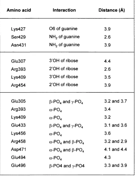
The molecular docking model predicts that the GTP substrate is located in the middle of the tunnel structure of the yeast RTPase (Fig. 1). The space-filling analysis suggests that the tunnel can accommodate the ribose, base, and phosphate moieties of the GTP molecule, thereby implying that the entire nucleotide, and not only the phosphates, is entering the tunnel (Fig. 1E). The molecular docking model provides instructive findings on the interaction between specific residues and the nucleotide substrate. For instance, multiple side chains (Summarized in Table 1) are contacting the a, p, and y phosphates of GTP. In addition to the Arg393, Lys456, and Arg458 residues that were previously observed in the crystal structure of the S. cerevisiae RTPase (15), six other amino acids appear to be involved in the coordination of the phosphates (Fig. 2). Some of these residues, such as Arg393, Lys409, Lys456, and Glu494 are solely contacting one phosphate, while others (Glu305, Glu433, Arg458, Asp471, and Glu496) are contacting two adjacent phosphates (Fig. 2A-D). Interestingly, all of these phosphate-contacting residues were previously shown to be essential for catalysis through mutational studies thereby highlighting their importance in catalysis (9, 17-19). Based on the crystal structure of the enzyme (15), some of these essential residues were previously proposed to be important for catalysis through their ability to make water-mediated contacts with the phosphate or the essential divalent metal ion (Glu433 and Glu494). Similarly, other residues such as Lys409 were thought to be indirectly involved in catalysis via their interactions with other essential side chains (15, 17). Although the role of these amino acids in making important
moiety of the nucleotide substrate. The previously observed ability of the TTM family of RTPases to hydrolyze tripolyphosphate (28-30), which completely lacks the sugar and base components of the nucleotide, is probably a reflection of the excessive number of interactions between the active site residues and the a-, (3-, and y-phosphates.
Analysis of the docking model between the enzyme and GTP suggests that only four amino acids are responsible for the coordination of the hydroxyl groups of the ribose. These are Glu307 and Lys409, which are contacting the 3'OH group, and Arg393 and Arg454, which are coordinating the 2'OH group of the sugar (Fig. 2A and 2D; Table 1). Previous mutational studies have shown that each of these four residues is essential for catalysis (9, 17-19). The molecular docking model also highlights key amino acids involved in the binding of the guanine base. These amino acids are either contacting the exocyclic 2-amino (Ser429, and Asn431) or the 6-oxo (Lys427) groups of the pyrimidine ring of guanine (Fig. 2A and 2D). Previous structure-function analyses of the amino acids that are contacting the guanine base in the docking model indicate that these residues are not essential for catalysis (17, 19). Moreover, pi-pi and cation-pi stacking interactions are also occurring between His411 (non-essential) and the pyrimidine ring of guanine, while Pro341 is engaged in hydrophobic interactions with the imidazole ring of guanine (Fig. 2A and 2C). The non-essentiality of the amino acids contacting the guanine base is not surprising since the enzyme can efficiently hydrolyze both purine and pyrimidine nucleotides. Both the coordination geometry and the nature of the amino acids contacting the purine/pyrimindine rings are likely modified according to the precise nature of the substrate.
Nucleotide analogues to probe the active site
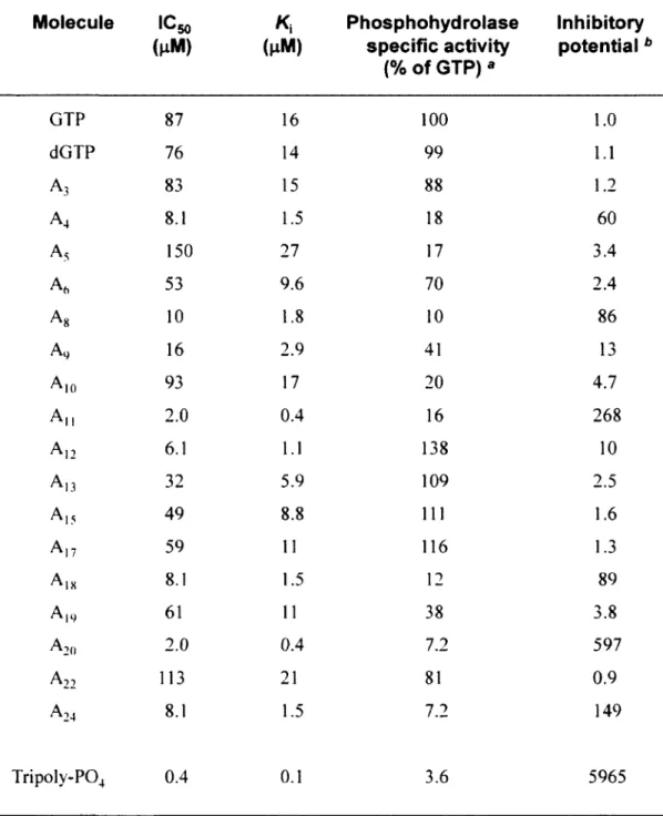
In order to experimentally confirm the docking model, and to gain additional insights on the molecular determinants involved in the formation of the enzyme-substrate complex, we have used nucleotide analogues to monitor their effects on the reaction chemistry. The nucleotide analogues displayed various modifications on both the ribose and the guanine base of GTP (Fig. 3). We initially monitored the ability of 17 analogues of GTP to inhibit the activity of the yeast RTPase by evaluating both the IC50 and K, values for each molecule (Table 2). The informative finding is that every analogue tested had the ability to inhibit the RTPase reaction, albeit to different extents, thus highlighting the high structural flexibility of the active site (Fig. 4A). All the nucleotide analogues used in the current study were competitive inhibitors of the RTPase reaction indicating that they bind to the active site of the enzyme. A typical example using the analogue A-12 is shown in figure 4B-E. In order to gain additional information on the functional flexibility of the catalytic center, we determined the ability of the various nucleotide analogues to be hydrolyzed by the enzyme. The
yeast RNA triphosphatase catalyzed the phosphohydrolysis of all nucleotide analogues tested in the current study with specific activity ranging from 7 to 138 % of GTP (Table 2). Interestingly, some of the analogues bound strongly to the enzyme active site (as evidenced from the low IC5o and K,
values) but were not efficiently hydrolyzed. These analogues (A4, Ag, A,,, A,8, A2o, and A24) had
IC50 values ranging from 2 to 16 u.M with low specific phosphohydrolase activities varying from 7 to 18% of the GTP substrate (Table 2).
Our study indicates that the active center of the yeast RTPase is highly flexible and can accommodate nucleotide substrates displaying a number of unusual chemical modifications. For instance, analogues harboring modifications on the hydroxyl moieties of the ribose (2'OH and 3'OH) were hydrolyzed with a high level of efficiency by the enzyme. The addition of a methyl group to either the 2' or 3' hydroxyls (A-13 and A-17) had no significant effect on the phosphohydrolase activity (Table 2). Most strikingly, A-12 which lacks both ribose hydroxyls (2',3'-dideoxy-GTP) was hydrolyzed very efficiently by the yeast RTPase. This was unexpected since amino acids contacting the ribose hydroxyls (Glu307, Arg393, Lys409, Arg454) were previously shown to be essential for catalysis through mutational studies (9, 17-19). However, we observe that two of these residues (Arg393 and Lys409) are also contacting the a-phosphate of the bound GTP, while Glu307 coordinates the essential divalent cation. Moreover, Arg454 forms a salt bridge with Glu492, an interaction which is important for the stabilization of the tunnel architecture (15, 17). Therefore, we conclude that the importance of these amino acids is not directly related to their ability to bind to the hydroxyls of the ribose, but rather lies in their other functions namely through the coordination of the a-phosphate and metal ion, or in the stabilization of the tunnel structure.
Some of the analogues used in the current study contain chemical modifications on the guanine base of the GTP molecule (Fig. 3). Although the S. cerevisiae RTPase is active on both purine and pyrimidine nucleotides (ATP, CTP and UTP were used as substrates with specific activities of 103, 103, and 109 % of the GTP substrate, respectively), analysis of the GTP analogues helped to illuminate the flexibility and complexity of the RTPase active site. For instance, the addition of a chemical group to the C8 position of guanine had a negative effect on the phosphohydrolysis activity. Analogues harboring such modification (A-10 and A-24) were used inefficiently by the enzyme. Although it can be argued that the addition of a bromo- or iodo- group at this position can potentially alter the electronic properties of the guanine ring, analysis of the docking model indicates that steric hindrance is the likely explanation for the limited hydrolysis of