Photocopying permitted by license only
Présence of yô TcR
+Tumor Infiltrating Lymphocytes (TILs) in One
Case of Pédiatrie Osteogenic Sarcoma
MICHEL MOUTSCHEN*, NATHALIE JACOBS*, CLAIRE HOYOUX*,
MARIE-THÉRÈSE MARTIN-SIMONET*, NICOLE SCHAAF-LAFONTAINE* and JACQUES BONIVER* *Department ofPathology, University of Liège, CHU Sart-Tilman, 4000 Liège, Belgium
^Department of Pediatrics, University of Liège, Hôpital de la Citadelle, 4000 Liège, Belgium
(Received June 18,1993; in final form November 3,1993)
Purpose: To characterize a(3 and yô TcR expression in cultured TILs from a metastatic osteogenic sar-coma.
Patient and methods: TILs were isolated from a lung metastasis of an osteogenic sarcoma in a 16-year-old f emale patient. Culture conditions were IL-2 and anti-CD3 MoAb+IL-IL-2. TcR expression and surface phenotype were studied by flow cytometry. 4h-chromium release assays were used to assess cy-totoxicity.
Results: IL-2-expanded tumor infiltrating T-lym-phocytes contained 50% yô TcR+ cells whereas yô TcR+ cells represented 20% of T-cells in cultures ex-panded with anti-CD3 MoAb+IL-2. The phenotype of yô TcR+ cells was CD3+, CD16-, CD4-, CD8-, and CD29+. AnH-CD3 MoAb + IL-2-expanded TILs me-diated a significant lysis of thé autologous tumor and of an allogeneic lymphoma and displayed a higher activity against thé NK-sensitive target K562. In cold target experiments, unlabeled autol-ogous tumor cells blocked thé lysis of K562, indi-cating that MHC-unrestricted effectors were involved in thé lysis of both targets.
Conclusions: This report demonstrates thé ex-pansion of large numbers of yô TcR+ T-cells after short-term culture of TILs from a metastatic os-teosarcoma. Further studies are needed to déter-mine thé rôle of yô TcR+ cells in host immune responses directed against this tumor.
Key words: Tumor infiltrating lymphocytes (TIL), gamma/delta T-cell receptor, osteogenic sarcoma
Corresponding author: Dr. Michel Moutschen
Department of Pathology, University of Liège, CHU Sart-Tilman, 4000 Liège, Belgium
INTRODUCTION
OLIGOCLONALITY OF TUMOR infiltrating lympho-cytes (TILs) has been demonstrated in several human tumors including lung cancer,1 melanoma,2 and ovary adenocarcinoma.3 It is a strong argument that TILs may be part of a spécifie immune response of thé host against thé tumor. Therefore, TILs could be viewed as tools to identify thé tumor antigens recognized by thé immune system. Animal models also suggest that in vitro ex-panded TILs could be used in adoptive cellular im-munotherapy protocols with a higher efficiency than lymphokine activated killer (LAK) cells generated from peripheral blood lymphocytes.4 In most cases, TILs are CD3+ T-cells expressing thé cc(i T-cell receptor (TcR), while yô TcR+ lymphocytes usually represent a minor subset of TILs (less than 5%).5-6
Despite their low frequency in TILs and peripheral blood, T-cells expressing thé yô TcR are of major interest in thé field of tumor immunity. After short-term culture in vitro, thèse lymphocytes develop MHC-unrestricted cytolytic activity against a variety of tumor cells of dis-tinct histologie origin.7-8 Récent reports hâve suggested that molécules related to thé 65-kDa heat shock protein found in mycobacterial extracts could be involved in thé non-MHC restricted lysis of some tumor cells by yô TcR+ lymphocytes.9-10
We hâve characterized thé phenotype and function of TILs from a lung metastasis of an osteogenic sarcoma in a 16-year-old patient. We observed a high frequency of yô TcR+ T-cells in thé cultured TILs from this tumor. TILs displayed a significant cytolytic activity of thé au-tologous tumor but also mediated non-MHC restricted cytotoxicity of K562 and of CESS, an allogeneic B-cell lymphoma.
PATIENT AND METHODS
Tumor. The tumor was a pulmonary metastasis of an osteogenic sarcoma. The patient was a 16-year-old girl who had developed an osteogenic sarcoma of thé
278 M. MOUTSCHEN ET AL.
metaphyseal région of thé proximal tibia two years ear-lier. Several pulmonary métastases were présent when thé primary lésion was diagnosed. The patient received a preoperative chemotherapy regimen including high dose methotrexate with citrovorum factor rescue and a combination of bleomycin, cyclophosphamide, and dactinomycin. Complète régression of thé pulmonary métastases was obtained. The primary tumor was re-sected, and thé biopsy spécimen showed less than 25% necrosis. Therefore, thé patient received several post-operative chemotherapy treatments with cisplatinum and adriamycin. She finally developed a large pul-monary metastasis, invading thé pleura and occupying thé anterior mediastinum. The metastasis was resected by anterior thoracotomy and sent without fixation to thé pathology department under aseptic conditions.
Préparation of cells. Cell suspensions were pre-pared by mechanical and enzymatic dissociation of thé tumor spécimen using an enzyme mixture containing collagenase and hyaluronidase (Sigma). Cell suspen-sions containing a mixture of tumor cells, lymphocytes, and monocytes were incubated with complète médium (CM) at 37°C in a humidified atmosphère of 5% CO2. Culture médium consisted of DMEM (Gibco), 30 U/ml penicillin-streptomycin (Gibco), l%nonessentialamino acids (Gibco), and 1 mM sodium pyruvate (Gibco). Heat-inactivated fêtai calf sérum (FCS) (30G, Gibco) was added at 10% to obtain complète médium (CM). Recombinant human IL-2 (rIL-2) was kindly provided by Glaxo Institute for Molecular Biology (AG, Genève, Switzerland) and used at 50 U/ml. For anti-CD3 + rlL-2 activated cultures, anti-CD3 MoAb (purified mouse IgGj., clone BMA030, Becton Dickinson, Erembodegem, Belgium) was added once at thé beginning of thé cul-tures at 10 ng/ml.
Antibodies andflow cytometry. FITC-conjugated-anti-CD4 (Mouse IgGj clone SK3). PE-conjugated-anti-CD8 (mouse IgGi clone SKI), FITC-conjugated anti-CD45RA (Mouse IgGj clone L48), FITC-conjugated and biotinylated-anti-CD3 (Mouse IgGj clone SK7), FITC-conjugated-anti-TcR-yôl (Mouse IgGt clone 11F2), purified anti-TcR-ap (Mouse IgGj clone WT31), and PE-conjugated anti-CD16 (Mouse IgGj clone B73.1) were purchased (Becton Dickinson, Erembodegem, Belgium). PE-conjugated anti-CD29 (Mouse IgGj clone 4B4) was purchased (Coulter, distributed by Analis, Namur, Belgium). Streptavidin-PE (Dako) was used to reveal bi-otinylated-anti-CD3. FITC-labeled-goat anti-mouse IgG (Dako) was used to reveal purified anti-TcR-ap. For flow cytometry analysis, cell suspensions were layered over Ficoll Hypaque and spun at 2000 rpm for 20 min. Viable lymphocytes were collected, washed, and incubated for 30 min. on ice with appropria te amounts of antibodies and second steps in CM with sodium azide 0.1%. Cells were washed twice and analyzed for fluorescence in-tensity on a FACScan (Becton Dickinson).
Cytotoxicity assays. Effector cells were washed, counted, and resuspended in CM at a concentration of 2.5 x 10⁵ cells/100 ul. Four sériai dilutions in triplicates were made in 96-well U-bottom plates (Nunc). Target cells were incubated with Na51CrO4 (Medgenix, Fleurus, Belgium) for 1 h at 37°C, washed three rimes, counted, and adjusted to obtain 10 x 103 cells/100 ul. This sus-pension was added to thé sériai dilutions of effector cells. After centrifugation, thé microplates were incubated for 4 h at 37°C, 100 pi of supernatant and were then recov-ered from each well; radioactivity was measured in a y-counter (Cobra auto-gamma Packard, Downers Grove, IL). Maximal chromium release was obtained by adding 100 ul of détergent (RBS10% Chemical products) to 100 ul of target cells suspension, and spontaneous release was given by thé incubation of target cells without ef-fectors cells. Percent cytotoxicity was calculated ac-cording to thé foliowing formula:
Expérimental mean CPM - Spontaneous mean CPM Maximal mean CPM - Spontaneous mean CPM
xlOO
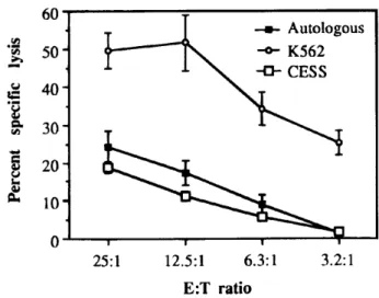
Target cells for cytotoxicity assays. K562 is an MHC-negative erythroleukemic cell Une commonly used to study natural killer (NK) activity. CESS is an EBV-in-duced B-cell lymphoma and is résistant to NK activity. Both cell lines were cultivated in RPMI-1640 with 10% FCS. Fresh tumor cells from thé biopsy spécimen did not grow in culture, therefore small tumor fragments were frozen after dissection. A few hours before thé cytolytic assay, tumor fragments were thawed and enzymatically dissociated to obtain cell suspensions. Thèse cells were washed and incubated at 37°C a few hours before ra-dioactive labeling. This procédure allowed an optimal viability of thé tumor cells. In thé cytotoxicity assay showed in Figure 1, thé spontaneous release was 1891 ± 98 cpm and thé maximum release was 10367 ± 1453 cpm (n=3). Thèse tumor cell suspensions were not de-pleted in lymphocytes and monocytes, however thé con-tribution of nontumor cells to Na51CrO4 uptake was considered to be negligible.
Cola target inhibition. Cytolytic specificity was eval-uated by adding unlabeled competing targets to thé 51Cr release assay. Varying ratios of cold targets were mixed with 5iCr-labeled targets (5 x 10³/well) in 100 pi CM. Effector lymphocytes (1 x 10⁵ cells/well) were then added and thé plates incubated for 4 h at 37°C. The per-cent inhibition of cytotoxicity (i) was calculated accord-ing to thé formula:
Vœnt.
where exp. is thé spécifie lysis observed in thé présence of cold targets and cont. is thé spécifie lysis observed without cold targets.
TABLE I
Sequential Surface Phenotypes of TILs from Osteogenic Sarcoma in Culture with rIL-2 (50 U/ml) or rIL-2 + BMA030 (10 ng/ml).
Percentage of Cells Expressing"
Culture Condition Dayof Culture CD3 apTcR yôTcR CD16 CD4 CD8 CD29 rIL-2 rIL-2+anti-CD3 rIL-2 rIL-2+anti-CD3 rIL-2+anti-CD3 10 10 28 28 49 63 81 98 98 99 34 67 48 75 73 30 18 46 21 28 34 15 2 1 1 11 31 6 3 4 23 32 40 75 69 NT NT 97 96 99 29 18 52 20 26
"Reactivity of cultured cells with spécifie MoAb was assessed by flow cytometry analysis. NT, not tested.
''Calculated proportion of CD3^ T-cells négative for CD4 and CD8 (double négative: DN) obtained with thé formula CD3 - (CD4+CD8).
RESULTS
1. Présence ofySTcR + T-cells in Cultured TILs After enzymatic dissociation of thé tumor and culture with rIL-2 (50 U/ml) or rIL-2 (50 U/ml) + anti-CD3 MoAb (10 ng/ml), tumor cells rapidly disappeared and activated lymphocytes were visible in thé wells. Viable lymphocytes were isolated and flow cytometry analy-sis was performed (Table I). After 10-day culture with IL-2,63% of TILs expressed CD3 but only 34% were rec-ognized by thé MoAb WT31 spécifie for an epitope as-sociated with thé cep TcR. The CD3+ ap TcR- T-cells,
representing 47% of CD3+ T-cells, expressed thé yô TcR. After initial stimulation with anti-CD3 MoAb, thé fre-quency of yô TcR+ cells was only 18%, representing 22%
of CD3+ T-cells. After 28 days, natural killer (NK) cells expressing CD16 had disappeared in both culture con-ditions and ail remaining lymphocytes expressed CD3. The proportion of CD3+ T-cells expressing thé yô TcR
re-mained around 50% in IL-2-expanded cultures and around 20% in anti-CD3 MoAb+IL-2-expanded TILs (Table I). Anti-CD3 MoAb expanded cultures displayed a 11.50-fold expansion of total lymphocyte counts be-tween day 10 and day 28, whereas IL-2 alone only in-duced a modest 1.70-fold expansion. Furthermore, thé viability of IL-2-expanded cultures decreased after day 28 while further expansion was observed in thé anti-CD3 MoAb+IL-2-expanded TILs. Therefore, although anti-CD3 MoAb acrivation was associated with a lower proportion of yô TcR+ cells, this culture condition
al-lowed a higher absolute expansion of thèse cells in com-parison with IL-2-expanded cultures.
The CD4 / CD8 ratio of TILs decreased in both culture conditions reaching 0.15 and 0.04 on day 28 for IL-2- and anti-CD3 MoAb+IL-2-expanded cultures respectively. Interestingly, thé number of CD3+ T-cells was higher
than thé number of cells expressing either CD4 or CD8 indicating that a large fraction of CD3+ T-lymphocytes
was négative for CD4 and CD8. At ail time points and in both culture conditions, mère was a perfect corréla-tion between thé frequency of double négative CD3+
T-cells (CD3 - (CD4 + CD8)) and thé frequency of yô TcR+
T-cells. Although three-color staining was not per-formed, this corrélation indicated that most yô TcR+
T-cells présent in thé cultures were CD4~ CD8~ as previously described for yô TcR+ T-cells found in thé
pe-ripheral blood.7'8 At day 28, ail lymphocytes displayed
a memory activated phenotype: CD45RA-CD29+. Taken together thèse results showed that thé yôTcR+ T-cell
pop-ulation was CD3+, CD16-, CD4-, CD8-, and CD29+.
2. Cytotoxic Activity ofTILs Expanded in Anti-CD3 MoAb+ IL-2
TILs cultured for 19 days in thé présence of anti-CD3 MoAb + IL-2 displayed a moderate but significant cy-tolytic activity against thé autologous tumor (25% spé-cifie lysis E:T 25:1) and against CESS, an EBV-induced B-cell lymphoma (20% spécifie lysis E:T 25:1) (Fig. 1). A strong activity was demonstrated against K562 (50% spécifie lysis E:T 25:1). The cytolytic activity of TILs
cul-—»- Autologous -o- K562 -0-CESS 25:1 12.5:1 6.3:1 E:T ratio 3.2:1
FIGURE 1 Cytotoxic activity mediated by anti-CD3 MoAb +
IL-2-expanded TILs (day 19) against ( —•—) autologous tumor cells, (—tr— ) K562, (—a—) CESS. Results represent mean spé-cifie lysis ± SD from thé triplicates of a 4 h-chromium release assay.
280 M. MOUTSCHEN ETAL.
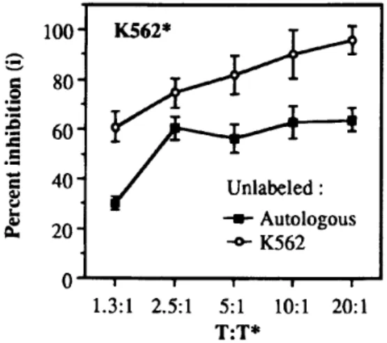
tured in IL-2 alone was not tested because of insuf ficient expansion in this condition. Afterward, thé cytolytic ac-tivity against thé autologous tumor declined while thé activity against K562 remained high (not shown). We used cold target inhibition experiments to détermine if thé effectors responsible for thé lysis of K562, assumed to be non-MHC restricted, could bind to thé autologous tumor cells. On day 33, thé addition of unlabeled autol-ogous tumor cells strongly inhibited thé lysis of K562 al-though less efficiently than unlabeled K562 cells (Fig. 2). This inhibition indicated that a large proportion of thé non-MHC restricted cytolytic effectors présent in thé anti-CD3 MoAb + IL-2-expanded cultures could bind to thé autologous tumor cells.
DISCUSSION
This report demonstrated thé présence of a large pro-portion of yô TcR+ T-cells within thé cultured TILs from a metastatic osteogenic sarcoma. To our knowledge, only two published reports hâve described large num-bers of yô îcR+ T-cells in TILs.n-12 Bachelez et al. reported frequencies of yÔ TcR+ T-cells between 15 and 25% in three cases of primary cutaneous melanoma.11 yS TcR+ T-cells were not found in metastatic lésions even in sub-cutaneous sites, therefore it was postulated that thé melanoma-associated yô TILs could dérive from thé large pool of yô TcR+ T-cells présent in normal epider-mis.11 Rivoltini et al. studied thé frequency of yô T-cells in IL-2-expanded TILs from 20 pédiatrie tumors in-cluding osteosarcomas, Wilms' tumors, neuroblas-tomas, ovarian teratocarcinomas and melanoma.12 Interestingly, thé only tumors in which large numbers
ioo-I 80-'•£ I60-£
40H 20-K562* Unlabeled : -•- Autologous -o-K562 1.3:1 2.5:1 5:1 T:T* 10:1 20:1FIGURE 2 Cold target inhibition of thé lysis of K562. Effectors
were anti-CD3 MoAb+ IL-2 expanded TILs (day 33). 51Cr labeled
K562 cells were mixed with effectors at E:T 20:1 with addition of competing unlabeled: (—•—) autologous tumor cells, and (—o—) K562 as indicated. T:T* represents thé ratio between un-labeled targets and 51Cr labeled K562 cells. The data represent
mean percent inhibition (i) ± SD from triplicates. i was calculated according to thé formula described in thé methods section.
of yô TILs were observed were two cases of metastatic osteogenic sarcoma with respectively 52% and 61% of T-cells expressing thé yô TcR.12 Unfortunately, no other data are available about TcR expression by TILs from os-teogenic sarcoma.
We did not study thé frequency of yô TcR+ T-cells in situ, before tumor dissociation and in vitro activation. Therefore, thé large number of yô TcR+ T-cells could be viewed as a preferential expansion due to in vitro cul-ture. Although we cannot formally rule out this hy-pothesis, it is important to note that using thé same protocol of isolation and in vitro expansion, we never de-tected any increase of CD3+ yÔ TcR+ cells in TILs from a large number of breast and gastrointestinal tumors.
The cultured TILs demonstrated a moderate lytic ac-tivity against thé autologous tumor which was signifi-cantly lower than that against thé NK sensitive target K562. A comparable autologous cytotoxicity and an ab-sence of tumor specificity were also found in most TILs cultures from pédiatrie tumors in Rivoltini's study, al-though thèse authors did not study autologous cyto-toxicity in thé cases of osteosarcomas with yô TcR+ TILs.12 Since our cytolytic assays were performed with non-sorted effector cells, our data are not sufficient to prove that thé cytolytic activity observed was actually due to yÔ TcR+ T-cells.
Récent évidence has shown that yô TcR+ lymphocytes may recognize a heat shock protein in human tumors.9'10 Therefore, thé présence of yô TcR-i- TILs in three cases of metastatic osteogenic sarcoma could be due to thé ex-pression of heat shock-related proteins by thèse tumor cells. It remains to define if such an expression would be intrinsic to this type of tumor or mostly related to thé intensive preoperative chemotherapy or radiotherapy regimens received by thé patients. Since MoAb directed against hspéO hâve been recently developed,13 our find-ings together with Rivoltini's report11 advocate for a careful analysis of hspôO surface expression in metasta-tic osteogenic sarcoma.
Acknowledgments: The authors want to thank Mrs. Elizabeth Franzen-Detroz for her expert technical assis-tance. This work is supported in part by thé Foundation for Scientific Médical Research (FRSM) and thé Centre Anticancéreux près l'Université de Liège. The work of Michel Moustschen is supported by grants from Télévie and thé Fonds National de la Recherche Scientifique (FRNS). The work of Nathalie Jacobs is supported by grants from thé Centre Anticancéreux près l'Université de Liège. The work of Marie-Thérèse Martin-Simonet is sup-ported by grants from Télévie.
REFERENCES
1. Yoshino I, Yano T, Yoshikai Y, et al.: Oligoclonal T lympho-cytes infiltrating human lung cancer tissues. Int J Cancer 1991; 47:654-658.
2. Morita T, Salmeron MA, Moser RP, et ai: Oligoclonal expan-sion of Vp 8+ cells in interleukin-2-activated T cells residing in
sub-cutaneous metastatic melanoma. Clin Exp Metastasis 1992; 10:69-76.
3. loannides CG, Freedman RS: Sélective usage of TCR Vp in tumor in specific CTL lines isolated from ovarian tumor-associated lymphocytes. Anticancer Res 1991; 11:1919-1925.
4. Rosenberg SA, Spiess P, Lafreniere R: A new approach to thé adoptive immunotherapy of cancer with tumor-infiltrating lym-phocytes. Science 1986; 233:1318-1321.
5. Nano M, Seki H, Mathioudakis G, et al: y/8 T cell antigen re-ceptors expressed on tumor-infiltrating lymphocytes from pa-tients with solid tumors. Eur } Immunol 1992; 22:679-687.
6. Alam SM, Clark JS, Leech V, et al: T cell receptor y/8 expres-sion on lymphocyte populations of breast cancer patients.
Immunol Lett 1992; 31:279-283.
7. Borst J, Van de Griend RJ, Van Oostveen J, et al: AT cell re-ceptor Y/CD3 complex found on peripheralblood cytotoxic T lym-phocytes. Nature 1987; 325:683-688.
8. Moingeon PS, Jitsukawa S, Faure F, et al: A y-chain complex forms a functional receptor on cloned human lymphocytes with natural killer-like activity. Nature 1987; 325:723-726.
9. Fisch P, Malkovsky M, Kovats S: Récognition by human Vy9/VS2 T cells of a GroEL homolog on Daudi Burkitt's lym-phoma cells. Science 1990; 250:1269-1272.
10. Kaur I, Voss SD, Gupta RS, et al: Human peripheral y8 T cells recognize hsp60 molécules on Daudi Burkitt's lymphoma cells. /
Immunol 1993; 150:2046-2055.
11. Bachelez H, Flageul B, Degos L, et al: TCR yS bearing T lym-phocytes infiltrating human primary cutaneous melanomas. /
Invest Dermatol 1992; 98:369-374.
12. Rivoltini L, Arienti F, Orazi A, et al: Phenotypic and func-tional analysis of lymphocytes infiltrating pédiatrie tumours with a characterization of thé tumour phenotype. Cancer Immunol
Immunother 1992; 34:241-251.
13. Jindal S, Dudani AK, Singh B, et al: Primary structure of a human mitochondrial protein homologous to thé bacterial and plant chaperonins and to thé 65-kilodalton mycobacterial antigen.