Open Archive Toulouse Archive Ouverte (OATAO)
OATAO is an open access repository that collects the work of some Toulouse
researchers and makes it freely available over the web where possible.
This is
an author'sversion published in:
https://oatao.univ-toulouse.fr/23113Official URL :
https://doi.org/10.1016/j.jse.2018.02.057
To cite this version :
Any correspondence concerning this service should be sent to the repository administrator: [email protected]
Krukenberg, Anna and McBirnie, Julie and Bartsch, Stefan and Böhler, Nikolaus and
Wiedemann, Ernst and Jost, Bernhard and Mansat, Pierre and Bellon-Champel, Pierre and
Angeloni, Renzo and Scheibel, Markus Sidus Stem-Free Shoulder System for primary
osteoarthritis: short-term results of a multicenter study. (2018) Journal of Shoulder and Elbow
Sidus Stem-Free Shoulder System for primary
osteoarthritis: short-term results of a multicenter
study
Anna Krukenberg
a, Julie McBirnie, MD
b, Stefan Bartsch, MD
c, Nikolaus Böhler, MD
d,
Ernst Wiedemann, MD
e, Bernhard Jost, MD
f, Pierre Mansat, MD, PhD
g,
Pierre Bellon-Champel, MD
h, Renzo Angeloni, MD
i, Markus Scheibel, MD
a,*
aDepartment of Shoulder and Elbow Surgery, Charité–Universitaetsmedizin Berlin, Berlin, Germany bDepartment of Orthopaedics, New Royal Infirmary of Edinburgh, Edinburgh, UK
c
Praxis am Wall Rinteln, Rinteln, Germany d
Allgemeines Krankenhaus der Stadt Linz, Kepler Universitaetsmedizin, Linz, Austria e
Orthopedic Surgery Center Munich (OCM), München, Germany f
Department of Orthopaedics and Traumatology, Kantonsspital St Gallen, St. Gallen, Switzerland g
Centre Hospitalier Universitaire Toulouse–Hôpital Purpan, Toulouse, France h
Groupe Chirurgical Thiers, Grenoble, France i
Universitaria Careggi–Centro Traumatologico Ortopedico, Florence, Italy
Background: The aim of this prospective multicenter study was to evaluate clinical and radiologic results
of a new metaphyseal anchored system. This system features a different anchor geometry that potentially leads to better bony integration and less loosening.
Methods: From November 2012 until December 2015, 148 patients (151 shoulders) were treated with the
Sidus Stem-Free Shoulder System at 9 centers in Europe. The main indication was primary osteoarthritis (80.1%). This analysis only includes patients diagnosed with primary osteoarthritis (n= 121).Aclinical evaluation was performed using the Constant-Murley score, Subjective Shoulder Value, American Shoulder and Elbow Surgeons Standardized Shoulder Assessment Form, and range of motion. Radiologic assessment was based on the occurrence of radiolucent lines and signs of implant migration, osteolysis, loosening, and heterotopic ossification.
This study was approved by the ethics committee of each institution:
• Ethikkommission, Ethikausschuss 4, Charité–Universitaetsmedizin Berlin, Berlin, Germany (No. EA4/021/13)
• Ethikkommission des Landes Oberoesterreich, Linz, Austria (No. B-38-12)
• Menistére de L’Enseignement Supérieur et de la Recherche, Paris, France (No. 12.513)
• Freiburger Ethikkommission International, Freiburg, Germany (No. CME2012-01E)
• Kanton St Gallen Ethikkommission, St Gallen, Switzerland (No. 12/128/1B)
• South East Scotland Research Ethics Committee 02, Edinburgh, UK (No. 12/SS/0154)
• Segreteria Comitato Ethico Sperimentazione Clinica, Firenze, Italy (No. CME2012-01E)
*Reprint requests: Markus Scheibel, MD, Department of Shoulder and Elbow Surgery, Charité–Universitaetsmedizin Berlin, Augustenburger Platz 1, D-13353 Berlin, Germany.
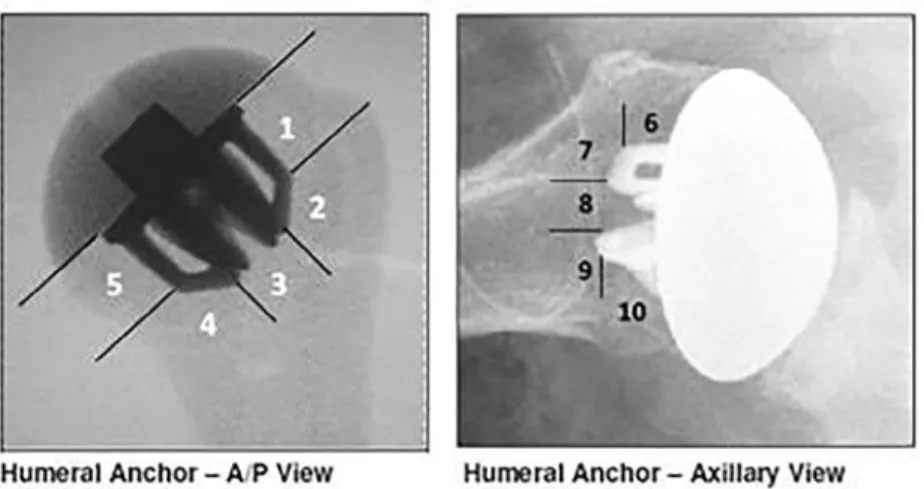
implant-bone interface of the humeral component was divided into 5 different zones in AP and axillary views (Fig. 1). The glenoid com-ponent was divided into 3 zones in AP view according to Lazarus et al.14
Implant description
The Sidus Stem-Free Shoulder System is a 2-component system con-sisting of a metaphyseal implanted anchor and the humeral head. The cross-shaped anchor creates a “press fit” when it is forced into the slightly undersized bed prepared in the metaphyseal bone, en-suring the primary stability of the implant. The anchor is made from a rough-blasted, forged titanium alloy (Protasul, 64WF; Zimmer Biomet) and has 4 fins to ensure good bony integration and to secure against rotation (Fig. 2). It is available in 3 diameters (small, medium, and large) and is not convertible to a reverse prosthesis. The humeral head is concentric and consists of a cobalt-chrome alloy (Protasul, 21WF; Zimmer Biomet). It is available in different diameters (38-52 mm) and heights (Fig. 3). The head and anchor are connected by a Morse taper connection. The Sidus system can be combined with 2 different and already known glenoid systems: Anatomical Shoul-der Glenoid (cemented keel and pegged; Zimmer Biomet) and Bigliani/ Flatow System (cemented keel or pegged; Zimmer Biomet).
Surgical technique
All surgical procedures were performed with patients under general anesthesia combined with an additional interscalene block or cath-eter for adequate intraoperative and postoperative pain relief. A deltopectoral approach was used in all patients. The subscapularis was released by a tenotomy or lesser tuberosity osteotomy accord-ing to Gerber et al10per surgeon preference. The long head of the
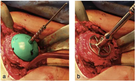
biceps underwent either tenotomy or tenodesis. After dislocation of the humeral head, a guide was positioned at the medial border of the insertion of the supraspinatus tendon and marked with a K-wire. The K-wire should exit at the posterior edge of the cartilage medial to the bare area. Afterward, the guide was removed, and a
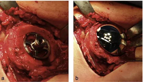
resec-tion guide was placed with another 2 K-wires along the anatomic neck (Fig. 4, a). By use of this resection guide, the inclination angle and retroversion can be determined even in severe cases of osteo-arthritis. Then, the humeral head was resected along the anatomic neck (Fig. 4, b). In the case of TSA, a metallic bone protector was placed on the resection plane while the glenoid was prepared. First, the capsule was released all around the glenoid. Then, the glenoid was prepared in a standard manner. After the glenoid was re-placed, the correct size of the humeral head was determined by using a trial head (Fig. 5, a). Via this model, a central wire was placed in the humerus and the metaphysis was prepared using a drill and an impaction instrument (Figs. 5, b, and 6, b). Then, the anchor was positioned in the metaphysis, and the humeral head was placed on top of the anchor (Fig. 7). Finally, subscapularis repair and wound closure were performed.
Postoperative rehabilitation
All patients were immobilized in a sling in internal rotation for 4 to 6 weeks. Simultaneously, physiotherapy was performed, but active motion and passive external rotation were not allowed for the first 6 weeks; only passive abduction, flexion, and internal rotation ex-ercises were permitted during this time. After 6 weeks, gradual progression of active motion was allowed. After full range of motion was achieved, strength exercises were allowed.
Statistical analysis
Statistical analysis was performed using the Wilcoxon rank sum test with a significance level of less than .05 and a confidence level of 0.95.
Results
Demographic data
In total, 151 Sidus systems were implanted in 148 patients. The main indication for joint replacement was primary os-teoarthritis (n= 121, 80.1%), other indications were post-traumatic arthritis (7.3%), focal avascular necrosis (3.3%), rheumatoid arthritis (2.6%), and instability arthropathy (1.3%). Of the 121 patients who underwent surgery for primary osteoarthritis, 105 (86.8%) were available for a 2-year follow-up evaluation, whereas 4 did not want to continue with this study and 12 were lost to follow-up. In the cohort of 105 pa-tients, there were 53 women (50.5%) and 52 men (49.5%), and the average age was 64 years (range, 40-79 years). TSA was performed in 73 cases, whereas hemiarthroplasty (HA) was performed in 32.Table IIIshows the distribution of glenoid morphology according to Walch et al.23
Clinical results
The average CS increased from 26 points (SD, 13 points) pre-operatively to 70 points (SD, 18.9 points) at 2 years’ follow-up (P< .001); the average ASES score, from 34 points (SD, 17.9 points) to 86 points (SD, 21.4 points) (P< .001); and the SSV, from 34% (SD, 17.2%) to 84% (SD, 17.5%)
Table II Exclusion criteria
The patient is unwilling or unable to give consent or to comply with the follow-up program.
The patient has any condition that would, in the judgment of the investigator, place the patient at undue risk or interfere with the study. Any patient who is institutionalized, is known to abuse drugs, is known to have alcoholism, or cannot understand what is required of him or her is excluded.
The patient is known to be pregnant or breastfeeding. The patient meets 1 of the following contraindications:
Soft or inadequate humeral bone (including osteoporosis and extensive avascular necrosis or rheumatoid arthritis) leading to poor implant fixation
Metaphyseal bony defect (including large cysts) Post-traumatic tuberosity nonunion
Signs of infection Irreparable cuff tear
Revision from failed stemmed prosthesis Charcot shoulder (neuroarthropathy)
(P< .001). Active forward elevation improved from 86° (SD, 29°) preoperatively to 144° (SD, 30.7°) at final follow-up, and active external rotation with the arm at the side improved from 13° (SD, 17.9°) to 41° (SD, 23.3°) (P< .001).
In this 2-year cohort, patients with TSA reached signifi-cantly higher scores than patients with HA (Table IV). In patients with TSA, the CS improved from 25 points preop-eratively to 70 points at 1-year follow-up and reached 75 points at final follow-up (P< .001). In patients with HA, the CS
improved from 27 points preoperatively to 59 points at 1-year and final follow-up (P< .001). The ASES score increased from 32 points preoperatively to 91 points at final follow-up in pa-tients with TSA and from 34 points to 86 points in papa-tients with HA (P< .001). The SSV improved from 32% preop-eratively to 87% in patients with TSA and from 38% to 75% in patients with HA (P< .001). Active range of motion dif-fered between TSA and HA: Flexion increased from 85° preoperatively to 150° postoperatively (P< .001) and exter-nal rotation with the arm at the side increased from 12° to 45° (P< .001) in patients with TSA, whereas in patients with HA, flexion improved from 89° to 129° (P< .001) and ex-ternal rotation from 15° to 34° (P< .006). Finally, 90.4% of
Figure 1 Radiologic classification in true anteroposterior (A/P) and axillary views (humeral classification by Zimmer Biomet).
Figure 2 Sidus anchor. D, diameter; H, height.
Figure 4 Implantation of Sidus Stem-Free Shoulder System: resection of humeral head.
Figure 5 Implantation of Sidus Stem-Free Shoulder System: head sampling (a) and preparation for anchor (b).
the patients with TSA and 53.1% of the patients with HA were very satisfied with the postoperative result.
Radiographic results
No cases of osteolysis, anchor migration, or implant loos-ening have been reported so far in this cohort. Around the humeral component, incomplete RLs of 1 mm each could be documented in 1 patient, in zones 2 and 6. Around the glenoid component, there were incomplete RLs of 1 mm each in 10 patients and complete RLs of 1 mm each in 6 patients. In 2 patients there were incomplete RLs of 2 mm each in zone 2. None of them have had clinical relevance yet.
Lower bone density or atrophy around the humeral com-ponent could be noted in 4 patients, occurring in zone 1 (n= 3), zone 4 (n= 1), zone 5 (n = 1), zone 6 (n = 1), zone 7 (n = 1), zone 8 (n= 1), zone 9 (n = 2), and zone 10 (n = 4). There was
no atrophy around the glenoid component. One patient had heterotopic ossification and inferior osteophytes (3-7 mm).
Adverse events
Within the study cohort of patients being treated for primary osteoarthritis, the complication rate was 6.7% and the revi-sion rate was 0%. One intraoperative fracture occurred at the greater tuberosity during anchor placement. Because there was no dislocation of the tuberosity and the anchor was well fixed, no further treatment was initiated. Postoperatively, temporary axillary nerve palsy was found in 1 patient, and temporary irritation of the plexus brachialis was found in 2 other patients. In 1 patient, insufficiency of the pectoralis major developed, without further treatment. One patient had deep vein throm-bosis postoperatively.
Discussion
The short-term results of this study showed that patients treated with the Sidus Stem-Free Shoulder System for primary os-teoarthritis achieved good clinical results after 2 years. Incomplete RLs of 1 mm each were noted in 0.95% of pa-tients on the humeral side and in 9.5% on the glenoid side. Incomplete RLs of 2 mm each were just found around the glenoid component in 1.9%. There were complete RLs of 1 mm each around the glenoid component in 5.7%. Atrophy or lower bone density was noted in 3.8%. Nevertheless, there were no signs of migration or loosening of the humeral or glenoid component. The complication rate was 6.7%, and no revisions have been performed.
To date, few articles have been published on stemless shoul-der arthroplasty. Studies of only 4 stemless prostheses have reported results at a minimum of 2 years’ follow-up.
The TESS prosthesis (Zimmer Biomet) was the first canal-sparing prosthesis and was introduced in 2004. The TESS group first published 3-year follow-up results of this prosthesis
Figure 7 Implantation of Sidus Stem-Free Shoulder System: impacted anchor (a) and humeral head (b).
Table III Glenoid morphology according to Walch et al23
TSA (n= 73) HA (n= 32) Type A1 18 (24.7%) 13 (40.6%) Type A2 19 (26%) 7 (21.9%) Type B1 8 (11%) 1 (3.1%) Type B2 26 (35.6%) 6 (18.8%) Type C 2 (2.7%) 5 (15.6%)
TSA, total shoulder arthroplasty; HA, hemiarthroplasty.
Table IV Comparison of TSA versus HA at 2 years’ follow-up
TSA HA P value
ASES score, points 90.6 74.6 <.001
CS, points 74.7 59 <.001
SSV, % 87.1 75.2 .0018
TSA, total shoulder arthroplasty; HA, hemiarthroplasty; ASES,
Ameri-can Shoulder and Elbow Surgeons Standardized Shoulder Assessment Form;
in 2010.13Between March 2004 and June 2005, 70 patients
(72 shoulders) were treated with the TESS prosthesis for primary or post-traumatic arthritis or osteonecrosis. In 61 pa-tients (63 shoulders), a 3-year follow-up evaluation was performed. The mean CS improved from 30 points preop-eratively to 75 points at 3 years postoppreop-eratively; active flexion, from 96° to 145°; and active external rotation, from 20° to 40°. Ninety percent of the patients were satisfied or very sat-isfied. Huguet et al13
could not find any signs of radiolucency, osteolysis, or stress shielding around the implant. In 5 pa-tients a small crack in the lateral cortex was noted, which healed within 2 months. Two patients needed revision because of a large hematoma and stiffness. The total revision rate was 11%. Berth and Pap3
were able to confirm the results of the TESS group in 2013. They compared the TESS prosthesis with a stemmed prosthesis (Affinis; Mathys, Bettlach, Swit-zerland) in patients who were treated for primary osteoarthritis. In total, 82 patients were included, 41 in each group, with a minimum follow-up period of 2 years. The mean CS in pa-tients with the TESS prosthesis improved from 30 points preoperatively to 55 points postoperatively; anteversion, from 81° to 116°; and external rotation, from 39° to 54°. Neither RLs around the humeral implant nor osteolysis was found, but there were RLs around the glenoid in 9 patients without any loosening or migration. There was 1 fissure of the glenoid, which healed without any additional therapy, and 1 temporary plexus neuropathy. The total revision rate was 0%. The results for the stemmed prosthesis group did not differ significantly. The Eclipse Stemless Prosthesis (Arthrex, Naples, Florida, USA) was introduced in 2005. Habermeyer et al12were the
first authors to publish the midterm results of a stemless pros-thesis. Between May 2005 and September 2008, 96 patients were treated with the Eclipse prosthesis, and 78 patients were available for a 5-year follow-up evaluation. The main indi-cations were primary osteoarthritis and post-traumatic arthritis. The mean CS improved from 46 points preoperatively to 65 points postoperatively; flexion, from 114° to 141°; and ex-ternal rotation, from 25° to 44°. In 1 patient an incomplete RL around the humeral component was seen, and in 3 pa-tients partial osteolysis without loosening was observed. Partial RLs were seen in 8.3% of TSA patients around the metal-backed glenoid and in 53.3% of patients with a cemented all-polyethylene glenoid. In 8.3%, loosening of the cementless glenoid component was observed. The overall revision rate was 9%. Similar results were described by Brunner et al4and
Uschok et al.22
The Affinis Short Stemless Shoulder (Mathys) was intro-duced in 2009. An Australian study group published early results in 2014.2A total of 97 patients were treated with this
implant. In this study, only 12 patients had undergone their 2-year follow-up visits at the time of publication. The mean CS improved from 25 points preoperatively to 86 points post-operatively; the ASES score, from 46 points to 96 points; and flexion of the arm, from 93° to 160°. Neither loosening nor implant migration was found. One patient needed revision surgery for rotator cuff failure.
The Simpliciti Canal-Sparing Shoulder Arthroplasty System (Wright Medical, Memphis, Tennessee, USA) was intro-duced in 2010. Churchill et al6 published 2-year follow-up
results. In their study, 157 patients were treated with the Simpliciti system at 14 study sites between July 2011 and No-vember 2012. Of these patients, 149 were available for the 2-year follow-up evaluation. In 96% primary osteoarthritis was the reason for shoulder arthroplasty, while post-traumatic osteoarthritis was observed in 4%. The mean CS increased from 44 points preoperatively to 81 points at 2 years post-operatively, reaching the highest scores compared with the other stemless systems. The mean ASES score improved from 38 points to 92 points; active flexion, from 103° to 147°; and external rotation, from 31° to 56°. No RLs, migration, sub-sidence, osteolysis, or loosening of the humeral component was found at 2 years’ follow-up. Five patients needed revi-sion, including conversion to a stemmed prosthesis because of poor bone quality (n= 1), change of the nucleus to a larger one (n= 1), conversion to reverse shoulder arthroplasty (n = 1), change of the humeral head because of infection (n= 1), and change of the glenoid because of loosening (n= 1). The overall revision rate was 2%.
The results of the Sidus Stem-Free Shoulder System in terms of clinical outcome and revision rate are comparable with those of other stemless prostheses. Nevertheless, there were more RLs than in other studies. However, it should be noted that those were mainly RLs of 1 mm each. In fact, there were just 2 cases with RLs of 2 mm. For now, there have been no signs of migration or loosening. However, further follow-up evaluation is required. In addition, lower bone density was seen in 3.8% of patients. The significance of this in terms of earlier loosening is not known yet and requires further follow-up evaluation.
This study has some limitations. The reported results are only short-term results, and further follow-up evaluation is needed. Furthermore, the evaluation was performed at 9 dif-ferent centers and by at least 9 difdif-ferent investigators, creating possible bias in evaluation. Furthermore, there was not an in-dependent reviewer who checked the results of the radiographic evaluation. Hence, there was no validation process to confirm that the data were correct other than each surgeon’s exper-tise. In the end, there are still questions unacknowledged in terms of further radiographic evaluation, such as re-creation of the glenohumeral anatomy. This should be part of further follow-up evaluation.
Conclusion
At 2 years postoperatively, the Sidus Stem-Free Shoulder System shows clinical scores comparable with other mar-keted stemless systems. There have been no instances of loosening or other major device-related complications. However, midterm and long-term results including a larger sample size are needed to confirm these short-term results.
Disclaimer
This study was financially supported by Zimmer Biomet (Warsaw, IN). Each author or institution received a payment for radiologic and clinical assessments per patient accord-ing to fair market value.
References
1. Athwal GS, Sperling JW, Rispoli DM, Cofield RH. Periprosthetic humeral fractures during shoulder arthroplasty. J Bone Joint Surg Am 2009;91:594.http://dx.doi.org/10.2106/JBJS.H.00439
2. Bell SN, Coghlan J. Short stem shoulder replacement. Int J Shoulder Surg 2014;8:72-5.http://dx.doi.org/10.4103/0973-6042.140113
3. Berth A, Pap G. Stemless shoulder prosthesis versus conventional anatomic shoulder prosthesis in patients with osteoarthritis: a comparison of the functional outcome after a minimum of two years follow-up. J Orthop Traumatol 2013;14:31-7.http://dx.doi.org/10.1007/s10195 -012-0216-9
4. Brunner UH, Fruth M, Rückl K, Magosch P, Tauber M, Resch H, et al. Die schaftfreie Eclipse-Prothese—Indikation und mittelfristige Ergebnisse: eine prospektive Multicenterstudie. Obere Extrem 2012;7:22-8 [in German].http://dx.doi.org/10.1007/s11678-011-0152-y
5. Campbell JT, Richard SM, Williams GR, Joseph P, Norris TR, Francisco CS. Periprosthetic humeral fractures: mechanisms of fracture and treatment options. J Shoulder Elbow Surg 1998;7:406-13.
6. Churchill RS, Chuinard C, Wiater JM, Friedman R, Freehill M, Jacobsen S, et al. Clinical and radiographic outcomes of the Simpliciti canal-sparing shoulder arthroplasty system: a prospective two-year multicenter study. J Bone Joint Surg Am 2016;98:552-60.http://dx.doi.org/10.2106/ JBJS.15.00181
7. Cil A, Veillette CJH, Sanchez-Sotelo J, Sperling JW, Schleck CD, Cofield RH. Survivorship of the humeral component in shoulder arthroplasty. J Shoulder Elbow Surg 2010;19:143-50.http://dx.doi.org/10.1016/ j.jse.2009.04.011
8. Constant CR, Murley AH. A clinical method of functional assessment of the shoulder. Clin Orthop Relat Res 1987;(214):160-4.
9. Franklin JL, Barrett WP, Jackins SE, Matsen FA. Glenoid loosening in total shoulder arthroplasty. J Arthroplasty 1988;3:39-46.
10. Gerber C, Pennington SD, Yian EH, Pfirrmann CA, Werner CM, Zumstein MA. Lesser tuberosity osteotomy for total shoulder arthroplasty.
Surgical technique. J Bone Joint Surg Am 2006;88(Suppl 1):170-7.
http://dx.doi.org/10.2106/JBJS.F.00407
11. Gilbart MK, Gerber C. Comparison of the subjective shoulder value and the Constant score. J Shoulder Elbow Surg 2007;16:717-21.http:// dx.doi.org/10.1016/j.jse.2007.02.123
12. Habermeyer P, Lichtenberg S, Tauber M, Magosch P. Midterm results of stemless shoulder arthroplasty: a prospective study. J Shoulder Elbow Surg 2015;24:1463-72.http://dx.doi.org/10.1016/j.jse.2015.02.023
13. Huguet D, DeClercq G, Rio B, Teissier J, Zipoli B. Results of a new stemless shoulder prosthesis: radiologic proof of maintained fixation and stability after a minimum of three years’ follow-up. J Shoulder Elbow Surg 2010;19:847-52.http://dx.doi.org/10.1016/j.jse.2009.12.009
14. Lazarus MD, Jensen KL, Southworth C, Matsen FA III. The radiographic evaluation of keeled and pegged glenoid component insertion. J Bone Joint Surg Am 2002;84:1174-82.
15. Lugli T. Artificial shoulder joint by Péan (1893): the facts of an exceptional intervention and the prosthetic method. Clin Orthop Relat Res 1978;(133):215-8.
16. Michener LA, McClure PW, Sennett BJ. American Shoulder and Elbow Surgeons Standardized Shoulder Assessment Form, patient self-report section: reliability, validity, and responsiveness. J Shoulder Elbow Surg 2002;11:587-94.http://dx.doi.org/10.1067/mse.2002.127096
17. Nagels J, Stokdijk M, Rozing PM. Stress shielding and bone resorption in shoulder arthroplasty. J Shoulder Elbow Surg 2003;12:35-9.http:// dx.doi.org/10.1067/mse.2003.22
18. Neer CS, Brown TH, McLaughlin HL. Fracture of the neck of the humerus with dislocation of the head fragment. Am J Surg 1953;85:252-8.
19. Phipatanakul WP, Bowen JM, Jobe CM. Removal of well-fixed flanged humeral prostheses may require humeral expansion. J Shoulder Elbow Surg 2009;18:724-7.http://dx.doi.org/10.1016/j.jse.2008.11.021
20. Raiss P, Edwards TB, Deutsch A, Shah A, Bruckner T, Loew M, et al. Radiographic changes around humeral components in shoulder arthroplasty. J Bone Joint Surg Am 2014;96:e54.http://dx.doi.org/ 10.2106/JBJS.M.00378
21. Seybold D, Geßmann J, Königshausen M, Schildhauer TA. Glenoidale und humerale revision nach schulterendoprothese. Obere Extrem 2016;11:210-7 [in German].http://dx.doi.org/10.1007/s11678 -016-0381-1
22. Uschok S, Magosch P, Moe M, Lichtenberg S, Habermeyer P. Is the stemless humeral head replacement clinically and radiographically a secure equivalent to standard stem humeral head replacement in the long-term follow-up? A prospective randomized trial. J Shoulder Elbow Surg 2017;26:225-32.http://dx.doi.org/10.1016/j.jse.2016.09.001
23. Walch G, Badet R, Boulahia A, Khoury A. Morphologic study of the glenoid in primary glenohumeral osteoarthritis. J Arthroplasty 1999;14:756-60.