HAL Id: dumas-00868068
https://dumas.ccsd.cnrs.fr/dumas-00868068
Submitted on 1 Oct 2013HAL is a multi-disciplinary open access archive for the deposit and dissemination of sci-entific research documents, whether they are pub-lished or not. The documents may come from teaching and research institutions in France or
L’archive ouverte pluridisciplinaire HAL, est destinée au dépôt et à la diffusion de documents scientifiques de niveau recherche, publiés ou non, émanant des établissements d’enseignement et de recherche français ou étrangers, des laboratoires
Corrélation radio-anatomique dans le carcinome hépato
cellulaire traité par transplantation hépatique : impact
de la chimioembolisation néoadjuvante
Laetitia Lecoq
To cite this version:
Laetitia Lecoq. Corrélation radio-anatomique dans le carcinome hépato cellulaire traité par transplan-tation hépatique : impact de la chimioembolisation néoadjuvante. Médecine humaine et pathologie. 2013. �dumas-00868068�
AVERTISSEMENT
Ce document est le fruit d'un long travail approuvé par le
jury de soutenance et mis à disposition de l'ensemble de la
communauté universitaire élargie.
Il n’a pas été réévalué depuis la date de soutenance.
Il est soumis à la propriété intellectuelle de l'auteur. Ceci
implique une obligation de citation et de référencement
lors de l’utilisation de ce document.
D’autre part, toute contrefaçon, plagiat, reproduction illicite
encourt une poursuite pénale.
Contact au SICD1 de Grenoble :
thesebum@ujf-grenoble.fr
LIENS
LIENS
Code de la Propriété Intellectuelle. articles L 122. 4
Code de la Propriété Intellectuelle. articles L 335.2- L 335.10
http://www.cfcopies.com/V2/leg/leg_droi.phpUNIVERSITE JOSEPH FOURIER FACULTE DE MEDECINE DE GRENOBLE
Année 2013 N°
THESE PRESENTEE POUR L’OBTENTION DU DOCTORAT DE
MEDECINE DIPLÔME D’ETAT
Laetitia LECOQ
Née le 11 Avril 1986 à Bron (69)
SOUTENUE PUBLIQUEMENT A LA FACULTE DE MEDECINE DE
GRENOBLE
Le 9 Septembre 2013
DEVANT LE JURY COMPOSE DE
:Monsieur le Professeur Jean Pierre ZARSKI
Président du jury
Monsieur le Professeur Christian LETOUBLON
Monsieur le Professeur Ivan BRICAULT
Madame le Docteur Nathalie STURM
Monsieur le Docteur Thomas DECAENS
CORRELATION RADIO-ANATOMIQUE
DANS LE CARCINOME
HEPATOCELLULAIRE TRAITE PAR
TRANSPLANTATION HEPATIQUE :
IMPACT DE LA CHIMIOEMBOLISATION
REMERCIEMENTS
Au Professeur Leroy, mon directeur de thèse, pour toutes ces heures passées sur ce travail, à
s’arracher les cheveux. Merci pour votre soutien, vos précieux conseils depuis le début. Parce que la thèse n’est pas qu’un travail solitaire et parce qu’il faut toujours un chef qui mène, dirige et oriente, merci pour tout.
Au Professeur Zarski, notre grand patron, pour votre soutien depuis ce tout premier jour et pour
m’avoir accompagnée pendant tout cet internat. Je n’aurai pas pu choisir quelqu’un d’autre comme président de jury.
Au Professeur Letoublon, le grand maître de la transplantation hépatique à Grenoble, celui par qui
tout a commencé. Merci de me faire l’honneur de faire partie de ce jury.
Au Professeur Bricault, pour toute l’aide précieuse apportée pendant tout cet internat, à relire des
scanners et faire des drainages en urgence. Parce que là aussi c’était une évidence que vous fassiez parti du jury, tellement nos disciplines sont intriquées.
Au Docteur Sturm, pour la grande spécialiste du foie que vous êtes. Parce que je n’ai même pas eu
besoin de faire relire les lames tellement tout est clair. Avec toute ma considération.
Au Docteur Decaens, que je n’ai pu connaître que par e-mail interposés, mais aussi par tous vos
articles que j’ai cités en référence. Merci de vous faire juge de ce travail, j’espère que vous serez indulgent en tant que grand spécialiste du sujet. En attendant de pouvoir travailler ensemble, merci.
A tous ceux qui m’ont aidé pour cette thèse.
A Jérôme Gendre qui a eu la patience de relire les scanners. A Alice qui m’a aidé dans les méandres de SPSS.
Aux infirmières de transplantation et spécialement à Annie qui m’a sorti les dossiers et les informations dont j’avais besoin.
A Mme Di Bartolomeo.
A Aurélie, parce qu’il faut bien te mettre à part, mais je n’en dirai pas plus pour ne pas tomber dans le pathos dégoulinant (je sais que tu serais bien à l’aise !).
A tous les médecins du service.
A Nicolas et Pr Bonaz qui m’ont initiée à la gastro (all right !), à Marie Noëlle, Caroline, Hichem, Aurora, Sandrine et Anca, à Victoire pour sa gentillesse et sa si grande générosité.
A tous ceux qui sont déjà partis, mes premières assistantes, Gaëlle et Anne Gaëlle, qui m’ont tout appris quand je ne savais rien, à Oana, au Dr Bichard.
A mes co-internes de gastro, parce que je me suis sentie bien seule au début… Mais qu’est ce que serait la gastro sans vous maintenant ! Sans les jérémiades de Gaël (que je vois déjà en train de dire « mais moi je me pains jamais ! », tu sais que tu seras toujours notre mascotte à toutes, seul mec entouré de toutes ces filles), sans les potins de Justine (je comprendrai jamais comment tu peux arriver à tout savoir !) et sans nos ptites nouvelles Bleuenn et Sandie, et nos plus vieilles Aude et Audrey, même si nos chemins n’ont fait que se croiser jusqu’à présent. Et pour Camille, et nos 2 années de folie qui nous attendent !
A tous mes autres co-internes dont j’ai croisé la route, parce qu’après tout vous avez été ma famille le temps d’un semestre.
A toute l’équipe du 7ème
étage pour m’avoir tant appris pendant ces quatre années. Pour les infirmières du service, d’endoscopie et de consultation, les secrétaires, les ARC, les cadres, les aides soignantes, les ASH, les brancardiers et j’espère que je n’ai oublié personne !
A toute l’équipe de gastro-entérologie d’Annecy, et à toute celle de Chambéry. A toute l’équipe de radiothérapie, et à toute celle de l’hôpital de jour d’oncologie.
A mes parents, qui me supportent et soutiennent depuis toujours. A mon grand frère, que JE supporte depuis toujours. A ma sœur, le 1er
(faux) docteur de la famille. Vous me connaissez assez bien pour savoir que je ne m’étendrai pas sur les mots pour dire tout le bien que je pense de vous.
A toute ma famille.
A mes amis. A Clo. A Anne Laure
A tous ceux que j’aurai pu oublier, parce que Justine n’a pas pu relire, et parce que je ne voudrais blesser personne !
A Vincent, parce que je ne trouverai jamais les bons mots, et parce que tu sais aussi combien je déteste ça ! Pour être là pour tout depuis plus de 10 ans, et pour encore bien longtemps j’espère.
Au ptit poussin Marius, qui est resté bien sage pendant ces longs mois et qui j’espère le restera pour quelques années encore…
TABLE DES MATIERES
REMERCIEMENTS ... 2
TABLE DES MATIERES ... 4
LISTE DES ABREVIATIONS ... 5
RESUME ... 6 INTRODUCTION ... 7 EPIDEMIOLOGIE... 8 ETIOLOGIE ... 8 DEPISTAGE ET SURVEILLANCE ... 8 Population à risque ... 8 Modalités de surveillance ... 9 DIAGNOSTIC DE CHC ... 9 CLASSIFICATION DU CHC ... 10 TRAITEMENT DU CHC ... 11 Transplantation Hépatique ... 11
Critères de sélection pour la TH ... 13
Score alpha-protéine ... 13
TRAITEMENT NEO-ADJUVANT ... 15
La CEL comme traitement d’attente ... 15
Downstaging ... 16 Critères d’inclusion ... 16 Critères d’efficacité ... 16 Résultats du downstaging ... 17 ARTICLE ... 18 DISCUSSION... 41
CORRELATION ENTRE L’IMAGERIE PREOPERATOIRE ET L’HISTOLOGIE ... 42
Corrélation radio-anatomique sur le nombre et la taille ... 42
Corrélation radio-anatomique sur les critères de sélection ... 43
Corrélation radio-anatomique après CEL ... 43
TRAITEMENT NEO-ADJUVANT D’ATTENTE VS DOWNSTAGING ... 45
LA REPONSE A LA CEL, LE MEILLEUR CRITERE DE SELECTION ? ... 46
REFERENCES ... 47
ANNEXES ... 53
LISTE DES ABREVIATIONS
AFP Alphafoeto protéine
AASLD American Association for the Study of Liver Diseases BCLC Barcelona Clinic Liver Cancer
CEL Chimioembolisation lipiodolée CHC Carcinome hépatocellulaire
EASL European Associtation for the Study of Liver
EORTC European Organisation for Research and Treatment of Cancer IRM Imagerie par resonance magnétique
MELD Model for End-stage Liver Disease
RECIST Response Evaluation Criteria in Solid Tumors TH Transplantation hépatique
RESUME
L’attribution des greffons hépatiques pour CHC repose en France depuis mars 2013 sur le score AFP, qui prend en compte le nombre et la taille des nodules viables en imagerie et le taux d’AFP. L’objectif de cette étude était d’évaluer la reproductibilité de ce score entre l’imagerie et l’histologie, et son impact sur la récidive et la survie après TH.
Cent malades consécutifs transplantés pour CHC dans un seul centre entre 2005 et 2012 ont été inclus. Une CEL néo-adjuvante était effectuée chez 54 malades. Une comparaison était effectuée entre la dernière imagerie préopératoire et l’explant sur un total de 322 nodules.
Une corrélation significative était observée pour le diamètre cumulé (r=0,77, p=0,001) et le nombre de nodules (r=0,77, p=0,001) entre l’imagerie et l’histologie. Quatre-vingt-seize nodules, essentiellement de petite taille (11,2 mm) étaient vus en histologie mais non sur l’imagerie, mais sans impact sur la récidive. La concordance entre le score AFP estimé par la radiologie et par l’histologie était excellente (90%). Elle était supérieure à celle observée pour les critères de Milan.
La nécrose tumorale moyenne était de 68% en imagerie, et 57% en histologie. Parmi les malades ayant obtenu une réponse tumorale complète, seuls 28% avaient une nécrose complète sur l’histologie. En utilisant la tumeur viable uniquement, la probabilité de récidive à 5 ans était significativement plus élevée chez les patients à haut risque dans le score AFP comparés à ceux à faible risque (33.3% vs 14.6%, p=0.02). Il n’y avait pas de différences sur la récidive entre les critères de Milan et le score AFP, mais le score AFP permettait de transplanter 13% de patients en plus par rapport aux critères de Milan, avec un bon pronostic.
Cette étude indépendante valide l’utilisation du score AFP et la prise en compte du tissu tumoral viable dans le bilan lésionnel.
EPIDEMIOLOGIE
Le carcinome hépatocellulaire (CHC) est le sixième cancer le plus fréquent dans le monde (749 000 nouveaux cas en 2008) et la troisième cause de mortalité par cancer. En 2008, il était responsable de 695 000 décès. Il est plus fréquent chez l’homme que chez la femme avec un ratio de 2.4 (478 000 décès par an dans le monde chez l’homme contre 217 000 chez la femme) [1].
Les pays en voie de développement sont les plus touchés. Les régions de forte incidence sont principalement l’Asie du Sud Est, l’Afrique centrale et l’Afrique de l’Ouest, qui représentent 85% des cas dans le monde. La distribution géographique de l’incidence est comparable à celle observée pour la mortalité (Annexe 1).
L’incidence du CHC est actuellement en augmentation dans le monde.
ETIOLOGIE
Le CHC est la plus fréquente des tumeurs primitives du foie. On retrouve dans environ 90% des cas un facteur de risque sous jacent. La cirrhose est le principal, et ce quelle que soit son origine, même si le risque est plus élevé chez les patients porteurs d’une hépatite virale. Le risque de développer un CHC est estimé entre 1 et 3% par an [2], mais ce chiffre peut augmenter si d’autres facteurs de risques sont associés tels que l’obésité ou le diabète.
DEPISTAGE ET SURVEILLANCE Population à risque
Pour que le dépistage soit à la fois efficace et peu coûteux, une population à risque a été définie.
Dans ses recommandations de pratique clinique de 2012, l’EASL [3] (annexe 2) a ainsi recommandé le dépistage :
- Pour les patients cirrhotiques Child-Pugh A et B
- Pour les patients cirrhotiques Child-Pugh C en attente de transplantation hépatique seulement. Les autres patients Child-Pugh C ne doivent pas être dépistés car ne pourront pas recevoir de traitement par la suite.
- Pour les patients porteurs d’une hépatite B sans cirrhose mais avec une hépatite B active ou des antécédents familiaux de CHC
L’AASLD avait également établi une population à risque dans ses recommandations de pratique clinique en 2005 [4]. Elle est relativement semblable à celle de l’EASL (annexe 3).
Modalités de surveillance
Un seul essai contrôlé randomisé a démontré l’efficacité d’un programme de surveillance. Cette étude chinoise [5] a inclus 18816 patients et a retrouvé une réduction de mortalité due au CHC de 37% avec un programme de surveillance incluant un dosage d’alphafoeto protéine (AFP) et une échographie tous les 6 mois.
Cette étude avait conduit l’AASLD [4] à recommander le dépistage du CHC chez les patients à risque par la réalisation d’une échographie abdominale semestrielle. L’EASL a également recommandé cette surveillance [3]. Le dosage de l’AFP couplé à l’échographie n’est pas conseillé étant donné sa faible sensibilité.
DIAGNOSTIC DE CHC
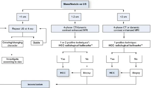
Le CHC a la particularité de pouvoir être diagnostiqué sur des critères non invasifs d’imagerie chez les patients cirrhotiques. L’AASLD en 2005 (Annexe 4) puis plus récemment l’EASL en 2012 (Figure 1) [3-4] ont publié les critères radiologiques nécessaires pour confirmer le diagnostic de CHC et ainsi éviter la biopsie hépatique.
Pour les nodules de moins de 1cm découverts en échographie, indication de surveillance par une nouvelle échographie à 4 mois. Si le nodule est inchangé, poursuite de la surveillance. Pour les nodules entre 1 et 2cm, indication de réaliser une imagerie complémentaire soit par scanner soit par IRM. Les caractéristiques typiques d’un CHC sont 1/ nodule hypervasculaire à la phase artérielle et 2/ présence d’un lavage au temps portal. Depuis 2012, une seule imagerie est suffisante dans les centres experts. Sinon 2 imageries (IRM et scanner) doivent être pratiquées.
Pour les nodules supérieurs à 2cm, le diagnostic de CHC est posé si la tumeur possède des caractéristiques typiques sur un seul examen d’imagerie.
Il n’y a plus nécessité d’avoir un taux d’AFP élevé pour affirmer le diagnostic grâce à l’évolution des performances d’imagerie diagnostique.
Le diagnostic histologique est nécessaire chez les patients non cirrhotiques, ou pour les nodules atypiques en imagerie qui ne correspondent pas aux critères précédemment décrits.
Figure 1. Critères diagnostiques du CHC selon l’EASL en 2012
* Une imagerie seulement est recommandée dans les centres experts
** Caractéristiques typiques du CHC : nodule hypervasculaire avec lavage veineux/portal
Figure extraite de Clinical Practice Guidelines : Management of Hepatocellular Carcinoma établi par l’EASL et l’EORTC, publié dans Journal of Hepatology, 2012 vol. 56 ; 908-943
Si le diagnostic de CHC est posé, le bilan d’extension devra comporter au minimum un scanner thoraco-abdomino-pelvien. D’autres examens complémentaires pourront être pratiqués en fonction des symptômes cliniques.
CLASSIFICATION DU CHC
Les classifications sont utilisées en cancérologie pour permettre d’établir un pronostic et de proposer le traitement le plus approprié en fonction de l’évolution de la maladie.
Habituellement la plus utilisée en cancérologie est la classification TNM (Tumor Nodes Metastasis). La 7ème et dernière édition date de 2010 (Annexe 5) [6]. Pour le CHC, cette classification TNM pose plusieurs problèmes. Premièrement elle prend en compte l’invasion vasculaire qui ne peut être appréciée que si les patients ont bénéficié d’une chirurgie. Mais surtout elle ne prend pas en compte le foie non tumoral, c’est à dire la maladie sous jacente du foie et sa gravité.
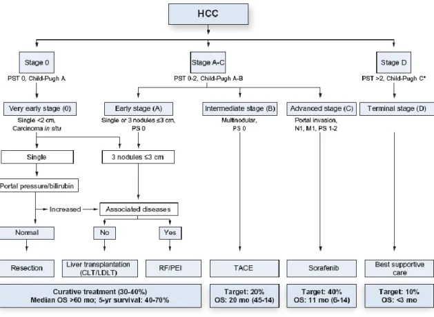
du patient, la sévérité de la cirrhose et l’extension tumorale. Dans la dernière version de cette classification parue en 2008 [8], 5 stades sont définis (BCLC 0, A, B, C et D) représentés dans la figure 2.
Figure 2. Classification BCLC (Barcelona Clinic Liver Cancer)
TRAITEMENT DU CHC
Grâce à cette classification BCLC, un algorithme thérapeutique a pu être établi (figure 3). Ainsi, on peut s’orienter :
- vers un traitement curatif pour les BCLC 0 et A avec la résection chirurgicale, la radiofréquence ou la transplantation hépatique
- vers la chimioembolisation pour les BCLC B
- vers la chimiothérapie, en première intention le sorafenib, pour les stades plus avancés (BCLC C) qui ont une atteinte vasculaire ou métastatique
- vers les soins de support exclusifs pour les patients BCLC D
Transplantation Hépatique
Les débuts de la transplantation hépatique (TH) pour le CHC furent difficiles. En effet, en l’absence de critères de sélection, les résultats étaient médiocres avec des taux de récidive élevés et des survies à 1 et 5 ans bien inférieures à celles observées dans la TH pour des pathologies bénignes.
Figure 3. Options thérapeutiques en fonction de la classification BCLC, selon l’EASL 2012
Figure extraite de Clinical Practice Guidelines : Management of Hepatocellular Carcinoma établi par l’EASL et l’EORTC, publié dans Journal of Hepatology, 2012 vol. 56 ; 908-943
En 1996, l’équipe de Mazzaferro [9] a établi les critères de Milan, qui régissent toujours l’attribution des greffons dans de nombreux pays. Cette étude prospective portait sur 48 patients qui avaient en imagerie un nodule de moins de 5cm ou 3 nodules de moins de 3cm. Une comparaison a été faite entre les 35 patients qui remplissaient ces critères sur l’explant et les 13 patients (27%) qui finalement sortaient des critères à l’analyse histologique. Il existait une différence significative à la fois pour la survie globale à 4 ans (85% contre 50%, p=0.01) et la survie sans récidive à 4 ans (92% contre 59%, p=0.002).
Cette étude a conduit à faire de la TH le traitement de choix des CHC de petite taille lorsque la résection est impossible, soit en raison de la gravité de la cirrhose sous jacente, soit en raison de la localisation de la tumeur.
L’objectif actuel de la TH pour le CHC est d’avoir des résultats similaires à la TH pour des pathologies non tumorales. Ainsi, la mortalité péri-opératoire, à 1 an et à 5 ans ne doit pas
Critères de sélection pour la TH (Tableau 1)
Depuis l’apparition des critères de Milan, de nombreuses équipes ont voulu élargir ces critères de sélection, considérés comme trop restrictifs [10].
L’équipe de Yao en 2001 [11] a défini les critères de San Francisco (UCSF, University of California, San Francisco) soit 1 nodule ≤ 6.5cm ou ≤ 3 nodules ≤ 4.5cm avec un diamètre cumulé total ≤ 8cm. Cette étude rétrospective portait sur 70 patients. Les 60 patients qui rentraient dans ces critères à l’anatomopathologie avaient une survie à 1 an significativement plus élevée que les 10 patients qui les dépassaient (90% contre 50%, p=0.0005).
Ces résultats ont ensuite été validés sur l’analyse de l’imagerie pré opératoire, les critères de San Francisco permettaient donc de sélectionner les patients avant la TH [12].
Par la suite, une grande étude rétrospective multicentrique [13] parue en 2009 et portant sur 1556 patients a établi les critères dits « up to seven » qui sont déterminés par l’absence d’invasion microvasculaire et le nombre total de nodule additionné à la taille du plus gros nodule qui ne doit pas excéder 7. La survie globale à 5 ans pour ce groupe de patients était de 71.2%, similaire à celle des patients dans les critères de Milan (73.3%) et bien supérieure à celle des patients excédant les 2 critères (48.1%).
Milan 1 nodule ≤ 5cm
ou ≤ 3 nodules ≤ 3cm
USCF
1 nodule ≤ 6,5cm ou ≤ 3 nodules ≤ 4,5cm avec un
diamètre cumulé total ≤ 8cm
Up to seven
Absence d'invasion microvasculaire Somme du nombre de nodules et de
la taille du plus gros nodule ≤ 7
Tableau 1. Les critères de sélection principalement utilisés pour le CHC dans la TH
UCSF, University of California, San Francisco
Score alpha-protéine
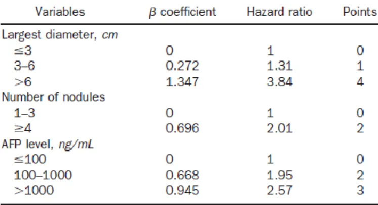
Plus récemment en 2012, l’équipe de Duvoux et al. [14] a publié le score « alpha-protéine ». Ce score a l’avantage de prendre en compte le nombre et la taille du plus gros nodule basés sur l’imagerie pré opératoire, mais aussi la concentration d’alphafoeto protéine (AFP).
Le taux d’AFP défini comme de mauvais pronostic varie selon les études, la limite n’étant pas bien établie et pouvant être fixée à 10ng/mL [16], 300ng/mL [17], 400ng/mL [18, 19], 455ng/mL [20] ou même 1000ng/mL [11] selon les auteurs.
Pour la pratique clinique courante, une version simplifiée du score alpha-protéine a été établie
(tableau 2). Une valeur supérieure à 2 définit le groupe de patients à fort risque, une valeur
inférieure ou égale à 2 celui à faible risque.
Tableau 2. Version simplifiée du score alpha-protéine
Tableau extrait de l’article de Duvoux C, Gastrenterology, 2012 ;143 :986-994
Les patients dans le groupe à faible risque du score alpha-protéine avaient un risque de récidive à 5 ans significativement plus faible que les patients dans le groupe à fort risque (respectivement 8.8% contre 50.6%, p<0.001). On retrouvait la même tendance pour la survie globale à 5 ans (respectivement 67.8% contre 47.5%, p=0.002).
En plus de ces bons résultats sur la survie et la récidive, le score alpha-protéine semble également supérieur aux critères de Milan. En effet, les patients inclus dans les critères de Milan mais avec un taux d’AFP > 1000ng/mL (donc dans le groupe à haut risque du score alpha-protéine) avaient un risque de récidive plus élevé que les patients ayant une AFP < 1000ng/mL (37.1% contre 13.3%, p<0.001). A l’opposé, les patients en dehors des critères de Milan mais avec un score alpha-protéine ≤ 2 et une AFP ≤ 100ng/mL avaient un risque de récidive significativement plus faible que les patients en dehors des 2 critères (14.4% contre 47.6%, p=0.006). La prise en compte de l’AFP avec des valeurs limites définies à 100ng/mL
Ceci a conduit à utiliser le score alpha-protéine comme critère de sélection pour la TH en France.
L’autre avantage de ce score est qu’il ne prend en compte que la tumeur viable en imagerie et non pas la tumeur totale. Ceci met donc en avant l’intérêt d’un traitement néo-adjuvant.
TRAITEMENT NEO-ADJUVANT
Le traitement des CHC en néo-adjuvant avant la TH est de pratique courante mais les preuves scientifiques restent faibles, comme l’atteste les recommandations de l’EASL-EORTC de 2012 [3] (Annexe 6).
La chimioembolisation lipiodolée (CEL) est actuellement le traitement le plus utilisé.
La CEL comme traitement d’attente
Malgré les faibles niveaux de preuve, l’EASL recommande de réaliser un traitement préopératoire seulement si la durée d’attente sur liste est estimée supérieure à 6 mois, afin d’éviter la progression tumorale [3].
En effet, dans le contexte actuel de pénurie de greffon, le risque de sortir de liste de TH en raison d’une attente trop longue sur liste est autour de 20%, pouvant aller même jusqu’à 46% [21].
Graziadei et al. [22] ont retrouvé que ce risque de sortir de liste était nul si les patients bénéficiaient d’une CEL préopératoire, malgré un temps d’attente sur liste de 200 jours en moyenne. D’autres études ont montré l’intérêt de la CEL comme traitement d’attente sur liste, permettant une stabilité voir une régression des tumeurs [23-25]
Même si il n’existe pas d’étude randomisée, le risque de sortir de liste est moins important après traitement néo-adjuvant, et ce d’autant plus que le temps d’attente sur liste est long.
Par contre, les effets de la CEL néo-adjuvante sur la survie et la récidive après TH sont beaucoup moins évidents [26-29].
Decaens et al. [26], retrouvaient dans une étude cas témoin portant sur 100 patients traités par CEL et 100 patients contrôles une survie globale à 5 ans identiques (59.4% contre 59.3%). Une autre étude cas témoin portant sur 31 patients [27] retrouvait également une absence de différence entre les patients traités par CEL et non traités sur la survie globale (84% contre 91%), la survie sans récidive (74% contre 85%) et la récidive après TH (23% contre 12%).
Seehorfer et al. [28] retrouvaient de même un taux de récidive et une survie à 5 ans similaires entre les patients ayant bénéficié d’une CEL ou non.
Dans toutes ces études rétrospectives, les patients inclus avaient les mêmes caractéristiques initialement.
Mais l’intérêt de la CEL est surtout d’induire une nécrose tumorale et donc d’essayer de faire accéder à la TH des patients qui sont initialement en dehors de tout critère et qui répondent efficacement à la CEL. Ce concept est connu sous le nom de downstaging,
Downstaging
Le concept de downstaging est plus récent. Deux revues sont parues récemment sur son intérêt dans le CHC [30,31].
Le premier à avoir étudié la CEL dans ce contexte est Majno [32]. Il a démontré que les patients qui avaient un downstaging des tumeurs > 3cm et une nécrose complète en anatomopathologie avaient une meilleure survie sans récidive que les patients ayant une réponse incomplète à la CEL ou pas de CEL.
Le taux de downstaging varie beaucoup d’une étude à l’autre, passant de 24 à 90% [22,24, 33,37]. Cette large fourchette est due à la grande disparité des critères à la fois d’inclusion et d’efficacité de la CEL.
Critères d’inclusion
Sur toutes les études étudiant le downstaging, chacune détermine les critères d’inclusion différemment. Ainsi, certains auteurs choisissent de ne pas mettre de limite de taille et de nombre [24,33,35], d’autres définissent de nouveaux critères d’inclusion [34,37] plus larges que ceux utilisés actuellement.
Critères d’efficacité
Concernant les critères d’efficacité qui définissent le downstaging, la plupart des études se basent sur les critères de Milan [24,34,37]. D’autres ont pris en compte les critères Response Evaluation Criteria in Solid Tumors (RECIST) [33,35,38]. Les études les plus récentes [34,37] ne prennent en compte que la tumeur viable et les zones de nécrose vues en imagerie sont exclues.
Résultats du downstaging
Dans l’étude de Millonig [38], la réponse tumorale complète ou partielle après CEL était associée à une meilleure survie globale à 1 an, 2 ans et 5 ans mais seulement pour le sous groupe de patients initialement dans les critères de Milan.
De nombreuses autres études ont montré l’efficacité d’un traitement néo-adjuvant également pour des patients initialement hors critères mais qui rentrent dans les critères de Milan après downstaging.
Ravaioli et al. [34] retrouvaient une survie sans récidive à 1 an et 3 ans similaire entre les patients ayant bénéficié d’un downstaging (78% et 71% respectivement) et les patients dans les critères de Milan initialement (80% et 71%, p=ns).
Yao et al. [37,39] ont étudié 61 patients excédant les critères de Milan. Les patients ayant un downstaging efficace ont été transplantés, la survie globale à 1 an et 4 ans était respectivement de 87.5% et de 69.3%.
Globalement, ces études montrent qu’un traitement néo-adjuvant doit être proposé à tout patient porteur de CHC non métastatique. Si un downstaging est acquis, une transplantation hépatique pourra être proposée car la survie et la récidive sont identiques à celles de patients transplantés dans les critères initialement [22,33,34,37,40]. Toute la difficulté réside dans le fait que le pronostic est principalement lié à l’analyse histologique et que les premiers critères étudiés (critères de Milan) étaient basés dessus. Il existe donc une nécessité de forte corrélation entre l’imagerie et l’anatomopathologie pour la détection des tumeurs (nombre et taille) mais également pour l’évaluation de la tumeur viable en imagerie après traitement néo-adjuvant.
Ce travail avait donc pour but :
- D’évaluer la corrélation entre l’imagerie et l’anatomopathologie sur le nombre et la taille des nodules
- D’évaluer la corrélation entre la tumeur viable en imagerie et la nécrose tumorale en anatomopathologie après CEL et ainsi évaluer l’intérêt de la CEL néo-adjuvante - D’évaluer le score alpha-protéine, en comparaison avec les critères de Milan, en
Use of the viable tumor as a predictor of hepatocellular carcinoma
recurrence after preoperative transarterial chemoembolization: a
radiological-pathological correlation
Laetitia Lecoq1, Nathalie Sturm², Jérôme Gendre3, Christian Letoublon4, Thomas Decaens1, Vincent Leroy1
1
Clinique d’hépato-gastro-entérologie, CHU de Grenoble, France ; ² Service d’anatomopathologie, CHU de Grenoble, France ; 3
Service de radiologie et imagerie médicale, CHU de Grenoble, France ; 4 Clinique de chirurgie digestive et de l’urgence, CHU
de Granoble, France Key words AFP score Liver transplantation Tumor downstaging Abbreviations
AFP, Alphafoeto protéine; AASLD, American Association for the Study of Liver Diseases; CT, Computerized Tomography; EASL, European Associtation for the Study of Liver; LT, Liver Transplantation; MELD, Model for End-stage Liver Disease; MRI, Magnetic Resonnance Imaging; RECIST, Response Evaluation Criteria in Solid Tumors; TACE, TransArterial ChemoEmbolization.
ABSTRACT
Nowadays, the liver organ allocation depends on Milan criteria which have been extended recently with AFP score, taking into account number and size of viable nodules and AFP level. The study was designed to evaluate the reproducibility of this criteria and the viable tumor after neo-adjuvant TACE, using a radiological-pathological correlation, and determine the impact on HCC recurrence and survival after liver transplantation.
One hundred consecutive patients transplanted for HCC in a unique centre between 2005 and 2012 were included. A neo-adjuvant TACE was performed for 54 patients. A comparison was made between the last imaging done before LT and histology, on a total of 322 nodules.
A significant correlation between imaging and histology was observed for the total tumor size (r=0.77, p=0.001) and the number of nodules (r=0.77, p=0.001). Ninety six nodules, mostly of small size (11.2mm) were seen on histology but not on imaging, with no impact on recurrence. There was a good concordance for Milan criteria between imaging and histology (81%) but lower than for AFP score (90%). The mean tumor necrosis was 68% on imaging and 57% on histology. Among the patients who had a complete tumor response on imaging, only 28% actually had a total tumor necrosis on histology. Using the viable tumor on imaging, the probability of recurrence at 5 years was significantly higher for patients at high risk in AFP score compared with low risk (33.3% vs 14.6%, p=0.02). There were no differences on recurrence between Milan criteria and AFP score, but AFP score allowed LT to 13% more patients compared with Milan criteria.
INTRODUCTION
Hepatocellular carcinoma (HCC) is the sixth most common cancer worldwide and the third most common cause of death from cancer [1]. Liver transplantation (LT) is an interesting therapeutic option for HCC, because it removes both the tumor and the underlying liver disease. However, because of donor shortage, LT for HCC is expected to reach a 5 year survival rate similar to the one achieved in benign liver disease, meaning around 70%.
In these conditions, LT is considered to be the best treatment for early-stage and unresectable HCC. Since 1996, the Milan criteria [9] (one nodule under 5cm or less than 3 nodules not exceeding 3cm) are used for the organ allocation in many countries, including United States, despite many recent studies showing an excellent 5 year survival with expended criteria [10]. Among them, the alpha-protein score [14] includes the total number of nodules, the size of the largest tumor and the alphafoetoprotein level (AFP). The criteria for number and size of nodules are less restrictive than Milan criteria and allow LT to a larger population with an excellent 5 year survival rate (67.8% for the low risk group). In the same time it excludes patients with AFP > 1000ng/ml, a well known poor prognostic factor [11,16-20]. This score takes account only the viable tumor on imaging, which reflects more the actual situation. Indeed, many programs choose to perform a neo-adjuvant treatment before LT, mainly Trans Arterial Chemo Embolization (TACE), because of the long waiting time on list and the risk of dropout. TACE is also used as downstaging, to reduce tumor size and allow patients outside the selection criteria to finally include it.
However, the benefit of neo-adjuvant TACE remains uncertain because no studies showed a benefit in survival [26-29]. Downstaging procedures are even more controversial, even if some studies suggest that recurrence and survival are similar to patients transplanted inside Milan criteria [22,33,34,37,40].
The purpose of this study was to evaluate the correlation between imaging and histology, including the evaluation of tumor response on imaging compared with tumor necrosis on the explant when a TACE was performed, and to estimate the impact on recurrence and survival after LT.
PATIENTS AND METHODS
Patient Population
Between January 2005 and August 2012, a total of 248 patients had undergone liver transplantation in Grenoble, France. Among them, we included all the patients with HCC on histology, either it was found before transplantation or not.
All patients had an imaging by CT completed with an MRI if necessary while listed. An imaging control by ultrasound, CT or MRI was performed every 3 months on the waiting list. The diagnosis of HCC was based on non invasive methods of AASLD (Association for the Study of Liver Diseases) practice guidelines [4]. Nodules between 1 and 2cm in cirrhotic liver with hypervascular and washout in the portal/venous phase in two dynamics imaging techniques or nodules superior to 2cm in cirrhotic liver either with typical features of HCC on one dynamic imaging or hypervascular nodules with AFP superior to 200ng/ml were considered as HCCs. In all other cases, a biopsy was performed.
Once patients were listed, a systematic preoperative treatment was proposed for patients with HCC, depending on the Child Pugh class.
The final patient population included 100 patients (40%), with a total number of nodules of 252 on imaging and 322 on histology.
Sixteen patients had an incidental HCC.
Imaging Technique
The abdomen was imaged from the dome of the diaphragm to the iliac crests.
CT examinations were performed with a multiphase contrast-enhanced spiral CT, on a 16 detector row Siemens® CT or a 64 detector row Philips® CT. The contrast was 100 or 150mL of XENETIX® 350 which was injected intraveinously at a rate of 4 mL per second. Noncontrast images with 3mm collimation were first performed followed by late arterial images with a 15 seconds delay and 3mm collimation and portal venous phase images with a 50 seconds delay and 3mm collimation. For patients who could not have CT (renal insufficiency, iode allergy) or when CT was not informative, a gadolinium-enhanced MRI was performed.
We studied the last imaging done before transplantation, either a CT for 87 patients (87%) or an MRI for 13 patients (13%). Seven patients who had neo-adjuvant treatment couldn’t have
One radiologist retrospectively reviewed CT for patients with a high discordance between imaging and pathology (15% of all CTs). He reviewed all images independently and was blinded to patient and histopathological outcomes.
If a neo-adjuvant treatment was performed (n=54), we studied on the last imaging both the total tumor (including necrotic nodules) and the viable tumor. We estimated the viable tumor with the residual lesion enhancement, just as described in mRECIST (modified Response Evaluation Criteria in Solid Tumors) [6], and the lipiodol fixation of the lesion. Patients with necrosis ≥ 95% were considered as complete tumor response.
Neo-adjuvant treatment protocol
Of the 54 patients who had a preoperative treatment, 51 (94%) had TACE, 2 patients (4%) had a percutaneous radiofrequency and 1 patient (2%) had both TACE and radiofrequency. For TACE, selective celiac and superior mesenteric angiography was performed to assess the hepatic vascular anatomy. The feeding arteries of the lesion were catheterized as selectively as possible in 27 patients, the other 25 patients having a global TACE. The chemoembolization solution containing 10 mL of lipiodol and 25 to 100 mg of doxorubicin and was injected under fluoroscopic control, just before mechanical obstruction.
Among the 27 patients who had a selective TACE, 8 patients had TACE with DC beads with diameters from 100 to 300µm or from 300 to 500µm charged with doxorubicin.
Thirty two patients (63%) underwent only 1 session, 17 patients 2 sessions (33%) and 2 patients 3 sessions (4%) (mean 1.41 ± 0.57)
Correlation with histopathology
All explanted livers were sliced serially at 5 to 10 mm intervals. First a macroscopic evaluation was performed to detect neoplastic nodules. Then all sections were embedded in paraffin and cut into 3 to 4µm serial sections for standard hematoxylin-eosin staining. All sections were carefully screened, and all tumors detected were noted for mapping. The necrosis percentage was defined as the volume of necrotic areas divided by the total tumor volume. Patients with necrosis ≥ 95% were considered as complete tumor response. The degree of tumor differentiation was graded according to Edmonson criteria. Patients who had multiple nodules with different differentiation were grouped according to the worst histologic grade.
Patient follow-up
All patients were followed up with clinical exam, biological test including AFP and an ultrasound every year at 3, 6, 9 and 12 months then every 6 months for life.
A routine thoraco-abdominal CT or MRI was performed every 6 months during 2 years or if symptoms appeared to detect HCC recurrence.
No adjuvant therapy was administrated after LT.
Since 2009, immunosuppression was replaced with mtor inhibitor (everolimus) instead of calcineurin inhibitor after 3 months if the tumor volume exceeded Milan criteria on histology.
Statistical analysis
All statistical analysis (chi-square test, Student test, Kaplan-Meier estimate, log rank test) were performed using IBM® SPSS program v21 for Windows®.
RESULTS
Patient and tumor characteristics
The patient characteristics are summarized in Table 1. Fifty four patients were in the neo-adjuvant treatment group and 46 patients did not have a neo-neo-adjuvant treatment. No differences were observed for age (median of 57 years), gender and AFP score between the 2 groups.
The most common etiology of cirrhosis was HCV (43%) in the neo-adjuvant group compared with alcohol (76%) in the other group (p=0.001).
The Child-Pugh score (48% of Child-Pugh C vs 7%) and the MELD score (17.6 vs 10.6) were significantly higher in the no preoperative treatment group (p<0.001), because whenever it was possible, a TACE was performed as a bridge to LT. The mean waiting time for LT was significantly lower (103 days vs 152 days) in the no preoperative treatment group (p=0.038) because the cirrhosis was more severe.
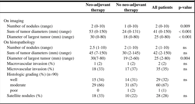
The tumor characteristics are summarized in Table 2. The main difference was observed on imaging with a significantly higher number of nodules (2 vs 1, p=0.009), sum of tumor diameters (53 vs 24mm, p=0.001) and diameter of largest tumor (30 vs 18mm, p=0.001) in the preoperative treatment group.
On the other hand, no differences were observed on histology between the 2 groups for number of nodules (2.5 vs 2, p=ns) or sum of tumor diameters (45 vs 30mm, p=ns). The only statistical difference was the diameter of largest tumor (30 vs 19mm, p=0.004) which was higher in the preoperative treatment group.
No other statistical differences were found on the histological characteristics such as macrovascular invasion, microvascular invasion, histological grading or satellite nodules.
Number of nodules
We first evaluated one of the most important predictive factors of recurrence, the total number of nodules, which included both viable and post TACE necrotic tumors.
For the 100 patients included in the study, 252 nodules were seen on imaging and 322 nodules on histology. On the 252 nodules seen on imaging, 26 nodules (10%) were not HCC on histology but adenoma or regeneration nodules. They were mainly hypervascular (96%), with washout in 46% of cases with a median size of 16mm (range 9-33mm).
For 43% of patients, the number of nodules seen on imaging was the same as the number seen on histology. The number of nodules was discordant between imaging and histology for 57% of patients. Imaging underestimated the number of nodules for 47% of patients and overestimated it for 10% of patients.
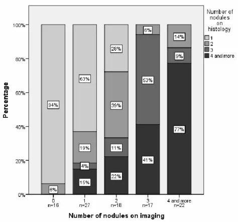
The figure 1 showed the correlation between imaging and histology for the total number of nodules. Sixteen patients (16%) had an incidental tumor, meaning a HCC found on the explant but not seen on imaging. Only one patient (6%) had 2 tumors on histology, all the others had just 1 nodule. For the non incidental tumors, the total number of nodules was in a majority of cases the same on imaging as on histology (63% for 1 nodule, 39% for 2 nodules, 53% for 3 nodules and 77% for more than 4 nodules). A high correlation was found with a Pearson correlation coefficient of 0.768 (p=0.001).
Size of nodules
Size of nodules is the second major predictive factor of recurrence after LT.
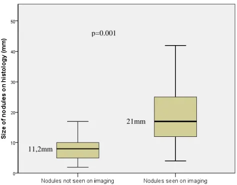
We first evaluated the characteristics of the 96 incidental nodules. The only difference between these nodules and the 226 nodules seen on imaging and histology was the size. The Figure 2 showed that incidental nodules were significantly smaller than those detected (median size 11mm vs 21mm, p=0.001), almost all of them were inferior to 2cm.
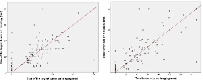
We then studied the size of the largest tumor and the total tumor size for the 100 patients included (figure 3). The Pearson correlation coefficient was 0.637 (p=0.001) for the size of the largest tumor and was 0.771 (p=0.001) for the total tumor size, proving an excellent correlation between imaging and histology.
Considering the 2 groups of patients (neo-adjuvant TACE or not), a good correlation was found for the size of nodules. For the 46 patients who did not have a neo-adjuvant treatment, the Pearson correlation coefficient was 0.514 (p=001) for the size of the largest tumor and 0.905 (p=0.001) for the total tumor size. For the 54 patients who had a neo-adjuvant TACE, it was respectively 0.678 (p=0.001) and 0.672 (p=0.001) for the size of the largest tumor and the total tumor size.
Looking only at the total tumor size, it was higher in the neo-adjuvant TACE group compared with the no neo-adjuvant, both on imaging (mean of 62mm vs 32mm, p=0.001) and on histology (mean of 59mm vs 44mm, p=0.06). However, total tumor size included both viable and necrotic tumor. Recent studies have demonstrated that we should take account only the
Viable tumor
We studied the viable tumor for patients who had a neo-adjuvant treatment (n=54).
As shown in Table 3, total tumor size using the whole tumor had an average of 62mm on imaging and 59mm on histology, but dropped to respectively 25mm and 31mm when only the viable tumor was taking account. The mean necrosis had an average of 68% ± 40% on imaging and 57% ± 40% on histology.
The Pearson correlation coefficient was 0.778 (p=0.001) for the total viable tumor size between imaging and histology and 0.627 for the evaluation of necrosis (p=0.001).
Despite the good correlation for the total viable tumor and the mean necrosis percentage, a large percentage of patients (45%) were overestimated by imaging. Indeed, patients were classified in 4 groups depending on the tumor percentage of necrosis: total necrosis (≥95%), partial necrosis (50-94% and 5-49%) and absence of necrosis (<5%). Only 47 patients were studied because of 7 missing data (no CT was done after TACE and viable tumor could not be compared with histology). Twenty patients (42%) were well classified between imaging and histology, but 21 patients (45%) had a tumor response overestimated on CT, and 6 patients (13%) were underestimated by imaging.
Regarding the total tumor response, 24 patients (44%) had a necrosis superior to 95% on imaging. It represented a mean necrosis of 68% on histology. Only 7 on the 24 patients (28%) also had a complete necrosis on histology. Consequently, 72% of patients were considered in remission but the CT overestimated the tumor response. Ten patients (42%) had a necrosis between 50% and 94%, 5 patients (21%) between 5% and 49% and 2 patients (8%) had a necrosis inferior to 4% on histology.
On histology, complete tumor necrosis was found in 15 patients (28%).
Classification according to predictive score
We evaluated 2 predictive scores of recurrence after LT, the gold standard (Milan criteria) and a more recent one (AFP score). Milan criteria are defined by 1 nodule under 5cm or not more than 3 nodules with a maximum diameter of 3cm. This score is currently used in many countries including USA. We also studied AFP score, based on the number and size of viable tumor on preoperative imaging and AFP level. This score is already used in France for the organ allocation.
We only used the viable tumor on last imaging before LT for classification, both for alpha-protein score and for Milan criteria.
Regarding the Milan criteria, 78% of the patients were inside on imaging and 71% on histology. The Pearson correlation index for Milan criteria between imaging and histology was 0.512, p=0.001. Nineteen patients (19%) had a discordance of classification between imaging and histology. Six patients were outside Milan criteria on imaging but turned inside on histology. For 4 patients, the cause of the discordance was the number of nodules overestimated on imaging and for the 2 remaining patients it was the size of the largest tumor also overestimated on CT. On the opposite, 13 patients were inside Milan criteria on imaging but finally turned outside on histology. In majority of cases (85%) the discordance was on the number of nodules because CT or MRI underestimated it. But as we have seen above, these nodules not detected on imaging were smaller, with a median size of 11mm.
Regarding the AFP score, 91% of the patients were inside on imaging and 85% on histology, this was consequently a less restrictive score than Milan criteria. The correlation was also excellent between imaging and histology with a Pearson correlation coefficient of 0.553 (p=0.001). The discordance between imaging and histology was mainly due to the size of nodules (87.5%), because this score did not give a limit for the number of nodules. AFP level excluded 4 patients on imaging (44%) and histology (27%).
Comparing the 2 scores, 15 patients were inside alpha-protein score and outside Milan criteria on imaging, including 3 recurrences (20%). On the opposite, only 2 patients were inside Milan criteria on imaging but were outside alpha-protein score because of AFP higher than 100ng/l, including 1 patient who had a recurrence (50%). Alpha-protein score was less restrictive and allowed LT to 13 patients more than Milan criteria on imaging.
Regarding the 16 patients who had an incidental tumor, they were all inside Milan criteria and AFP score on histology. None of these patients had a microvascular invasion and AFP was always under 20ng/L. No recurrence was observed in this group, confirming the good prognosis of these incidental tumors.
Impact on survival and recurrence
Eleven recurrences occurred during the follow up period, 9 were dead and 2 in remission in March 2013. Seven recurrences (64%) were a metastasis evolution, 2 were a hepatic recurrence, 2 were infiltrative HCC not seen on imaging before transplantation.
The 5 year recurrence free survival was similar between patients who had a preoperative TACE and those who did not (respectively 60.2% vs 67.2%, p=0.18).
We then studied the impact of the nodules not seen on imaging. We compared the probability of recurrence at 5 years for patients with underestimated number of nodules on imaging (n=47, 6.4%) and patients with no discordance between imaging and histology (n=43, 28.5%, p=0.04). These nodules not seen on imaging did not impact the risk of recurrence, they were even of better prognosis because it included the 16 patients with incidental tumors.
Regarding the Milan criteria, patients who met the criteria on histology had a 5 year recurrence free survival of 66.1% compared to 55.7% for patients outside (p=0.30). This was similar for Milan criteria on imaging (respectively 62.5% for patients inside vs 65.5% for patients outside, p=0.75).
For AFP score, the 5 year recurrence free survival was 62.9% for patients with a low risk compared with 66.7% for patients at high risk (p=0.11) on histology and 64.8% compared with 60% on imaging (p=0.71).
The figure 5 showed the Kaplan Meier probability of recurrence according to Milan criteria and alpha-protein score, both on imaging and histology. The probability of recurrence at 5 years was respectively 32.2% for patients outside Milan criteria on imaging and 11.1% for patients inside on imaging (p=0.065). On histology, the difference was significant between patients outside (probability of recurrence at 5 years of 32.5%) and inside (9.8%, p=0.025). Regarding AFP score, the Kaplan Meier probability of recurrence at 5 years was 33,3% (n=9) for patients at high risk on imaging and 14,6% (n=91) for patients at low risk (p=0,023). The probability of recurrence at 5 years was also significantly higher for patients at high risk on histology (35% n=15) compared with patients at low risk (12.3% n=85) (p=0.01).
DISCUSSION
Nowadays the diagnosis of HCC is based on non invasive imaging criteria in patients with cirrhosis [3,7]. These imaging, represented mainly by CT multidetector row and MRI, need to be very efficient in order not to mistreat patients.
A few studies tried to determine sensitivity and specificity of imaging making a correlation with the explant analysis after LT [43-45]. Luca et al [43] showed a overall sensitivity of multidetector row CT of 78% for nodules under 10mm and 98% for nodules above 20mm. Lu et al. [45] compared 2 imaging techniques (multidetector row CT and MRI) with a sensitivity around 80% for the number of nodules and more than 85% for the size of nodules for both imaging. Our study also confirmed the good correlation between imaging and histology, both for total number of nodules and for the size of the largest tumor (p<0.0001, figure 1 and 2). Despite the good correlation, 47% of patients had a number of nodules underestimated by CT. But the discordance was mainly due to small nodules not detected on imaging significantly smaller than nodules seen on histology (11mm vs 21mm, p=0.001). These nodules did not impact on survival and recurrence. Boin et al. [44] also found the same results with 42.2% of patients outside Milan criteria on the explant but the 10 year survival rate was similar (57% for patients inside Milan versus 47% for patients outside). Sugimashi et al. [46] also concluded that the detection of small HCCs (<10mm) and well-differentiated HCCs was significantly lower and these nodules could drop patients outside Milan criteria on histology, but the recurrence-free survival rate was similar to the patients inside Milan criteria on histology.
The lack of sensitivity for small nodules detection was the main reason why patients dropped outside Milan criteria on histology. Indeed, in 85% of cases, patients were inside criteria on imaging but outside on histology because of the number of nodules underestimated by CT. But this concerned only 13 patients, and our study showed an excellent correlation for Milan criteria between imaging and histology (p=0.001).
However, Milan criteria are considered to be too restrictive. Many studies in the past years summarized in a review [47] tried to find a better selection system to avoid the exclusion of too many patients and in the same time achieve a 5 year survival rate around 70%. Nowadays,
addition of the number of nodules and the size of the largest tumor can not exceed 7) [13]. More recently, the alpha-protein score was released, based both on imaging (number and size of nodules) and AFP level [14]. This is an interesting score because AFP is known to be a poor prognostic factor in many studies [11,16-20]. It also has the advantage to take account only the viable tumor. In our study, we showed a good correlation between imaging and histology for this score (p=0.001). Most of all, this score permitted to allow LT to 13 patients more (13%) than with Milan criteria.
Regarding the neo-adjuvant treatment and the viable tumor, the first to evaluate his effect was Majno [32]. In his study, 27% of patients had a complete tumor necrosis and 52% had a downstaging. No significant difference was observed in survival between patients with TACE or without, although the total tumor size was significantly higher in patients who received TACE (62mm vs 32mm, p<0.0001). In our study, complete necrosis on histology was seen in 28% of the treated lesions and the mean necrosis was 57%. This was quite similar to other reports with complete necrosis from 15% to 43% (23,27,36,37,48-51], with an average around 35%, and a mean necrosis found on histology from 57% to 72.5% [48,49,52-54]
We did not find significant differences for recurrence between patients with neo-adjuvant TACE or not (p=0.63), as in other studies [26-29].
Even if impact on long term outcome is uncertain, EASL [3] recommends a preoperative treatment if the waiting list exceeds 6 months, to avoid dropout.
In our study, we chose to use only the viable tumor after TACE even for the Milan criteria, including consequently patients who had an efficient downstaging. This was already studied by some authors like Ravaioli et al. [34] who compared a group inside Milan criteria and a group initially outside conventional criteria but within Milan criteria after the downstaging procedure. The 1 and 3 years disease free survival were comparable (respectively 80% and 71% in the Milan group and 78% and 71% in the downstaging). Yao et al. [37] had the same conclusion. The 1 and 4 year recurrence free survival were respectively 96% and 92% after downstaging. More recently, a study from Otto et al. [42] demonstrated that response to pre-transplant TACE was a better predictive factor than being in the Milan criteria on the imaging at referral. If selection criteria were expanded to response to pre-transplant TACE taking account the viable tumor, it could permit to patients initially outside criteria and who had an efficient downstaging to benefit from LT.
The most important issue is to correlate radiological and pathological tumor response to neo-adjuvant TACE. Indeed, imaging for evaluation of necrosis after TACE seems to be less efficient than without preoperative treatment. In our study, 72% of patients who had a neo-adjuvant treatment with a complete tumor response on imaging had an overestimation of the treated lesions compared to the explant analysis.
Hunt et al. [55] showed the same results with a sensitivity of 36% and a specificity of 57% for CT. This was not better for MRI with sensitivity and specificity of respectively 43% and 75%. These results were far from those without TACE, with sensitivity and specificity close to 80% like seen above. In 2013, Bargellini et al. [41] published a study with good results using mRECIST criteria to assess tumor response. Sensitivity and specificity of CT in detecting complete necrosis were 87.5% and 68.9% respectively, but CT overestimated response in 21.9% of patients.
In order to better correlate tumor response, some authors tried to define more specific criteria than mRECIST. A recent study from Kwan et al. [56] evaluated the radiological predictive factors of necrosis on the explant. On post TACE CT, a lack of residual contrast enhancement, a decrease in the lesion size, a high lesion density due to accumulation of ethiodized oil and a diffuse distribution of ethiodized oil throughout the lesion were correlated with near complete tumor necrosis on histology.
In conclusion, we showed a good correlation between imaging and histology, for both nodule and tumor size. Imaging tended to underestimate the number of nodules, but these nodules were small and did not impact on recurrence. We validated the use of only the viable tumor for selection criteria and not the whole tumor, which could allow LT to all patients who had an efficient downstaging. The 2 predictive scores studied, Milan criteria and alpha-protein score, showed both acceptable 5 year recurrence free survival. This last score could be validated for the organ allocation because it reflects more the actual situation taking account only the viable tumor with less restrictive selection criteria. The main issue remains to better correlate imaging and histology after TACE, even if great advances have been made in the last few years. Indeed, imaging rather overestimated the tumor response after neoadjuvant treatment and we should be careful not to temporarily contraindicate those patients who have no viable tumor seen on imaging but not complete necrosis on histology at last.
Age, years (range) 57 (43-68) 57 (45-66) 57 (43-68) ns Sex (%) Male 49 (91) 39 (85) 88 (88) ns Female 5 (9) 7 (15) 12 (12) Etiology of cirrhosis (%) Alcohol 19 (36) 35 (76) 55 (55) <0.001 HBV 9 (17) 2 (5) 29 (29) HCV 23 (43) 6 (13) 11 (11) Others 3 (5) 2 (4) 5 (5) Child (%) A 39 (73) 2 (4) 42 (42) <0.001 B 10 (20) 22 (48) 32 (32) C 4 (7) 22 (48) 26 (26)
MELD, median (range) 10 (6-25) 15 (8-44) 12 (6-44) <0.001 Mean waiting time, days (range) (n=96) 146 (5-444) 56 (2-614) 97 (2-614) 0.038 Distribution of Alpha fetoprotein (%)
<10 ng/mL 25 (46) 31 (67) 66 (66) ns 10-400 ng/mL 25 (46) 12 (26) 37 (37)
>400 ng/mL 4 (8) 3 (7) 7 (7)
Alpha fetoprotein (range) 11.5 (1-1250) 6 (1-1774) 8 (1-1774) ns
Neo-adjuvant therapy (n=54)
No neo-adjuvant
therapy (n=46) All patients p-value Table 1 - Patient demographics
On imaging
Number of nodules (range) 2 (0-10) 1 (0-10) 2 (0-10) 0.009
Sum of tumor diameters (mm) (range) 53 (0-150) 24 (0-131) 41 (0-150) < 0.001
Diameter of largest tumor (mm) (range) 30 (0-80) 18 (0-80) 25 (0-80) < 0.001
On histopathology
Number of nodules (range) 2.5 (1-10) 2 (1-10) 2 (1-10) ns
Sum of tumor diameters (mm) (range) 45 (7-150) 30 (2-145) 42 (2-150) ns
Diameter of largest tumor (mm) (range) 30(7-80) 19 (2-60) 25 (2-80) 0.004
Macrovascular invasion (%) 1 (2) 1 (2) 2 (2) ns Microvascular invasion (%) 18 (33) 17 (37) 35 (35) ns Histologic grading (%) (n=90) well 15 (34) 14 (31) 29 (32) ns moderate 29 (66) 31 (67) 60 (67) poor 0 1 (2) 1 (1) Satellite nodules (%) 18 (33) 10 (22) 28 (28) ns
Table 2 - Tumor characteristics Neo-adjuvant
therapy
No neo-adjuvant
Total tumor size (mm)
(mean, standard deviation) 62 ± 38 59 ± 39 Total viable tumor size
(mm) (mean, standard deviation)
25 ± 40* 31 ± 43 Percentage of necrosis
(mean, standard deviation) 68 ± 40* 57 ± 40
Table 3 - Evaluation of the total tumor size for patients
who had a preoperative treatment (n=54)
Imaging Histology
* viable tumor on imaging was estimated with a residual lesion enhancement and the lipiodol fixation of the nodule (n=47)
Figure 1. Radio-anatomic correlation for the number of nodules The Pearson correlation coefficient is 0.768 (p=0.001)
Figure 2. Size comparison between nodules not seen on imaging and histology and nodules seen only on histology and missed on imaging
p=0.001
11,2mm
Figure 3. Correlation between imaging and histology for size of the largest tumor and total tumor size.
Pearson correlation coefficient = 0.637 (p=0.001) for size of the largest tumor. Pearson correlation coefficient = 0.771 (p=0.001) for total tumor size
Figure 4. Comparison of 2 predictive scores on imaging and histology (Milan criteria and alpha-protein score)
Figure 5. Probability of recurrence according to Milan Criteria and Alpha-protein score both on imaging and histology. The probability of recurrence at 5 years is noted.
32,2% p=0.023 p=0.065 p=0.025 32,5% 9,8% 33,3% 14,6% p=0.01 35% 12,3%
Cette étude a permis de soulever plusieurs problèmes actuels rencontrés dans la TH pour le CHC :
- la corrélation entre l’imagerie et l’histologie qui doit être la plus forte possible, y compris après traitement préopératoire
- l’intérêt d’un traitement néo-adjuvant, soit comme traitement d’attente sur liste, soit dans le cadre d’un « downstaging »
- la nécessité de critères de sélection à la fois rigoureux pour éviter la récidive post opératoire mais également pas trop restrictifs pour éviter d’exclure de la TH des patients qui auraient eu une excellent survie en post TH.
CORRELATION ENTRE L’IMAGERIE PREOPERATOIRE ET L’HISTOLOGIE Corrélation radio-anatomique sur le nombre et la taille
Globalement, il existe une bonne corrélation entre l’imagerie et l’anatomopathologie avec des index de corrélation excellents. Ces bons résultats sont retrouvés malgré une sous estimation du nombre de nodules par l’imagerie (47% des patients).
Des résultats semblables sont retrouvés dans la littérature [43-45].
Lu et al. [45] retrouvaient pour le nombre de nodule une sensibilité de 83.6% et de 75.8% respectivement pour le scanner et l’IRM. La sensibilité pour la taille était de 88.8% pour le scanner et 88.7% pour l’IRM.
Dans l’étude de Boin et al. [44], le scanner avait pour la taille des nodules une sensibilité à 83.3% et une spécificité à 96%.
Les discordances retrouvées concernent le plus souvent les CHC de petite taille et bien différenciés [43,46].
Ainsi, Sugimashi et al. [46] montraient que la détection préopératoire des CHC < 1cm et bien différenciés était significativement réduite, mais ceux-ci n’affectaient pas le pronostic final. Dans l’étude de Luca et al. [43], la sensibilité du scanner pour la détection des nodules de CHC passait de 78% pour les tumeurs < 1cm jusqu’à 98% pour celles > 20mm.
Dans notre étude également les nodules non détectés en imagerie étaient significativement plus petits (11 vs 21mm) et n’aggravaient pas le pronostic.
Corrélation radio-anatomique sur les critères de sélection
Il faut noter que les critères de Milan, ainsi que les critères up to seven, ont été validés sur des données histologiques et non morphologiques, d’où l’importance d’une corrélation la plus précise possible.
De nombreuses études ont montré qu’il existe des discordances entre imagerie et histologie pour les critères de sélection, qui vont surtout dans le sens de la sous estimation du CHC en imagerie [9,51,57]. Déjà en 1996 Mazzaferro et al. [9] retrouvaient dans son étude parue dans le New England Journal of Medecine 27% de patients sous estimés qui sortaient des critères de sélection à l’anatomopathologie. Baccarani et al. [57] retrouvaient également que 26% des patients qui étaient dans les critères de Milan à l’imagerie sortaient des critères en histologie.
Dans une récente étude parue en 2013 [51], 38% des patients étaient sous estimés en imagerie pour les critères de Milan contre seulement 5% qui étaient surestimés.
Mais même si il peut y avoir des discordances de classification entre imagerie et histologie, cela ne change pas au final le pronostic, la plupart des études montrant des survies similaires entre les patients qui sont dans les critères à l’histologie et ceux qui sortent finalement des critères [44,46,57,58]
Corrélation radio-anatomique après CEL
L’évaluation de la réponse tumorale sur l’imagerie est évaluée par les critères mRECIST qui ont supplantés les critères RECIST, comme recommandé par l’EASL [3] (tableau 3). Ils ne prennent en compte que la tumeur rehaussée par le produit de contraste. L’étude la plus intéressante sur les critères mRECIST est celle de Bargellini et al. [41]. Cette étude portait sur 178 patients. Les patients ont été divisés en 2 groupes : soit répondeurs (réponse complète ou partielle selon mRECIST) soit non répondeur (maladie stable ou en progression). Sur la dernière imagerie avant TH, le taux de répondeur était de 78.1% avec 48.3% de réponse complète. En comparant avec l’anatomopathologie, il existait une bonne corrélation pour 67.4% des patients, une sous estimation de la réponse en imagerie dans 10.1% des cas et une surestimation chez 21.9% des patients. La sensibilité et la spécificité du scanner pour détecter une réponse complète était respectivement de 87.5% et de 68.9%.