Journal of Fundamental and Applied Sciences is licensed under aCreative Commons Attribution-NonCommercial 4.0 International License.Libraries Resource Directory. We are listed underResearch Associationscategory.
ANTIOXIDANT, ANTIBACTERIAL AND CELL TOXICITY EFFECTS OF POLYPHENOLS FROM AHMEUR BOUAMER GRAPE SEED EXTRACTS
Z. Ghouila1,2,*, S. Laurent3,5, S. Boutry3, L.Vander Elst3,5, F. Nateche4, R. N. Muller3,5, A. Baaliouamer2
1
Scientific and Technical Research Center in Physical and Chemical Analysis, B.P 248 Alger RP 16004 Algiers, Algeria
2
USTHB, Faculty of Chemistry, B.P N°32 El-Alia 16111 Bab Ezzouar, Algiers, Algeria
3
Department of General, Organic and Biomedical Chemistry, University of Mons, Belgium
4
USTHB, Faculty of Biology, B.P N°32 El-Alia 16111 Bab Ezzouar, Algiers, Algeria
5
Center for Microscopy and Molecular Imaging, Académie Wallonie- Bruxelles, Gosselies, Belgium
Received: 01 September 2016 / Accepted: 26 December 2016 / Published online: 01 January 2017
ABSTRACT
In this work and for the first time, significant concentrations of total polyphenols and flavonoids from Vitis vinifera L. grape seed extracts were obtained (256.15 ± 17.40 mg GAE/gdmand 14.08 ± 0.64 mg CE/gdm, respectively).The LC/MS analysis revealed richness in
procyanidins. For antioxidant, antimicrobial and antifungal effects, the grape seed extract (GSE) responded positively. At 100 μg/mL, GSE induced a moderate toxicity of the order of 3.88% on 3T6 cells at the first 24 hours of treatment, whereas, its prolonged effect to 48 hours reduced this toxicity to less than 0.5%. As for the anti-proliferative effect on tumoral cell lines, a cell death of 18.39% to 23.79% and 10.30% to 20.37% was registered respectively for HeLa and BCPAP cells during 24 and 48 hours of treatment. Consequently, it is possible to consider using GSE at lower concentrations as an anti-proliferative agent without losing sight of its benefic effect on healthy cells.
Keywords: grape seed extract, 3T6 cell, antioxidant, antibacterial, anti-proliferation.
Author Correspondence, e-mail:zghouila@yahoo.com doi:http://dx.doi.org/10.4314/jfas.v9i1.24
ISSN 1112-9867
1. INTRODUCTION
Owing to the multiple chronic degenerative diseases widespread across the world, interest is now focused on products and foods that can play a protective role in the pathogenesis of these diseases [1-3].The antioxidants naturally present in foods (fruits, vegetables and plants) are nowadays the miracle products to deal with the formation of oxidative species [4,5]. Among the many phytochemical compounds consumed in our diet, polyphenols are the most important group due to their antioxidant properties [6,7]. The chemical structure of polyphenols allows them to act by different mechanisms, such as: scavengers of free radicals, chelators of metal ions such as iron and copper, and also inhibitors of the enzymes responsible for the production of free radicals [8,9].The imbalance between the formation and removal of ROS (reactive oxygen species) causes damage to the cells at the nucleic acids, proteins and membrane lipids, thus leading to many health problems associated with ageing (carcinogenesis, cardiovascular and coronary diseases) [10]. Previous research reports the action of grape seed extract in many health related areas due to its antioxidant effect [11]. In this work, polyphenols extracted for the first time from Ahmeur Bouamer grape seeds were tested on 3T6 fibroblasts. In order to eliminate any exogenous effects that could cause damage to fibroblasts and to confirm that the studied effect does result from the seed extract, the following precautions were taken to perform this work: (a) The use of ethanol to dissolve the grape seed extract was avoided because of its toxic effect on fibroblasts even at low doses and at lowest applied time [12]; (b)The tested products (GSE and synthetic antioxidants) on 3T6 fibroblasts were all dissolved in the growing medium in order to avoid osmotic pressure changes due to the organic and aqueous solvents that may cause damage to cells [13]; (c) The culture medium (DMEM) used in the treatment phase of the cells does not contain pyruvate and therefore gives an important energetic intake that contributes to the proliferation of cells [14]. In order to compare the effect of GSE, the effect of three compounds were tested on 3T6 cells: phenolic compounds, antioxidant compounds used in the pharmaceutical and food industry (BHA, BHT and vitamin E) and 2,2’-azinobis(3-ethylbenzothiazoline-6-sulfonic acid diammonium (a powerful antioxidant normally used to measure antioxidant activity). In the light of the obtained results on healthy cells, it could be envisaged a possible use of GSE as an
anti-proliferative agent on tumoral cells. Human cancer cell lines are frequently used and considered as experimental models [15] and the results obtained on cell line are sometimes directly extrapolated for in vivo cancers [16]. In this context, the effects of GSE (100 μg/mL) on HeLa (cell line derived from a cervical cancer of the uterus) and BCPAP (cell line derived from a papillary thyroid cancer) were tested. In a first step, the determination of total polyphenols and total flavonoids of grape seed extract from the Ahmeur Bouamer variety obtained by maceration was performed and the antioxidant activity by the DPPH and ABTS methods was also evaluated. The antimicrobial and antifungal activity of GSE was studied and the major components of the extract were identified by LC/MS.
2. MATERIALS AND METHODS 2.1. Materials
Embryo fibroblast cell line 3T6 and tumoral cell lines HeLa and BCPAP were obtained from
Invitrogen™ (Life technologies, UK). Dulbecco's modified Eagle's medium (DMEM), RMPI
1640 medium, sodium pyruvate 100 mM, streptomycin-penicillin (10000 μg/mL streptomycin - 10000 Units/mL Penicillin), Fetal Bovine Serum (heat inactivated FBS), TrypLE™ Express (1X) and Trypan Blue Solution 0.4% were purchased from Gibco® and PlasmocinTM 50 mg
from Invitrogen™ (Life technologies UK). PBS buffer 1%, butylhydroxytoluene (BHT),
butylhydroxyanisole (BHA), Folin-Ciocalteu reagent, 2, 2’-azinobis
(3-ethylbenzothiazoline-6-sulfonic acid diammonium salt) (ABTS+), Trolox and hydrogen peroxide 30% were purchased from Sigma-Aldrich (Co., USA). Catechin (CAT), Quercetin (QUER) and Gallic Acid (GA) were purchased from Extra syntheses (France, Lyon). All other chemicals used are of analytical grade.
2.1.1. Preparation of cell culture
The embryo fibroblast cell line 3T6 was cultured in Dulbecco's modified Eagle's medium (DMEM) with pyruvate (1%) supplemented with 10% fetal bovine serum and 1% streptomycin-penicillin (10000 μg/mL streptomycin - 10000 Units/mL Penicillin). HeLa and BCPAP tumor cells lines were cultured in RMPI 1640 medium supplemented with 10% fetal
bovine serum and 1% streptomycin-penicillin. Cells were incubated at 37oC in 5% CO2
atmosphere and the medium was changed every 2-3 days.
2.1.2. Preparation of grape seed extracts (GSE)
Ahmeur Bouamer grapes were harvested in September 2010 at Benchicao in the Medea hills (80 Km South of Algiers). Grape seeds were removed from berries, washed and dried between two filter papers for 24 hours. The dry seeds were ground into a fine powder and stored hermetically at 4°C prior to use. Liquid solid extraction was used to extract polyphenols from seeds. Approximately 10 g of milled grape seeds were extracted by 50% of aqueous ethanol solution, at room temperature for 15 min in the ratio of 1:20 (w/v). The ethanolic extract was centrifuged (IEC CL31, Thermo Scientific, Belgium) for 20 min on 3500 g at 20°C and then filtered on Whatman filter paper #4 (9.0 cm diameter). The alcoholic phase was evaporated and the extract was lyophilized to eliminate water phase and obtain a solid extract.
2.2. METHODS
2.2.1 Total phenolic and flavonoid content
The total phenolic content (TPC) was determined using the Folin-Ciocalteu reagent method [17]. The absorbance was measured at 767 nm spectrophotometrically (LAMBDA 25/35/45 UV/Vis Spectrophotometer, Perkin-Elmer, USA) and quantification was done on the basis of a standard curve of gallic acid. The results are expressed as milligrams of gallic acid equivalent (GAE) per gram of dry matter (DM). Total flavonoid content (TFC) of the GSE was determined by a colorimetric method [18]. The absorbance was measured spectrophotometrically at 417 nm. Catechin was used as standard for the calibration curve. Flavonoid content was expressed as milligrams of Catechin equivalents per gram of dry matter (DM). All samples were run in triplicate.
2.2.2. Dpph radical scavenging assay
Antiradical activity was determined using the procedure described by Brandwilliams et al. [19] with minor modifications. An aliquot of 25 μL of extract was added to 975 μL of DPPH
by extracts was evaluated spectrophotometrically at 517 nm against the absorbance of the DPPH radical after 30 minutes. The percentage of antioxidant activity (% AA) was calculated for the extracts as follows:
1
Extract100
BlankA
% AA
A
(eq 1)ABlank: absorbance of blank at initial time reaction
AExtract: absorbance of extract at reaction time. 2.2.3. Abts radical scavenging assay
The ABTS assay described by Francisco and Resurreccion [20] with some modifications was used based on the relative ability of antioxidant to scavenge the radical ABTS+, which results from the reaction of ABTS (7 mM) with potassium persulphate (2.45 mM). The mixture was left to stand in the dark at room temperature for 12-16 h before use and the ABTS+ solution was diluted with ethanol to a maximum absorbance of 0.80 ± 0.02 at 748 nm. In the 6th minute after adding the sample to the ABTS radical, the extract's ability to scavenge the radical was determined spectrophotometrically by an absorbance measurement, at a wave length of 748 nm (maximum absorbance). The percentage of antioxidant activity (%AA) of the extracts was calculated using the eq 1. For both methods, DPPH and ABTS+, a standard curve drawn up for solutions of the synthetic vitamin E (Trolox) at 1200 μg/mL with appropriate dilution was used to calculate the IC50%value. The results are expressed as μg TE
per gram of sample (dried matter).Values are reported as means ± SD of three determinations.
2.2.3. LC and LC/MS analysis
The analysis of phenolic compounds was carried out on a LC chain Waters equipped with quaternary pump, photodiode array detector (PDA), in line degasser (Waters, Brussels, Belgium) controlled by the M-power software. 20 μL of GSE were injected on Microsorb C18
column (5 μm, 250mm×4.6 mm, Varian), eluted by a gradient with 0.05% aqueous TFA (v/v,
solvent A), acetonitrile with 0.05% TFA (solvent B), gradient: initial 85% A, 15% B in 22 min to 63% A, 37% B, in 3 min to 30% A, 70% B, in 5 min to 10% A, 90% B in 5 min to 85% A, 15% B in 5 min and held isocratic at 85% for 5 min; detection at 280 nm; flow rate
0.6 mL/min. The ZQ electrospray ionization mass spectrometer (single quad, waters -Micromass, Manchester, UK) operating in positive ion mode was used to validate peak identities with the following mass conditions: capillary voltage 2.26 kV; source temp 100°C; gas flow 650 L/h; probe temp 350°C and scan range m/z from 100 to 1500.
2.2.4. Antimicrobial and antifungal activities
The antimicrobial and antifungal activities of the GSE were evaluated according to the well diffusion methods [21]. All the microorganisms were kindly obtained from the Microbiology laboratory (Faculty of Biology, University of Science and Technology Houari Boumedienne, Algeria). Three Gram-positive (Staphylococcus aureus ATCC® 29213, Staphylococcus aureus ATCC® 43300 and Microccocus luteus ATCC® 9341) and two Gram-negative (Pseudomonas aeruginosa ATCC® 27853, Escherichia coli ATCC® 25922) bacteria were used for the antibacterial activity assay. Two phytopathogenic fungi strains (Aspergillus niger and Fusarium oxysporum) were also investigated. The bacterial and fungi strains were grown at 37°C for 24 hours on Mueller Hinton Agar and at 28°C for 48-72 hours on Potato Dextrose Agar medium, respectively.The antibacterial activity was measured by diameter of the inhibition zone formed around the well. Muller Hinton agar was poured into Petri dishes and seeded with 18-20-hours-old cultures of microbial inoculum (0.5 Mc Farland standard of 1 × 10⁸ cfu/mL, then diluted at 10−2 in sterile physiological water). The plates were allowed to stand for 10–15 minutes to allow for culture absorption. Wells, 6 mm in diameter, were cut
into the agar media. GSE at concentration of 100 and 1000 μg/mL were prepared in methanol. These test samples (100 μL) were poured into the wells and Petri plates were then incubated at 37°C for 24 hours. 100 μl of methanol was filled in one well and used as negative control.
The plates were then incubated at 37°C for 24 hours, the inhibition zones were measured using a caliper. For the antifungal activity, the fungi strains were grown at 28°C for 48-72 hours on Potato Dextrose Agar medium with the same preparation as described previously. The antifungal activity of the GSE can be assessed by the inhibition of mycelial growth of the fungus and is observed as a zone of inhibition near the wells. All samples were tested in triplicates.
GSE solution with various concentrations was prepared in DMEM medium without pyruvate
one hour before cell incubation. The solution was filtered on 0.22 μm filter sterilization
membrane. Cultured cells were incubated with various concentrations of GSE (ranging from
100 to 1000 μg/mL) respectively for two times (24 and 48 hours) at 37°C in 6-well plates.
Fibroblasts (3×10⁵cells/ well) were plated at 97% confluence in 6-well plates in DMEM medium with pyruvate and allowed to adhere for at least 24 hours. The following day, the medium was removed and changed by the DMEM medium without pyruvate containing GSE extract with varying doses (100 - 1000 μg/mL) for treatment of the cells for 24 hours. First the cells were washed with PBS 1% (2 mL/well) and harvested using TrypLe (1 mL/well), the cell wells were incubated at 37°C until the cells had visibly detached (observed at 5 min intervals). The culture cell solution was pipetted, transferred in falcon tube of 15 mL and centrifuged at 3000 g for 5 min. The supernatant was removed, the cell pellet was reclaimed and the cell wells were again thoroughly washed with DMEM medium (2 mL/well), transferred in the same falcon tube and centrifuged at 3000 g for 5 min. The supernatant was removed; the cell pellet was resuspended in fresh growth medium and pipetted repeatedly to break up any clumps that may be present. Cell viability was measured by the trypan blue exclusion method using a hemocytometer with appropriate dilution factor [22]. The dye exclusion test is based upon the concept that viable cells do not take up impermeable dyes (like Trypan Blue), but dead cells are permeable and take up the dye. 20 μL of cell suspension
was taken from the falcon tube and added to 20 μL of 0.4% trypan blue and mixed gently by pipetting the liquid up and down. 10 μL of the mixture was then deposited on a counting
(Neubauer cell) and cells were observed under a microscope (Eclipse E400, Nikon Instruments INC, USA) at magnification of 20× counted and the percentage of cell viability was calculated by the following formula:
( eq 2)
The same protocol was applied to test some chemical antioxidants like GA, CAT, QUER, BHA, BHT, TROLOX and ABTS+identically prepared in DMEM medium without pyruvate
produced by the GSE on 3T6 cells. For the anti-proliferation test on tumoral cells line HeLa and BCPAP, 3×10⁵ cells/well were plated at 85% confluence in 6-well plates in RPMI 1600 medium - supplemented with 10% fetal bovine serum and 1% streptomycin-penicillin (10000
μg/mL streptomycin-10000 Units/mL Penicillin) - and allowed to adhere for at least 24 hours.
The second day, after the cells were detached and counted as described for the 3T6 cells, both
tumoral cells were treated with GSE at 100 μg/mL for 24 and 48 hours.
2.3. Statistic al analysis
The results are expressed as mean ± standard deviation (SD) of triplicate determination. All
figures are represented with error bars corresponding to the standard deviation. Student’s t-test
or a one-way analysis of variance (ANOVA) was used to compare the data, and all the tests were considered statistically significant at p < 0.05.
3. RESULTS AND DISCUSSION
3.1. Total phenolic, total flavonoid and antioxidant activity
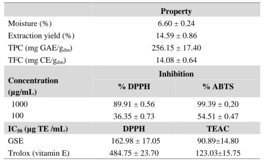
As illustrated in Table1, high values of TPC and TFC (256.15 ± 17.40 mg GAE/gdmand 14.08
± 0.64 mg CE/gdm, respectively) were obtained in grape seeds extract (GSE). In addition, GSE
at 1000 μg/mL exhibited a strong antioxidant activity exceeding 90% inhibition by DPPH and
ABTS assays. Compared to the IC50 of vitamin E obtained by the two antioxidant methods,
GSE shows a stronger reducing capacity than that of vitamin E at low concentrations. This gives GSE the opportunity to be a promising antioxidant and be regarded as a safe food additive and an alternative to synthetic antioxidants used in the food industry.
Table 1. Chemical properties and antioxidant activities of Ahmeur Bouamer grape seed extract. Property Moisture (%) 6.60 ± 0.24 Extraction yield (%) 14.59 ± 0.86 TPC (mg GAE/gdm) 256.15 ± 17.40 TFC (mg CE/gdm) 14.08 ± 0.64 Concentration (µg/mL) Inhibition % DPPH % ABTS 1000 100 89.91 ± 0.56 36.35 ± 0.73 99.39 ± 0,20 54.51 ± 0.47 IC50(µg TE /mL) DPPH TEAC GSE 162.98 ± 17.05 90.89±14.80 Trolox (vitamin E) 484.75 ± 23.70 123.03±15.75
3.2. Identification of procyanidins from GSE
The positive electrospray mass spectrum of GSE shows the presence of ions between m/z 100 and 1500 (Figure 1).These ions were identified as the protonated molecules [M+H]+ of different procyanidins with their three characteristic fragmentation pathways, which have been previously described as: quinone methide (QM), Retro-Diels–Alder (RDA) and heterocyclic ring fission (HRF) cleavage [23]. As summarized in Table 2, nongalloyllated procyanidins Pn and monogalloyllated PnG1 with (n=2-4) were identified in GSE. The major
ions observed at m/z 289, 579, 867 and 1155, showing a series with a mass difference of 288 Da, could be attributed to the [M+H]+ nongalloylated B-type procyanidin. Also, with the same mass difference, the ions at m/z 731, 1018 and 1307 indicate the presence of monogalloyllated B-type procyanidin. At m/z 559, nongalloyllated dimer A-type with the fragment 433, 407, 388, 289, 270 was identified in the extract. This dimer has been reported before by Appeldoorn et al. [24] in peanut skins extract. This rich composition in procyanidins with high degrees of polymerization (DP) may explain the high antioxidant activities obtained from the extract, since the chemical structure of procyanidins allows them to easily scavenge free radicals due essentially to the presence of the galloyl group and the number and position of hydroxyl groups [25].
Fig. 1. ESI-MS spectra of peak 1 [M+H]+ions of monogalloylated tetramer [P4G+H] (m/z=1307) with:
[P4G+H-water] (m/z=1289), [P4G+H-GAres] (m/z=1155), [P4G+H-GAres-288DA] (m/z=867),
[P4G+H- QM] (m/z=729) and (m/z=579) and digalloylated dimer P2G. QM (quinone-methide);
-GAres (loss of gallic acid residue).
Table 2. Some procyanidins identified at 278 nm on Ahmeur Bouamer grape seed extract
Pic Tr
(min)
Molecular weight
[M+H]+ Product ions DP Type Identity
1 4.991 1306 1307 1155,867, 729, 579, 409, 291/289 4 B P4G1 2 5.293 578 579/578/577 555/556, 409, 289, 271 2 B P2 3 6.721 578 57 579/578/577 296,179 2 B P2 4 7.705 866 867 731, 579, 427, 289, 247 3 B P3 5 10.250 1306 1324/1325 (1307+H2O) 867,707, 598, 407,291, 245 4 B P4G1 6 12.323 730 731 577, 453, 287, 207 2 B P2G1 7 16.523 1018 1018 577, 559, 443, 409, 289, 254 3 B P3G1 8 17.459 1306 1346(1307+K+) 731,579/577,449, 289 4 B P4G1 9 19.775 578 559(578- H2O ) 433, 407, 388, 289, 270 2 A P2 10 20.426 1154 1003(1155-GAres) 731, 451, 425, 289,270 4 B P4
-GAres(152 Da) loss of Gallic acid residue; - GA unit (170 Da) loss of Gallic acid units; K+ ion adduct.
3.3. Antimicrobial and antifungal activities
The extract was subjected to antimicrobial and antifungal tests. The results shown in Table 3 confirmed that GSE responds positively to all of them except for Staphylococcus aureus
ATCC® 43300 known to be resistant to methicillin [26]. The diameters of the inhibition growth zone for Gram positive and Gram negative were comprised between 15–17 mm and 15–20 mm, respectively. This result shows an important sensitivity of these bacterial species
to GSE at the concentration of 1000 μg/mL. The same results were obtained by Radovanovic
et al. [27] from wine extract against the Staphylococcus aureus and Escherichia coli bacteria. These strong sensitivities of the bacteria to GSE may be related to the inhibition of the hydrolytic enzymes (proteases and carbohydrolases) or other interactions capable of inactivating microbial adhesins, transport proteins and cell envelope due to the composition of extract in procyanidines with high DP, as stated by Cowan [28]. Some authors have studied the relationship between compound structure and antimicrobial activity and conclude that the number of hydroxyls and the degree of polymerization might be pivotal for the antimicrobial activity of phenolic compounds [29]. Shoko and others have previously revealed that three hydroxyl groups of the compounds were effective for antibacterial activity and all the substituents of the benzene rings were effective against Staphylococcus aureus [30]. GSE at 100 μg/mL remains moderately active against the bacteria tested (11-14 mm of inhibition zone). The extract may be more effective than some antibiotics at precise doses, such as
Penicillin G (6μg or 10 UI) and Oxacillin (5μg), which are not active against Escherichia coli
ATCC 25922 and Pseudomonas aeruginosa ATCC 27853, as well as Methicillin (5μg) against Staphylococcus aureus ATCC 25922 [31].
GSE was also submitted to the antifungal tests against Aspergillus niger which is considered as the main cause of the majority of fungal infections [32]. An inhibition zone of 15.00 ± 0.81 mm was obtained against this fungi, indicating that the sensitivity of Aspergillus niger to GSE
at 1000 μg/mL may be considered as positively important. Faced with Fusarium oxysporum,
the causal agent of Fusarium one of the cryptogramic diseases of vines [33], GSE showed a significant resistance against this agent by developing an inhibition zone around the mycelium
of 18.00 ± 0.82 mm for a concentration of 1000 μg/mL. Although the Algerian vineyards are
not affected by this fungus, its effect is however ubiquitous in palm groves that are experiencing serious contamination by Bayoud disease [34]. Given its resistance against these fungi, a possible use of GSE as a natural bactericide agent may be considered.
Table 3. Anti-microbial and anti-fungal activities of Ahmeur Bouamer grape seed extract
3.4. Cytotoxicity testing
The evaluation of cytotoxicity on 3T6 cells of the various concentrations of GSE after 24 hours of treatment is illustrated in Figure 2a. High concentrations of GSE (1000 and 500
μg/mL) caused mortal damage to 3T6 cells with less than 50% of the cells being viable (25.00 ± 1.01% and 50.00 ± 2.30%, respectively). For concentrations of 250 and 150 μg/mL, a
toxicity of 10 to 24% was found (73.07 ± 2.33% and 87.83 ± 2.58% for viable cells,
respectively, vs. 97.01 ± 0.23% for the negative control). For 100 μg/mL of GSE treatment,
the toxicity decreases to about 3.88% (93.13 ± 1.56% for viable cells vs 97.01 ± 0.23% for the negative control). The prolonged effect to 48 hours of the same concentrations led to an
increase in toxicity for the high concentrations of GSE (500 and 1000 μg/mL). Nevertheless,
for the other concentrations, the toxicity considerably decreased and became less than 0.5%
for 100 μg/mL (96.59 ± 1.18% for viable cells vs. 97.01 ± 0.23% for the negative control).
Inhibition zone diameter (mm)
Bacteria Concentration of GSE (µg/mL) 1000 100 GRAM+ Microccocus luteus ATCC® 9341 16.00±1.41 11.33±0.47 Staphylococcus aureus ATCC® 29213 15.00±0.81 13.66±1.24 Staphylococcus aureus ATCC® 43300 NI NI GRAM-Eschericia coli ATCC® 25992 18.66±1.25 13.33 ± 1.88 P.aeruginosa ATCC® 27853 14.66±1.24 11.33 ± 0.47 Fungi Aspergillus niger 15.00±0.81 10.33 ± 0.81 Fusarium oxysporum 18.00 ±0.82 10.33 ± 0.47
3.5. Effect of some synthetic antioxidants on 3t6 fibroblasts
The exposure of 3T6 cells to some flavonoids and synthetic antioxidants such as Gallic acid, Catechin, Quercetin, BHA, BHT, ABTS and Trolox at a concentration of 100 μg/mL showed a sensitivity of cells in the majority of treatments (Figure 2b). High toxicity (between 20% and 95%) was noted with Trolox treatment and Gallic acid. As for the two flavonoid compounds, Catechin and Quercetin, a toxicity of less than 6% was been registered on 3T6 cells. This toxicity continued to decrease to less than 2.40% with BHT and ABTS. For BHA, a beneficial effect was noted without any toxicity on 3T6 cells (97.26 ± 0.13% vs. 97.01 ± 0.23% for the negative control). It appears from the results that, compared to antioxidants like Gallic acid, Catechin, Quercetin and Trolox, GSE has shown a low toxicity while, compared to BHT and ABTS, the toxicity was slightly higher (3.88% for GSE vs. 2.40% for BHT and ABTS). Compared to the known toxicity of the synthetic compounds like BHA and BHT, it’s more advisable to replace them with GSE which present the advantage to be a natural product.
Contr ol 100 µg /mL 150 µg /mL 250 µg /mL 500 µg /mL 1000 µ g/mL 0 50 100 % o f C el l V ia bi lty GSE concentration 24 h 48 h a Contr ol GSE 1 00µg/m l GA 10 0µg/m l CAT 1 00µg/m l QUER 100µ g/ml BHA 1 00µg/m l BHT 1 00µg/m l ABTS 100µ g/ml TROL OX 10 0µg/m l 0 50 100 % O f C el l V ia bi lit y b
Fig. 2. (a) Cytotoxicity testing: Effect of 24 and 48 hours of GSE treatment on 3T6 cells, (b)
Comparison between the effect of GSE (100 µg/mL) and that of some synthetic antioxidant compounds (100 µg/mL) on 3T6 cells. Values are mean ± SD of three independent experiments. P<0.05 for (a) and P <0.01 for (b) compared with negative control group.
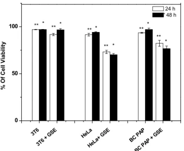
3.6 Anti-proliferative effect
Anti-proliferation was assessed on two different tumoral cells: a HeLa cell line derived from a cervical cancer of the uterus and a BCPAP cell line derived from a papillary thyroid cancer. As shown in Figure 3, a high sensitivity to GSE treatment for both tumoral cells was observed. A cell mortality of 18.39% and 23.79% was respectively reached for 24 and 48 hours of treatment (73.13 ± 1.79% of cell viability in HeLa cells treated for 24 hours vs. 91.52 ± 1.46% of cell viability in HeLa cells without treatment and 70.20 ± 1.27% of cell viability in HeLa cells treated for 48 hours vs. 93.99 ± 0.69% of cell viability in HeLa cells without treatment). BCPAP cell death increased from 10.30% to 20.37% for GSE pretreatment times of 24 and 48 hours, respectively (82.23 ± 3.14% of cell viability in BCPAP cells treated for 24 hours vs. 93.53 ± 0.47% of cell viability in BCPAP cells without treatment and 76.62 ± 2.82% of cell viability in BCPAP cells treated for 48 hours vs. 96.99 ± 1.49% of cell viability in BCPAP cells without treatment). HeLa and BCPAP cells showed respectively a proliferation of 2.47% and 3.46% in 24 hours. Given the rapid growth of these tumoral cells, GSE at a low concentration (100 μg/mL) has a clear effect on them since the anti–proliferation is respectively two-fold higher (5.37%) and three-fold higher (9.01%) than the normal growth for the HeLa and BCPAP cells in 24 hours. In this context, it is possible to consider the use of GSE at low concentrations as an anti-proliferative agent without losing sight of its beneficial effect on healthy cells since it does not cause significant toxicity. On 3T6 cells, only 3.88% of cell mortality was observed after 24 hours of treatment (93.13 ± 1.56% vs. 97.01 ± 0.23% of cell viability in 3T6 alone) and it continued to decrease to less than 0.5% after 48 hours of treatment (96.59 ± 1.18% vs. 97.01 ± 0.23% of cell viability in 3T6 alone).
3T6 3T6 + G SE HeLa HeLa + GSE BC PAP BC PA P + GS E 0 50 100 * * * * * ** ** ** ** ** % O f Cel l V iab ili ty 24 h ' 48 h ** *
Fig. 3. Comparative effect of GSE (100µg/mL) on healthy cells (3T6) and two tumor cells lines (HeLa
and BCPAP). Values are mean ± SD of three independent experiments. ** P<0.01 of GSE
treatment for 24 h vs. control group for 24 h (cells without treatment), *P< 0.05 of GSE
treatment for 48 h vs. control group for 48 h (cells without treatment).
4. CONCLUSION
In this present work, rich extracts of procyanidins were obtained. These constituents give good results in antioxidant effect, compared to Trolox. The antimicrobial activity of GSE, against seven microorganisms also presents relatively strong effects. Furthermore, it is possible to envisage using GSE at a low concentration as an anti-proliferative agent, since it does not cause significant toxicity and it can regenerate cells. These findings establish a basis for a possible exploitation of this native variety as an alternative to synthetic products.
5. ACKNOWLEDGEMENTS
This work was financially supported by the Ministry of Higher Education and Scientific Research of Algeria and by the ARC (research contract AUWB- 2010—10/15-UMONS-5), FNRS and ENCITE programs, and was performed in the framework of COST TD 1004 and CM1006. Ahmeur Bouamer grapes were harvested with the collaboration of the Technological Institute of Fruit Arboriculture and Vine from Benchicao, Medea (Algeria).
6. REFERENCES
[1] Block G., Patterson B., Subar A. Fruit, vegetables, and cancer prevention: a review of the epidemiologicalevidence. Nutr Cancer. 18: 1–29 (1992)
[2] Saini R., Garg V., Dangwal K. Effect of extraction solvents on polyphenolic composition and antioxidant, antiproliferative activities of Himalyan bayberry (Myrica esculenta). Food Sci Biotechnol. 4: 887-894 (2013)
[3] Xiao Y., Wang L., Rui W., Li W., Chen X., Jiang M., Dong M. Enhancement of antioxidant capacity of soy whey by fermentation with Lactobacillus plantarum B1-6. J Funct Food. 12: 33-44 (2015)
[4] Baldrick F.R., Elborn J.S., Woodside J.V., Treacy K., Bradley J., Patterson C.C., Schock B.C., Ennis M., Young I.S., McKinley M.C. Effect of fruit and vegetable intake on oxidative stress and inflammation in COPD: a randomized controlled trial. Eur Respir J.6:1377-1384 (2012)
[5] Apostolou A., Stagos D., Galitsiou E., Spyrou A., Haroutounian S., Portesis N., Trizoglou I., Hayes A.W., Tsatsakis A.M., Kouretas D. Assessment of polyphenolic content, antioxidant activity, protection against ROS-induced DNA damage and anticancer activity of Vitis vinifera stem extracts. Food Chem Toxicol. 61: 60-68 (2013)
[6] Felice F., Zambito Y., Di Colo G., D’Onofrio C., Fausto C., Balbarini A., Di Stefano R. Red grape skin and seeds polyphenols: Evidence of their protective effects on endothelial progenitor cells and improvement of their intestinal absorption. Eur J Pharm Biopharm. 80: 176–184 (2012)
[7] Razak D.L.A., Rashid N.Y.A., Jamaluddin S.A., Sharifuddin S.A., Long K. Enhancement of phenolic acid content and antioxidant activity of rice bran fermented with Rhizopus oligosporus and Manascus purpureus. Biocatal Agr Biotechnol. 4: 33-38 (2015)
[8] Gutteridge J.M.C., Halliwell B. Free radicals and antioxidants in the year 2000. A historical look to the future. Ann NY Acad Sci. 899: 136-147 (2000)
[9] Halliwell B., Gutteridge J.M.C. Free radicals in biology and medicine. 4th ed., Oxford University Press, UK (2006)
peroxide, hydroxyl radicals, and singlet oxygen. J Agr Food Chem. 48, 11: 5677-84 (2000) [11] Shi J., Yu J., Pohorly J.E., Kakuda Y.J. Polyphenolics in grape seeds-biochemistry and functionality. Med Food. 4: 291-299 (2003)
[12] Nash R., Krishnamoorthy M., Jenkins A., Csete M. Human embryonic stem cell model of ethanol-mediated early developmental toxicity. Exp Neuro. 1: 127-35 (2012) [13] Beney L., Gervais P. Influence of the fluidity of the membrane on the response of microorganisms to environmental stresses. Appl Microbiol Biot. 57: 34-42 (2001)
[14] Irani N., Beccaria A.J., Wagner R. Expression of recombinant cytoplasmic yeast pyruvate carboxylase for the improvement of the production of human erythropoietin by recombinant BHK-21 cells. J Biotechnol. 93: 269-282 (2002)
[15] Van Staveren W.C., Solis D.Y., Hebrant A., Detours V., Dumont J.E., Maenhaut C. Human cancer cell lines: experimental models for cancer cells in situ? For cancer stem cells? Biochim Biophys Acta. 1795: 92–103 (2009)
[16] Wang H., Mannava S., Grachtchouk V., Zhuang D., Soengas M.S., Gudkov A.V. c-Myc depletion inhibits proliferation of human tumor cells at various stages of the cell cycle. Oncogene. 27: 1905–1915 (2008)
[17] Waterhouse A.L. Determination of Total Polyphenols in Food.Curr Protoc in Food Anal Chem., I1.1.1-I1.1.8, John Wiley & Sons, Inc (2002)
[18] Brandwilliams W., Cuvelier M.E., Berset C. Use of a free-radical method to evaluate antioxidant activity. LWT-Food Sci Technol. 28: 25-30 (1995)
[19] Zhishen J., Mengeheng T., Jianming W. The determination of flavonoid contents in mulberry and their scavenging effects on superoxide radicals. Food Chem. 64:555–559 (1999) [20] Francisco M.L.DL., Resurrection A.V.A. Total phenolics and antioxidant capacity of heat treated peanut skin. J Food Compos Anal. 22: 16-24 (2009)
[21] Berghe V.A., Vlletinck A.J. Screening methods for antibacterial and antiviral agents from higher plants. Method Plant Biochem. 6: 47-68 (1991)
[22] Strober W. Trypan blue exclusion test of cell viability. Curr Protoc Immunol. Appendix 3B.(2001)
MA. Evidence for galloylated type-A procyanidins in grape seeds. Food Chem.105: 1457–1467 (2007)
[24] Appeldoorn M.M., Sanders M., Vincken J.P., Cheynier V., Le Guernevé C., Hollman P.C.H., Gruppen H. Efficient isolation of major procyanidin A-type dimers from peanut skins and B-type dimers from grape seeds. Food Chem. 11: 7713–7720 (2009)
[25] Rice-Evans C.A., Miller N.J., Paganga G. Antioxidant properties of phenolic compound. Trends Plant Sci. 2: 152 (1997)
[26] Laurent F., Chardon H., Haenni M., Bes M., Reverdy M.E., Madec J.Y., Lagier E., Vandenesch F., Tristan A. New European methicillin resistant Staphylococcus aureus harboring mecA-variant gene: human and animal isolates in France. Emerg Infect Dis. 18: 1465-1467 (2012)
[27] Radovanovic A., Radovanovic B., Jovancicevic B. Free radical scavenging and antibacterial activities of southern Serbian red wines. Food Chem. 117: 326–331 (2009) [28] Cowan M.M. Plant products as antimicrobial agent. Clin Microbiol Rev. 12: 564-582 (1999)
[29] Tagurt T., Tanaka T., Kouno I. Antimicrobial activity of 10 different plant polyphenols against bacteria causing food-borne disease. Biol Pharm Bull. 27: 1965–1969 (2004)
[30] Shoko T., Soichi T., Megumi M.M., Eri F., Jun K., Michiko W. Isolation and identification of an antibacterial compound from grape and its application to foods. Nippon Nogeik Kaishi. 73: 125–128 (1999)
[31] CLSI. Clinical and Laboratory Standards Institute Performance Standards for Antimicrobial Susceptibility Testing; Seventeenth Informational Supplement document M100-S17, vol 27, No 1. Clinical and Laboratory Standards Institute, 940 West Vally Road, Suite 1400, Wayne, Pennsylvania 19087-1898 USA (2007)
[32] Groll AH., Shah PM., Mentzel C., Schneider M., Nuebling G., Huebner K. Trends in the postmortem epidemiology of invasive fungal infections at a university hospital. J Infect. 13: 23–32(1996)
[33] Larignon P. Maladies cryptogamiques du bois de la vigne: symptomatologie et agents pathogènes. (2012)http://www.vignevin.com.
[34] Djerbi M., Sedra MH., El Idrissi Ammari MA. Caractéristiques culturales et identification du Fusarium oxysporum f. sp. albedinis, agent causal du Bayoud. Ann Inst Nat Rech Agro. Tunisie. 58: 1-8 (1985)
How to cite this article:
Ghouila Z. Laurent S. Boutry S. Vander Elst L. Nateche F. Muller N.R and Baaliouameur A. Antioxidant, Antibacterial and Cell Toxicity Effects of Polyphenols From Ahmeur Bouamer Grape Seed Extracts. J. Fundam. Appl. Sci., 2017, 9(1), 392-410.