http://jfm.sagepub.com/
Journal of Feline Medicine and Surgery
http://jfm.sagepub.com/content/15/7/631
The online version of this article can be found at:
DOI: 10.1177/1098612X13489228
2013 15: 631
Journal of Feline Medicine and Surgery
Radford, Etienne Thiry, Uwe Truyen and Marian C Horzinek
Gruffydd-Jones, Margaret J Hosie, Albert Lloret, Hans Lutz, Fulvio Marsilio, Karin Möstl, Maria Grazia Pennisi, Alan D
Katrin Hartmann, Diane Addie, Sándor Belák, Corine Boucraut-Baralon, Herman Egberink, Tadeusz Frymus, Tim
Toxoplasma Gondii Infection in Cats: ABCD guidelines on prevention and management
technique does not amount to an endorsement of its value or quality, or the claims made by its manufacturer. those of the authors and the inclusion in this publication of material relating to a particular product, method or of animals and interpretation of published materials lies with the veterinary practitioner. The opinions expressed are from actions or decisions based on information contained in this publication; ultimate responsibility for the treatment
arising country. The authors, editors, owners and publishers do not accept any responsibility for any loss or damage advertising material, it is the responsibility of the reader to check that the product is authorised for use in their own bear this in mind and be aware of the prescribing laws pertaining to their own country. Likewise, in relation to Furthermore, drugs may be mentioned that are licensed for human use, and not for veterinary use. Readers need to formulations that are not available or licensed in the individual reader's own country.
The Journal of Feline Medicine and Surgery is an international journal and authors may discuss products and Disclaimer
Published by:
International Society of Feline Medicine
American Association of Feline Practitioners
and
http://www.sagepublications.com
can be found at: Journal of Feline Medicine and Surgery
Additional services and information for
http://jfm.sagepub.com/cgi/alerts Email Alerts: http://jfm.sagepub.com/subscriptions Subscriptions: http://www.sagepub.com/journalsReprints.nav Reprints: http://www.sagepub.com/journalsPermissions.nav Permissions:
What is This?
- Jun 27, 2013
Version of Record
>>
at Universite de Liege on September 3, 2013 jfm.sagepub.com
C L I N I C A L
R E V I E W
European Advisory Board on Cat Diseases www.abcd-vets.org
Corresponding author: Katrin Hartmann Email: Hartmann@uni-muenchen.de
TOXOPLASMA GONDII INFECTION IN CATS
ABCD guidelines on prevention
and management
Katrin Hartmann, Diane Addie, Sándor Belák, Corine Boucraut-Baralon, Herman Egberink, Tadeusz Frymus, Tim Gruffydd-Jones, Margaret J Hosie, Albert Lloret, Hans Lutz, Fulvio Marsilio, Karin Möstl, Maria Grazia Pennisi, Alan D Radford, Etienne Thiry, Uwe Truyen and Marian C Horzinek
Overview: Toxoplasma gondii infection is
common in cats, but the clinical disease is rare. Up to 50% of cats, especially free-roaming ones, have antibodies indicating infection and the presence of cystic stages.
Disease signs: Clinical signs only appear in few cats when they become immunosuppressed – in these situations cystic stages can be reactivated. Commonly affected are the central nervous system (CNS), muscles, lungs and eyes.
Human infection: Cats can pose a risk for humans when they shed oocysts. However, this happens only once in their lifetime, usually only for 3–10 days after ingestion of tissue cysts. Thus, cats that have antibodies to T gondii no longer shed oocysts, and do not pose a risk to humans.
Agent properties
Toxoplasma gondii is an obligate intracellular coccidian parasite that can
infect virtually all species of warm-blooded animals, including people. Domestic cats and other felids are the natural hosts – non-feline species serve only as intermediates.1,2
Three infectious structures can be distinguished: sporozoites in oocysts, tachyzoites (the actively multiplying stage), and bradyzoites (the slowly multiplying stage) enclosed in tissue cysts. Oocysts are excreted in faeces, whereas tachyzoites and bradyzoites are found in tissues and milk.1,2
Pathogenesis
Enteroepithelial life cycle
This cycle is found only in the feline host. Most cats are infected by ingesting intermediate hosts – typically rodents – infected with tissue cysts. Bradyzoites are released in the stomach and intestine from the tissue cysts when digestive enzymes dissolve the cyst wall. They enter epithelial cells of the small intestine and give rise to schizonts, initiate five types of predetermined asexual stages, and merozoites released from the schizonts eventually form male and female gamonts. After fertilisation, a wall is formed around the fertilised macrogamont to form an oocyst. Oocysts are round to oval, 10 x 12 μm in size, and are still unsporulated (not infectious) when passed in fae-ces. After exposure to air and moisture for 1–5 days they sporu-late to contain two sporocysts, each with four sporozoites.1,2 European Advisory Board on Cat Diseases
The European Advisory Board on Cat Diseases (ABCD) is a body of experts in immunology, vaccinology and clinical feline medicine that issues guidelines on prevention and management of feline infectious diseases in Europe, for the benefit of the health and welfare of cats. The guidelines are based on current scientific knowledge of the diseases and available vaccines concerned.
The latest version of the Toxoplasma gondii infection in cats guidelines is available at www.abcd-vets.org
632
JFMSCLINICAL PRACTICEThe cycle is usually completed within 3–10 days of ingestion of tissue cysts, which is the route of infection in up to 97% of naive cats. In the rare event that cats ingest oocysts or tachyzoites, formation of new oocysts is delayed and shedding can occur for up to 18 days (occasionally longer). However, only 20% of cats fed oocysts will shed.1,2
Extraintestinal life cycle
The extraintestinal development of T gondii is the same for all hosts, including cats, dogs, and people, irrespective of whether tissue cysts or oocysts have been ingested. After the ingestion of oocysts, sporozoites hatch in the lumen of the small intestine and enter intes-tinal cells, including those in the lamina pro-pria. Sporozoites divide into two by an asexual process known as endodyogeny, thereby becoming tachyzoites. These are lunate (falciform) in shape, approximately 6 x 2 μm, and multiply in almost any cell of the body. When the cell ruptures, releasing the tachyzoites, these infect new cells. Otherwise, tachyzoites multiply intracellularly for an undetermined period, and eventually encyst. Tissue cysts vary in size from 15–60 μm and usually conform to the shape of the para-sitised cell. Tissue cysts are formed mainly in the CNS, muscles and visceral organs, and probably persist for the life of the host. They can be reactivated after immunosuppression, which may then lead to clinical signs.1,2
Parasitaemia during pregnancy of the host can cause placentitis and spread of tachy-zoites to the fetus. Many kittens born to queens infected with T gondii during gestation become infected transplacentally or when suckling. Clinical signs are common in these kittens, varying with the stage of gestation at the time of infection; some of these newborn kittens shed oocysts.1,2
Epidemiology
Antibody prevalence to T gondii varies geo-graphically: in Portugal, 24% of cats had anti-bodies in one study;3in the USA, 16–40% were antibody-positive, depending on the state.2 However, only 3/326 faecal samples from cats in California and 1/252 in Switzerland contained
T gondii oocysts.4The annual burden in the envi-ronment is about 90–5000 oocysts per square metre.5The age of the cat does not play a role in the frequency of T gondii shedding, but the sea-son does: shedding is more common between July and December in the northern hemisphere.6 The three major modes of transmission of
T gondii in all host species are congenital
infec-tion, ingestion of infected tissue, and inges-tion of oocyst-contaminated food or water.2 Less important are blood transfusions and
organ transplantations.1,2 Lactogenic transmis-sion is suspected because the organism has been detected in queen’s milk.7
T gondii blocks the innate aversion of rats for
cat urine, instead making them attracted by the feline pheromone, which can increase the likelihood of a cat capturing an infected rat. This reflects adaptive ‘behavioural manipula-tion’ by T gondii in optimising the chances of completing the parasite’s life cycle: it repro-duces only in the feline intestine. The behav-ioural manipulation hypothesis postulates that a parasite will specifically manipulate host conduct essential for its transmission. However, the neural circuits for innate fear, anxiety and acquired fright all overlap, raising the possibility that T gondii can disrupt all of these non-specifically.8 Experimental infec-tions have shown that T gondii can change chemical messages in the CNS that affect rodent behaviour because the infection can lead to cyst formation in the CNS with pro-duction of tyrosine hydroxylase, resulting in a lack of dopamine.9–14
Meat contaminated with T gondii cysts has been the primary source of infection in people, and antibody prevalence in humans is relatively high. Exposure from oocyst-contaminated soil or water is common. Indeed, water-borne outbreaks of toxoplasmo-sis have been reported worldwide and support the theory that exposure to environmental oocysts poses a significant health risk.15
Clinical signs
Clinical signs develop very rarely in infected cats and are caused by inflammation and tissue necrosis resulting from intracellular growth of tachyzoites.2Congenital infection tends to be more serious than infection of the adult cat.2
Clinical toxoplasmosis develops during dissemination and intracellular replication of tachyzoites. It usually originates from reactiva-tion of a latent infecreactiva-tion rather than after a newly acquired infection. If a carrier cat is immunosuppressed, bradyzoites in tissue cysts replicate rapidly and disseminate again as tachyzoites. Clinical toxoplasmosis has been documented in some cats infected with feline immunodeficiency virus (FIV) or feline leukaemia virus (FeLV) [EBM grade III].16 Commonly used doses of glucocorticoids do
Congenital
infection tends
to produce
more serious
clinical disease
than infection
of the
adult cat.
The three major modes of transmission of
T gondii in all host species are congenital infection,
ingestion of infected tissue, and ingestion of
oocyst-contaminated food or water.
R E V I E W/ABCD guidelines on Toxoplasma gondii infection
at Universite de Liege on September 3, 2013 jfm.sagepub.com
not predispose to reactivation of T gondii cysts.17However, administration of ciclosporin to cats with renal transplants or dermatologi-cal disease has been associated with clinidermatologi-cal manifestations [EBM grade IV].18–20
The most commonly affected tissues are the CNS, muscles (Figure 1), lungs (Figure 2) and eyes. Hepatic and pancreatic involvement is less likely. Cats with toxoplasmosis show neurological signs (eg, seizures, ataxia), mus-cle hyperaesthesia, dyspnoea, uveitis, icterus, diarrhoea, fever, depression, anorexia and weight loss.2Transplacentally or lactogenically infected kittens develop more severe signs and frequently die of pulmonary or hepatic disease [EBM grade III].21 Immune complex forma-tion22 and deposition in tissues, as well as delayed hypersensitivity reactions, can be involved in chronic forms of toxoplasmosis. Since T gondii is not cleared from the body, either naturally or through treatment, toxo-plasmosis can recur.
Immunity
Immunity to T gondii in the cat is poorly under-stood. In the mouse and in humans, it is highly dependent on cell-mediated effector responses.23 All infected cats develop IgG and IgA anti-bodies; about 80% also have IgM antibodies. IgG can take 4–6 weeks to appear, and maxi-mal antibody titres are achieved within 2–3 weeks of first appearance.2
Diagnosis
Oocyst shedding is diagnosed by microscopy of faecal samples. Diagnosis of the disease is only confirmed when the organism is found in body fluids or tissue. If suitable samples cannot be taken, a tentative diagnosis is sometimes based on rising IgM titres, exclusion of other causes for the clinical signs, and a favourable clinical response to anti-T gondii drugs [EBM grade II].1,2
Figure 1 Myositis in a cat caused by T gondii cysts. The cat presented in lateral recumbency, was unable to get up, and showed severe muscle hyperaesthesia. Courtesy of Katrin Hartmann, Ludwig Maximilians University, Munich, Germany
Figure 2Thoracic radiograph (laterolateral view) of a cat with pulmonary toxoplasmosis. Courtesy of Katrin Hartmann, Ludwig Maximilians University, Munich, Germany
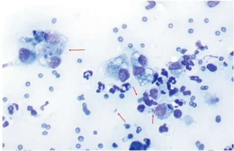
Figure 3 Cytology of a fine-needle aspirate from a cat with pulmonary toxoplasmosis and lung consolidation, showing numerous intracellular and extracellular T gondii tachyzoites and cysts (arrows). Courtesy of George Reppas, Vetnostics, Australia
Detection of
tachyzoites in
tissue samples
provides
definitive
confirmation of
the diagnosis
of the disease
toxoplasmosis.
EBM gradesThe ranking system for grading the level of evidence of various statements within this article is described on page 533 of this Special Issue.
634
JFMSCLINICAL PRACTICER E V I E W/ABCD guidelines on Toxoplasma gondii infection
Detection of oocysts in faeces
T gondii oocysts are 10 μm in size and best
demonstrated by centrifugation using Sheather’s sugar solution (saccharose solution with a specific gravity of 1.27 g/ml) during the shedding period. T gondii oocysts are mor-phologically indistinguishable from those of
Hammondia hammondi, Besnoitia oryctofelisi and Besnoitia darlingi.2A cesium chloride method for easy purification of T gondii oocysts from faeces of infected cats has been described.15 Detection of tachyzoites
Ante-mortem diagnosis of clinical toxoplas-mosis ideally is based on the detection of the organism by cytology or polymerase chain reaction (PCR) [EBM grade III]. Tachyzoites can be detected in various tissues and body fluids during acute illness (Figure 3). They are rarely found in blood, but occasionally in cerebrospinal fluid (CSF) or aqueous humour, fine-needle aspirates of organs (eg, lymph nodes), and transtracheal or bronchoalveolar lavage fluid. Detection of tachyzoites results in a definitive diagnosis. Alternatively, a PCR can be performed using CSF, aqueous humour or bronchoalveolar lavage fluid.
Detection of antibodies
Using the immunofluorescence assay (IFA), antibodies of the IgM, IgG and IgA isotypes can be detected. For assessing human health risks, antibody test results from healthy cats are useful. An antibody-negative cat can be shedding oocysts (early during infection, before antibodies have had time to develop) and will likely shed oocysts if exposed for the first time. An antibody-positive cat does not shed oocysts: antibodies need 2–3 weeks to develop, by which time cats usually no longer shed; and a cat sheds only once in its lifetime. It is also unlikely to shed oocysts if re-exposed or immunosuppressed [EBM grade II].1
Antibodies are commonly found in both healthy and sick cats. Thus, their presence does not prove clinical toxoplasmosis. Antibodies of the IgM class are also common-ly detected in healthy cats and do not corre-late with clinical signs.
Treatment
Clindamycin is the treatment of choice24 and should be administered at 10–12 mg/kg orally q12h for 4 weeks (Table 1) [EBM grade III]. Cats with systemic disease and uveitis should be treated with clindamycin in combination with topical, oral or parenteral glucocorticoids, to avoid secondary glaucoma and lens luxa-tion [EBM grade III].25 Prednisolone acetate (1% solution) applied topically to the eye three to four times daily is generally sufficient.
Clinical signs not involving the eyes or the CNS usually begin to resolve within the first 2–3 days of clindamycin administration. CNS and ocular toxoplasmosis tend to respond more slowly. In cases of pulmonary toxo -plasmosis, radiographic abnormalities might not resolve for several weeks. The prognosis is usually poor in pulmonary or hepatic disease, particularly in immunocompromised animals.26
Prevention of infection
Preventing toxoplasmosis in cats involves measures intended to reduce the incidence of infections and the shedding of oocysts into the environment. Cats should preferably be fed commercially available, processed food. Prevalence of feline T gondii infection is high-er in countries whhigh-ere raw meat is fed. Freezing or irradiation can kill tissue cysts without affecting meat quality. Pets should be prevented from hunting and eating intermedi-ate hosts (rodents) or mechanical vectors, such as cockroaches and earthworms. If meat is fed, it should be thoroughly cooked, even if frozen. Cats should be prevented from enter-ing buildenter-ings where food-producenter-ing animals are housed or where feed storage areas are located.1
Drug Comment ABCD recommendation EBM grade
Antiparasitic therapy
Clindamycin 10–12 mg/kg PO q12h
for 4 weeks (treatment of choice)
III
Symptomatic therapy
Prednisolone acetate (1%) For cats with Toxoplasma-induced uveitis (to avoid secondary glaucoma and lens luxation)
Use in addition to systemic antibiotic treatment
Apply topically to the eye q6–8h
IV Treatment of toxoplasmosis
Table 1
Antibodies are commonly found in both
healthy and sick cats. Thus, their presence
does not prove clinical toxoplasmosis.
at Universite de Liege on September 3, 2013 jfm.sagepub.com
Public heath issues have come into focus (Box 1), partly because of the increasing number of immunocompromised persons (eg, after infection with human immunodeficiency virus [HIV]). Also, recent research linking psychological and cognitive disorders (eg, reduced IQ, as evidenced by psychomotor and verbal intelligence tests) to T gondii infection have contributed. A recent survey among obstetricians and gynaecologists in the USA to determine their knowledge and practices relating to toxoplasmosis prevention and testing found that most overestimated the risk of cat ownership versus environmental risk factors.27A systematic review of risk factors for pregnant women
is available;28it reports a relatively low risk associated with cat
ownership [EBM grade I].
Veterinarians commonly get questions from clients as to whether or not to get rid of their cat during pregnancy. If hygiene recommendations are followed (Boxes 2 and 3), the risk of transmission is low (Box 4).
P u b l i c h e a l t h c o n s i d e r a t i o n s
Box 1
Routes of infection for humans
Common routes< Ingestion of meat containing tissue cysts is the most common route of infection. Thorough cooking or freezing for several days will kill tissue cysts.29–32
< Ingestion of sporulated oocysts, either from the
environment (eg, through contact with contaminated soil) or from faeces of shedding cats, is the second most common route. This may also happen when eating unwashed fruit or vegetables. Infection via the environment is more common than direct infection through cat contacts. Less common routes
< Ingestion of sporulated oocysts through contact with contaminated water.
< Ingestion of raw (unpasteurised) goat’s milk.
< Inhalation of sporulated oocysts on dust particles (rare).
Box 2
Recommendations to reduce the risk
of parasite transmission from cat to owner
< Litter trays should be emptied daily so that oocysts do not have sufficient time (24 h) to sporulate.
< Gloves should be worn when handling cat litter, and hands should be washed thoroughly after cleaning of litter trays.
< Litter tray liners should be used, if possible, and the tray cleaned regularly with detergent and scalding water.
< Cat litter should be disposed in sealed plastic bags.
< Children’s sandpits should be covered when not in use, to prevent cats from using them.
< Only properly cooked food or commercial cat food should be fed.
< Hands should be washed after contact with a cat (especially before eating).
Box 4
Evidence that contact with cats does
not increase the risk of T gondii infection
33< Cats shedding oocysts in faeces are rare.34In one
study, only about 1/250 cats shed oocysts.4 < Contact with cats has no influence on the probability of
people developing antibodies to T gondii, whereas the consumption of raw meat significantly increases the risk of acquiring the infection.35
< Veterinarians working with cats are not more likely to become infected with T gondii or to suffer from toxoplasmosis than the general population, including people without cat contacts.36–38
< Stroking a cat will not spread the infection. Even when cats are shedding oocysts in their faeces, oocysts cannot be found on their coat.39Studies in dogs have shown that
oocysts do not sporulate on their fur, and the same is probably true for cats.40
< Cat ownership does not increase the risk of toxoplasmosis in people with HIV infection. Although toxoplasmosis is more common in HIV-infected persons, the disease results from reactivation of a previous infection rather than from acquiring a new infection.
< Most people are infected with T gondii through ingestion of undercooked meat, especially goat, mutton and pork. The risk of infection from cats is low, except for young children playing in soil contaminated with sporulated oocysts.41 < Bites or scratches from an infected cat do not transmit the
infection.
< Infected cats under treatment with immunosuppressive drugs at standard doses do not start shedding oocysts in their faeces.17
< Infected cats also do not re-shed oocysts in their faeces when they become immunosuppressed due to infection with FIV or FeLV.42Cats infected with FIV or FeLV that are
subsequently infected with T gondii do not shed oocysts for any longer or in any greater numbers than other cats.2,43 < Newly identified strains of T gondii are highly infectious for
species other than cats; thus, cats might actually become less important in the spread of this infection.
Box 3
Additional advice for households
with immunocompromised persons or
pregnant women
< Immunosuppressed persons and pregnant women should avoid contact with cat litter.
< Cats should be kept indoors to prevent them hunting and eating intermediate hosts such as voles and mice.
< Cats should not be fed raw or partially cooked meat.
< Cats should be discouraged from eating insects (eg, cockroaches).
< Cats should be tested for T gondii antibodies; their presence indicates past infection. These cats will not be a source of infection as they have completed their period of oocyst shedding.
< Cats without antibody will have not been infected previously and, when newly infected, will shed oocysts in their faeces for a short time. They should, therefore, be kept indoors during the phase of immunosuppression or pregnancy of the owner.
636
JFMSCLINICAL PRACTICER E V I E W/ABCD guidelines on Toxoplasma gondii infection
< T gondii infection is common in cats. The clinical picture toxoplasmosis, however, is rare.
< Up to 50% of cats have antibodies, which show they are infected and harbour T gondii cysts.
< Clinical signs usually occur when cats become immunosuppressed, due to the reactivation of cystic stages.
< Commonly affected sites are the CNS, muscles, lungs and eyes.
< Cats shedding oocysts can pose a risk to humans. However, they shed only once in their lifetime, and usually for only 3–10 days after ingestion of tissue cysts.
< Cats with T gondii antibodies no longer shed oocysts; they neither are nor will become a zoonotic risk.
< A definitive diagnosis of toxoplasmosis relies on the detection of the organism in body fluids or tissues.
< The treatment of choice for cats with the disease toxoplasmosis is clindamycin.
KEY
POINTS
Funding
The authors received no specific grant from any funding agency in the public, commercial or not-for-profit sectors for the preparation of this article. The ABCD is supported by Merial, but is a scientifically independent body.
Conflict of interest
The authors do not have any potential conflicts of interest to declare.
References
1 Dubey JP. Toxoplasma update. Proceedings of the WSAVA Congress; 2005, Mexico City, Mexico.
2 Dubey JP and Lappin MR. Toxoplasmosis and
neosporosis. In: Greene CE (ed). Infectious
disease of the dog and cat. Philadelphia: WB Saunders, 2006, pp 754–775.
3 Duarte A, Castro I, Pereira da Fonseca IM, Almeida V, Madeira de Carvalho LM, Meireles J, et al. Survey of infectious and parasitic
dis-eases in stray cats at the Lisbon Metropolitan Area, Portugal. J Feline Med Surg 2010; 12:
441–446.
4 Berger-Schoch AE, Herrmann DC, Schares G, Muller N, Bernet D, Gottstein B, et al.
Prevalence and genotypes of Toxoplasma
gondii in feline faeces (oocysts) and meat from
sheep, cattle and pigs in Switzerland. Vet Parasitol 2011; 177: 290–297.
5 Dabritz HA, Miller MA, Atwill ER, Gardner IA, Leutenegger CM, Melli AC, et al. Detection of
Toxoplasma gondii-like oocysts in cat feces
and estimates of the environmental oocyst burden. J Am Vet Med Assoc 2007; 231:
1676–1684.
6 Herrmann DC, Pantchev N, Vrhovec MG, Barutzki D, Wilking H, Frohlich A, et al.
Atypical Toxoplasma gondii genotypes identified in oocysts shed by cats in Germany. Int J Parasitol 2010; 40: 285–292.
7 Powell CC, Brewer M and Lappin MR.
Detection of Toxoplasma gondii in the milk of experimentally infected lactating cats. Vet Parasitol 2001; 102: 29–33.
8 Vyas A, Kim SK, Giacomini N, Boothroyd JC and Sapolsky RM. Behavioral changes induced
by Toxoplasma infection of rodents are highly specific to aversion of cat odors. Proc Natl Acad Sci U S A 2007; 104: 6442–6447.
9 Flegr J and Havlicek J. Changes in the
person-ality profile of young women with latent toxo-plasmosis. Folia Parasitol 1999; 46: 22–28.
10 Hrda S, Votypka J, Kodym P and Flegr J.
Transient nature of Toxoplasma gondii-induced behavioral changes in mice. J Parasitol
2000; 86: 657–663.
11 Havlicek J, Gasova ZG, Smith AP, Zvara K and Flegr J. Decrease of psychomotor performance
in subjects with latent ‘asymptomatic’ toxo-plasmosis. Parasitology 2001; 122: 515–520.
12 Webster JP. Rats, cats, people and parasites: the
impact of latent toxoplasmosis on behaviour. Microbes Infect 2001; 3: 1037–1045.
13 Flegr J, Havlicek J, Kodym P, Maly M and Smahel Z. Increased risk of traffic accidents
in subjects with latent toxoplasmosis: a retrospective case-control study. BMC Infect Dis 2002; 2: 11.
14 Flegr J, Preiss M, Klose J, Havlicek J, Vitakova M and Kodym P. Decreased level of psycho
-biological factor novelty seeking and lower intelligence in men latently infected with the protozoan parasite Toxoplasma gondii Dopamine, a missing link between schizo-phrenia and toxoplasmosis? Biol Psychol 2003;
63: 253–268.
15 Staggs SE, See MJ, Dubey JP and Villegas EN.
Obtaining highly purified Toxoplasma gondii oocysts by a discontinuous cesium chloride gradient. J Vis Exp 2009; 33: pii, 1420.
16 Davidson MG, Rottman JB, English RV, Lappin MR and Tompkins MB. Feline
immunodefi-ciency virus predisposes cats to acute general-ized toxoplasmosis. Am J Pathol 1993; 143:
1486–1497.
17 Lappin MR, Dawe DL, Windl PA, Greene CE and Prestwood AK. The effect of
glucocorti-coid administration on oocyst shedding, serol-ogy, and cell mediated immune responses of cats with recent or chronic toxoplasmosis. J Am Anim Hosp Assoc 1992; 27: 625–632.
at Universite de Liege on September 3, 2013 jfm.sagepub.com
18 Beatty J and Barrs V. Acute toxoplasmosis in
two cats on cyclosporin therapy. Aust Vet J
2003; 81: 339.
19 Last RD, Suzuki Y, Manning T, Lindsay D, Galipeau L and Whitbread TJ. A case of fatal
systemic toxoplasmosis in a cat being treated with cyclosporin A for feline atopy. Vet Dermatol 2004; 15: 194–198.
20 Barrs VR, Martin P and Beatty JA. Antemortem
diagnosis and treatment of toxoplasmosis in two cats on cyclosporin therapy. Aust Vet J
2006; 84: 30–35.
21 Dubey JP, Lappin MR and Thulliez P. Diagnosis
of induced toxoplasmosis in neonatal cats. J Am Vet Med Assoc 1995; 207: 179–185.
22 Lappin MR, Cayatte S, Powell CC, Gigliotti A, Cooper C and Roberts SM. Detection of
Toxoplasma gondii antigen-containing
immune complexes in the serum of cats. Am J Vet Res 1993; 54: 415–419.
23 Sanchez Y, Rosado Jde D, Vega L, Elizondo G, Estrada-Muniz E, Saavedra R, et al. The
unex-pected role for the aryl hydrocarbon receptor on susceptibility to experimental toxoplasmo-sis. J Biomed Biotechnol Epub 11 January 2010.
DOI: 10.1155/2010/505694.
24 Davidson MG. Toxoplasmosis. Vet Clin North
Am Small Anim Pract 2000; 30: 1051–1062.
25 Lappin MR, Greene CE, Winston S, Toll SL and Epstein ME. Clinical feline toxoplasmosis.
Serologic diagnosis and therapeutic manage-ment of 15 cases. J Vet Intern Med 1998; 3: 139–143.
26 Dubey JP, Lindsay DS and Lappin MR.
Toxoplasmosis and other intestinal coccidial infections in cats and dogs. Vet Clin North Am Small Anim Pract 2009; 39: 1009–1034, v.
27 Jones JL, Krueger A, Schulkin J and Schantz PM.
Toxoplasmosis prevention and testing in pregnancy, survey of obstetrician-gynaecolo-gists. Zoonoses Public Health 2010; 57: 27–33.
28 Leroy V and Hadjichristodoulou C. Systematic
review of risk factors for Toxoplasma gondii infection in pregnant women. Bordeaux,
France, The Eurotoxic Group, 2005.
29 Lunden A and Uggla A. Infectivity of
Toxoplasma gondii in mutton following
cur-ing, smokcur-ing, freezing or microwave cooking. Int J Food Microbiol 1992; 15: 357–363.
30 Dubey JP. Longterm persistence of Toxo
-plasma gondii in tissues of pigs inoculated
with T gondii oocysts and effect of freezing on viability of tissue cysts in pork. Am J Vet Res
1988; 49: 910–913.
31 Dubey JP. Toxoplasma gondii oocyst survival
under defined temperatures. J Parasitol 1998;
84: 862–865.
32 Dubey JP, Kotula AW, Sharar A, Andrews CD and Lindsay DS. Effect of high temperature on
infectivity of Toxoplasma gondii tissue cysts in pork. J Parasitol 1990; 76: 201–204.
33 Elmore SA, Jones JL, Conrad PA, Patton S, Lindsay DS and Dubey JP. Toxoplasma gondii:
epidemiology, feline clinical aspects, and pre-vention. Trends Parasitol 2010; 26: 190–196.
34 Hill SL, Cheney JM, Taton-Allen GF, Reif JS, Bruns C and Lappin MR. Prevalence of enteric
zoonotic organisms in cats. J Am Vet Med Assoc
2000; 216: 687–692.
35 Flegr J, Hrda S and Tachezy J. The role of
psychological factors in questionnaire-based studies on routes of human toxoplasmosis transmission. Cent Eur J Public Health 1998; 6:
45–50.
36 Behymer RD, Harlow DR, Behymer DE and Franti CE. Serologic diagnosis of
toxoplasmo-sis and prevalence of Toxoplasma gondii anti-bodies in selected feline, canine, and human populations. J Am Vet Med Assoc 1973; 162:
959–963.
37 Sengbusch HG and Sengbusch LA. Toxoplasma
antibody prevalence in veterinary personnel and a selected population not exposed to cats. Am J Epidemiol 1976; 103: 595–597.
38 DiGiacomo RF, Harris NV, Huber NL and Cooney MK. Animal exposures and antibodies
to Toxoplasma gondii in a university popula-tion. Am J Epidemiol 1990; 131: 729–733.
39 Dubey JP. Duration of immunity to shedding
of Toxoplasma gondii oocysts by cats. J Parasitol 1995; 81: 410 –415.
40 Lindsay DS, Dubey JP, Butler JM and Blagburn BL. Mechanical transmission of Toxoplasma
gondii oocysts by dogs. Vet Parasitol 1997; 73:
27–33.
41 Wallace MR, Rossetti RJ and Olson PE. Cats and
toxoplasmosis risk in HIV-infected adults. JAMA 1993; 269: 76–77.
42 Lappin MR. Cat ownership by immuno
-suppressed people. In: August JR (ed).
Consultations in feline medicine. 4th ed. Philadelphia: WB Saunders, 2001, pp 18–27. 43 Lappin MR, George JW, Pedersen NC, Barlough
JE, Murphy CJ and Morse LS. Primary and
sec-ondary Toxoplasma gondii infection in normal and feline immunodeficiency virus-infected cats. J Parasitol 1996; 82: 733–742.
Available online at jfms.com