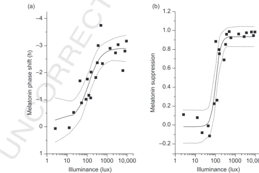
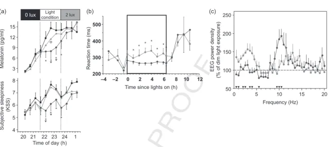
Can light make us bright? Effects of light on cognition and sleep.
Texte intégral
Figure
Documents relatifs
In section 3, we will discuss the coupling of ddCOSMO with an existing code and review what are the quantities that the existing code needs to assemble in order to compute
apical membranes of Drosophila epithelial cells and required for organization of epithelia. The Crumbs complex: from epithelial-cell polarity to 1019.
Epidemiologic studies have provided evidence that high ambient concentrations of this air pollutant are associated with an increased rate of asthma exacerbations, increased
Je m’apprêtais à leur raconter que des vilaines choses, il s’en était passé dans le coin ces dernières années, et toujours avec des personnes isolées,
A radical generated on the surface of a protein can: (1) be immediately and fully repaired by direct reaction with an antioxidant; (2) react with dioxygen to form the corre-
Ainsi la forte augmentation des prix du quinoa et la mécanisation des pratiques agricoles, vécues comme des opportunités par les agriculteurs, menacent l’équilibre fragile
In this work we use the mean-field approach to derive the differential equa- tions which describe the process of information dissemination. We obtain a couple of differential
In particular, for GM, a nutation structure appears near the center of the hole and persists for excita- tion times as long as 400 @sec in spite of a much shorter (21. 7 @sec)
