Titre:
Title:
Full range of wettability through surface modification of single-wall carbon nanotubes by photo-initiated chemical vapour deposition
Auteurs:
Authors:
Seyedehsan Hosseininasab, Nathalie Faucheux, Gervais Soucy et Jason Robert Tavares
Date: 2017
Type: Article de revue / Journal article
Référence:
Citation:
Hosseininasab, S., Faucheux, N., Soucy, G. & Tavares, J. R. (2017). Full range of wettability through surface modification of single-wall carbon nanotubes by photo-initiated chemical vapour deposition. Chemical Engineering Journal, 325, p. 101-113. doi:10.1016/j.cej.2017.05.034
Document en libre accès dans PolyPublie
Open Access document in PolyPublie
URL de PolyPublie:
PolyPublie URL: http://publications.polymtl.ca/2790/
Version: Version finale avant publication / Accepted versionRévisé par les pairs / Refereed Conditions d’utilisation:
Terms of Use: CC BY-NC-ND
Document publié chez l’éditeur officiel Document issued by the official publisher
Titre de la revue:
Journal Title: Chemical Engineering Journal (vol. 325)
Maison d’édition:
Publisher: Elsevier
URL officiel:
Official URL: https://doi.org/10.1016/j.cej.2017.05.034
Mention légale:
Legal notice: "In all cases accepted manuscripts should link to the formal publication via its DOI"
Ce fichier a été téléchargé à partir de PolyPublie, le dépôt institutionnel de Polytechnique Montréal
This file has been downloaded from PolyPublie, the institutional repository of Polytechnique Montréal
Full Range of Wettability through Surface Modification of Single-Wall Carbon
Nanotubes by Photo-Initiated Chemical Vapor Deposition
Seyedehsan Hosseininasab a, Nathalie Faucheux b, Gervais Soucy b and Jason R. Tavares a,*
a
Department of Chemical Engineering, Polytechnique Montreal, Montreal, Québec H3T 1J4, Canada
b Department of Chemical and Biotechnological Engineering, Université de Sherbrooke, Sherbrooke, Québec J1K 2R1
Canada
ABSTRACT
Single-wall carbon nanotubes
(SWCNTs) have various remarkable properties, which make them a promising candidate for many applications. However, their inherent hydrophobicity have limited their commercial use in optical, biological, and electrical applications. Photo-initiated chemical vapor deposition (PICVD) using syngas is proposed as a novel, affordable, and versatile method to tailor SWCNT wettability through the addition of oxygen-containing functional groups. Following PICVD surface treatment, X-ray
photoelectron spectroscopy, water contact angle measurements (CA), thermogravimetric analysis, Raman spectroscopy and transmission electron microscopy confirm controlled oxygenation of the SWCNT surface. Indeed, this novel approach allows to reproducibly make SWCNT having surfaces properties ranging from superhydrophilic (CA<5°) to superhydrophobic (CA>150°), including any intermediate values, by simply varying operational parameters such as molar ratio of the syngas precursor, photo-polymerization time and reactor pressure (about normal conditions).
Keywords: PICVD, Surface modification, Oxygen functional group, Superhydrophilic, Superhydrophobic
*Revised Manuscript (clean for typesetting)
List of abbreviations and parameters S-T-SWCNT Syngas Treated Single-Wall Carbon Nanotubes TACVD Thermal Activated Chemical Vapour Deposition T-SWCNT Treated Single-Wall Carbon Nanotubes CA Contact Angle TEM Transmission Electron Microscopy DTGA Differential Thermogravimetric Analysis TGA Thermogravimetric Analysis EDS Energy Dispersive Spectroscopy UV Ultra-Violet
HR High-Resolution XPS X-ray Photoelectron Spectroscopy HR-XPS High Resolution X-ray Photoelectron
Spectroscopy sl
Solid-Liquid Interfacial Energy
Ozone/UV Treatment by Ozone Under UV Light ld Dispersive Component of Liquid Surface Tension
O-T-SWCNT Ozone Treated Single-Wall Carbon Nanotubes lp Polar Component of Liquid Surface Tension
PICVD Photo-Initiated Chemical Vapour Deposition s Total Free Surface Tension of Solid
PECVD Plasma Enhanced Chemical Vapour Deposition sd Dispersive Components of Solid Surface Tension
PI Photo-Initiator sp Polar Components of Solid Surface Tension
P-SWCNT Pure Single-Wall Carbon Nanotubes Liquid Contact Angle SWCNT Single-Wall Carbon Nanotubes A Advancing Contact Angle
Syngas/PICVD Syngas Photo-Initiated Chemical Vapour
Deposition R
Receding Contact Angle
1. Introduction
Single wall carbon nanotubes (SWCNTs) have various properties of interest, such as high
mechanical and electrical conductivity, remarkable thermal stability (up to 2800 C under vacuum)
[1, 2], proportionally lower weight than steel and titanium (typical materials in bone applications) [3,
4] and the highest Young’s modulus among all different types of composites and nano-materials (>
1-5 TPa) [1, 4]. These individual properties make them promising candidates for a wide range of applications such as aerospace, nanocomposites, biomedical and tissue engineering, to name only a few [5-7]. These materials have also shown potential to be used in bone applications due to their
similarities with triple helix collagen fibrils, in terms of size and shape (diameter of SWCNTs ranges
between 0.7-1.5 nm) [8-10]. Not only can these materials enhance the mechanical properties of
biomaterials [11], they can also stimulate bone regeneration [12]. Therefore, they may be effective
for use in different bone substitutes, such as scaffolds and fillers.
Despite all the above-mentioned properties and potential of SWCNTs for different applications such as reinforcements in polymer nanocomposites and biomaterials synthesis, their inherent hydrophobicity and insolubility are the most challenging features that need to be addressed
[2]. For example, in the case of nanocomposites, one of the most important factors that should be
considered is the homogeneous dispersion of SWCNT nanofillers [7]. Untreated SWCNTs tend to
aggregate due to their high surface area to volume ratio and strong van der Waals interactions while the resulting aggregation negatively overshadows mechanical, electrical and thermal properties
Various approaches exist to modify the CNT’s surface properties and address such
surface-based problems, including surfactants, oxidation, sonication and functionalization [1]. Of these,
surface functionalization has been identified as a promising approach [1]. Functionalization implies
the covalent grafting of a specific chemical functional group to the surface. Intuitively, the addition of oxygen-based functional groups would seem like a viable route to decrease hydrophobicity and,
possibly, make them more compatible to be used in nanocomposites [7] and bio-applications [8].
Covalent functionalization, in which functional groups (such as amino andcarboxylic acid
groups) are grafted onto SWCNT walls, help overcome attractive forces can prevent this
agglomeration and lead to better dispersion [7, 13]. Moreover, the higher reactivity of treated
SWCNTs, as well as their increased interfacial bonding and load transfer with the surrounding polymer resulting from reactive functional groups (oxygen- or nitrogen-containing groups) after
treatment leads to an increase in mechanical and electrical properties of nanocomposites [7]. Given
the wide range of polymer materials (hydrophobic and hydrophilic) and subsequent filler-matrix interactions, a technique capable of tailoring SWCNT wettability is required.
In the case of biomedical applications, there are significant contradictions in the literature
concerning the effect of such SWCNT functionalization on their cytotoxicity [16]. For example,
some indicate that functionalization of SWCNTs using carboxylic acid groups reduces cell viability
and proliferation [17-19], whereas Montes-Fonseca et al. (2012) reported less cytotoxicity for
functionalized CNTs [20]. A deep examination of the literature reveals that the contradiction comes
from different CNT properties (i.e. wettability, functional group charging, variation of SWCNT size,
degree of purity and effect of surface energy, etc.) [21]. Furthermore, the contradictions regarding
cell response to functionalized SWCNTs in the literature can likely be attributed to the fact that individual reports do not study surface treatments over wide ranges (i.e. no attention typically paid to the extent of functionalization) – essentially, a single treatment is applied and the cytotoxicity is analyzed [22, 23]. Beyond the issue of cytotoxicity, applications such as the selective binding of specific blood proteins (that can have polar and non-polar components) to CNTs can necessitate
hydrophobic or hydrophilic surfaces [24, 25]. Therefore, before any practical biomaterial
applications of CNTs can be considered, it is necessary to address the issue of surface wettability. Specifically, there is a need to study a surface modification technique capable of controlling the extent of the functionalization. Such a method should allow a “full range” of properties (from superhydrophilic to superhydrophobic, for example) using a similar reaction scheme and functional groups.
and solvent-free (gas-phase) methods. Solvent-based approaches suffer from multi-step and complex preparation methodologies and can damage the CNT structure. Furthermore, separation of the treated nanomaterials from the solvent, as well as the management of solvent waste, can be a
time-consuming and significant processing issue, which is generally dodged in the literature [26-30].
Gas-phase surface modification techniques can be classified according to the energy source applied for
reaction initiation, including thermally activated chemical vapor deposition (TACVD) [31],
plasma-enhanced CVD (PECVD) [32] and photo-initiated CVD (PICVD) [27]. Table 1 presents a detailed
comparison between the current functionalization methods for CNTs.
Unlike TACVD (which uses heat, problematic for temperature-sensitive substrates) and PECVD (which requires specialized and costly plasma conditions), PICVD uses light to initiate organic deposition reactions. In this process, radicals can be generated by exposure to ultraviolet (UV) light, launching a series of heterogeneous reactions on the surface of a substrate in parallel with gas phase reactions. PICVD has been gaining interest for the surface treatment of nanomaterials because of its simple procedure, low cost, possibility of operation at or near atmospheric pressure
(depending on the wavelength selected) [27, 32], higher intermolecular cross-linking [33], low
energy consumption [27, 32] and, most importantly, ability to adjust the desired degree of
functionalization [33].
A key parameter for PICVD is the selection of an appropriate monomer to impart the desired functional groups and surface properties (hydrophilic or hydrophobic). The selected monomer should therefore contain the desired chemical moieties, as well as contain groups capable of forming radicals following exposure to light at the selected wavelength. Ozone is a UV sensitive molecule
that can be used as a precursor for oxidative attacks on SWCNTs [34]. It absorbs light significantly
at 253.7 nm (peak emission in Hg discharge lamps), with an efficient quantum yield [34].
Accordingly, it can be a good precursor to treat SWCNTs under PICVD, given that Hg discharge lamps are commercially available at low cost (in the form of germicidal lamps) and light at 253.7 nm (UVC) is readily transmitted through common materials such as quartz.
Beyond oxidative attacks with ozone (which are limited to imparting hydrophilic functionalities on CNTs), it is possible to form organic coatings with UVC light, namely by using
syngas as a precursor [27]. Our research team has previously demonstrated that syngas (CO and H2)
could be used in PICVD to form hydrophilic and hydrophobic surfaces on copper substrates by
manipulating treatment pressure (near atmospheric) and CO/H2 ratio [27]. In this process, radicals
produced from the dissociation of a photo-initiator (PI) and the precursor mixture lead to a
Surface wettability can be investigated through contact angle (CA) measurements, namely through two approaches: static and dynamic methods. Static (or “sessile drop”) CA is a measure of the angle between a liquid droplet (of known polarity) and the surface of interest. This method has been applied extensively by CNT researchers - Woo et al. (2010) found that the static CA with
water of CNT films oxidized at 400 C for 30 min under air flow was 96.9 [35]. Koumoulos and
Charitidis (2017) measured the static CA to assess the wettability of CNT arrays synthesized by
CVD, finding values of. 136 and 150 for water and glycerol droplets, respectively [36]. Liu et al.
(2017) surface treated CNT films by atmospheric pressure plasma using a mixture of helium and oxygen, leading to a static contact angle decrease from 105° to 80°, 64°, and 47° only after 0.1, 0.2,
and 0.3 s treatments, respectively [37]. Indeed, oxygen plasma functionalization is a common
method to make CNTs hydrophilic [38, 39] Similiarly, Wang et al (2009) measured the wettability
of graphene and graphene oxide thin films, reporting surface energies of 46.7 and 62.1 mJ/m2,
respectively [40]. Alternatively, the dynamic CA assesses the angle between the liquid and the
surface under moving boundaries (three phases: air, liquid and solid film). The dynamic CA is
defined through three major parameters: advancing CA ( A), receding CA ( R), and CA hysteresis.
A indicates the wetting ability of a liquid on a solid film upon initial contact, whereas R describes
the difficulty of removing the liquid from the surface. CA hysteresis is defined as the difference
between the two ( A - R). Dynamic CA serves as an indicator of surface quality in terms of
inhomogeneity of chemical treatment, roughness, and stability [41-43]. For example, Lau et al
(2003) employed dynamic CA to investigate the stability and wettability behavior of aligned CNT forests coated by poly(tetrafluoroethylene)(to make superhydrobic surfaces through a combination
of chemical and roughness changes) [44]. Aside from CA approaches, an inverse gas
chromatography-surface energy analyzer (IGC-SEA) may be used to assess expected wettability
[45, 46]. For example, Li et al.(2016) measured surface energy of carbon fibres coated with carbon
nanotubes. They could successfully fabricate CNT coated Carbon fibres by electrospray method resulted in a multi-scale hierarchal structure without any tensile strength changes of fibres [47]. Both CA and IGC-SEA approaches applied to CNT rely on measurements of the nanomaterials in a bucky paper configuration (or deposited/grown on a surface) – in other words, in aggregate form. To measure the wettability of individual CNTs, indirect measurements must be applied, typically
with the CNT in a dispersed state – these can range from stability of CNT in suspension [48] to
measurements of the zeta potential (with corrections applied for high-aspect ratio particles) [49].
never been investigated, and shows potential as a viable method to systematically modify CNTs over a wide range of surface energy. Given the importance of process control and materials customization in the fields of chemical and materials engineering, we consider this exploration to be significant. Hence, the main goal of this study is the surface treatment of SWCNTs by syngas/PICVD to allow the addition of oxygen-containing functional groups and thus obtain different degrees of functionalization and, consequently, various surface energies. In this regard, syngas/PICVD is compared with an ozone/UV-based (ozone/UV) treatment.
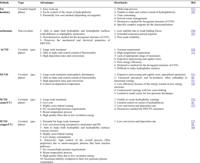
Table 1.Comparison between some common functionalization methods of nanoparticles and carbon nanotubes
Methods Type Advantages Drawbacks Ref.
Wet chemistry
Covalent (liquid
phase) 1- Easy scale-up 2- Facile control of the extent of hydrophilicity 3- Potentially low-cost method (depending on reagents)
1- Multi-step process
2- Unable to make and control extend of hydrophobicity 3- Time consuming
4- Solvent waste management
5- Destructive method for hexagonal structure of CNTs 6- Specific complex reagents for the functionalization
[27-29]
Surfactants Non-covalent 1- Able to make both hydrophilic and hydrophobic surfaces (with different or amphiphilic molecules)
2- Non-destructive method for the hexagonal structure of CNTs 3- Preserves the mechanical and electrical properties of SWCNTs
1- Low stability due to weak binding forces 2- Extended sonication period required 3- Poor scale reliability
[50-52]
TACVD Covalent
(gas-phase) 1- Large scale treatment 2- Able to make and control extend of functionality 3- High deposition rates and conversions
1- Vacuum requirement 2- High temperature requirement 3- Lack of appropriate range of monomers 4- Expensive (processing and capital costs) 5- Poor energy efficiency
6- Destructive method for the hexagonal structure of CNTs 7- Difficult to make hydrophobic surfaces
[28, 31]
PECVD Covalent
(gas-phase) 1- Large scale treatment (atmospheric discharges) 2- Able to make and control extend of functionality 3- High deposition rates and conversion 4- Control on deposition temperature
1- Expensive (processing and capital costs, specialized operation) 2- Unreacted precursors and by-products, often embedded in functional coating
3- Low efficiency because of the energy wasted on low energy electrons
4- Unstructured coatings with low cross-linking 5- Limited to small scales for low-pressure discharges
[31, 48, 53-55] PICVD (ozone/UV) Covalent
(gas-phase) 1- Large scale treatment 2- Low cost 3- Highly cross-linked coating 4- No vacuum/high pressure requirements 5- Room temperature process
6- High quality films due to low excitation energy
1- Unable to create hydrophobic coatings 2- Limited control on extent of hydrophilicity 3- Low conversion and deposition rate 4- Destructive method for SWCNT structures
[33, 56, 57] PICVD (syngas/UV) Covalent
(gas-phase) 1- Potential for large scale treatment 2- Low cost processing (inexpensive monomers and PI) 3- Able to make both hydrophilic and hydrophobic surfaces (various extents)
4- Highly cross-linked coating 5- Low energy consumption
6- Selectivity; high control of the overall process (film properties) due to mono-energetic photons that limit reaction pathways
7- No vacuum/high pressure requirements 8- Room temperature process
9- High quality films due to low excitation energy 10- Increased stability compared to their low-pressure plasma counterparts
1- Low conversion and deposition rate [27, 28, 58]
2. Materials and Methods 2.1. Materials
Pure SWCNTs (P-SWCNTs) grown by radio frequency induction thermal plasma were
purchased from Raymor-NanoIntegris (96.5% w/w, Quebec, Canada). The main impurities
remaining in the purchased P-SWCNTs are Fe, Ni, Y and amorphous carbon (as identified by XPS
and TEM measurements). Hydrogen peroxide (H2O2, 50% (w/w)) and n-hexadecane (≥99%,
EMD-Millipore) were purchased from Fisher Scientific (Montreal, Quebec). Ozone was generated by an
Ozone Solutions TS-40 ozone generator (20 g/h), with ambient air as the gas source. Syngas (CO
and H2, Air Liquide, chemically pure) and argon (Air Liquide, 99.9%), were used as the
functionalization precursors and purging gas, respectively. Two 96 cm-long UVC germicidal lamps (Model T-97505-80, Cole-Parmer Inc) were used for all experimental treatments (main emission
peak at 253.7 nm, irradiance of 0.01 W/cm2 at 3.5 cm, measured with an ILT1700 Radiometer
equipped with a SED240 detector from International Light Technologies).
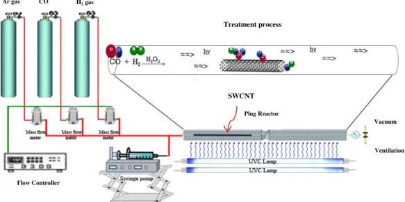
Fig. 1. Schematic of the PICVD setup.
2.2. PICVD Setup
The PICVD reactor used consists of two UVC lamps and a 25-mm internal diameter quartz
tube reactor (Fig.1). A syringe pump was used for H2O2 injection and Brooks mass flow controllers
Treatment process Vacuum Ventilation Plug Reactor Flow Controller Ar gas CO gas H2 gas SWCNT
(series 5850E) were used to inject precursors (CO and H2). A centralized vacuum system was
applied when working at sub-atmospheric pressure (down to -20 kPa).
2.3. Surface Modification
To modify SWCNT surfaces, the method developed by Dorval Dion et al. (2014) was used
[27]. Accordingly, P-SWCNTs (in the form of bucky paper) were placed inside the quartz reactor,
which was then closed and sealed. Within the same reactor, SWCNTs were treated with one of two different precursors: ozone or syngas. In the case of ozone treatments, the reactor was purged for two minutes with argon at a rate of 3 L/min then connected to the ozone generator. Ozonated air (5-7% w/w ozone, 500 ml/min) was fed to the reactor under different conditions presented in Table 2.
In Table 2, experimental treatments 2 to 6 are related to the SWCNTs treated by ozone/UV while
experiments 7 to 27 are dedicated to investigating the effects of treatment time, pressure, position of samples inside the reactor and hydrogen peroxide injection. In the case of syngas/PICVD
treatments, the reactor was also purged with argon, then CO and H2 precursors (total flow rate of
350 ml/min) were introduced at various ratios and conditions, as per Table 2. H2O2 was injected
simultaneously via the syringe pump as a radical photo-initiator (PI) at a rate of 1 μL/s, directly into the syngas stream. Each experimental treatment was repeated at least 3 times. Six independent
variables, including precursor molar ratio (H2/CO), pressure, treatment time, flow rate of precursors
and use of PI and position of samples inside the reactor were studied. According to the desired surface functionality, the process can be operated under slight pressure (up to +15 kPa) or slight vacuum (down to -20 kPa). Hydrophilic surfaces are typically obtained under slight vacuum, using hydrogen peroxide, and near the reactor inlet (16 cm), while hydrophobic surfaces are achieved
under pressure closer to the outlet (82 cm), based on previous work performed with copper [27].
During treatment, the surface temperature was monitored using an IR Infrared thermometer. Treatment started at room temperature (~25 C). Upon activating the UVC lamps, the temperature increased to ~52 °C over the first 15 min, and remained constant until the end of treatment (temperature increase attributable to heat emission from the UVC lamps).
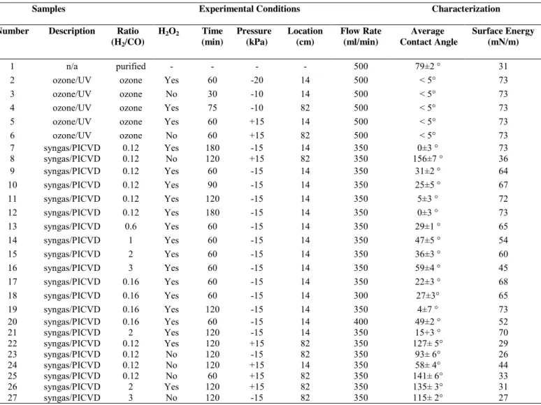
Table 2. Experimental treatment conditions with corresponding wettability (contact angle/surface energy)
Samples Experimental Conditions Characterization Number Description Ratio
(H2/CO) H2O2 Time (min) Pressure (kPa) Location (cm) Flow Rate (ml/min) Average Contact Angle Surface Energy (mN/m) 1 n/a purified - - - - 500 79±2 ° 31
2 ozone/UV ozone Yes 60 -20 14 500 < 5° 73 3 ozone/UV ozone No 30 -10 14 500 < 5° 73 4 ozone/UV ozone Yes 75 -10 82 500 < 5° 73 5 ozone/UV ozone Yes 60 +15 14 500 < 5° 73 6 ozone/UV ozone No 60 +15 82 500 < 5° 73 7 syngas/PICVD 0.12 Yes 180 -15 14 350 0±3 ° 73 8 syngas/PICVD 0.12 No 120 +15 82 350 156±7 ° 36 9 syngas/PICVD 0.12 Yes 60 -15 14 350 31±2 ° 64 10 syngas/PICVD 0.12 Yes 90 -15 14 350 25±5 ° 67 11 syngas/PICVD 0.12 Yes 120 -15 14 350 5±3 ° 72 12 syngas/PICVD 0.12 Yes 180 -15 14 350 0±3 ° 73 13 syngas/PICVD 0.6 Yes 60 -15 14 350 29±1 ° 65 14 syngas/PICVD 1 Yes 60 -15 14 350 47±5 ° 54 15 syngas/PICVD 2 Yes 60 -15 14 350 36±3 ° 60 16 syngas/PICVD 3 Yes 60 -15 14 350 59±4 ° 45 17 syngas/PICVD 0.16 Yes 60 -15 14 350 22±3 ° 68 18 syngas/PICVD 0.16 Yes 60 -15 14 300 27±3° 65 19 syngas/PICVD 0.16 Yes 120 -15 14 350 4±7 ° 73 20 syngas/PICVD 0.16 Yes 60 -15 14 400 49±2 ° 52 21 syngas/PICVD 2 Yes 120 -15 14 350 15+3 ° 70 22 syngas/PICVD 0.12 Yes 120 +15 82 350 127± 5° 29 23 syngas/PICVD 0.12 No 120 -15 82 350 93± 6° 26 24 syngas/PICVD 0.12 No 120 +15 14 350 58± 4° 44 25 syngas/PICVD 0.12 No 60 +15 82 350 141± 6° 33 26 syngas/PICVD 2 Yes 120 +15 82 350 135± 3° 31 27 syngas/PICVD 3 No 120 -15 82 350 115± 2° 27 2.4. Surface Characterization
X-ray photoelectron spectroscopy (XPS) was performed with a VG ESCALAB 3 MKII system using Mg K x-rays, with a pass energy of 100 eV and energy step size of 1 eV for survey scans. High-resolution (HR) spectra of treated SWCNT (T-SWCNT) buckypapers were obtained at a pass energy of 20 eV, in increments of 0.05 eV. Some of the XPS characterizations were also performed by Kratos Ultra DLD system with a pass energy of 160 eV and step size of 1 eV for survey scans and a pass energy of 20 eV and step size of 0.05 eV for HR spectra, using Al K
x-rays. Peak fitting was performed as described by Yang and Sacher [59], using the Wagner table for
sensitivity factors. Background correction was based on the Shirley method, used within the Thermo
Surface wetting was assessed through sessile drop contact angle (CA) measurements using a FDS OCA DataPhysics TBU 90E tensiometer. 2 L droplets of water (as a polar liquid) or n-hexadecane (as a nonpolar liquid) were deposited on 3 different areas of the samples, 3 successive times, in order to obtain the average CA. The exposure time between the droplet and the surface was considered 3 s for all experimental treatments. The surface energy was obtained by applying
the Owens-Wendt method (Equation 1) to CA measurements ( with the two different liquids
(water/polar and n-hexadecane/nonpolar) [60]. The total free surface energy (γs) was obtained by
gathering the polar (γsp) and dispersive (γsd) components based on Equation 2. Table S1 in
supplementary results presents the related information of the liquids.
(1)
(2)
Where sl, ld and lp are the solid-liquid interfacial energy, the total dispersive component
and the polar component of liquid surface tension, respectively.
Transmission electron microscopy (TEM, JEOL model JEM2100F) was used to investigate the effect of treatment on SWCNT morphology. The TEM grids used were coated with a lacey carbon film (D20045 grids with formwar substrates, Ni mesh 400, SOQUELEC International). To collect samples, T-SWCNTs were dispersed in methanol at a concentration of 1 mg/mL; TEM grids were then briefly dipped in this suspension (~ 1 s) and analyzed after drying.
The integrity of SWCNT samples was assessed by Raman spectroscopy using a Renishaw Invia Reflex Raman microscope equipped with an argon laser (514 nm), scanning in the range of
300-3500 cm-1. Peak deconvolution was completed through the Renishaw Wire 3.4 software.
Gaussian and Lorentzian line shapes were employed for all G and D bands, respectively, as this combination provides appropriate fits for Raman bands, especially to identify disorder in SWCNTs [61].
Thermal stability was assessed through thermogravimetric analysis (TGA, Model Q500, TA instruments) under air atmosphere. The temperature range was 30-800 ◦C, with a heating rate of 10 ◦ C/min for all samples. All experimental treatments were carried out with a platinum TGA pan (4 to 5 mg).
3.1. Physical Characterization
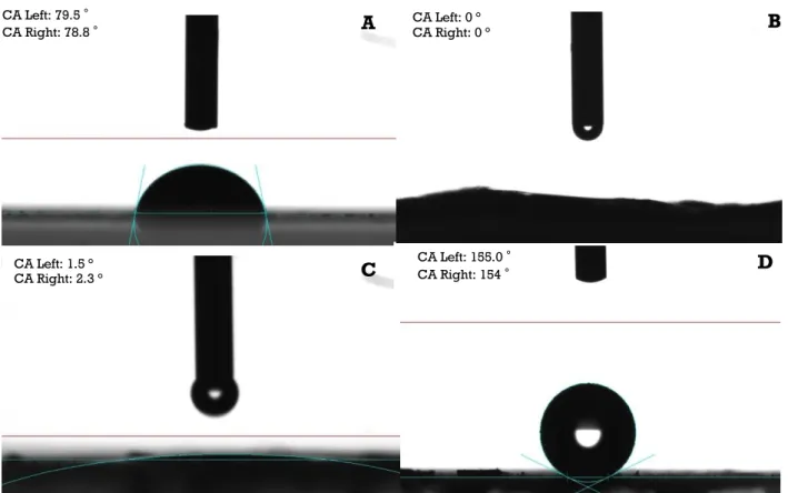
To analyse the efficiency of ozone/UV and syngas/PICVD treatments on SWCNTs, we have first determined the physical properties of the treated samples. Table 2 presents the experimental conditions, related water contact angle and surface energy results for ozone/UV and syngas/PICVD treatments. The average water CA of P-SWCNT samples was 79 2° (Fig. 2A). For all SWCNTs treated by ozone/UV (Experimental treatments 2-6), the water CA was <5° (below the detection limit of the instrument), irrespective of treatment time, pressure or position (Fig. 2B). This behavior is
expected: ozone peak absorption overlaps with that of the UVC lamps used [62]. These results are
consistent with those of Wang et al. (2010), where they showed that ozone/UV treatments of vertically aligned multi-walled carbon nanotubes for 5 min could lead to superhydrophilic surface
behavior (reactor operating with 0.2 L/min flow of oxygen gas supply at 50 C) [62]. Since the peak
absorption cross section of ozone is found at 253.7 nm, treatments under UVC light ( = 200 to 300
nm) cause it to break down into oxygen gas (O2) and reactive O radicals. These can react with defects
sites (or dangling bonds) on the P-SWCNT surface, leading to the formation of -COOH, OH, and CO
groups [62]. While ozone/UV allow for the possibility of sidewall functionalization on SWCNTs,
longer ozone/UV treatments can actually destroy the SWCNTs’ hexagonal carbon structure [63, 64].
While these experiments and previous works show ozone/UV’s ability to produce superhydrophilic
SWCNT surfaces [62, 65], it highlights its inability to vary surface energy over a wide range even by
changing effective parameters such as treatment time, pressure and position inside the reactor (Experimental treatment 2 to 6).
Syngas/PICVD, on the other hand, can generate superhydrophilic surfaces (Table 2, treatment 7, CA<5°, Fig. 2C), all the way to superhydrophobic (Table 2, treatment 8, CA>150°, Fig. 2D), mainly by varying sample position from 14 to 82 cm, treatment pressure from -15 to +15 kPa and absence of hydrogen peroxide as PI. The key parameters influencing hydrophobicity are pressure and location. These are illustrated by comparing treatment 8 and 23: save for pressure, both experiments have the same conditions – a lower pressure (- 15 kPa) leads to more hydrophilic behavior. Similarly, experiment 24, compared to experiment 8, changes only location, showing that positioning the sample closer to the inlet results in hydrophilic behavior. The effect of position can be attributed to the accessibility of syngas-derived radicals, activated monomers and PI-derived species (like OH*)
in the reactor [27, 66], while treatment pressure affects the organic chain growth rate and collisions
characterization show that H2O2 addition tends to make surfaces more hydrophilic, given that it
photo-dissociates under UVC light (peak between 180 and 200 nm) to generate two hydroxyl radicals (OH ) that can actively participate in the radical-driven syngas and form additional oxygen-containing (hydrophilic) groups [27, 34].
Experimental treatments 9 to 12 (Table 2) show that SWCNTs become more hydrophilic as a function of treatment time (from 31° at 60 min to 0° at 180 min), likely as a result of additional
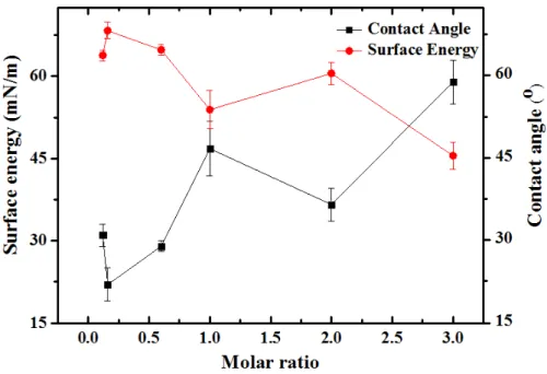
functional group deposition. Experimental treatments 9, 13 to 17, show that the H2/CO molar ratio
plays a significant role in the syngas/PICVD treatment (p<0.05). Indeed, molar ratio is known to play a significant role in binary precursor systems [10, 48, 56, 70]. Several authors used binary
mixtures consisting of an oxygen-containing monomers (e.g. O2 or N2O) and a hydrocarbon (e.g.
CH4 or C2H4) to treat SWCNTs [10, 48, 56, 70] and showed that the concentration of certain
functionalities (mostly hydroxyl, carbonyl and carboxylic acid groups) can increase along with an
increase in the ratio of oxygen- or nitrogen-containing precursors to hydrocarbons. Fig. 3 shows
CA Left: 1.5 °
CA Right: 2.3 ° C
CA Left: 79.5 °
CA Right: 78.8 ° A CA Left: 0 ° CA Right: 0° B
CA Left: 155.0 °
CA Right: 154 ° D
Fig. 2. CA measurement obtained by tensiometry A) P-SWCNT (Experimental treatment 1), B) SWCNTs treated by ozone/UV for 1h (Experimental treatment 2), C) SWCNTs treated by syngas/PICVD under vacuum for 3h (Experimental treatment 7), D) SWCNTs treated by syngas/PICVD under pressure for 2h (Experimental treatment 8).
CA Left: 0 °
that the CA of SWCNTs treated by syngas/PICVD (experimental treatments of 13 to 17 in addition to experimental treatments 9) generally increase as a function of the molar ratio (H2/CO), likely
because fewer oxygen-containing groups are present to increase wetting as the ratio increases. This is confirmed with the overlaid surface tension results. The experimental treatments in Fig. 3 are performed at a constant pressure and reactor position – varying these parameters in combination
with molar ratio allows for a full range of surface energies to be attained ranging from 0 ° to 156 °
(Table 2).
Beyond monomer ratio, our results show that total flow rate in the PICVD reactor can play a role in the final surface properties, given that it will impact residence time in the reactor and, therefore, the probability of collisions between the radicals and the substrate. Except for experimental treatments 18 and 20, all treatments were performed with a total flow rate of 350 ml/min. These two experimental treatments (at 300 and 400 mL/min, respectively), when compared to experiment 17, served to investigate the effect of flow rate. Both experimental treatments 18 and 20 yielded CA higher than at the nominal 350 ml/min flow rate (p-value < 0.05), indicating that hybrid effects may be at play – the reaction domain may switch from deficient to efficient regions (similar to the effect of position). The specific effect of residence time is studied by Farhanian et al (2017) [58]. They reported an increase in film thickness as a function of residence time for a
syngas/PICVD system treating flat surfaces [58].
Fig. 3. Black squares: Contact angle measurements of T-SWCNTs obtained by tensiometry in terms of molar ratio of H2/CO; Red circles: Surface energy measurements that have been plotted in terms of molar ratio. Error bars show
It is pertinent to note that the surface energy values presented in Table 2, computed using the Owens-Wendt model (equations 1 and 2), do not consider surface roughness. For this reason, some aberrant values are possible; for example, the surface energy calculated by the tensiometer’s software package for experimental treatment 8 (showing superhydrophobic behavior with a contact angle of 156°) is 36 mN/m, nearly unchanged from the far more hydrophilic P-SWCNT (treatment 1, with a contact angle of 79°, 31 mN/m). Given that PICVD treatments on flat surfaces have shown
the formation of nanostructured, island-like deposits [58, 71, 72], it is very likely that the increase
in roughness plays a large role in the observed wetting behavior.
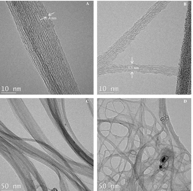
TEM characterization was done to provide more insight regarding the morphology and structure deposited coatings of T-SWCNTs by syngas/PICVD. Fig. 4 show the morphology of SWCNTs before (A,C) and after (B,D) treatment by syngas/PICVD under slight vacuum (-15 kPa, experimental treatment 11). The principal change observed is the appearance of an apparently polymeric layer on the treated sample. The SWCNT diameter grew from 1.4 nm before treatment to 5.3 nm after treatment.
3.2. Chemical Characterization
We have determined the chemical properties of SWCNTs treated by ozone/UV or syngas/PICVD treatments using XPS. During ozone/UV treatment, the relative oxygen atomic Fig. 4. TEM micrographs of A: P-SWCNTs (Experimental treatment 1), B: Syngas Treated SWCNTs (Experimental treatment 11), C: P-SWCNTs with lower magnification (Experimental treatment 1), D: Syngas treated in lower magnification (Experimental treatment 11).
B
5.3 nm 1.4 nm
A
percentage obtained by XPS survey spectra increased to 20.8% after 30 min (Experimental treatment 3) from untreated P-SWCNTs’ 6.9% oxygen content, due to the successful deposition of
oxygen-containing groups on the SWCNT surfaces by PICVD (Supplementary Fig. S1)
(p-value<0.05). HR-XPS fitting of the C1s peak (Supplementary Fig. S2) shows 6 major
functionalities compared to P-SWCNTs: A) 284.6 eV, corresponding to C=C (sp2); B) 285.7 eV,
corresponding to C=C (sp2) with defects and C–C (sp3); C) 286.5 eV, assigned to C–O; D) 288 eV,
assigned to C=O and transition of C=C with defects; E) 289 eV, corresponding to O–C=O;
and F) 291.3 eV, assigned to highly delocalized transition of C=C [73]. Successful
incorporation of oxygen-containing groups (mostly –COOH, C=O and -OH) on the SWCNT
surfaces after ozone/UV treatment is confirmed, in agreement with previous studies [57]. Raja et al.
(2014) had reported successful incorporation of oxygen-containing groups such as carboxylic acid, hydroxyl and carbonyl groups to SWCNTs while they treated SWCNT under ozone/UV or UV and
benzophenone [57]. Experimental treatments 2 to 4 were also used to investigate the effect of
treatment time in the case of SWCNTs treated by ozone/UV-treated SWCNT (O-T-SWCNT) by applying 60, 30, and 75 min, respectively. As seen in Supplementary Fig. S3 (inset), oxygen content reaches a plateau ca. 20-26% after 30 min. However, Supplementary Fig. S3 (over plot of C1s HR-XPS spectra of SWCNTs treated by ozone/UV with 60, 30, and 75 min) did not show a significant difference as a function of treatment time. Vautard et al. (2012) reported gas-phase surface treatment of carbon nanofibers in a reactive ozone environment with residence times on the order of 2 min. Their results are in agreement with ours, showing successful incorporation of
oxygen-containing groups onto carbon nanofibers [73].
The oxygen content of syngas/PICVD-treated SWCNTs (S-T-SWCNTs, Experimental treatment 1) climbed to 60.1%, with the balance being carbon (18.1%) and iron (21.8%) (Supplementary Fig. S1). The presence of iron (binding energy of 714.4 eV) can be attributed to iron
pentacarbonyl (Fe(CO)5) in the CO tank, a common impurity that forms over time in steel tanks
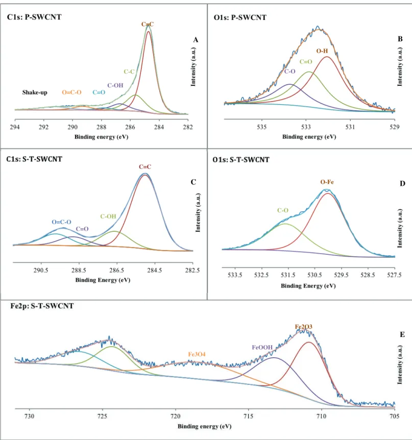
exposed to CO; it can readily decompose under the action of heat or UVC light [74]. Fig. 5A and 5B
show C1s and O1s HR-XPS spectra of P-SWCNTs to provide comparison with the treated SWCNTs. According to the C1s HR-XPS spectra (Fig. 5C), the syngas-based coating was composed of C-C, hydroxyl (-OH), carboxylic acid (COOH), and carbonate groups, corresponding to binding energies of 285, 286.7, 288.9, and 289.8 eV, respectively. Interestingly, the C1s peak for the treated sample became wider, compared to the untreated sample’s more sharp and narrow peak, implying that the
S-T-SWCNTs have two major peaks at binding energies of 530 and 531.65 eV, which are assigned to
O-Fe (or O-Fe2O3) and C-OH, respectively (though Fe(OH)3, phenol and O=C-O-Fe are also possible)
[73]. Based on the Fe2p high resolution XPS spectra (Fig. 5E), deposited iron species consisted of
Fe2O3, FeOOH, and Fe3O4 based on peaks at binding energies of 710.55, 713.55, and 718.75 eV,
respectively [73]. The Fe2O3 and Fe3O4 are likely the result of decomposition initiated by the 185 nm
peak (as discussed earlier), or through degradation of Fe(CO)2 radicals through collision with
reactive species and hydrogen peroxide. Overall, the iron content is essentially in the form of
Fig. 5. A: High resolution C1s spectra of P-SWCNT (Experimental treatment 1); B: High resolution O1s spectra of P-SWCNT (Experimental treatment 1); C: High resolution C1s spectra of S-T-SWCNT (Experimental treatment 11); D: High resolution O1s spectra of Treated SWCNT (Experimental treatment 11); E: High resolution Fe2P spectra of T-SWCNT (Experimental treatment 11). A B D E C C=C C-OH C=O O=C-O O-H C=O C-O C=C C-C C-OH C=O O=C-O Shake-up C-O O-Fe Fe3O4 FeOOH Fe2O3
To elucidate the role and effect of each individual precursor (H2, CO, H2O2) on coating
2
composition, we performed some blank experiments on glass substrates (thereby eliminating
3
interactions that may occur with SWCNT) (supplementary Table S2). The C1s HR-XPS spectra for
4
bare glass (supplementary Fig. S4A) showed four peaks at binding energies of 285, 286.75, 288.78
5
and 288.46 eV, assigned to aliphatic carbon or C-C, C-OH and carboxylic acid (COOH) groups,
6
respectively (carbon sourced from airborne contaminants). The O1s spectra (supplementary Fig. S4B)
7
showed two peaks at binding energies of 531.30 and 532.62 eV, corresponding to OH and SiO2
8
groups, respectively. After treatment with CO, H2 and H2O2, the same results and structure as those
9
observed on SWCNTs were obtained. Compared to bare glass, the C1s spectra (supplementary Fig.
10
S4C) showed two additional peaks, namely C-Fe and carbonyl groups at binding energies of 284.1
11
and 290.26 eV, respectively. Moreover, treated glass exhibited decreased hydroxyl groups and
12
significantly increased carboxylic groups. The O1s spectra (supplementary Fig. S4D) showed three
13
new peaks at binding energies of 530.18, 531.67 and 532.61 eV assigned to O-Fe, C-OH groups and
14
O=C-O-Fe, respectively.
15
In the absence of H2O2 (CO and H2 only), treatment lead to the appearance of two extra peaks
16
compared to bare glass, at binding energies of 284.33 and 289.55 eV (supplementary Fig. S4E). These
17
correspond to C-Fe (or C-Si, as Si could be still seen after treatment in survey peaks), and carbonate
18
groups (adsorbed CO2), respectively. The spectra are generally similar to the CO+H2+H2O2 case, with
19
increased carboxylic (COOH or COOC) and decreased carbon-bonded hydroxyl groups in the
20
structure. The O1s spectra (supplementary Fig. S4F) also showed three peaks at binding energies of
21
530.18, 531.67 and 532.61 eV assigned to O-Fe, OH and O=C-O-Fe, respectively. While the
22
chemical differences appear small, the film produced in the absence of H2O2 showed significant
23
morphological differences: it appeared as a powder adsorbed on the substrate (not a bound film) and
24
could be removed by shaking lightly. Based on this observation, we can conclude that H2O2 plays a
25
determinant role as both a photo-initiator and during film formation.
26
Knowing that CO can absorb light below 200 nm [27] (including the 185 nm emission peak
27
from the Hg lamps), and that it contains Fe(CO)5, it may lead to deposition without the addition of H2.
28
When combining CO with H2O2 (without hydrogen), the C1s spectra (supplementary Fig. S4G) shows
29
two peaks not present for bare glass at binding energies of 283.9 and 287.3 eV, assigned to C-Fe and
30
C=O, respectively. The peak at binding energy 289.3 eV was also quite significant compared to its
(supplementary Fig. S4H) showed three peaks (530.19, 532.06 and 533.05 eV) assigned to Fe-O (or
33
Fe(OH)O or Fe3O4), O-Si and chemisorded H2O. As expected, combining H2O2 with only H2 lead to
34
no deposit and HR-XPS spectra (supplementary Fig. S4I and S4J) identical to bare glass.
35 36 37
3.3. Thermal Stability and Defects 38
39
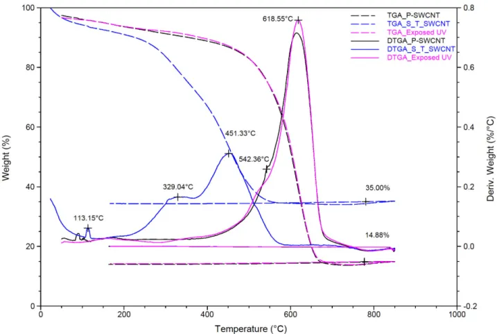
In order to quantify deposited functionalities on SWCNTs, we performed TGA analyses. Fig.
40
6 shows TGA and Derivative Thermogravimetric Analysis (DTGA) of P-SWCNTs (experimental
41
treatment 1), S-T-SWCNTs (CO+H2+H2O2, experimental treatment 11) and SWCNT exposed to
42
UVC light as a blank experiment. The DTGA graph of P-SWCNT shows two peaks around 618.5
43
and 542.4 C, assigned to SWCNTs with sp2 hybridization and disordered SWCNTs with sp3
44
hybridizations, respectively [76]. The P-SWCNTs showed 77.2% carbon with sp2 hybridization and
45
14.9% residue which corresponds to metallic and inorganic impurities. There are no significant
46
differences or decomposition shifts between P-SWCNT and SWCNT exposed to UV light. Using
47
the mass loss of P-SWCNTs at 360 C as a reference (10%), the mass loss of S-T-SWCNTs is about
48
26%, meaning that treatment leads to decomposition at a lower temperature. This is consistent with
49
previous work where it was shown that a higher amount of functionalities leads to a lower
50
degradation temperature because of more treatment-induced physical defects on the tubes’ surfaces
51
and end-caps [57]. The DTGA of S-T-SWCNT shows two new peaks around ca. 113°C and 329°C
52
which are related to oxygen-containing groups and carbon coating on SWCNTs, respectively [77].
53
The SWCNT peak is also shifted to lower temperatures (from 618.5 to 451.3°C) after treatment
54
(consistent with mass loss results). S-T-SWCNTs presented 0.7% humidity, 19.5% deposited carbon
55
with sp3 hybridization, 38.7% carbon with sp2 hybridization and 35% metallic residue. The increase
56
of iron content agrees with XPS results (presence of iron).
57 58 59 60 61 62 63
65
Fig. 6.TGA/DTGA graphs comparing thermal mass loss of P-SWCNTs (Experimental treatment 1), SWCNTs exposed 66
to UV and S-T-SWCNTs treated (Experimental treatment 11). 67
68
We characterized T-SWCNTs by Raman spectroscopy to study their hexagonal structure
69
after treatments. Table 3 (extracted from Fig. S5 in supplementary results) shows the Raman
70
spectrum results used to assess SWCNT integrity. The ratio of D- to G-bands in Raman spectra
71
presents defects produced in the hexagonal structure [61]. This ratio is lower for P-SWCNT and
72
increases as a function of treatment time, confirming the tensiometry and XPS findings. If we
73
assume that the D/G ratio is assigned strictly to presence of functionalities producing structural
74
defect sites, the Raman results are also in agreement with the TEM observations (longer treatment
75
leads to a thicker, amorphous carbon coating – from experimental treatment 1 (0 min) to
76
experimental treatment 9 (60 min) to experimental treatment 11 (120 min) to experimental treatment
77
12 (180 min)) [10]. In the case of 2h treatments, two different molar ratios, namely 0.12 and 2, were
78
evaluated (Experiments 11 and 21). The higher molar ratio (Experimental treatment 21) leads to
79
higher D/G ratio, even higher than that of the long treatment time at low ratio (Experimental
organic film with a higher amount of saturated carbon (with oxygen carried away, likely in the form
82
of water).
83 84
Table 3. D/G band ratio of SWCNTs before and after treatment at different time obtained by Raman spectra 85
Samples Related Experimental Treatment Time (h) D/G Bond Ratio
P-SWCNT 1 - 0.29 S-T-SWCNT 9 1 0.34 S-T-SWCNT 11 2 0.36 S-T-SWCNT 12 1 0.43 S-T-SWCNT 21 2 0.58 86 87
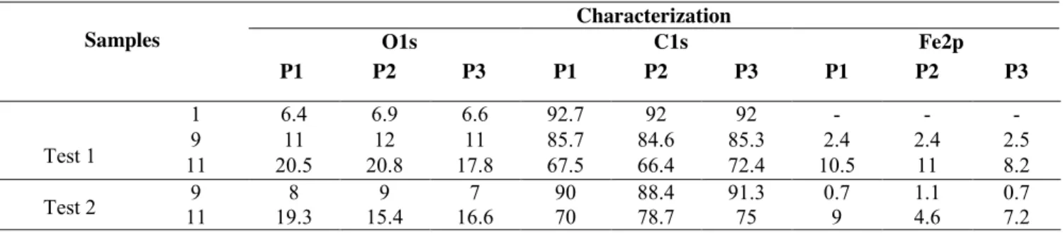
3.4. Treatment Homogeneity and Dispersibility of SWCNTs 88
89
Homogeneous treatment of SWCNT buckypapers will be an important factor in many
90
applications, including polymer nanocomposites or sensors, in which unfunctionalizaed areas may
91
deteriorate mechanical and electrical properties and overshadow the SWCNT’s performance
92
improvements [78]. We have performed XPS mapping of three typical SWCNT samples to verify
93
homogeneity of surface coatings (Table 4, experimental treatments 1, 9 and 11) at three different
94
regions on the samples (the head/position P1, middle/P2 and end/P3). Carbon, oxygen and iron
95
content is nearly identical at all positions, confirming that the treatment is homogeneous.
96 97
Table 4. XPS mapping results and their experimental conditions 98 Samples Characterization O1s C1s Fe2p P1 P2 P3 P1 P2 P3 P1 P2 P3 Test 1 1 6.4 6.9 6.6 92.7 92 92 - - - 9 11 12 11 85.7 84.6 85.3 2.4 2.4 2.5 11 20.5 20.8 17.8 67.5 66.4 72.4 10.5 11 8.2 Test 2 11 9 19.3 8 15.4 9 16.6 7 90 70 78.7 88.4 91.3 75 0.7 9 4.6 1.1 0.7 7.2 99 100
However, this homogeneity may not hold over the full thickness of the bucky paper sample
101
used. Given that many biological assays are conducted SWCNTs in suspension,
through-the-102
thickness heterogeneity may be an issue. To evaluate dispersion, samples exhibiting
103
superhydrophilic behaviors (Experimental treatment 11) were dispersed in deionized water at a
concentration of 0.05 g/mL. After sonicating for 60 minutes, the samples were allowed to settle.
105
Untreated SWCNT samples from areas not reached by the PICVD treatment settled out, while
106
treated S-T-SWCNTs remained in suspension (Fig. 7A). Sediments were removed through
107
decantation and the supernatants remained stable for at least 24 h (Fig. 7B). The “supernatant
108
SWCNTs” are the samples that could be used for cytotoxicity assessments before applying them in
109
biomedical devices. Furthermore, the treatment resists sterilization by immersion of the sample
110
container in a boiling water bath for 30 min (variation in CA <5° before and after boiling, data not
111
reported).
112
Beyond sterilization in suspension, the S-T-SWCNTs exhibited exceptional stability,
113
hydrophobic samples (from experimental treatment 8) remained at 151 2 after 7 days (ambient
114
conditions), while hydrophilic samples (from experimental treatments 11 and 12) increased to 7 4 .
115
This so-called “hydrophobic recovery” of the hydrophilic-treated sample is a well-documented
116
ageing phenomenon, dependent on absorption of contaminations, reorientation and reconstruction of
117
dangling groups on the surface [13, 46]. However, this phenomenon can be significantly reduced in
118
cross-linking modified surfaces [79]. These dispersion assays demonstrate the change in wettability
119
of individual S-T-SWCNT; dispersion into a wide variety of solvents is the focus of on-going work.
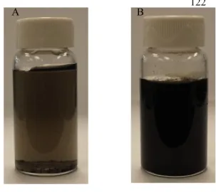
120 121 122 122 123 124 125 126 127 128 129 130 131 132 133 A B A
Fig. 7.A) Dispersion of T-SWCNTs in deionized water after 24h, B) Well dispersion of T-SWCNTs under vacuum in deionized water upon treatment.
4. Conclusions
135 136
In this study, we compared treatment of SWCNTs by ozone/UV and syngas/PICVD. The
137
main objective of this project was to functionalize SWCNTs with various surface energies ranging
138
from superhydrophilic to superhydrophobic. Although treatment of SWCNTs by ozone/UV was
139
straightforward and superhydrophilic SWCNTs could be obtained in a short time, this method could
140
not meet the main goal of the project. Syngas/PICVD on the other hand provides an efficient method
141
to fabricate functionalized SWCNTs with oxygen-containing groups in a full range of surface
142
energies (ranging from superhydrophilic to superhydrophobic behaviour). Molar ratio of the H2/CO
143
precursor mix, pressure and treatment time were found to have significant effects during treatment.
144
The PICVD induced coating on the SWCNTs was chemically characterized and proved to be
145
homogeneous (on the surface) and exceptionally stable (including being subjected to boiling for
146
sterilization purposes). This approach is therefore viable as a technique to be used in various
147
applications such as nanocomposites, aerospace and bio applications, in which various degrees of
148
functionality (and consequently surface energies) are desirable. Furthermore, this method will be
149
applicable to investigate the true effect of surface energy on cytotoxicity, which will be the focus of
150 subsequent work. 151 152 Acknowledgments 153 154
We wish to acknowledge the financial support provided by Fonds de recherche du Québec
155
(FRQNT), as well as moral support from Polytechnique Montreal and Université de Sherbrooke. We
156
would also like also to thank the Université de Sherbrooke Materials Characterization Laboratory,
157
the Polytechnique thin films group (GCM) and the (CM). The authors acknowledge the
158
collaboration of Dr Josianne Lefebvre (Polytechnique Montreal University) for her assistance in
159 analyzing XPS results. 160 161 References 162 163
[1] M.C. Serrano, M.C. Gutiérrez, F. del Monte, Role of polymers in the design of 3D carbon nanotube-based scaffolds 164
for biomedical applications, Prog. Polym. Sci. 39 (2014) 1448-1471. 165
[2] E.T. Thostenson, Z.F. Ren, T.W. Chou, Advances in the science and technology of carbon nanotubes and their 166
composites: a review, Compos. Sci. Technol. 61 (2001) 1899-1912. 167
[3] L.P. Zanello, B. Zhao, H. Hu, R.C. Haddon, Bone cell proliferation on carbon nanotubes, Nano Lett. 6 (2006) 562-168
[4] P.D. Bradford, A.E. Bogdanovich, Electrical conductivity study of carbon nanotube yarns, 3-D hybrid braids and 170
their composites, J. Compos. Mater. 42 (2008) 1533-1545. 171
[5] B. Kateb, M. Van Handel, L. Zhang, M.J. Bronikowski, H. Manohara, B. Badie, Internalization of MWCNTs by 172
microglia: possible application in immunotherapy of brain tumors, Neuroimage 37 Suppl 1 (2007) S9-17. 173
[6] N. Saito, Y. Usui, K. Aoki, N. Narita, M. Shimizu, N. Ogiwara, K. Nakamura, N. Ishigaki, H. Kato, S. Taruta, M. 174
Endo, Carbon nanotubes for biomaterials in contact with bone, Curr Med Chem 15 (2008) 523-527. 175
[7] J.M. Wernik, S.A. Meguid, Recent Developments in Multifunctional Nanocomposites Using Carbon Nanotubes, 176
Applied Mechanics Reviews 63 (2010) 050801. 177
[8] Y. Alinejad, N. Faucheux, G. Soucy, Preosteoblasts behavior in contact with single-walled carbon nanotubes 178
synthesized by radio frequency induction thermal plasma using various catalysts, J Appl Toxicol 33 (2013) 1143-1155. 179
[9] B. Zhao, H. Hu, S.K. Mandal, R.C. Haddon, A bone mimic based on the self-assembly of hydroxyapatite on 180
chemically functionalized single-walled carbon nanotubes, Chem. Mater. 17 (2005) 3235-3241. 181
[10] J.C. Ruiz, P.L. Girard-Lauriault, F. Truica-Marasescu, M.R. Wertheimer, Plasma- and vacuum-ultraviolet (VUV) 182
photo-polymerisation of N- and O-rich thin films, Radiat. Phys. Chem. 79 (2010) 310-314. 183
[11] N. Ogihara, Y. Usui, K. Aoki, M. Shimizu, N. Narita, K. Hara, K. Nakamura, N. Ishigaki, S. Takanashi, M. 184
Okamoto, H. Kato, H. Haniu, N. Ogiwara, N. Nakayama, S. Taruta, N. Saito, Biocompatibility and bone tissue 185
compatibility of alumina ceramics reinforced with carbon nanotubes, Nanomedicine : nanotechnology, biology, and 186
medicine 7 (2012) 981-993. 187
[12] S. Giannona, I. Firkowska, J. Rojas-Chapana, M. Giersig, Vertically aligned carbon nanotubes as cytocompatible 188
material for enhanced adhesion and proliferation of osteoblast-like cells, J. Nanosci. Nanotechnol. 7 (2007) 1679-1683. 189
[13] Y. Wang, J. Wu, F. Wei, A treatment method to give separated multi-walled carbon nanotubes with high purity, 190
high crystallization and a large aspect ratio, Carbon 41 (2003) 2939-2948. 191
[14] J. Zhao, Light Scattering Characterization of Carbon Nanotube Dispersions and Reinforcement of Polymer 192
Composites, Department of Chemical and Materials Engineering, University of Cincinnati, 2006. 193
[15] S. Z hang, T. Shao, S.S. Kaplan-Bekaroglu, T. Karanfil, The Impacts of Aggregation and Surface Chemistry of 194
Carbon Nanotubes on the Adsorption of Synthetic Organic Compounds, Environ. Sci. Technol. 43 (2009) 5719–5725. 195
[16] M. Foldvari, M. Bagonluri, Carbon nanotubes as functional excipients for nanomedicines: II. Drug delivery and 196
biocompatibility issues, Nanomedicine : nanotechnology, biology, and medicine 4 (2008) 183-200. 197
[17] Z. Liu, X. Dong, L. Song, H. Zhang, L. Liu, D. Zhu, C. Song, X. Leng, Carboxylation of multiwalled carbon 198
nanotube enhanced its biocompatibility with L02 cells through decreased activation of mitochondrial apoptotic pathway, 199
J Biomed Mater Res A 102 (2014) 665-673. 200
[18] G. Vuković, A. Marinković, M. Obradović, V. Radmilović, M. Čolić, R. Aleksić, P.S. Uskoković, Synthesis, 201
characterization and cytotoxicity of surface amino-functionalized water-dispersible multi-walled carbon nanotubes, 202
Appl. Surf. Sci. 255 (2009) 8067-8075. 203
[19] D. Liu, C. Yi, D. Zhang, J. Zhang, M. Yang, Inhibition of proliferation and differentiation of mesenchymal stem 204
cells by carboxylated carbon nanotubes, ACS nano 4 (2010) 2185-2195. 205
[20] S.L. Montes-Fonseca, E. Orrantia-Borunda, A. Aguilar-Elguezabal, C. Gonzalez Horta, P. Talamas-Rohana, B. 206
Sanchez-Ramirez, Cytotoxicity of functionalized carbon nanotubes in J774A macrophages, Nanomedicine : 207
nanotechnology, biology, and medicine 8 (2012) 853-859. 208
[21] P. Newman, A. Minett, R. Ellis-Behnke, H. Zreiqat, Carbon nanotubes: their potential and pitfalls for bone tissue 209
regeneration and engineering, Nanomedicine : nanotechnology, biology, and medicine 9 (2013) 1139-1158. 210
[22] C.B. Dong, A.S. Campell, R. Eldawud, G. Perhinschi, Y. Rojanasakul, C.Z. Dinu, Effects of acid treatment on 211
structure, properties and biocompatibility of carbon nanotubes, Applied Surface Science 264 (2013) 261-268. 212
[23] S. Vardharajula, S.Z. Ali, P.M. Tiwari, E. Eroglu, K. Vig, V.A. Dennis, S.R. Singh, Functionalized carbon 213
nanotubes: biomedical applications, Int J Nanomedicine 7 (2012) 5361-5374. 214
[24] C. Ge, J. Du, L. Zhao, L. Wang, Y. Liu, D. Li, Y. Yang, R. Zhou, Y. Zhao, Z. Chai, C. Chen, Binding of blood 215
proteins to carbon nanotubes reduces cytotoxicity, Proceedings of the National Academy of Sciences of the United 216
States of America 108 (2011) 16968-16973. 217
[25] R.J. Chen, Y. Zhang, D. Wang, H. Dai, Noncovalent sidewall functionalization of single-walled carbon nanotubes 218
for protein immobilization, J. Am. Chem. Soc. 123 (2001) 3838-3839. 219
[26] Z. Rastian, A.A. Khodadadadi, F. Vahabzade, Y. Mortazavi, Functionalization of Multi -Walled Carbon Nanotubes 220
for Lipase Immobilization, JMTI 1 (2013) 54-71. 221
[27] C.A. Dorval Dion, W. Raphael, E. Tong, J.R. Tavares, Photo-initiated chemical vapor deposition of thin films using 222
syngas for the functionalization of surfaces at room temperature and near-atmospheric pressure, Surf. Coat. Technol. 223
[28] C.A. Dorval Dion, J.R. Tavares, Photo-initiated chemical vapor deposition as a scalable particle functionalization 225
technology (a practical review), Powder Technology 239 (2013) 484-491. 226
[29] K. Balasubramanian, M. Burghard, Chemically functionalized carbon nanotubes, Small 1 (2005) 180-192. 227
[30] A.H. Lu, E.L. Salabas, F. Schuth, Magnetic nanoparticles: synthesis, protection, functionalization, and application, 228
Angewandte Chemie 46 (2007) 1222-1244. 229
[31] K.L. Choy, Chemical vapour deposition of coatings, Prog. Mater Sci. 48 (2003) 57-170. 230
[32] C.A. Dorval Dion, J.R. Tavares, Photo-initiated chemical vapor deposition as a scalable particle functionalization 231
technology (a practical review), Powder Technol. 239 (2013) 484-491. 232
[33] E. Kasparek, J.R. Tavares, M.R. Wertheimer, P.-L. Girard-Lauriault, Sulfur-Rich Organic Films Deposited by 233
Plasma- and Vacuum-Ultraviolet (VUV) Photo-Polymerization, PLASMA PROCESS POLYM (2016) n/a-n/a. 234
[34] H. Okabe, Photochemistry of small molecules, Wiley New York1978. 235
[35] J.Y. Woo, D. Kim, J. Kim, J. Park, C.S. Han, Fast and Efficient Purification for Highly Conductive Transparent 236
Carbon Nanotube Films, Journal of Physical Chemistry C 114 (2010) 19169-19174. 237
[36] E.P. Koumoulos, C.A. Charitidis, Surface analysis and mechanical behaviour mapping of vertically aligned CNT 238
forest array through nanoindentation, Applied Surface Science 396 (2017) 681-687. 239
[37] X. Liu, F. Xu, K. Zhang, B. Wei, Z. Gao, Y. Qiu, Characterization of enhanced interfacial bonding between epoxy 240
and plasma functionalized carbon nanotube films, Composites Science and Technology 145 (2017) 114-121. 241
[38] S. Min, J. Kim, C. Park, J.-H. Jin, N.K. Min, Long-term stability of superhydrophilic oxygen plasma-modified 242
single-walled carbon nanotube network surfaces and the influence on ammonia gas detection, Applied Surface Science 243
410 (2017) 105-110. 244
[39] M. Garzia Trulli, E. Sardella, F. Palumbo, G. Palazzo, L.C. Giannossa, A. Mangone, R. Comparelli, S. Musso, P. 245
Favia, Towards highly stable aqueous dispersions of multi-walled carbon nanotubes: the effect of oxygen plasma 246
functionalization, Journal of colloid and interface science 491 (2017) 255-264. 247
[40] S. Wang, Y. Zhang, N. Abidi, L. Cabrales, Wettability and surface free energy of graphene films, Langmuir 25 248
(2009) 11078-11081. 249
[41] A. Paul, POLYMER FUNCTIONALIZED SINGLE-WALLED CARBON NANOTUBE COMPOSITES AND 250
SEMI-FLUORINATED QUATERNARY AMMONIUM POLYMER COLLOIDS AND COATINGS, Oklahoma State 251
University, ProQuest Dissertations, 2012, pp. 194. 252
[42] M. AfzaliTabar, M. Alaei, R.R. Khojasteh, F. Motiee, A.M. Rashidi, Preference of multi-walled carbon nanotube 253
(MWCNT) to single-walled carbon nanotube (SWCNT) and activated carbon for preparing silica nanohybrid pickering 254
emulsion for chemical enhanced oil recovery (C-EOR), J. Solid State Chem. 245 (2017) 164-173. 255
[43] B.A. Kakade, V.K. Pillai, Tuning the wetting properties of multiwalled carbon nanotubes by surface 256
functionalization, Journal of Physical Chemistry C 112 (2008) 3183-3186. 257
[44] K.K.S. Lau, J. Bico, K.B.K. Teo, M. Chhowalla, G.A.J. Amaratunga, W.I. Milne, G.H. McKinley, K.K. Gleason, 258
Superhydrophobic carbon nanotube forests, Nano Letters 3 (2003) 1701-1705. 259
[45] R. Rajkhowa, A. Kafi, Q.T. Zhou, A. Kondor, D.A.V. Morton, X.G. Wang, Relationship between processing, 260
surface energy and bulk properties of ultrafine silk particles, Powder Technology 270 (2015) 112-120. 261
[46] I.A. Sacui, R.C. Nieuwendaal, D.J. Burnett, S.J. Stranick, M. Jorfi, C. Weder, E.J. Foster, R.T. Olsson, J.W. 262
Gilman, Comparison of the properties of cellulose nanocrystals and cellulose nanofibrils isolated from bacteria, tunicate, 263
and wood processed using acid, enzymatic, mechanical, and oxidative methods, ACS Appl Mater Interfaces 6 (2014) 264
6127-6138. 265
[47] Q.X. Li, J.S. Church, M. Naebe, B.L. Fox, Interfacial characterization and reinforcing mechanism of novel carbon 266
nanotube - Carbon fibre hybrid composites, Carbon 109 (2016) 74-86. 267
[48] L. Vandsburger, E.J. Swanson, J. Tavares, J.L. Meunier, S. Coulombe, Stabilized aqueous dispersion of multi-268
walled carbon nanotubes obtained by RF glow-discharge treatment, JNR 11 (2009) 1817-1822. 269
[49] B. White, S. Banerjee, S. O'Brien, N.J. Turro, I.P. Herman, Zeta-potential measurements of surfactant-wrapped 270
individual single-walled carbon nanotubes, Journal of Physical Chemistry C 111 (2007) 13684-13690. 271
[50] P. Bilalis, D. Katsigiannopoulos, A. Avgeropoulos, G. Sakellariou, Non-covalent functionalization of carbon 272
nanotubes with polymers, Rsc Advances 4 (2014) 2911-2934. 273
[51] L. Chen, H. Xie, W. Yu, Functionalization Methods of Carbon Nanotubes and Its Applications, Carbon Nanotubes 274
Applications on Electron Devices (2011). 275
[52] Y.L. Zhao, J.F. Stoddart, Noncovalent functionalization of single-walled carbon nanotubes, Acc. Chem. Res. 42 276
(2009) 1161-1171. 277
[53] Q. Chen, L. Dai, M. Gao, S. Huang, A. Mau, Plasma Activation of Carbon Nanotubes for Chemical Modification, J. 278
Phys. Chem. 105 (2001) 618-622. 279