Caspase-8-Dependent HER-2 Cleavage in Response to Tumor Necrosis Factor
␣
Stimulation Is Counteracted by Nuclear Factor
B through
c-FLIP-L Expression
Vale´rie Benoit,
1Alain Chariot,
1Laurence Delacroix,
2Vale´rie Deregowski,
1Nathalie Jacobs,
2Marie-Paule Merville,
1and Vincent Bours
11Laboratory of Medical Chemistry and Human Genetics, Center for Molecular and Cellular Therapy and2Center for Research in Experimental Cancerology, University of Liege, Liege, Belgium
ABSTRACT
The oncoprotein HER-2/neu is a prosurvival factor, and its
overexpres-sion has been correlated with poor prognosis in patients with breast
cancer. We report that HER-2 is a new substrate for caspase-8 and that
tumor necrosis factor
␣ (TNF-␣) stimulation leads to an early
caspase-8-dependent HER-2 cleavage in MCF7 A/Z breast adenocarcinoma cells
defective for nuclear factor
B (NFB) activation. We show that the
antiapoptotic transcription factor NF
B counteracts this cleavage
through induction of the caspase-8 inhibitor c-FLIP. Our results also
demonstrate that this HER-2 cleavage contributes to the TNF-
␣-induced
apoptosis pathway because ectopic expression of an uncleavable HER-2
protects NF
B-defective cells against TNF-␣-mediated cell death.
There-fore, we propose an original model in which NF
B exerts a new
antiapo-ptotic function by counteracting TNF-
␣-triggered cleavage of the HER-2
survival factor.
INTRODUCTION
Tumor necrosis factor
␣ (TNF-␣) plays a pivotal role in the control
of cell proliferation and inflammation by regulating proapoptotic and
antiapoptotic signaling pathways through binding to two distinct
receptors, TNF receptor 1 (TNF-R1) and TNF receptor 2 (TNF-R2;
Refs. 1–5). TNF-
␣ binding triggers receptor trimerization and
subse-quent recruitment of various signaling proteins to the receptor
cyto-plasmic domains. Depending on the nature of the receptor and the
adaptor proteins, TNF-
␣ can exert opposite effects (6–8). Activation
of the TNF-R2 produces an antiapoptotic and proinflammatory
cas-cade, whereas activation of the TNF-R1 leads to antagonist events,
namely, apoptosis through recruitment and activation of caspase-8 (9)
as opposed to the activation of the antiapoptotic and proinflammatory
transcription factor nuclear factor
B (NFB; Refs. 6–8). Numerous
NF
B target genes counteract the TNF-␣-activated apoptosis pathway
and include caspase inhibitors such as c-IAP1 (10), c-IAP2 (11, 12),
XIAP (13), and c-FLIP (14); Bcl-2 family members such as A1
(15–17) or Bcl-x
L(18, 19); and other proteins like A20 (20 –22) or
MnSOD (23–25). These opposite events can explain that TNF-
␣ is a
poor inducer of apoptosis in the absence of RNA or protein synthesis
inhibitors (26 –28).
Cancers are characterized by an increased proliferation and a
de-creased apoptotic rate (29). Among the proteins implicated in this
dysregulation, HER-2/neu (ErbB-2) plays a major role in some
ovar-ian and breast cancer cells. The Her-2 oncogene encodes a
transmem-brane receptor protein structurally related to the epidermal growth
factor receptor (30, 31). HER-2 is overexpressed in approximately
one-third of the primary breast carcinomas (32) and in ovarian
carci-nomas (31, 33–35), and its expression is correlated with poor
prog-nosis and decreased overall and disease-free survival (36 –38). HER-2
overexpression has been demonstrated to enhance proliferative,
met-astatic, and prosurvival signals in breast cancer cell lines (39 – 41) and
to induce resistance to hormonal therapy, paclitaxel, and TNF-
␣ (31,
39, 42, 43), although one report did not find any modification of TNF
response in relation with HER-2 expression (44). Moreover, the
anti-HER-2 antibody trastuzumab has clinical activity either alone or
combined with chemotherapy in HER-2-expressing breast cancers
(45–50).
Because the link between HER-2 overexpression and NF
B
cur-rently is unclear, we studied the effect of TNF-
␣ treatment and NFB
activation on HER-2 expression in MCF7 A/Z breast adenocarcinoma
cells. We demonstrated that TNF-
␣ stimulation leads to HER-2
cleav-age through a caspase-8-dependent pathway in cells defective for
NF
B activation. Therefore, our results provide evidence for HER-2
being a newly described caspase-8 substrate and demonstrate that
NF
B-dependent inhibition of HER-2 cleavage could be a novel
mechanism for NF
B antiapoptotic role.
MATERIALS AND METHODS
Reagents. Human recombinant TNF-
␣ was purchased from Roche
(Mann-heim, Germany). Cycloheximide and actinomycin D were from Sigma (St.
Louis, MO), and BAY 11–7085 was obtained from Biomol (Plymouth
Meet-ing, PA). Recombinant caspase-8 and caspase inhibitors were purchased from
Calbiochem (La Jolla, CA).
Cell Cultures and Transfections. MCF7 A/Z breast cancer cells (supplied
by Dr. Mareel, University of Ghent, Belgium) were maintained in RPMI 1640
medium without phenol red supplemented with 10% stripped fetal bovine
serum, 1%
L-glutamine (200 m
M), penicillin (100 IU), and streptomycin (100
g/ml). For the stably transfected cell lines (pcDNA3 and IB␣ MT), culture
medium was supplemented with geneticin (G418, 500
g/ml; Roche).
For DNA transfection, cells were plated at 7
⫻10
5cells per
35-mm-diameter well culture dishes and transfected 24 h later with FuGENE,
accord-ing to the protocol provided by the manufacturer (Roche).
Protein Extraction and Western Blot Analysis. Whole cell extracts were
obtained by resuspending the PBS-washed cellular pellets in SDS 1%. The
lysates then were boiled for 10 min, and protein amounts were quantified with
Micro BCA Protein Assay reagent (Pierce, Rockford, IL) using a BSA
stand-ard solution as reference.
Protein extracts were separated on SDS-PAGE gels and blotted onto an
Immobilon P membrane (polyvinylidene diflouride; Millipore, Bedford, MA).
The membranes then were blocked in Tris-buffered saline/Tween 20% buffer
plus 5% nonfat dry milk, incubated for 2 h with the first antibody, washed with
Tris-buffered saline/Tween 20%, and incubated for 1 h with the second
horseradish peroxidase-conjugated antibody (DAKO, Glostrup, Denmark).
The reaction was revealed with the enhanced chemoluminescence detection
method (ECL kit; Amersham Pharmacia Biotech, Piscataway, NJ).
The following antibodies were used for Western blot analysis: rabbit
poly-Received 9/16/03; revised 1/8/04; accepted 2/9/04.
Grant support: This research was supported by the Leon Fredericq Foundation, the
Centre Anticance´reux pre`s l’Ulg (Liege, Belgium), and by grants from Te´le´vie and the National Fund for Scientific Research (Belgium).
The costs of publication of this article were defrayed in part by the payment of page charges. This article must therefore be hereby marked advertisement in accordance with 18 U.S.C. Section 1734 solely to indicate this fact.
Note: V. Benoit is Research Assistant, N. Jacobs is Senior Research Assistant, and A.
Chariot and M-P. Merville are Research Associates at the National Fund for Scientific Research (Belgium).
Requests for reprints: Vincent Bours, Laboratory of Medical Chemistry and Human
Genetics, CHU B35, Sart Tilman, 4000 Liege, Belgium. Phone: 32-43-66-81-44; Fax: 32-43-66-81-46; E-mail: [email protected].
clonal anti-HER-2 (Upstate Biology, Lake Placid, NY), mouse monoclonal
antiactin (Sigma), mouse monoclonal anti-poly(ADP-ribose) polymerase
(PARP; PharMingen, San Diego, CA), and mouse monoclonal XIAP (R&D,
Minneapolis, MN). Mouse monoclonal anti-c-FLIP (sc-5276), rabbit
poly-clonal anti-c-IAP-1 (sc-7943), and anti-c-IAP-2 (sc-7944) were obtained from
Santa Cruz Biotechnology (Santa Cruz, CA).
Real-Time Quantitative PCR. Total RNA was extracted using RNeasy
columns from Qiagen (Valencia, CA) according to the manufacturer’s
recom-mendations. After DNase treatment, RNAs were eluted and quantified using a
spectrophotometer. One
g of RNA then was reverse transcribed using the
first-strand cDNA synthesis kit for reverse transcription-PCR (Roche).
The quantitative PCR reaction samples involved 2
l of 20⫻ diluted
cDNAs, 2
l of 10⫻ SYBR Green PCR mix buffer, 1.6 l MgCl
225 m
Mand
7
Mof each primer. The number of cycles was selected to allow linear
amplification of the cDNAs under study. For quantitative PCR, the GAPDH
housekeeping gene was used as a control. Quantification was performed with
the LightCycler PCR Technology (Roche). The primer sequences were as
follows: GAPDH, 5
⬘-ATGGGGAAGGTGAAGGTGGTC-3⬘ and
5⬘-TGATG-GCATGGACTGTGG-3
⬘; and HER-2, 5⬘-AGACGAAGCATACGTGA-3⬘ and
5
⬘-GTACGAGCCGCACATC-3⬘.
Caspase-8 Activity. To evaluate caspase-8 activity, pcDNA3 MCF7 A/Z
cells pretreated or not with cycloheximide (CHX) and I
B␣ MT cells were
stimulated with TNF-
␣. Cell lysates then were incubated for 3 h at 37°C with
a caspase-8 fluorogenic substrate, Ac-IEPD-AMC (Alexis Biochemicals,
Lausen, Switzerland). The fluorescence of the generated cleaved product then
was measured using a spectrofluorometer (380 nm and 460 nm). The
experi-mental procedure followed the manufacturer’s recommendations.
FLIP Antisense Experiments. FLIP antisense, 5
⬘-ACTTGTCCCTGCTC-CTTGAA-3
⬘; sense, 5⬘-TTCAAGGAGCA GGGACAAGT-3⬘; or scrambled,
5
⬘-ATCACGTATCGTCGCTTCTC-3⬘ oligonucleotides bearing
phosphoro-thioate linkages were delivered into cells by lipofection with FuGENE (Roche)
at a final concentration of 10
Mfor 8 h before TNF-
␣ treatment.
Generation of HER-2 Mutants. We screened the HER-2 amino acid
sequence for aspartate residues and identified six putative caspase-8 cleavage
sites. For each site, we mutated the aspartate residue into an alanine using the
Quick Change XL site-directed mutagenesis kit (Stratagene, Cedar Creek, TX)
following the manufacturer’s instructions. Twenty-seven nucleotide-long
oli-gonucleotides carrying a single nucleotide mutation in the middle were
gen-erated in this purpose, and the mutations were GCT (A) instead of GAT (D) for
the mutations D837A, D1012A, D1019A, D1087A, and D1125, and GCC (A)
instead of GAC (D) for the mutation D115A.
In Vitro Proteolysis Assays. In vitro proteolysis assays were performed as
described previously (51). HER-2 coding sequence (a gift from Dr. Di Fiore,
Bethesda, MD) was subcloned by PCR in a PCR-XL-TOPO vector using the
TOPO XL PCR cloning kit (Invitrogen, Carlsbad, CA). This plasmid was
mutated in vitro using the Quick Change XL site-directed mutagenesis kit
(Stratagene) following the manufacturer’s instructions.
C-termini were amplified by PCR and translated in vitro in the presence of
[
35S]methionine using the TNT T7 Quick for PCR DNA kit (Promega,
Mad-ison, WI) and incubated with recombinant caspase-8 (100 units) or cellular
extracts (50
g) at 37°C for 2 h.
After TNF-
␣ stimulation, cells were harvested, washed with PBS, and
centrifuged. The cellular pellets then were resuspended in hypotonic buffer (10
m
MHEPES, 40 m
Mglycerophosphate, 50 m
MNaCl, 2 m
MMgCl
2, 5 m
MEGTA, 1 m
MDTT, 1 m
Mphenylmethylsulfonyl fluoride, and 0.5
g/ml
aprotinin). After freezing and thawing, the lysates were cleared by
centrifu-gation. The extracts were incubated in the presence of the
35S-labeled HER-2
C-termini with or without a 15-min pretreatment with caspase-8 inhibitor. The
samples then were boiled and analyzed by SDS-PAGE.
Thymidine Incorporation. MCF7 A/Z I
B␣ cells were seeded in 96-well
plates (5000 cells/well). Twenty-four h later, cells were transfected with the
pcDNA3 empty vector or with an expression vector coding for HER2 WT or
MT. Twenty-four h after transfection, [H
3]thymidine (0.4
Ci/well; Sigma)
was added in the culture medium, and cells were incubated for 5 h. Cells then
were lysed, and DNA was transferred on a Unifilter GC plate (Perkin Elmer,
Boston, MA) using a Packard Filter Mate. After plate drying, Microscint O
(Perkin Elmer) was added, and [H
3]thymidine incorporation was assessed
using a microplate scintillation counter (Top count; Perkin Elmer).
RESULTS
TNF-
␣-Mediated HER-2 Degradation in NFB-Defective Cells.
To study the effects of TNF-
␣ and NFB on HER-2 expression levels,
we used MCF7 A/Z mammary adenocarcinoma cells stably
trans-fected either with an empty vector (pcDNA3 or control cells) or with
an expression vector encoding I
B␣ mutated on serines 32 and 36
(I
B␣ MT or MT cells), an NFB super-repressor that inhibits NFB
nuclear translocation and biological activity and leads to increased
apoptotic response to TNF-
␣ (52).
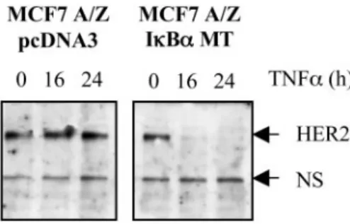
Both cell lines were stimulated with TNF-
␣ for up to 24 h. HER-2
expression was investigated by Western blot analysis and remained
constant in the control cell line. By contrast, an important decrease of
HER-2 levels was observed in the cell overexpressing the mutated
form of I
B␣ (Fig. 1).
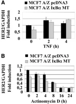
To determine the influence of transcriptional regulation on
de-creased HER-2 protein, MCF7 A/Z pcDNA3 or I
B␣ MT was treated
with TNF-
␣ for 2 or 4 h, and levels of HER-2 transcripts were
assessed by quantitative real-time PCR using HER-2-specific primers
and GAPDH housekeeping gene as controls. No difference in HER-2
mRNA levels was observed when both cell lines were stimulated by
TNF-
␣ (Fig. 2A). Moreover, TNF-␣ did not modify the transcriptional
activity on the HER-2 proximal promoter as judged by luciferase
assays (data not shown). To inhibit RNA neosynthesis, control and
I
B␣ MCF7 A/Z cells were preincubated with actinomycin D for 1 h
before a TNF-
␣ time course stimulation and real-time PCR
experi-ment were performed with HER-2 and GAPDH primers. We observed
that the degradation rate of HER-2 mRNA was identical in both cell
lines (Fig. 2B). Therefore, these results demonstrate that in our
ex-perimental conditions, TNF-
␣-mediated HER-2 down-regulation was
not the consequence of decreased RNA levels.
To investigate whether a difference in HER-2 protein stability was
involved in the TNF-
␣-mediated HER-2 disappearance in IB␣ MT
MCF7 cells, we first incubated both cell lines with CHX before
TNF-
␣ stimulation. HER-2 protein levels were assessed using
West-ern blot analysis. In both cell lines, HER-2 protein was not detectable
beyond 4 h of TNF-
␣ stimulation (Fig. 3A). However, HER-2 protein
is highly stable because we detected its expression
⬎24 h after CHX
alone in both cell lines (Fig. 3B and data not shown).
TNF-
␣-stimulated control cells then were preincubated with CHX
or BAY 11–7085, a NF
B inhibitor, and HER-2 protein levels were
assessed by Western blot analysis. In agreement with our previous
results, we observed a decrease in HER-2 protein levels in response to
TNF-
␣ only after NFB or protein synthesis inhibition (Fig. 3B),
indicating that an NF
B-dependent protein synthesis occurs in MCF7
A/Z control cells and counteracts the TNF-
␣-mediated HER-2
degra-dation.
Fig. 1. Tumor necrosis factor␣ (TNF-␣)-mediated HER-2 degradation in nuclear factorB (NFB)-defective cells. pcDNA3 or IB␣ MT MCF7 A/Z cells were treated with TNF-␣ (100 units/ml) for the indicated times. Ten g of total protein extracts were analyzed by Western blot analysis with a specific anti-HER-2 COOH-terminal antibody. A nonspecific band is shown as loading control.
HER-2 Cleavage by
␣-Activated Caspase-8 in NFB-Defective
Cells. We hypothesized that decreased HER-2 levels in response to
TNF-
␣ might be a consequence of protease activation. To test such a
hypothesis, we preincubated cells with protease inhibitors and tested
their ability to counteract the TNF-
␣-mediated HER-2 decay.
Phen-ylmethylsulfonyl fluoride (a serine protease inhibitor) and
hydroxam-ate (a specific inhibitor of some metalloproteases) did not prevent
HER-2 degradation in response to TNF-
␣ stimulation in cells
over-expressing the mutated I
B␣ (data not shown).
Because it has been demonstrated recently that HER-2 may be a
caspase substrate in geldanamycin and staurosporin-treated cells, we
investigated this pathway (53). We observed endogenous caspase-8
cleavage and activation following TNF-
␣ stimulation in MT cells and
CHX-pretreated control cells (Fig. 4, A and B), and the kinetics of this
activation perfectly paralleled HER-2 decay and PARP cleavage
(compare Fig. 4 and Fig. 5).
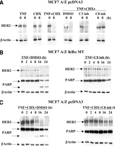
Moreover, z-IETD-fmk, a specific caspase-8 inhibitor, blocked
TNF-
␣- and CHX-induced HER-2 degradation in control cells,
whereas caspase-3 or caspase-9 inhibitors did not have any effect (Fig.
5A and data not shown). Furthermore, MCF7 A/Z MT cells or
CHX-pretreated control cells were stimulated with TNF-
␣ in the presence of
DMSO or z-IETD-fmk, and the kinetics of HER-2 protein degradation
and PARP cleavage were compared. Fig. 5, B and C, shows that in all
of our experimental conditions, HER-2 and PARP cleavages occurred
simultaneously and were inhibited similarly by the caspase-8
inhibi-tor. The efficiency of the z-IETD-fmk inhibitor was confirmed by
Western blot analysis revealed with a caspase-8-specific antibody
(data not shown).
Taken together, these data indicated that caspase-8 is responsible
for the TNF-
␣-induced HER-2 degradation in NFB-deficient cells.
Inhibition of Caspase-8-Mediated HER-2 Cleavage by NF
B-Regulated c-FLIP. Our data indicated that caspase-8-induced HER-2
cleavage only occurred in cells in which NF
B activation or protein
synthesis was inhibited. This cleavage is prevented in control cells
through a TNF-
␣-induced NFB pathway leading to the expression of
caspase-inhibiting proteins.
To identify the protein products of these NF
B-regulated genes,
control and MT cells were treated with TNF-
␣, and total cellular
lysates were analyzed by Western blot analysis revealed with specific
antibodies directed against well-known NF
B-regulated caspase
in-hibitors. As shown in Fig. 6, c-IAP-1, c-IAP-2, and XIAP protein
levels were similar in untreated or TNF-
␣-stimulated control or MT
MCF7 A/Z cells. However, c-FLIP-L protein amounts were increased
markedly in TNF-
␣-stimulated control cells, whereas the expression
of this protein decreased rapidly following treatment of MT cells
(Fig. 6).
Because c-FLIP-L is a well-known caspase-8 inhibitor and an
Fig. 2. HER-2 mRNA expression and stability. A, pcDNA3 or IB␣ MT MCF7 A/Zcells were stimulated by tumor necrosis factor␣ (TNF-␣; 100 units/ml) for the indicated times. Quantitative real-time PCR was performed using HER-2-specific oligonucleotides and glyceraldehyde-3-phosphate dehydrogenase (GAPDH) as internal control. B, control or MT cells were treated with actinomycin D (5g/ml) for the indicated times. Quanti-tative real-time PCR was performed as in A.
Fig. 3. HER-2 protein stability. A, control or MT cells were incubated with cyclohex-imide (CHX; 50g/ml) for 1 h before tumor necrosis factor ␣ (TNF-␣; 100 units/ml) time course stimulation. Teng of total cellular extract were subjected to anti-HER-2 Western blot analysis. B, pcDNA3 MCF7 A/Z cells were incubated up to 24 h with CHX (50
g/ml), BAY 11–7085 (10 M), or TNF-␣ (100 units/ml) either alone or in combination. For combined treatment, cells were preincubated with CHX or BAY 11–7085 for 1 h before TNF-␣ stimulation. Ten g of total cellular extracts were used for Western blot analysis with a specific anti-HER-2 antibody or an antiactin antibody for loading control.
Fig. 4. Caspase-8 cleavage and activation in response to tumor necrosis factor␣ (TNF-␣). A, caspase-8 cleavage. pcDNA3 MCF7 A/Z cells were stimulated with cyclo-heximide (CHX; 50g/ml) and TNF-␣ (100 units/ml; left) or TNF-␣ only (middle), and IB␣ MT MCF7 A/Z cells were stimulated with TNF-␣. Proteolysis of caspase-8 in total cellular extracts (10g) was assessed by Western blot analysis. Caspase-8 proform is indicated by an arrow, and asterisks show the cleaved fragments. B, caspase-8 activity. pcDNA3 MCF7 A/Z cells were stimulated with CHX (50g/ml) and TNF-␣ (100 units/ml) or TNF-␣ only, and IB␣ MT MCF7 A/Z cells were stimulated with TNF-␣ as indicated. Cellular extracts were incubated with a fluorogenic caspase-8 substrate. Fluo-rescence of the generated cleaved product was assessed with a spectrofluorometer. The results are expressed in fold activation compared with untreated cells. A representative experiment out of three reproducible measurements is represented.
NF
B target gene product, we investigated whether modulation of its
expression affects HER-2 cleavage (14, 54, 55). Specific antisense
oligonucleotides targeting c-flip mRNA efficiently decreased c-FLIP
protein expression in control cells, whereas scrambled or sense
oli-gonucleotides did not (Fig. 7A). The inhibition of c-FLIP expression
in control cells was correlated with TNF-
␣-induced PARP cleavage
and HER-2 degradation (Fig. 7A). Interestingly, the inhibition of
c-FLIP expression by the same antisense oligonucleotides also led to
TNF-
␣-induced HER-2 degradation in BT-474 and SKBR3 breast
cancer cells, which are known to overexpress HER-2 (Fig. 7B).
As additional evidence for c-FLIP-dependent inhibition of HER-2
cleavage, the opposite experiment was performed by reintroducing
c-FLIP-L exogenous transient expression in I
B␣ MT cells. We
demonstrated that such ectopic c-FLIP expression partially inhibited
TNF-
␣-mediated HER-2 cleavage (Fig. 8). IB␣ MT cells were
transfected with either an empty vector or a c-FLIP expression vector
and stimulated with TNF-
␣. Cellular extracts then were analyzed by
Western blot analysis for c-FLIP and HER-2 expression. In
mock-transfected cells (Fig. 8A), HER-2 completely disappeared after 16 h
of TNF-
␣ treatment, whereas it still could be observed in
c-FLIP-expressing cells (Fig. 8B).
These data clearly indicated that c-FLIP expression was induced
through NF
B activation in TNF-␣-stimulated control cells and that
this protein inhibited the caspase-8-mediated HER-2 cleavage.
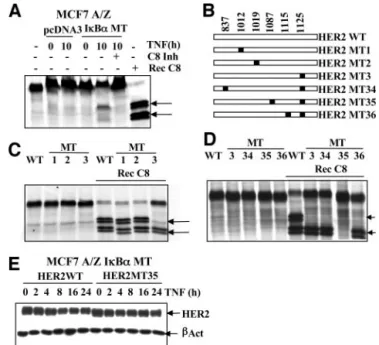
Identification of HER-2 Cleavage Sites Targeted by Caspase-8.
HER-2 degradation by caspase-8 did not generate any detectable
cleavage product on immunoblot analyses performed with cellular
extracts, even in the presence of proteasome inhibitors (data not
shown). Therefore, HER-2 cleavage also was investigated in vitro.
The HER-2 cytoplasmic domain was translated in vitro, and in vitro
proteolysis assays were performed in the presence of hypotonic
cel-lular extracts from control or MT cells, either untreated or TNF-
␣
stimulated for 10 h. As shown in Fig. 9A, S
35-labeled HER-2
COOH-terminus domain was cleaved in two fragments after incubation with
cellular extracts from TNF-
␣-treated IB␣ MT cells. This cleavage
was inhibited by preincubation of these extracts with the caspase-8
inhibitor, and similar cleavage fragments were generated after HER-2
COOH-terminus domain incubation with recombinant caspase-8
(Fig. 9A).
We localized six potential caspase cleavage sites in the HER-2
COOH-terminal domain, and the aspartate residues from these sites
were mutated into alanines (Fig. 9B). The mutants MT1, MT2, and
MT3 designated proteins where the residues 1012, 1019, and 1125
were mutated, respectively. In vitro proteolysis assays showed that the
D1125A (MT3) mutant was partially resistant to proteolysis by
re-combinant caspase-8 (Fig. 9C). Therefore, we proceeded with
addi-tional mutations and generated double mutants carrying the D1125A
Fig. 6. Expression of nuclear factor B (NFB)-controlled caspase-8 inhibitors. pcDNA3 and IB␣ MT MCF7 A/Z cells were stimulated with tumor necrosis factor ␣ (TNF-␣; 100 units/ml) for up to 24 h. Total extracts (10 g) were analyzed by Western blot analysis with anti-c-IAP-1, anti-c-IAP-2, anti-XIAP, anti-c-FLIP-L, and antiactin antibodies, respectively.
Fig. 7. Increased HER-2 cleavage by inhibition of c-FLIP expression. A, pcDNA3 MCF7 A/Z cells were transfected with c-FLIP-L antisense (AS), sense (S), or scrambled (SC) oligonucleotides. Eight h after transfection, cells were left untreated or were stim-ulated with tumor necrosis factor␣ (TNF-␣; 100 units/ml) for an additional 24 h. Total extracts (10g) were evaluated by Western blot analysis with anti-HER-2, anti-c-FLIP-L, anti-poly(ADP-ribose) polymerase (PARP), and antiactin antibodies, respectively. B, BT-474 and SKBR3, two HER-2-overexpressing cell lines, were handled as above. Total cellular extracts (5g) were submitted to an HER-2 and actin Western blot analysis. Fig. 5. HER-2 cleavage by caspase-8. A, pcDNA3 MCF7 A/Z cells were stimulated by
TNF-␣ (100 units/ml), cycloheximide (CHX; 50 ı`g/ml), or both for 8 h (left). CHX- and TNF-␣-costimulated cells were preincubated with a caspase-3- or caspase-8-specific inhibitor (20M) or with vehicle (DMSO) for 1 h (right). HER-2 expression was evaluated by Western blot analysis performed with total protein extracts (10g). B, IB␣ MT MCF7 A/Z cells were stimulated with tumor necrosis factor␣ (TNF-␣; 100 units/ml) for indicated times in the absence (left) or presence (right) of the caspase-8 inhibitor (20
M). Western blot analysis was carried out with 10-g total extracts. Proteins were probed with anti-HER-2, anti-poly(ADP-ribose) polymerase (PARP), and antiactin antibodies as indicated. C, pcDNA3 MCF7 A/Z cells were costimulated with TNF-␣ (100 units/ml) and CHX (50g/ml) for the indicated times in the absence (left) or presence (right) of caspase-8 inhibitor (20M). Western blot analysis was performed as described in B.
mutation plus a mutation of aspartate 837 (MT34), 1087 (MT35), or
1115 (MT36; Fig. 9B). The double-mutant MT35, carrying D1125A
and D1087A substitutions, was resistant completely to recombinant
caspase-8-induced proteolysis (Fig. 9D).
The wild-type and the HER-2 double mutants then were cloned into
a pcDNA3 expression vector, and we verified that the mutated form
was still uncleavable by recombinant caspase-8 during in vitro
pro-teolysis assay (data not shown). The HER-2 wild-type and MT35
double mutant then were expressed exogenously in MCF7 A/Z I
B␣
MT cells by transient transfection. Total cellular extracts from
TNF-␣-treated cells were evaluated by immunoblot analyses, and these
experiments showed that exogenously overexpressed wild-type
HER-2 was cleaved partially in response to TNF-
␣ treatment, whereas
the MT35 mutant was not (Fig. 9E).
Involvement of HER-2 Cleavage in TNF-
␣-Induced Cell Death.
To test the biological significance of these observations, I
B␣ MT
MCF7 A/Z cells were transfected transiently with an empty
expres-sion vector, an expresexpres-sion vector coding for the wild-type HER-2, or
with an expression vector coding for the MT35 HER-2. Cells then
were stimulated for 2 days with TNF-
␣ and living cells counted by
trypan blue exclusion. As shown in Fig. 10A, most control
pcDNA3-transfected cells were killed by TNF-
␣ treatment. Exogenously
ex-pressed wild-type HER-2 clearly protected MCF7 A/Z cells from
TNF-
␣-induced cytotoxicity because the proportion of living cells
was increased from 26 to 38% of the control. Interestingly, cell
survival was additionally and reproducibly increased following
exog-enous expression of the MT35 HER-2 mutant (49% of the untreated
control). These transfected cells also were pretreated with the
anti-HER-2 monoclonal antibody trastuzumab before TNF-
␣ stimulation.
This antibody clearly suppressed HER-2-mediated cytoprotection
against TNF-
␣-induced cell death because it reduced the percentages
of living cells to 26% after transfection of the wild-type HER-2 vector
and to 30% after transfection of the mutated HER-2 vector (Fig. 10A).
For this experiment, transduction efficiency was controlled by
trans-fecting in parallel an expression vector coding for the green
fluores-cent protein and counting positive cells by fluorescence-activated cell
sorting. In representative experiments,
⬃50% of the cells were
trans-fected (data not shown).
We checked that the increased living cell numbers were not related
to HER-2-induced proliferation by measuring thymidine
incorpora-tion following transfecincorpora-tion of MCF7 A/Z cells with the same
expres-sion vectors. As shown in Fig. 10B, there was not any significant
difference in thymidine incorporation between control and
HER-2-overexpressing cells.
These data proved that HER-2 expression protected I
B␣ MT cells
against TNF-
␣-induced cell death. Therefore, the TNF-␣-mediated
HER-2 cleavage in these cells may participate in the apoptotic
pathway.
DISCUSSION
The apoptosis relies on the activity of caspases, a growing family of
aspartyl-specific cysteine proteases that are essential for
death-recep-tor proximal events and the execution of apoptosis by cleavage of
broad-spectrum substrates. Among these “death substrates” are
mol-ecules involved in DNA repair like PARP, structural proteins, lamin,
and focal adhesion kinase (56), oncoproteins such as Bcl-2 (57), and
signaling proteins (58). Caspase-mediated cleavage of signaling
mol-ecules such as caspase-activated DNase (59, 60), STAT-1 (61), Raf-1,
Akt (62), and c-Abl (63) leads to either their activation or inactivation.
Because caspases are responsible for the onset of apoptosis,
identifi-cation of their cellular substrates is essential and provides insights into
the downstream events involved in apoptosis signaling. Among
po-tential caspase substrates, a recent study showed that geldanamycin or
staurosporin treatment of SKBR-3 human breast cancer cells led to
HER-2 proteolytic cleavage, which was inhibited partially by a
pleio-tropic caspase inhibitor (53). Our results clearly identified the
onco-protein HER-2 as a caspase-8 substrate. Interestingly, HER-2
cleav-age by caspase-8 occurred early after TNF-
␣ treatment, suggesting
that this cleavage might be involved in proapoptotic signal
pro-gression.
It was reported previously that HER-2 proteolytic fragments were
degraded rapidly but could be observed in the presence of proteasome
inhibitors (55). Nevertheless, in our experimental conditions,
frag-ments derived from HER-2 cleavage were not detectable even in the
presence of various protease inhibitors. Therefore, we decided to
investigate HER-2 cleavage by in vitro proteolysis assays. This
tech-nique allowed us to observe cleaved fragments, identify two sites
Fig. 8. Inhibition of HER-2 cleavage by c-FLIP expression. IB␣ MT MCF7 A/Z cellswere transfected with pcDNA3 (A) or c-FLIP-L (B) expression vectors. Twenty-four h after transfection, cells were stimulated with TNF-␣ (100 units/ml) for indicated times. Western blot analysis was performed with total extracts (10g) using anti-HER-2, anti-c-FLIP-L, and antiactin antibodies.
Fig. 9. Identification of the HER-2 cleavage sites. A, pcDNA3 and IB␣ MT MCF7 A/Z cells were stimulated with TNF-␣ (100 units/ml) for 10 h or left untreated, and hypotonic extracts were prepared.35S-labeled in vitro translated HER-2 COOH-terminus
was incubated at 37°C for 2 h with either recombinant caspase-8 or hypotonic extracts in the presence or absence of caspase-8 inhibitor as indicated. Samples then were separated using SDS-PAGE. Cleaved bands are marked with an arrow. B, schematic representation of the in vitro mutagenesis performed using the HER-2 COOH-terminus template. White rectangles represent the COOH-terminal part of HER-2. Black squares and numbers indicate the aspartate to alanine substitutions. C and D, wild-type (WT), single-mutant (MT; C), or double-mutant (D)35S-labeled in vitro translated HER-2 C-termini were
loaded untreated or were incubated with recombinant caspase-8 for 2 h at 37°C. Samples then were separated using SDS-PAGE. Arrows indicate the cleaved bands. E, IB␣ MT MCF7 A/Z cells were transfected with expression vectors encoding wild-type HER-2 (HER-2WT) or the double-mutant 35 (HER-2MT35). Eight h after transfection, cells were stimulated by TNF-␣ (100 units/ml), and total extracts (10 g) were analyzed by Western blot analysis using specific anti-HER-2 and antiactin antibodies.
targeted by caspase-8, and generate a mutant resistant to proteolytic
cleavage in vitro and in cells.
It was shown previously that TNF-
␣ treatment led to a decrease in
HER-2 mRNA synthesis (64) or in protein level expression (65). We
did not observe any effect of TNF-
␣ on HER-2 transcription level.
These discrepancies probably rely on differences in cellular models.
Nevertheless, for the first time, we report a caspase-dependent
cleav-age of the HER-2 protein following TNF-
␣ treatment.
HER-2 expression enhances proliferative, metastatic, and
prosur-vival signals in breast cancer cells and is correlated with a poor
prognosis in breast and ovarian adenocarcinomas (39 – 41).
Further-more, HER-2 expression induces a resistance to cancer therapy, and
its inhibition by a specific antibody, trastuzumab, has clinical activity
either alone or in combination with chemotherapy in
HER-2-express-ing breast cancers (45– 49). Moreover, it has been demonstrated that
HER-2 overexpression can induce resistance to TNF-
␣ stimulation
(43) and that trastuzumab can restore the cytotoxic response (50). Our
results raise the hypothesis that a caspase-8-mediated HER-2 cleavage
following TNF-
␣ stimulation could participate in the subsequent
apoptosis. Overexpression of a wild-type HER-2 protein significantly
inhibits TNF-
␣-induced cell death, and survival is further increased
following overexpression of an uncleavable HER-2 protein,
demon-strating that HER-2 cleavage contributes to apoptosis.
HER-2 expression inhibits cell death by inducing antiapoptotic
pathways, such as Bcl-2 and Bcl-XL up-regulation (66), or by
acti-vation of the Akt/NF
B prosurvival cascade (67–69). NFB is an
extensively described antiapoptotic transcription factor whose nuclear
DNA binding is potently and rapidly induced by TNF-
␣ in almost all
of the cell lines (70). Constitutive NF
B activation has been observed
in a wide variety of cancers and is associated with a resistance to
apoptosis because many of its target genes code for antiapoptotic
molecules (10 –25). In our model, TNF-
␣ stimulation leads to NFB
activation and subsequent expression of one of its target genes,
c-FLIP (14, 54). c-FLIP is a caspase-8 inhibitor and therefore
coun-teracts HER-2 cleavage triggered by TNF-
␣ stimulation (55). This
c-FLIP-mediated cleavage inhibition does not occur when NF
B
activity is blocked by I
B␣ MT expression or a chemical inhibitor
like BAY 11–7085, demonstrating that it requires the integrity of the
NF
B pathway.
Because we demonstrated that HER-2 cleavage might participate in
TNF-
␣-induced apoptosis, our results suggest that NFB-dependent
inhibition of HER-2 cleavage is a novel mechanism for NF
B
anti-apoptotic role. Interestingly, we also observed a HER-2 cleavage in
response to daunorubicin through an NF
B-independent mechanism,
suggesting that this cleavage could be a general feature of apoptosis.
3These results currently are under investigation.
In conclusion, we provide evidence, for the first time, that
caspase-8 cleaves the HER-2 oncoprotein in response to TNF-
␣
stimulation, and we propose an original model in which NF
B exerts
a new antiapoptotic function through c-FLIP-induced expression and
subsequent inhibition of TNF-
␣-triggered cleavage of the HER-2
survival factor (Fig. 11). Therefore, this antiapoptotic mechanism
3Unpublished observations.
Fig. 10. HER-2 expression and cell survival. A, IB␣ MT MCF7 A/Z cells were transfected with an empty vector or expression vectors encoding wild-type HER-2 (HER-2WT) or the double mutant (HER-2MT35). Eight h after transfection, cells were treated with TNF-␣ for 24 h, and a trypan blue exclusion counting of the living cells was performed. In the same experiment, cells also were pretreated for 24 h with the trastu-zumab monoclonal antibody before TNF-␣ stimulation, as indicated in the figure. The data are representative of three independent experiments and are expressed as percentage of the values observed with control cells. B, IB␣ MT MCF7 A/Z cells were transfected as in A or left untransfected (T0). Twenty-four h after transfection, [H3]thymidine was
added in culture medium for 5 h. [H3]thymidine incorporation then was evaluated and
expressed as percentage of the values observed in the control cells.
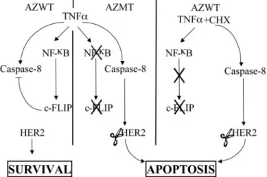
Fig. 11. A new model for proapoptotic and antiapoptotic pathways in response to tumor necrosis factor␣ (TNF-␣). In TNF-␣-stimulated MCF7 A/Z cells, nuclear factor B (NFB) activation leads to c-FLIP expression, inhibition of the caspase-8-mediated HER-2 cleavage, and cell survival. In contrast, in IB␣ MT and cycloheximide (CHX)-pretreated pcDNA3 MCF7 A/Z cells, c-FLIP expression in response to TNF-␣ is pre-vented by NFB or protein synthesis inhibition, and TNF-␣ stimulation triggers caspase-8 activation, subsequent HER-2 cleavage, and apoptosis.
underlines the opportunity to evaluate the NF
B activation status and
the HER-2 expression level in breast cancer cells and opens new ways
to develop combined anticancer therapies for HER-2-overexpressing
cancers. Because NF
B inhibitors currently are being evaluated as
anticancer therapeutic agents, our results indicate a putative novel
activity for these agents. Anti-HER-2 antibodies and NF
B inhibitors
possibly may be combined with chemotherapy or biological modifiers
for the management of HER-2-overexpressing cancers. It also would
be most interesting to determine whether a similar effect could be
observed with other receptors belonging to the EGFR family.
ACKNOWLEDGMENTS
We thank Dr. Jacques Piette for the critical reading of the manuscript.
REFERENCES
1. Barbara JA, Smith WB, Gamble JR, et al. Dissociation of TNF-␣ cytotoxic and proinflammatory activities by p55 receptor- and p75 receptor-selective TNF-␣ mu-tants. EMBO J 1994;13:843–50.
2. Lewis M, Tartaglia LA, Lee A, et al. Cloning and expression of cDNAs for two distinct murine tumor necrosis factor receptors demonstrate one receptor is species specific. Proc Natl Acad Sci USA 1991;88:2830 – 4.
3. Heller RA, Song K, Fan N, Chang DJ. The p70 tumor necrosis factor receptor mediates cytotoxicity. Cell 1992;70:47–56.
4. Loetscher H, Pan YC, Lahm HW, et al. Molecular cloning and expression of the human 55 kd tumor necrosis factor receptor. Cell 1990;61:351–9.
5. Schall TJ, Lewis M, Koller KJ, et al. Molecular cloning and expression of a receptor for human tumor necrosis factor. Cell 1990;61:361–70.
6. Karin M, Lin A. NF-B at the crossroads of life and death. Nat Immunol 2002;3: 221–7.
7. Sartorius U, Schmitz I, Krammer PH. Molecular mechanisms of death-receptor-mediated apoptosis. Chembiochem 2001;2:20 –9.
8. Baud V, Karin M. Signal transduction by tumor necrosis factor and its relatives. Trends Cell Biol 2001;11:372–7.
9. Rath PC, Aggarwal BB. TNF-induced signaling in apoptosis. J Clin Immunol 1999; 19:350 – 64.
10. Wang CY, Mayo MW, Korneluk RG, Goeddel DV, Baldwin AS Jr. NF-B antiapo-ptosis: induction of TRAF1 and TRAF2 and c-IAP1 and c-IAP2 to suppress caspase-8 activation. Science 1998;281:1680 –3.
11. Chu ZL, McKinsey TA, Liu L, Gentry JJ, Malim MH, Ballard DW. Suppression of tumor necrosis factor-induced cell death by inhibitor of apoptosis c-IAP2 is under NF-B control. Proc Natl Acad Sci USA 1997;94:10057–62.
12. Hong SY, Yoon WH, Park JH, Kang SG, Ahn JH, Lee TH. Involvement of two NF-B binding elements in tumor necrosis factor ␣-, CD40-, and Epstein-Barr virus latent membrane protein 1-mediated induction of the cellular inhibitor of apoptosis protein 2 gene. J Biol Chem 2000;275:18022– 8.
13. Stehlik C, de Martin R, Kumabashiri I, Schmid JA, Binder BR, Lipp J. Nuclear factor (NF)-B-regulated X-chromosome-linked iap gene expression protects endothelial cells from tumor necrosis factor␣-induced apoptosis. J Exp Med 1998;188:211–6. 14. Kreuz S, Siegmund D, Scheurich P, Wajant H. NF-B inducers upregulate cFLIP, a cycloheximide-sensitive inhibitor of death receptor signaling. Mol Cell Biol 2001; 21:3964 –73.
15. Grumont RJ, Rourke IJ, Gerondakis S. Rel-dependent induction of A1 transcription is required to protect B cells from antigen receptor ligation-induced apoptosis. Genes Dev 1999;13:400 –11.
16. Lee HH, Dadgostar H, Cheng Q, Shu J, Cheng G. NF-B-mediated up-regulation of Bcl-x and Bfl-1/A1 is required for CD40 survival signaling in B lymphocytes. Proc Natl Acad Sci USA 1999;96:9136 – 41.
17. Zong WX, Edelstein LC, Chen C, Bash J, Gelinas C. The prosurvival Bcl-2 homolog Bfl-1/A1 is a direct transcriptional target of NF-B that blocks TNF␣-induced apoptosis. Genes Dev 1999;13:382–7.
18. Khoshnan A, Tindell C, Laux I, Bae D, Bennett B, Nel AE. The NF-B cascade is important in Bcl-xL expression and for the anti-apoptotic effects of the CD28 receptor in primary human CD4⫹ lymphocytes. J Immunol 2000;165:1743–54.
19. Chen C, Edelstein LC, Gelinas C. The Rel/NF-B family directly activates expression of the apoptosis inhibitor Bcl-x(L). Mol Cell Biol 2000;20:2687–95.
20. Krikos A, Laherty CD, Dixit VM. Transcriptional activation of the tumor necrosis factor␣-inducible zinc finger protein, A20, is mediated by B elements. J Biol Chem 1992;267:17971– 6.
21. Sarma V, Lin Z, Clark L, et al. Activation of the B-cell surface receptor CD40 induces A20, a novel zinc finger protein that inhibits apoptosis. J Biol Chem 1995;270: 12343– 6.
22. Jaattela M, Mouritzen H, Elling F, Bastholm L. A20 zinc finger protein inhibits TNF and IL-1 signaling. J Immunol 1996;156:1166 –73.
23. Jones PL, Ping D, Boss JM. Tumor necrosis factor␣ and interleukin-1 regulate the murine manganese superoxide dismutase gene through a complex intronic enhancer involving C/EBP- and NF-B. Mol Cell Biol 1997;17:6970–81.
24. Delhalle S, Deregowski V, Benoit V, Merville MP, Bours V. NF-B-dependent MnSOD expression protects adenocarcinoma cells from TNF-␣-induced apoptosis. Oncogene 2002;21:3917–24.
25. Bernard D, Monte D, Vandenbunder B, Abbadie C. The c-Rel transcription factor can both induce and inhibit apoptosis in the same cells via the upregulation of MnSOD. Oncogene 2002;21:4392– 402.
26. Beg AA, Baltimore D. An essential role for NF-B in preventing TNF-␣-induced cell death. Science 1996;274:782– 4.
27. Wang CY, Mayo MW, Baldwin AS Jr. TNF- and cancer therapy-induced apoptosis: potentiation by inhibition of NF-B. Science 1996;274:784–7.
28. Van Antwerp DJ, Martin SJ, Kafri T, Green DR, Verma IM. Suppression of
TNF-␣-induced apoptosis by NF-B. Science 1996;274:787–9.
29. Krammer PH. CD95(APO-1/Fas)-mediated apoptosis: live and let die. Adv Immunol 1999;71:163–210.
30. Coussens L, Yang-Feng TL, Liao YC, et al. Tyrosine kinase receptor with extensive homology to EGF receptor shares chromosomal location with neu oncogene. Science 1985;230:1132–9.
31. Slamon DJ, Godolphin W, Jones LA, et al. Studies of the HER-2/neu proto-oncogene in human breast and ovarian cancer. Science 1989;244:707–12.
32. Press MF, Pike MC, Chazin VR, et al. Her-2/neu expression in node-negative breast cancer: direct tissue quantitation by computerized image analysis and association of overexpression with increased risk of recurrent disease. Cancer Res 1993;53: 4960 –70.
33. Salomon DS, Brandt R, Ciardiello F, Normanno N. Epidermal growth factor-related peptides and their receptors in human malignancies. Crit Rev Oncol Hematol 1995; 19:183–232.
34. Tyson FL, Boyer CM, Kaufman R, et al. Expression and amplification of the HER-2/neu (c-erbB-2) protooncogene in epithelial ovarian tumors and cell lines. Am J Obstet Gynecol 1991;165:640 – 6.
35. Zhang X, Silva E, Gershenson D, Hung MC. Amplification and rearrangement of c-erb B proto-oncogenes in cancer of human female genital tract. Oncogene 1989;4: 985–9.
36. Berchuck A, Kamel A, Whitaker R, et al. Overexpression of HER-2/neu is associated with poor survival in advanced epithelial ovarian cancer. Cancer Res 1990;50:4087– 91.
37. Ross JS, Fletcher JA. The HER-2/neu oncogene in breast cancer: prognostic factor, predictive factor, and target for therapy. Stem Cells 1998;16:413–28.
38. Slamon DJ, Clark GM, Wong SG, Levin WJ, Ullrich A, McGuire WL. Human breast cancer: correlation of relapse and survival with amplification of the HER-2/neu oncogene. Science 1987;235:177–182.
39. Hung MC, Zhang X, Yan DH, et al. Aberrant expression of the c-erbB-2/neu protooncogene in ovarian cancer. Cancer Lett 1992;61:95–103.
40. Ignatoski KM, Maehama T, Markwart SM, Dixon JE, Livant DL, Ethier SP. ERBB-2 overexpression confers PI 3⬘ kinase-dependent invasion capacity on human mammary epithelial cells. Br J Cancer 2000;82:666 –74.
41. Tzahar E, Yarden Y. The ErbB-2/HER2 oncogenic receptor of adenocarcinomas: from orphanhood to multiple stromal ligands. Biochim Biophys Acta 1998;1377: M25–37.
42. Yu D, Jing T, Liu B, et al. Overexpression of ErbB2 blocks Taxol-induced apoptosis by upregulation of p21Cip1, which inhibits p34Cdc2 kinase. Mol Cell 1998;2: 581–91.
43. Hudziak RM, Lewis GD, Shalaby MR, et al. Amplified expression of the HER2/ ERBB2 oncogene induces resistance to tumor necrosis factor␣ in NIH 3T3 cells. Proc Natl Acad Sci USA 1988;85:5102– 6.
44. Egeblad M, Jaattela M. Cell death induced by TNF or serum starvation is independent of ErbB receptor signaling in MCF-7 breast carcinoma cells. Int J Cancer 2000;86: 617–25.
45. Sliwkowski MX, Lofgren JA, Lewis GD, Hotaling TE, Fendly BM, Fox JA. Non-clinical studies addressing the mechanism of action of trastuzumab (Herceptin). Semin Oncol 1999;26:60 –70.
46. Baselga J, Norton L, Albanell J, Kim YM, Mendelsohn J. Recombinant humanized anti-HER2 antibody (Herceptin) enhances the antitumor activity of paclitaxel and doxorubicin against HER2/neu overexpressing human breast cancer xenografts. Can-cer Res 1998;58:2825–31.
47. Baselga J, Tripathy D, Mendelsohn J, et al. Phase II study of weekly intravenous trastuzumab (Herceptin) in patients with HER2/neu-overexpressing metastatic breast cancer. Semin Oncol 1999;26:78 – 83.
48. Pegram MD, Slamon DJ. Combination therapy with trastuzumab (Herceptin) and cisplatin for chemoresistant metastatic breast cancer: evidence for receptor-enhanced chemosensitivity. Semin Oncol 1999;26:89 –95.
49. Pegram MD, Lipton A, Hayes DF, et al. Phase II study of receptor-enhanced chemosensitivity using recombinant humanized anti-p185HER2/neu monoclonal an-tibody plus cisplatin in patients with HER2/neu-overexpressing metastatic breast cancer refractory to chemotherapy treatment. J Clin Oncol 1998;16:2659 –71. 50. Hudziak RM, Lewis GD, Winget M, Fendly BM, Shepard HM, Ullrich A. p185HER2
monoclonal antibody has antiproliferative effects in vitro and sensitizes human breast tumor cells to tumor necrosis factor. Mol Cell Biol 1989;9:1165–72.
51. Tang G, Yang J, Minemoto Y, Lin A. Blocking caspase-3-mediated proteolysis of IKK suppresses TNF-␣-induced apoptosis. Mol Cell 2001;8:1005–16.
52. Delhalle S, Deregowski V, Benoit V, Merville MP, Bours V. NF-B-dependent MnSOD expression protects adenocarcinoma cells from TNF-␣-induced apoptosis. Oncogene 2002;21:3917–24.
53. Tikhomirov O, Carpenter G. Caspase-dependent cleavage of ErbB-2 by geldanamycin and staurosporin. J Biol Chem 2001;276:33675– 80.
54. Micheau O, Lens S, Gaide O, Alevizopoulos K, Tschopp J. NF-B signals induce the expression of c-FLIP. Mol Cell Biol 2001;21:5299 –305.
55. Krueger A, Baumann S, Krammer PH, Kirchhoff S. FLICE-inhibitory proteins: regulators of death receptor-mediated apoptosis. Mol Cell Biol 2001;21:8247–54. 56. Wen LP, Fahrni JA, Troie S, Guan JL, Orth K, Rosen GD. Cleavage of focal adhesion
kinase by caspases during apoptosis. J Biol Chem 1997;272:26056 – 61.
57. Kirsch DG, Doseff A, Chau BN, et al. Caspase-3-dependent cleavage of Bcl-2 promotes release of cytochrome c. J Biol Chem 1999;274:21155– 61.
58. Nicholson DW. Caspase structure, proteolytic substrates, and function during apo-ptotic cell death. Cell Death Differ 1999;6:1028 – 42.
59. Sakahira H, Enari M, Nagata S. Cleavage of CAD inhibitor in CAD activation and DNA degradation during apoptosis. Nature 1998;391:96 –9.
60. Enari M, Sakahira H, Yokoyama H, Okawa K, Iwamatsu A, Nagata S. A caspase-activated DNase that degrades DNA during apoptosis, and its inhibitor ICAD. Nature 1998;391:43–50.
61. King P, Goodbourn S. STAT1 is inactivated by a caspase. J Biol Chem 1998;273: 8699 –704.
62. Widmann C, Gibson S, Johnson GL. Caspase-dependent cleavage of signaling pro-teins during apoptosis. A turn-off mechanism for anti-apoptotic signals. J Biol Chem 1998;273:7141–7.
63. Barila D, Rufini A, Condo II, et al. Caspase-dependent cleavage of c-Abl contributes to apoptosis. Mol Cell Biol 2003;23:2790 –9.
64. Kalthoff H, Roeder C, Gieseking J, Humburg I, Schmiegel W. Inverse regulation of human ERBB2 and epidermal growth factor receptors by tumor necrosis factor␣. Proc Natl Acad Sci USA 1993;90:8972– 6.
65. Kumar R, Mendelsohn J. Reduced expression of c-erbB2 gene product in human mammary carcinoma SK-BR-3 cells treated with interferon-␥ and tumor necrosis factor-␣. Anticancer Res 1994;14:1001–8.
66. Kumar R, Mandal M, Lipton A, Harvey H, Thompson CB. Overexpression of HER2 modulates bcl-2, bcl-XL, and tamoxifen-induced apoptosis in human MCF-7 breast cancer cells. Clin Cancer Res 1996;2:1215–9.
67. Zhou BP, Hu MC, Miller SA, et al. HER-2/neu blocks tumor necrosis factor-induced apoptosis via the Akt/NF-B pathway. J Biol Chem 2000;275:8027–31. 68. Pianetti S, Arsura M, Romieu-Mourez R, Coffey RJ, Sonenshein GE. Her-2/neu
overexpression induces NF-B via a PI3-kinase/Akt pathway involving calpain-mediated degradation of IB-␣ that can be inhibited by the tumor suppressor PTEN. Oncogene 2001;20:1287–99.
69. Bhat-Nakshatri P, Sweeney CJ, Nakshatri H. Identification of signal transduction pathways involved in constitutive NF-B activation in breast cancer cells. Oncogene 2002;21:2066 –78.
70. Osborn L, Kunkel S, Nabel GJ. Tumor necrosis factor␣ and interleukin 1 stimulate the human immunodeficiency virus enhancer by activation of the nuclear factorB. Proc Natl Acad Sci USA 1989;86:2336 – 40.