https://doi.org/10.1007/s00415-018-8849-0
ORIGINAL COMMUNICATION
Resistance to eye opening in patients with disorders of consciousness
Hjalmar Jochem van Ommen1,2 · Aurore Thibaut1,3 · Audrey Vanhaudenhuyse1,4 · Lizette Heine1,5 · Vanessa Charland‑Verville1 · Sarah Wannez1 · Olivier Bodart1 · Steven Laureys1 · Olivia Gosseries1 Received: 16 November 2017 / Revised: 11 March 2018 / Accepted: 26 March 2018 / Published online: 5 April 2018
© Springer-Verlag GmbH Germany, part of Springer Nature 2018
Abstract
Introduction Resistance to eye opening (REO) is a commonly encountered phenomenon in clinical practice. We aim to investigate whether REO is a sign of consciousness or a reflex in severely brain-injured patients.
Methods We recorded REO in chronic patients with disorders of consciousness during a multimodal diagnostic assess-ment. REO evaluations were performed daily in each patient and clinical diagnosis of unresponsive wakefulness syndrome (UWS), minimally conscious state with (MCS+) or without (MCS−) preserved language processing was made using the Coma Recovery Scale-Revised (CRS-R).
Results Out of 150 consecutive patients, 79 patients fit inclusion criteria. REO was seen in 19 patients (24.1%). At the group level, there was a significant relationship between the presence of REO and the level of consciousness. We also observed a difference in the repeatability of REO between patients in UWS, MCS− and MCS+. Out of 23 patients in UWS, six showed REO, in whom five showed atypical brain patterns activation.
Conclusion Our findings suggest a voluntary basis for REO and stress the need for multiple serial assessments of REO in these patients, especially since most patients show fluctuating levels of consciousness.
Keywords Disorders of consciousness · Resistance to eye opening · Unresponsive wakefulness syndrome · Minimally conscious state
Introduction
Resistance to eye opening (REO) concerns the strong clo-sure of the already closed eyelids by a patient while trying to open the eyes for the ophthalmological examination. It was first described by Charles Miller Fisher [1] and is often
seen in conversion disorders such as psychiatric coma and non-epileptic seizures [2, 3]. Although it is usually consid-ered a voluntary, albeit motiveless, response comparable to gegenhalten [4], it has also been considered to be a reflex in reports of anoxic and ischemic basal nuclei pathology [5,
6], brainstem lesions due to stroke or demyelination [7], and even paraneoplastic syndromes [8, 9]. Furthermore, reflex closure of one eye upon touch of the eyelid has also been described in preterm born children [10]. The hyperexcitable orbicularis oculi (or glabellar tap) reflex and the blink reflex in untreated Parkinson’s disease [11] have historically been considered identical to the resistance to eye opening that Miller Fisher described [12]. Thus, there might be two dif-ferent presentations of REO, namely voluntary and reflexive. At present, empirical data regarding REO in chronic dis-orders of consciousness (DOC) such as the unresponsive wakefulness syndrome (UWS, also called vegetative state [13]) and the minimally conscious state (MCS [14]) have not been studied. UWS patients only show reflexive behav-iors whereas patients in MCS show fluctuating but repro-ducible signs of consciousness such as visual pursuit and
* Olivia Gosseries ogosseries@uliege.be
1 GIGA-Consciousness, Coma Science Group, and Neurology
Department, University Hospital of Liege, University of Liege, Avenue de l’Hopital, 1, Liege, Belgium
2 Department of Intensive Care, MC Slotervaart, Amsterdam,
The Netherlands
3 Spaulding Neuromodulation Center, Spaulding Rehabilitation
Hospital, Harvard Medical School, Boston, MA, USA
4 GIGA-Consciousness, Department of Algology
and Palliative Care, University Hospital of Liege, University of Liege, Liege, Belgium
5 Auditory Cognition and Psychoacoustics Team, Lyon
responses to command. Patients who show behaviors such as visual pursuit or automatic motor reactions, are considered MCS− while patients who show signs of preserved language processing are considered MCS+ [15]. Misdiagnosis among these disorders is frequent and can lead to serious medical and ethical issues [16].
The aim of the present study is to investigate if REO is present in patients in chronic UWS, MCS− and MCS+ and if so, whether it can help to distinguish between these states. Our test hypothesis is that it should be possible to distinguish between reflex and voluntary: in the case of a volitional response, REO should be seen repeatedly during multiple assessments in patients with higher levels of consciousness.
Methods
During a period of 28 months, we selected all patients over 16 years of age in UWS, MCS− and MCS+ who were admitted to our hospital for a multimodal diagnostic workup (including positron emission tomography and magnetic reso-nance imaging scanning). Patients were excluded when in the acute phase (i.e., within 30 days of brain injury), diag-nosed other than DOC, were sedated, medically unstable, or could not be assessed more than once to assess repeat-ability. The diagnosis was based on repeated assessments with the Coma Recovery Scale-Revised (CRS-R) [17], in which patients are tested extensively on different clinical subscales of consciousness, namely auditory, visual, motor and oromotor/verbal functions, communication and arousal.
REO was defined as a forceful closure of one or both eyes upon manual opening of the upper eyelid on either side by the examiner [1]. Assessments of CRS-R and the presence of REO were performed daily by experienced neuropsycholo-gists. The repeatability of REO was defined as the number of CRS-R assessments in which REO was observed divided by the total number of CRS-R assessments. Differences in the presence and repeatability of REO between the UWS, MCS− and MCS+ patient groups were examined using Chi square, Kruskal–Wallis H and Mann–Whitney U tests. Two-tailed p values of < 0.05 were considered significant. For statistical analysis, IBM SPSS version 19.0 was used (IBM Belgium).
Most patients underwent a positron emission tomography that was performed and analysed as in our previous studies [18, 19]. Briefly, patients were scanned after the injection of fluorodeoxyglucose and brain areas showing significantly preserved or decreased metabolism were detected in each patient compared to a group of healthy controls. Typi-cally, patients in MCS show preserved metabolic activity in the associative fronto-parietal network, while patients in UWS show no preserved metabolism in this network [19]. A few patients also underwent a magnetic resonance
imaging during which they were instructed to perform men-tal imagery tasks, done as previously published [20]. Clini-cal reports of these neuroimaging findings were reviewed and compared to the REO data. The study was approved by the Ethics Committee of the Medical School of the Univer-sity of Liège and informed consents were obtained from the patient’s legal surrogates.
Results
Of a total of 150 screened patients, 79 patients (36 females, median age 37, interquartile range (IQR) 28–52 years) fit the inclusion criteria. The median interval between brain injury and assessment was 15 months (IQR 1–41 months). Etiolo-gies were traumatic brain injury (TBI) (n = 42, 53%), post-anoxic encephalopathy due to cardiac arrest (n = 25, 32%), ischemic or hemorrhagic stroke (n = 6, 8%), subarachnoid hemorrhage (n = 5, 6%), encephalitis (n = 2, 3%), and hypo-glycemia (n = 1, 1%). Two patients had suffered from cardiac arrest after traumatic brain injury. Of these 79 patients, 23 were diagnosed in UWS (29%), 15 in MCS− (19%) and 41 in MCS+ (52%).
REO was observed in 19 out of 79 patients (24.1%) during one or more behavioral assessments: six out of 23 patients in UWS (26%), eight out of 15 patients in MCS− (53%) and five out of 41 patients in MCS+ (12%) (Table 1). The Chi-square test of independence showed a significant relationship between the presence of REO and the level of consciousness (χ2 = 10.25, df = 2, p = 0.006). There was no difference in the
presence of REO between patients with and without TBI (χ2 = 1.23, df = 1, p = 0.27).
A Kruskal–Wallis H test showed that there was a sta-tistically significant difference in the repeatability of REO between patients in UWS, MCS− and MCS+ (χ2 = 6.01,
df = 2, p = 0.049, with a mean rank repeatability of 9.8%
in UWS patients, 7.1% in MCS− patients and 14.9% in MCS+ patients). Performing Mann–Whitney U post hoc tests revealed that there was a significant difference between patients in MCS− and MCS+ (Z = − 2.21, p = 0.027), while repeatability of REO between UWS and MCS− and between UWS and MCS+ did not differ (Z = − 1.10, p = 0.27, and
Z = − 1.74, p = 0.082, respectively, see Fig. 1).
Among the six patients in UWS with REO, five showed neuroimaging results that were more compatible with the diagnosis of MCS (Fig. 2). Using positron emission tomog-raphy, four patients presented relatively preserved metab-olism in the fronto-parietal network (Fig. 2a) (as usually observed in patients in MCS) and one patient was able to respond to command using a motor imagery task (i.e., imag-ine playing tennis) when assessed with functional magnetic resonance imaging, which indicates that this patient pre-sented a cognitive-motor dissociation (Fig. 2b) [19–21].
Only one of these patients recovered from UWS on re-exam-ination 6 months later (two passed away and three remained clinically in UWS). Of the patients in UWS without REO, 5 out of 17 also showed atypical brain pattern that was more compatible with the diagnosis of MCS, which is in line with
previous studies [19]. This atypical brain metabolism was thus seen more often in UWS patients with REO (83%) than without REO (29%) (χ2 = 5.25, df = 1, p = 0.02).
Discussion
In this study, REO was observed in about one out of four patients with disorders of consciousness, which is quite fre-quent considering how scarce the literature is on this subject. It is, therefore, important to know how to interpret this find-ing in this challengfind-ing population. In their landmark book on disorders of consciousness, Plum and Posner make a distinction between voluntary and reflex REO, as they state that in the assessment of comatose patients “strong resist-ance to eyelid opening and then rapid closure, is usually voluntary, suggesting that the patient is not truly comatose. However, lethargic patients with either metabolic or struc-tural lesions may resist eye opening, as do some patients with a non-dominant parietal lobe infarct” [22]. This diffi-culty is confirmed by our results in patients in DOC, where the clinical finding of REO at the individual level cannot reliably distinguish between voluntary or reflex behavior (and thus conscious versus unconscious patients), since 6 out of 23 UWS patients presented REO. CRS-R assessments provided a clear diagnosis of UWS in all six patients, but,
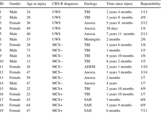
Table 1 Characteristics of the patients displaying resistance to eye opening
Repeatability is defined by REO observed/total number of assessments
CRS-R Coma Recovery Scale—revised, UWS unresponsive wakefulness syndrome, MCS− minimally
con-scious state minus, MCS+ minimally concon-scious state plus, TBI traumatic brain injury, ADEM acute demy-elinating encephalomyelitis, SAH (aneurysmal) subarachnoid hemorrhage
ID Gender Age at injury CRS-R diagnosis Etiology Time since injury Repeatability
1 Male 24 UWS TBI 2 years 4 months 1/12
2 Male 28 UWS TBI 3 years 0 months 4/9
3 Female 36 UWS Anoxia 5 years 8 months 2/12
4 Female 49 UWS Anoxia 38 days 1/4
5 Male 40 UWS Anoxia 7 years 11 months 2/13
6 Male 53 UWS Meningitis 2 months 2/6
7 Female 28 MCS− TBI 1 years 6 months 1/8
8 Male 73 MCS− TBI 1 months 1/5
9 Male 34 MCS− TBI 8 years 10 months 1/6
10 Male 13 MCS− TBI 8 years 2 months 1/5
11 Female 26 MCS− ADEM 2 years 1 months 1/10
12 Female 47 MCS− Anoxia 1 years 3 months 3/14
13 Female 59 MCS− Anoxia 2 months 1/7
14 Male 47 MCS− Anoxia 4 years 1/7
15 Male 32 MCS+ TBI 2 years 10 months 4/9
16 Female 22 MCS+ TBI 1 years 10 months 1/7
17 Female 43 MCS+ SAH 3 months 6/8
18 Female 64 MCS+ SAH 3 years 9 months 6/9
19 Female 47 MCS+ SAH 6 months 7/11
Fig. 1 Repeatability of resistance to eye opening between the three different states of consciousness. Values are expressed as medians and interquartile range (IQR). UWS unresponsive wakefulness syn-drome, MCS− minimally conscious state minus, MCS+ minimally conscious state plus
interestingly, neuroimaging results showed atypical brain activation in 5 of these patients (as well as 5 out of 17 UWS patients without REO). For such UWS patients who show a dissociation between behavior and brain results, the diag-nosis of MCS* has been suggested [18, 23], and for patients who show specific brain activation during mental imagery tasks, the diagnosis of cognitive-motor dissociation has been proposed [21]. Regarding REO outcome, only one of these patients in UWS recovered signs of consciousness 6 months post-evaluation, which indicates that there is no prognostic value of REO in chronic patients in UWS in the present dataset.
At the group level, we, however, observed a significant relationship between the presence of REO and the level of consciousness. REO repeatability was also the highest in patients in MCS+, suggesting a link between the number of times REO is seen and the level of consciousness. This is comparable to the way the CRS-R is used, where consistent or reproducible findings of even one specific behavior are enough to indicate a certain level of consciousness. Our find-ings, therefore, suggest a voluntary basis for REO when it is seen multiple times in a single patient, and stress the need to assess REO repeatedly in DOC patients, especially since
most patients with chronic DOC have fluctuating levels of consciousness.
Voluntary forceful eye closure is mediated by sensory input through spino-thalamical and thalamo-cortical con-nectivity with the somatosensory cortex and motor control from precentral gyrus and corticobulbar tract. Functional magnetic resonance imaging studies have shown that during voluntary blinking, increased activity is especially seen in the medial parts of precentral gyrus, superior frontal gyrus and occipital cortex; the latter probably due to visual activa-tions [24]. Further studies should, therefore, look into the correlation between the localization of brain lesions and the presence and repeatability of REO. Although this study used a large population of DOC, results should be read with cau-tion due to the heterogeneity in etiologies, time since onset and brain damage.
Acknowledgements The study was supported by the University and University Hospital of Liège, the Belgian National Funds for Scien-tific Research (FRS-FNRS), Human Brain Project (EU-H2020-fet-flagship-hbp-sga1-ga720270), Luminous project (EU-H2020-fetopen-ga686764), the French Speaking Community Concerted Research Action (ARC-06/11-340), the James McDonnell Foundation, Mind Science Foundation, IAP research network P7/06 of the Belgian Fig. 2 Neuroimaging results in UWS patients with resistance to eye
opening. The results of the six patients in UWS with REO are dis-played (a PET standardized uptake values—SUV, b fMRI motor
imagery task) and compared to PET-SUV typically observed in patients with UWS and MCS. UWS unresponsive wakefulness syn-drome, MCS minimally conscious state
Government (Belgian Science Policy), the European Commission, the Public Utility Foundation ‘Université Européenne du Travail’, “Fon-dazione Europea di Ricerca Biomedica”, and the BIAL Foundation. OB is a research fellow, AT, VCV and OG are post-doctoral fellows, and SL is research director at FRS-FNRS. The authors thank Prof. Pierre Maquet from the Neurology department, University Hospital of Liège, as well as the whole Neurology staff, patients and their families. Author contributions The authors have contributed to conception and design of the study (HJvO, SL, OG), acquisition and analysis of data (HJvO, AT, AV, LH, VCV, SW, OB, OG), writing of the manuscript (HJvO, OG), creation of the figures (HJvO, OG) and critical revision of the manuscript for intellectual content (AT, AV, LH, VCV, SW, OB, SL).
Compliance with ethical standards
Conflicts of interest The authors declare that they have no conflict of interest.
Ethical standards The study was approved by the Ethics Committee of the Medical School of the University of Liège and written consents were obtained from the legal representative of each patient.
References
1. Miller Fisher C (1963) Reflex blepharospasm. Neurology 13:77. https ://doi.org/10.1212/WNL.13.1.77
2. DeToledo JC, Ramsay RE (1996) Patterns of involvement of facial muscles during epileptic and nonepileptic events: review of 654 events. Neurology 47:621–625. https ://doi.org/10.1212/ WNL.47.3.621
3. Young JL, Rund D (2010) Psychiatric considerations in patients with decreased levels of consciousness. Emerg Med Clin North Am 28:595–609. https ://doi.org/10.1016/j.emc.2010.03.010 4. Hurwitz TA (2011) Psychogenic unresponsiveness. Neurol Clin
29:995–1006. https ://doi.org/10.1016/j.ncl.2011.07.006 5. Larumbe R, Vaamonde J, Artieda J, Zubieta JL, Obeso JA (1993)
Reflex blepharospasm associated with bilateral basal ganglia lesion. Mov Disord 8:198–200. https ://doi.org/10.1002/mds.87008 0215
6. Grandas F, López-Manzanares L, Traba A (2004) Transient blepharospasm secondary to unilateral striatal infarction. Mov Disord 19:1100–1102. https ://doi.org/10.1002/mds.20109 7. Jankovic J, Patel SC (1983) Blepharospasm associated with
brainstem lesions. Neurology 33:1237. https ://doi.org/10.1212/ WNL.33.9.1237
8. Iizuka T, Sakai F, Ide T, Monzen T, Yoshii S, Iigaya M, Suzuki K, Lynch DR, Suzuki N, Hata T, Dalmau J (2008) Anti-NMDA receptor encephalitis in Japan: long-term outcome without tumor removal. Neurology 70:504–511. https ://doi.org/10.1212/01. wnl.00002 78388 .90370 .c3
9. Choe CH, Gausas RE (2012) Blepharospasm and apraxia of eyelid opening associated with anti-Hu paraneoplastic antibodies: a case report. Ophthalmology 119:865–868. https ://doi.org/10.1016/j. ophth a.2011.10.008
10. Minkowski M (1921) Sur les mouvements, les reflexes, et les reac-tions musculaires du foetus humain de 2 a 5 mois et leurs relareac-tions avec le systeme nerveux foetal. Rev Neurol (Paris) 37:1105
11. Deuschl G, Goddemeier C (1998) Spontaneous and reflex activity of facial muscles in dystonia, Parkinson’s disease, and in normal subjects. J Neurol Neurosurg Psychiatry 64:320–324. https ://doi. org/10.1136/jnnp.64.3.320
12. Pearce J, Aziz H, Gallagher JC (1968) Primitive reflex activity in primary and symptomatic Parkinsonism. J Neurol Neurosurg Psychiatry 31:501–508
13. Laureys S, Celesia GG, Cohadon F, Lavrijsen J, León-Carrión J, Sannita WG, Sazbon L, Schmutzhard E, von Wild KR, Zeman A, Dolce G (2010) Unresponsive wakefulness syndrome: a new name for the vegetative state or apallic syndrome. BMC Med 8:68. https ://doi.org/10.1186/1741-7015-8-68
14. Giacino JT, Ashwal S, Childs N, Cranford R, Jennett B, Katz DI, Kelly JP, Rosenberg JH, Whyte J, Zafonte RD, Zasler ND (2002) The minimally conscious state: definition and diagnostic criteria. Neurology 58:349–353
15. Bruno M-A, Vanhaudenhuyse A, Thibaut A, Moonen G, Laureys S (2011) From unresponsive wakefulness to minimally conscious PLUS and functional locked-in syndromes: recent advances in our understanding of disorders of consciousness. J Neurol 258:1373– 1384. https ://doi.org/10.1007/s0041 5-011-6114-x
16. Gosseries O, Di H, Laureys S, Boly M (2014) Measuring con-sciousness in severely damaged brains. Annu Rev Neurosci 37:457–478. https ://doi.org/10.1146/annur ev-neuro -06201 2-17033 9
17. Seel RT, Sherer M, Whyte J, Katz DI, Giacino JT, Rosenbaum AM, Hammond FM, Kalmar K, Pape TL-B, Zafonte R, Biester RC, Kaelin D, Kean J, Zasler N (2010) Assessment scales for disorders of consciousness: evidence-based recommendations for clinical practice and research. Arch Phys Med Rehabil 91:1795– 1813. https ://doi.org/10.1016/j.apmr.2010.07.218
18. Bodart O, Gosseries O, Wannez S, Thibaut A, Annen J, Boly M, Rosanova M, Casali AG, Casarotto S, Tononi G, Massimini M, Laureys S (2017) Measures of metabolism and complexity in the brain of patients with disorders of consciousness. NeuroImage Clin 14:354–362. https ://doi.org/10.1016/j.nicl.2017.02.002 19. Stender J, Gosseries O, Bruno M-A, Charland-Verville V,
Van-haudenhuyse A, Demertzi A, Chatelle C, Thonnard M, Thibaut A, Heine L, Soddu A, Boly M, Schnakers C, Gjedde A, Laureys S (2014) Diagnostic precision of PET imaging and functional MRI in disorders of consciousness: a clinical validation study. Lancet (Lond, Engl) 384:514–522. https ://doi.org/10.1016/s0140 -6736(14)60042 -8
20. Monti MM, Vanhaudenhuyse A, Coleman MR, Boly M, Pickard JD, Tshibanda L, Owen AM, Laureys S (2010) Willful modula-tion of brain activity in disorders of consciousness. N Engl J Med 362:579–589. https ://doi.org/10.1056/NEJMo a0905 370 21. Schiff ND (2015) Cognitive motor dissociation following
severe brain injuries. JAMA Neurol 72:1413–1415. https ://doi. org/10.1001/jaman eurol .2015.2899
22. Posner JB, Saper CB, Schiff ND, Plum F (2007) Plum and Pos-ner’s diagnosis of stupor and coma, 4th edn. Oxford University Press, New York
23. Gosseries O, Zasler ND, Laureys S (2014) Recent advances in disorders of consciousness: focus on the diagnosis. Brain Inj 28:1141–1150. https ://doi.org/10.3109/02699 052.2014.92052 2 24. Kato M, Miyauchi S (2003) Functional MRI of brain activation
evoked by intentional eye blinking. Neuroimage 18:749–759. https ://doi.org/10.1016/S1053 -8119(03)00005 -3