fmicb-10-03060 January 23, 2020 Time: 17:51 # 1 REVIEW published: 24 January 2020 doi: 10.3389/fmicb.2019.03060 Edited by: Maria J. Buzon, Vall d’Hebron Research Institute (VHIR), Spain Reviewed by: Alberto Bosque, The George Washington University, United States Marta Massanella, University of Montreal Hospital Centre (CRCHUM), Canada Maria Abad, The University of North Carolina at Chapel Hill, United States *Correspondence: Carine Van Lint cvlint@ulb.ac.be
†These authors have contributed
equally to this work
Specialty section: This article was submitted to Virology, a section of the journal Frontiers in Microbiology Received: 18 October 2019 Accepted: 18 December 2019 Published: 24 January 2020 Citation: Ait-Ammar A, Kula A, Darcis G, Verdikt R, De Wit S, Gautier V, Mallon PWG, Marcello A, Rohr O and Van Lint C (2020) Current Status of Latency Reversing Agents Facing the Heterogeneity of HIV-1 Cellular and Tissue Reservoirs. Front. Microbiol. 10:3060. doi: 10.3389/fmicb.2019.03060
Current Status of Latency Reversing
Agents Facing the Heterogeneity of
HIV-1 Cellular and Tissue Reservoirs
Amina Ait-Ammar
1†, Anna Kula
2†, Gilles Darcis
3, Roxane Verdikt
1, Stephane De Wit
4,
Virginie Gautier
5, Patrick W. G. Mallon
5, Alessandro Marcello
6, Olivier Rohr
7and
Carine Van Lint
1*
1Service of Molecular Virology, Department of Molecular Virology (DBM), Université Libre de Bruxelles (ULB), Gosselies, Belgium,2Malopolska Centre of Biotechnology, Laboratory of Virology, Jagiellonian University, Krakow, Poland,3Infectious Diseases Department, Liège University Hospital, Liège, Belgium,4Service des Maladies Infectieuses, CHU Saint-Pierre, Université Libre de Bruxelles, Bruxelles, Belgium,5UCD Centre for Experimental Pathogen Host Research (CEPHR), School of Medicine, University College Dublin, Dublin, Ireland,6Laboratory of Molecular Virology, International Centre for Genetic Engineering and Biotechnology (ICGEB), Trieste, Italy,7Université de Strasbourg, EA7292, FMTS, IUT Louis Pasteur, Schiltigheim, France
One of the most explored therapeutic approaches aimed at eradicating HIV-1 reservoirs
is the “shock and kill” strategy which is based on HIV-1 reactivation in latently-infected
cells (“shock” phase) while maintaining antiretroviral therapy (ART) in order to prevent
spreading of the infection by the neosynthesized virus. This kind of strategy allows for the
“kill” phase, during which latently-infected cells die from viral cytopathic effects or from
host cytolytic effector mechanisms following viral reactivation. Several latency reversing
agents (LRAs) with distinct mechanistic classes have been characterized to reactivate
HIV-1 viral gene expression. Some LRAs have been tested in terms of their potential
to purge latent HIV-1 in vivo in clinical trials, showing that reversing HIV-1 latency is
possible. However, LRAs alone have failed to reduce the size of the viral reservoirs.
Together with the inability of the immune system to clear the LRA-activated reservoirs
and the lack of specificity of these LRAs, the heterogeneity of the reservoirs largely
contributes to the limited success of clinical trials using LRAs. Indeed, HIV-1 latency is
established in numerous cell types that are characterized by distinct phenotypes and
metabolic properties, and these are influenced by patient history. Hence, the silencing
mechanisms of HIV-1 gene expression in these cellular and tissue reservoirs need
to be better understood to rationally improve this cure strategy and hopefully reach
clinical success.
Keywords: HIV-1, latency reversing agents, cure, latency, reservoirs, heterogeneity
INTRODUCTION
Approximately, 36.9 million people in the world are living with HIV, while there were 33.3 million
in 2010, showing that HIV is still a global public health problem (UNAIDS 2018).
The development of ART has allowed the suppression of virus replication to undetectable levels.
Thus, ART reduces HIV-associated morbidity, prolongs survival, prevents transmission, and results
fmicb-10-03060 January 23, 2020 Time: 17:51 # 2
Ait-Ammar et al. LRAs Facing HIV-1 Reservoirs Heterogeneity
in a partial reconstitution of the immune system, as measured
by an increase in circulating CD4
+T cells. However, ART can
cause cumulative toxic effects with the emergence of HIV-1
drug-resistant variants (
Hu and Kuritzkes, 2014
). Moreover, ART is
unable to achieve complete virus eradication since it targets
only actively replicating virus, and as such, treatment needs
to be taken life-long. The main barrier to an HIV cure is the
formation of stable reservoirs of latent HIV-1 that are defined as
a cell type or an anatomical site in which integrated proviruses
persist (
Eisele and Siliciano, 2012
). The latent reservoirs are
highly heterogeneous and composed of multiple cell types, such
as macrophages, but the best-characterized ones are a small
population of HIV-1-infected memory CD4
+T cells. In addition
to cellular reservoirs, GALT, CNS, genital tract, and lymph nodes
are the main anatomical reservoirs of HIV-1 (
Coombs et al., 2003
;
Gras and Kaul, 2010
;
Yukl et al., 2013
). The role of the respiratory
tract (
Cribbs et al., 2015
), liver (
Penton and Blackard, 2014
),
kidney (
Winston et al., 2001
), and adipose tissue (
Couturier
et al., 2015
) as HIV-1 reservoirs is also gaining importance.
Moreover, an anatomical site in which ART penetration is limited
(called a sanctuary) or a site which is immune-privileged (such
as B cell follicular centers within lymph nodes, testis, and the
brain) may allow for residual replication contributing to viral
persistence (
Connick et al., 2007
;
Chomont et al., 2009
;
Buzón
et al., 2011
;
Eisele and Siliciano, 2012
;
Fletcher et al., 2014
).
Together, these reservoirs can be induced to actively produce
viruses by various cellular stimuli and therefore represent one
potential source of rebound of viremia after ART interruption
(
Chun et al., 1997
;
Davey et al., 1999
;
Beliakova-Bethell et al.,
2017
;
Ganor et al., 2019
).
Abbreviations:5-AzaC, 5-azacytidine; 5-azadC, 5-aza-20
deoxycytidine; 50 LTR, 50 Long Terminal Repeat; 7SK snRNP, 7SK small nuclear ribonucleoprotein; APOBEC3A, Apolipoprotein B mRNA editing enzyme, catalytic polypeptide-like 3 A; ART, Antiretroviral therapy; ASP, Antisense protein; BETis, Bromodomain and Extraterminal (BET) bromodomain inhibitors; B-HIVE, Barcoded HIV ensembles; CA-US, Cell associated unspliced; CCR5/6/7, CC chemokine receptor 6/7; CGi, CpG island; CSF, Cerebrospinal fluid; CTIP2, COUP-TF (Chicken Ovalbumin Upstream Promoter Transcription Factor)-Interacting Protein 2; CXCR3/5, C-X-C C-X-Chemokine Receptor3/5; C-X-CNS, central nervous system; DC-X-Cs, dendritic cells; DNA, Deoxyribonucleic acid; DNMT, DNA methyl-transferase; DSIF, DRB (5,6-dichloro-1-beta-D-ribofuranosylbenzimidazole) Sensitivity Inducing Factor; EDITS, Envelope Detection by Induced Transcription-based Sequencing; ESEtat, Exonic Splice Enhancer tat; ESR-1, Estrogen receptor-1; EZH2, Enhancer Of Zeste 2 Polycomb Repressive Complex 2 Subunit; fDCs, follicular dendritic cells; GALT, Gut-Associated Lymphoid Tissue; HDAC, histone deacetylase; HIV, human immunodeficiency virus; HLA-DR, Human Leukocyte Antigen – DR isotype; HMBA, Hexamethylene bisacetamide; HP1, heterochromatin Protein 1; HSCs, hematopoietic Stem Cells; IL-2/4/7/15, Interleukin 2/4/7/15; ITIM, Immunoreceptor Tyrosine-based Inhibitory Motif; KAP1, KRAB (Krüppel-Associated Box) domain-(Krüppel-Associated Protein 1; LAG-3, Lymphocyte Activation Gene-3; LARA, latency and reversion assay; lncRNA, Long non-coding RNA; LRA, latency-reversing agents; MAPK, mitogen-activated protein kinase; MATR3, Matrin 3; mDCs, myeloid dendritic cells; miRNA, Micro ribonucleic acid; MMQO, 8-methoxy-6-methylquinolin-4-ol; Nef, negative regulatory factor; NELF, Negative Elongation Factor; NFAT, Nuclear Factor of Activated T-cells; NF-κB, Nuclear Factor-kB; PBMCs, peripheral blood mononuclear cells; PCAF, p300/CBP (CREB Binding Protein)-Associated Factor; PD-1, programmed cell death-1; PHA, phytohemagglutinin; PMA, phorbol myristate acetate; PML, promyelocytic leukemia protein; PRC2, polycomb repressive complex 2; PSF, polypyrimidine tract-binding protein associated splicing factor; PTB, Polypyrimidine Tract Binding protein; P-TEFb, Positive Transcription Elongation Factor b; qPCR, Quantitative polymerase chain reaction; QVOA, quantitative viral outgrowth
Viruses have developed different mechanisms to escape the
host immune system. In addition to the rapid evolution of viral
variants, the establishment of viral latency in the infected cells is
one of these mechanisms (
Deng et al., 2015
). Latently-infected
cells contain stably-integrated, replication-competent proviruses
repressed by a plethora of silencing mechanisms operating at the
transcriptional and post-transcriptional levels. The number of
latently-infected cells carrying replication-competent proviruses
is extremely low with a majority of defective proviruses (
Ho
et al., 2013
;
Bruner et al., 2016, 2019
). Significant progress has
been made in the development of various therapeutic approaches
that target HIV-1 and prevent disease progression. Currently,
two strategies are under investigation in order to reach
long-term control of viral replication in the absence of ART: the
first strategy is aiming at achieving a sterilizing cure (i.e., a
total elimination of the virus from the human body), whereas
the second is a functional cure (i.e., a remission or long-term
control of HIV-1 in the absence of ART, without loss of CD4
+T cells, no clinical progression, lack of HIV-1 transmission, and
a reduction of the size of the reservoirs). Given the difficulty
of achieving a sterilizing cure, one specific approach for a
functional cure, called the “shock and kill” strategy, has become
one of the major focus of attention (
Thorlund et al., 2017
).
However, it now appears that the main barrier to reaching
success with this “shock and kill” strategy is the heterogeneity
of the latent HIV-1 reservoirs that is reflected by the diversity
of infected cell types residing in the blood and in the different
tissues and by the complexity of the molecular mechanisms
governing latency and most likely differing from one cell to
the other.
The present review will discuss the heterogeneity of the
HIV-1 reservoirs that has been highlighted by studies using
LRAs. We will describe the determinants responsible for these
heterogeneous responses to LRAs, including the diversity of cell
types composing the reservoirs (their origin, their differentiation,
and their activation state). We will briefly present the multiple
molecular mechanisms governing latency, the HIV-1 diversity
within the latent and reactivated reservoirs, the patient-to-patient
and cellular variations in response to LRAs. Finally, we will
discuss why a better understanding of these elements is crucial
for reaching an HIV-cure.
assays; Rev, regulator of virion expression; RNA, ribonucleic acid; RNAPII, RNA polymerase II; RT-ddPCR, teverse transcription droplet digital PCR; SAHA, suberoylanilide hydroxamic acid; SIV, simian immunodeficiency virus; SMAC, second mitochondrial activator of caspases; SMYD2, SET (Suppressor of variegation, Enhancer of Zeste, Trithorax) and MYND (Myeloid-Nervy-DEAF1) domain-containing protein 2; Sp1, specificity protein 1; STAT5, signal transducer and activator of transcription 5; Suv39H1, Suppressor of variegation 3-9 homolog 1; TAR, transactivation response element; Tat, transactivator of transcription; TCM, central memory CD4+ T cells; TCR, T Cell Receptor; TEM, effector memory CD4+ T cells; Tfh, T follicular helper cells; Th17, T helper cells 17; TIGIT, T-cell immunoreceptor with Ig ITIM domains; Tim-3, T cell immunoglobulin and mucin 3; TILDA, Tat/rev-induced limiting dilution assay; TN, Naïve CD4+ T cells; TNFα, tumor necrosis factor alpha; Treg, Regulatory CD4+ T cells; TRIM19, Tripartite Motif 19/22; TSA, trichostatin A; TSCM, stem cell-like memory CD4+ T cells; TSS, transcription start site; TTD, terminally differentiated CD4+T cells; TTM, Transitional memory CD4+ T cells; UNAIDS, United Nations Programme on HIV and AIDS; UPF-1, up-frameshift protein 1.
fmicb-10-03060 January 23, 2020 Time: 17:51 # 3
Ait-Ammar et al. LRAs Facing HIV-1 Reservoirs Heterogeneity
HETEROGENEOUS COMPOSITION OF
HIV-1 RESERVOIRS
CD4
+
T Cell Reservoirs
HIV-1 infects subsets of CD4
+T lymphocytes, leading either
to productively-infected cells or, rarely, to latently-infected cells.
HIV-1 infected CD4
+T cells can be distinguished either by their
state of differentiation, by their function, or by the markers they
express on their surface.
CD4
+T Cell Differentiation Subsets
CD4
+T cells infected with HIV-1 can be grouped by their state
of differentiation into naive T cells (T
N, CD45RA
+, CD62L
+,
and CD95
−) and memory CD4
+T cells, which can be further
divided into four subpopulations: central memory T cells (T
CM,
CD45RA
−, CCR7
+, and CD27
+), effector memory T cells
(T
EM, CD45RA
−, CCR7
−, and CD27
−), transitional memory
T cells (T
TM, CD45RA
−, CCR7
−, and CD27
+), and stem
cell-like memory T cells (T
SCM, CD45RA
+, CCR7
+, CD27
+, and
CD95
+). T
Ncells that are more resistant to HIV-1 infection than
memory CD4
+T cells and as such contain lower levels of
HIV-1 DNA (
Venanzi Rullo et al., 2019
), produce as many virions
(
Zerbato et al., 2019
) or greater (
Venanzi Rullo et al., 2019
)
amount of replication-competent HIV-1 than the memory CD4
+T cells following latency reversal with LRAs, indicating that T
Ncells are an important reservoir of latent HIV-1. Moreover, the
total proportion of T
Nincreases under ART regimen compared
to memory T cells (
Wightman et al., 2010
). However, clonal
expansion is more frequently observed in memory than in T
Ncells, thus contributing to HIV-1 persistence (
von Stockenstrom
et al., 2015
). Indeed, homeostatic proliferation driving clonal
expansion has been evidenced by pioneering work of
Chomont
et al. (2009)
that identified T
CMand T
TMas the main reservoir
for HIV-1 infection. These two major subgroups are a more
stable reservoir for HIV-1 than T
EMwhich are short-lived and,
unlike T
CMand T
TM, express markers of activation
(HLA-DR) (
Pardons et al., 2019
) and might be more sensitive to
programmed cell death (
Riou et al., 2007
;
Chomont et al., 2009
;
Cockerham et al., 2014
). It has been previously shown by the
group of Sarah Palmer that T
EMwhich are in an activation
state carry the highest number of intact proviruses compared to
T
CM, T
TM, and T
N(
Hiener et al., 2017
). This finding suggests
that the resting phenotype of CD4
+T cells (i.e., characterized
by no expression of activation markers) is not a prerequisite
for latent infection. These differences could be explained by
patient-specific variations related to the duration of infection
before ART initiation or to the duration of ART. Moreover, some
evidence supports the theory that HIV-1 can establish latent
infection in actively replicating CD4
+T cells, suggesting that
HIV-1 infection of both resting and activated primary CD4
+T cells could result in latency (
Chavez et al., 2015
). The T
TD,
corresponding to aged T-cell populations that reflect HIV-1
disease progression (
Cao et al., 2009
) and in which integrated
HIV-1 DNA is also detected, are a very small reservoir with a
reduced frequency in HIV-1 individuals under suppressive ART
and improved CD4
+T cell counts (
Chomont et al., 2009
;
Behrens
et al., 2018
). Finally, T
SCMare permissive to HIV-1 infection and
contribute to its persistence by their capacity of self-renewal and
prolonged survival rate. The proportion of viral DNA associated
with these T
SCMcells is higher than in T
CM. Although T
SCMare
latently-infected, they represent only a small fraction of the total
reservoir (
Gattinoni et al., 2011
;
Buzon et al., 2014
).
CD4
+T Cell Functional Subsets
Different CD4
+T cell subsets that are generated from T
Nwere
shown to be HIV-1 infected. For instance, resting CD4
+CD25
+T
regwere found to be sensitive to HIV-1 infection acting as a viral
reservoir in patients under long ART (
Tran et al., 2008
). These
cells are characterized by a lesser response to T cell activation
which limits virus expression and inhibits CD8
+T-cell cytotoxic
function (
Tran et al., 2008
;
Pardons et al., 2019
). Additionally,
the gamma-delta T cells (
γδ T) which represent approximately
2 to 10% of total circulating CD3
+T lymphocytes and whose
majority express TCR from V
δ2 variable regions (hereafter
referred as Vδ2 cells), are classified as memory cells according to
the expression of CD45RO and CD27 markers (
Miyawaki et al.,
1990
). These V
δ2 cells have been documented to be productively
infected and depleted upon HIV-1 infection (
Li and Pauza, 2011
).
More recently, it has been demonstrated that V
δ2 cells in
ART-treated patients with complete suppression of HIV-1 plasma
viremia harbor latent HIV-1 that can replicate following
ex vivo
stimulation indicating that peripheral V
δ2 T cells are a potential
HIV-1 reservoir (
Soriano-Sarabia et al., 2015
). Also, Th17 CCR6
+memory CD4
+T-cell subsets in the blood and colon are
long-lived cells that act as HIV-1 reservoirs during ART (
Gosselin et al.,
2010, 2017
;
Pardons et al., 2019
). In addition, T follicular helper
cells (T
fh) from the germinal center and peripheral blood (pT
fh)
are highly susceptible to HIV-1 infection holding
replication-competent virus and serve as reservoirs during ART (
Perreau
et al., 2013
;
Pallikkuth et al., 2015
;
Kohler et al., 2016
;
Pardons
et al., 2019
). These cells are characterized by surface expression
of CXCR5 and PD-1, reside in the lymph node follicles in
immediate anatomical proximity to B cells, and support the
germinal center reaction essential for the generation of effective
humoral immunity. Notably, the group of Matthieu Perreau, by
investigating lymph node T
fh(expressing CXCR5 and PD-1) and
pT
fh(expressing CXCR3), has shown that these subpopulations
are the major sources of infectious replication-competent HIV-1
(
Banga et al., 2016b, 2018
).
Very recently, resident memory CD4
+T cells (T
RM), present
in tissues such as the lower female genital tract has been described
as a critical HIV-1 reservoir in cervical mucosa (
Cantero-Pérez
et al., 2019
). Interestingly, cervical tissues from aviremic
ART-treated HIV-1 infected woman contained higher viral DNA
content compared to blood samples and showed that CD4
+T
RMharboring viral DNA and viral RNA are the main contributors
to this reservoir.
Markers of Latently-Infected CD4
+T Cells
Studies investigating the role in latency of activation markers
such as HLA-DR and immune checkpoint molecules (i.e.,
PD-1, LAG-3, TIGIT and Tim-3) have indicated that these
markers are preferentially expressed at the surface of memory
fmicb-10-03060 January 23, 2020 Time: 17:51 # 4
Ait-Ammar et al. LRAs Facing HIV-1 Reservoirs Heterogeneity
CD4
+T cells (T
CMand T
TM) harboring latent HIV-1 provirus
(
Fromentin et al., 2016
;
Evans et al., 2018
;
Pardons et al.,
2019
). Although several studies, including those carried on
SIV-infected macaques, have demonstrated that cells expressing these
markers carry latent, replication-competent integrated viral DNA
(
Chomont et al., 2009
;
Hurst et al., 2015
;
Banga et al., 2016b
;
Fromentin et al., 2016
;
McGary et al., 2017
), the replication
competence of the integrated proviruses and the contribution of
the cells bearing these markers to the latent reservoir still need to
be fully elucidated.
Recently, the expression of CD32a has been reported as
a potential marker of memory CD4
+T cells harboring a
replication-competent latent virus in aviremic patients under
ART (
Descours et al., 2017
;
Darcis et al., 2019
). The role of
CD32a as a cellular marker of HIV-1 reservoirs has been the
subject of several works (
Abdel-Mohsen et al., 2018
;
Martin et al.,
2018
;
Osuna et al., 2018
;
Thornhill et al., 2019
). A complete
study presented at CROI by Darcis et al. (CROI 2019, Poster
346 - CD32
+CD4
+T cells are enriched in HIV-1 DNA) showed
that active CD4
+T cells co-expressing HLA-DR and CD32a are
highly enriched with HIV-1 DNA.
The integrin
α4β7 has been shown on a T cell subset that
is highly susceptible to HIV-1 infection (
Cicala et al., 2009
;
Sivro et al., 2018
). Moreover, the integrin
α4β1 was shown
to be expressed by more than 70% of infected cells both in
untreated and ART-suppressed individuals (
Pardons et al., 2019
).
Integrins mediate the adhesion and transendothelial migration
of lymphocytes facilitating their homing to GALT (for
α4β7)
and to the inflamed central nervous system/bone marrow (for
α4β1) suggesting a role in HIV-1 persistence by enhancing
T cell expansion.
In addition, high levels of CD2 receptor expression on latently
infected resting memory CD4
+T cells in virally suppressed
HIV-1-infected subjects has been also identified (
Iglesias-Ussel
et al., 2013
). Moreover, CD30 receptor was identified on
transcriptionally active CD4
+lymphocytes in individuals on
suppressive ART suggesting that it might be a marker of residual,
transcriptionally active HIV-1 infected cells in the setting of
suppressive ART (
Hogan et al., 2018
). Last but not least,
very recently the B lymphocyte antigen CD20 has also been
identified as a marker of transcriptionally-active HIV-infected
cells (
Serra-Peinado et al., 2019
).
Non-T Cell Reservoirs
Even though memory CD4
+T cells are a long-term cellular
reservoir for HIV-1, they are not the only source of viral rebound
during treatment interruption. Macrophages, DCs, and tissue
macrophages, such as microglial cells, are part of the viral
reservoir (
Kumar et al., 2014
;
Kandathil et al., 2016
;
Honeycutt
et al., 2017
;
Schwartz et al., 2019
).
Indeed, overwhelming evidence supports the notion that
tissues, such as the CNS (
Canestri et al., 2010
;
Gras and
Kaul, 2010
), lymph nodes (
Sewald et al., 2015
), testes (
Darcis
et al., 2016
;
Jenabian et al., 2016
), gut (
Yukl et al., 2013
),
genital tract (
Iversen et al., 2004
;
Marcelin et al., 2008
;
Sheth
et al., 2009
), and lungs (
Costiniuk and Jenabian, 2014
), serve
as HIV-1 sanctuaries that counteract viral eradication. In this
regard, the group of Morgane Bomsel has recently reported that
urethral tissue macrophages expressing IL-1 receptor, CD206,
and IL-4 receptor, but not CD163, constitute a
replication-competent HIV-1 reservoir (
Ganor et al., 2019
). Importantly,
lipopolysaccharides specifically reactivate the production of
replication-competent infectious proviruses from these tissue
macrophages (
Ganor et al., 2019
).
In the case of circulating monocytes, they are more resistant
to HIV-1 infection and their contribution as a viral reservoir
is controversial and remains debatable. Monocytes have been
proposed as a vehicle of HIV-1 dissemination throughout the
body upon their entry in tissues where they differentiate into
macrophages. Especially, due to their ability to cross the
blood-tissue barrier, monocytes consequently, spread the infection into
sanctuaries such as the brain (
Pulliam et al., 1997
;
Valcour et al.,
2009, 2010, 2013
;
Williams et al., 2014
). These characteristics
indicated the monocytes as an important viral reservoir.
However, even if several studies have reported persistent infection
of monocytes in ART treated individuals (
Lambotte et al.,
2000
;
Sonza et al., 2001
;
Gibellini et al., 2008
), others failed to
detect HIV-1 in circulating monocytes (
Almodóvar et al., 2007
).
A recent work from the group of Nicolas Chomont indicated
that monocyte infection is infrequent, and they highlighted the
importance of using flow cytometry cell-sorting to minimize
contamination by CD4
+T cells (
Massanella et al., 2019
).
Follicular dendritic cells (fDCs) in lymphoid tissues
specialized in the trapping and retention of antigens in the
form of immune complexes on their surface, including HIV-1
virions, can serve as a potential viral reservoir (
Smith et al.,
2001
). Using specific and sensitive next-generation
in situ
hybridization approach,
Deleage et al. (2016)
documented the
importance of B cell follicles in active, latent, and persistent
infections by analyzing lymphoid tissues from macaques
prior to and during ART. These fDCs could thus transfer the
virus to T cells present in the follicles of secondary lymphoid
organs (
Heesters et al., 2015
). In addition, myeloid dendritic
cells (mDCs) located in the lymph nodes may support a
very low level of viral replication and have a role in HIV-1
latency (
Shen et al., 2014
). However, the mechanism of viral
persistence in these cells is not yet clearly understood [reviewed
in
Kandathil et al. (2016)
].
Viral DNA has also been detected in hematopoietic stem cells
(HSCs) from ART-treated patients, which could demonstrate
their involvement in HIV-1 persistence (
Sebastian et al., 2017
;
Zaikos et al., 2018
). Despite the limited infection and detection of
HSCs in a subset of patients (
McNamara et al., 2013
), their role as
a viral reservoir may be crucial. Finally, HIV-1 can infect kidney
allografts after transplantation despite undetectable viremia
thereby suggesting that podocytes can serve as viral reservoirs
and revealing once again the heterogeneous composition of the
HIV-1 reservoir (
Canaud et al., 2014
).
In conclusion, multiple cell subsets being present at various
cellular differentiation states, either resting or activated, can serve
as HIV-1 reservoir. All these cells contribute to the complexity
and heterogeneity of the reservoirs of latent HIV-1. It is very likely
that different molecular mechanisms of latency establishment
fmicb-10-03060 January 23, 2020 Time: 17:51 # 5
Ait-Ammar et al. LRAs Facing HIV-1 Reservoirs Heterogeneity
and persistence are involved in different latently-infected cell
types and even may vary among cells of the same lineage.
COMPLEXITY OF MOLECULAR
MECHANISMS REGULATING
HIV-1 LATENCY
It is well established that repression of HIV-1 expression
is mediated through a plethora of molecular mechanisms
both for the establishment and for the maintenance of
post-integration latency (summarized in Figure 1). In fact, HIV-1
latency is a multifactorial phenomenon, controlled by several
interlinked mechanisms operating at the transcriptional and
post-transcriptional levels [reviewed in
Van Lint et al., 2013
)]
and influenced by the transcriptional program of the host cell
(
Bradley et al., 2018
). For instance, some studies have suggested
that HIV-1 viral latency is related to the integration site and
orientation of the provirus (
Greger et al., 1998
;
Han et al.,
2008
;
Lenasi et al., 2008
;
Chen et al., 2017
;
Einkauf et al.,
2019
). HIV-1 integration seems random but favors the introns
of transcriptionally-active genes located in gene dense regions
in the outer shell of the nucleus close to the nuclear pores
(
Schröder et al., 2002
;
Han et al., 2004
;
Marini et al., 2015
;
Singh
et al., 2015
;
Chen et al., 2017
). The importance of transcriptional
interference as a mechanism suppressing the expression of
the integrated provirus (including steric hindrance, convergent
transcription, and enhancer trapping) has also been proposed
(
Greger et al., 1998
;
Han et al., 2008
;
Lenasi et al., 2008
) [reviewed
in
Colin and Van Lint (2009)
].
Moreover, during latency, the HIV-1 promoter is heavily
controlled
by
epigenetic
mechanisms,
including
DNA
methylation (
Blazkova et al., 2009
;
Kauder et al., 2009
;
Chávez
et al., 2011
) and histone post-translational modifications, such as
histone acetylation (
Lusic et al., 2003
;
Jiang et al., 2007
;
Tyagi and
Karn, 2007
;
Li et al., 2018
), methylation (
du Chéné et al., 2007
;
Marban et al., 2007
;
Imai et al., 2010
;
Friedman et al., 2011
;
Ding
et al., 2013
;
Tchasovnikarova et al., 2015, 2017
;
Boehm et al., 2017
;
Nguyen et al., 2017
;
Zhang et al., 2017
;
Huang et al., 2019
), and
crotonylation (
Jiang et al., 2018
). The degree of DNA methylation
on the HIV-1 promoter was long considered controversial due
to conflicting observations in patients (
Blazkova et al., 2009,
2012
;
Kauder et al., 2009
;
Ho et al., 2013
;
Weber et al., 2014
).
However, this heterogeneity in promoter methylation profile
between patients has now been explained by the duration of
the infection (
Palacios et al., 2012
) and/or the duration of ART
therapy (
Trejbalová et al., 2016
) which impacts the accumulation
of cytosine methylation on the 5
0LTR. Still, the exact mechanisms
of this DNA methylation accumulation in the latent reservoir of
HIV-1-infected individuals remain unclear.
Additionally, an antisense transcript originating from the
3
0LTR (named the
ASP RNA) (
Zapata et al., 2017
) was
recently shown to recruit the PRC2 repressor complex to
the HIV-1 5
0LTR, increasing the repressive epigenetic mark
H3K27me3 while reducing RNAPII occupancy at the viral
promoter and promoting the establishment and maintenance
of HIV-1 latency at the epigenetic level (
Zapata et al., 2017
).
An HIV-encoded antisense long non-coding RNA directs the
same epigenetic silencing mechanisms by recruiting and guiding
chromatin-remodeling complex consisting of proteins such as
DNMT3a, EZH2, and HDAC-1 to the viral promoter leading to
transcriptional HIV-1 latency (
Saayman et al., 2014
).
HIV-1 latency is also associated with poor availability of
transcriptional activating factors including NF-
κB, NFAT, and
STAT5 due to their cytoplasmic sequestration (
Williams et al.,
2006
;
Della Chiara et al., 2011
;
Hakre et al., 2012
;
Bosque
et al., 2017
) and with a low expression level of the viral
transactivator Tat (
Burnett et al., 2009
;
Weinberger et al.,
2005
). The interaction of the cellular transcriptional cofactor
CTIP2 with HP1
α is involved in the relocation of Tat to
transcriptionally inactive regions of the chromatin (
Rohr et al.,
2003
). However, HIV-1 counteracts CTIP2 mediated-repression
by promoting its degradation by the HIV-1 accessory protein
Vpr (
Forouzanfar et al., 2019
). Another recent finding has
revealed that APOBEC3A, the restriction factor that suppresses
HIV-1 infection in macrophages, maintains HIV-1 latency by
recruiting KAP1 and HP1 inducing repressive histone marks
(
Taura et al., 2019
). In addition, the importance of TRIM22,
known to inhibit the binding of Sp1 to the 5
0LTR promoter
(
Turrini et al., 2015
) and to colocalize with HIV-1 transcriptional
repressors TRIM19/PML (
Forlani et al., 2017
), contributes to the
maintenance of HIV-1 latency (
Turrini et al., 2019
).
During latency and even after transcription initiation, the
RNAPII is paused during early elongation, constraining its
processivity and resulting in repression of HIV-1 gene expression
(
Adams et al., 1994
). RNAPII terminates prematurely and
accumulates at specific positions of the HIV-1 promoter that
overlap with the position of the repressive nucleosome nuc-1,
positioned in the 5
0LTR just after the TAR region, inducing a
block of transcriptional elongation (
Verdin et al., 1993
). This
inhibition of HIV-1 transcription elongation is regulated by the
binding of negative factors such as NELF and DSIF to the RNAPII
(
Yamaguchi et al., 1999
;
Jadlowsky et al., 2014
) and by the limited
availability of the cellular transcription factor P-TEFb due to its
sequestration by the 7SK snRNP complex (
Nguyen et al., 2001
;
Yang et al., 2001
;
Cherrier et al., 2013
;
Eilebrecht et al., 2014
).
P-TEFb is composed of CyclinT1 and CDK9 and requires the
CDK9 Thr-186 phosphorylation for its activity to mediate the
phosphorylation of the C-terminal domain of the paused RNAPII
antagonizing its negatives factors (
Wei et al., 1998
;
Ramakrishnan
et al., 2009
). The level of CDK9 phosphorylation has been found
to be lower in resting CD4
+T cells harboring latent HIV-1
(
Budhiraja et al., 2013
;
Ramakrishnan et al., 2015
). Furthermore,
CTIP2 binds to the 7SK snRNP complex to inhibit P-TEFb and
HIV-1 transcription in microglial cells (
Cherrier et al., 2013
;
Eilebrecht et al., 2014
).
Abortive TAR transcripts have been reported even during
transcriptional elongation block (
Adams et al., 1994
;
Core
and Lis, 2008
;
Kaiser et al., 2017
). In a very elegant manner,
the group of Steven Yukl has evidenced that multiple blocks
to transcription and even to splicing and export exist in
patient cells. They developed a panel of RT-ddPCR assays
to measure at the same time different HIV-1 transcripts
(
Telwatte et al., 2018
;
Yukl et al., 2018
) in patient-derived
fmicb-10-03060 January 23, 2020 Time: 17:51 # 6
Ait-Ammar et al. LRAs Facing HIV-1 Reservoirs Heterogeneity
FIGURE 1 | Schematic representation of the different transcriptional and post-transcriptional blocks involved in HIV-1 latency. During HIV-1 latency, several blocks preventing viral production have been described. These are represented by the methylation of the two CGIs surrounding the HIV-1 TSS and the deposit of repressive epigenetic marks (histone deacetylation and methylation) maintaining the repressive nucleosome nuc-1 positioned in the HIV-1 50LTR promoter just downstream the
TSS. The transcription initiation is also blocked because of the cytoplasmic sequestration of the positive NF-κB heterodimer p50–p65 and the phosphorylated NFAT and STAT5. The presence of repressive factors (such as CTIP2, TRIM22 and the binding of the homodimer p50–p50 to the NF-κB binding sites in the HIV-1 promoter) acts negatively on HIV-1 transcription initiation. The RNAPII, with its phosphorylated serine 5 (S5P) residue in its C-terminal domain, pauses and accumulates at the promoter-proximal region due to the binding of the negative factors NELF and DSIF. The elongation is also blocked by the absence of the master regulator of viral transcription Tat and by the sequestration of the positive transcriptional elongation factor P-TEFb into the inactive complex named 7SK snRNP. The splicing and export of HIV-1 transcripts are inefficient during latency due to the low expression level of post-transcriptional factors such as PTB, MATR3, and PSF. Finally, translation of viral transcripts could be inhibited by mechanisms involving mRNA degradation and sequestration in cytoplasmic granules.
cells. They quantified the transcripts suggestive of transcriptional
interference (U3-U5), initiation (TAR), 5
0elongation
(R-U5-pre-Gag), distal transcription (Nef), completion (U3-polyA), and
multiple splicing (Tat-Rev) (
Yukl et al., 2018
). Using blood
CD4
+T cells from HIV-1 individuals under ART, they showed
a greater block to HIV-1 transcriptional elongation, distal
HIV-1 transcription, completion, and multiple splicing than to
transcriptional initiation (
Yukl et al., 2018
). This transcription
profiling approach highlighted the blocks at distinct stages of
HIV-1 transcription and splicing (which may be governed by
different mechanisms), thereby underlying the heterogeneity of
HIV-1 latency in CD4
+T cells.
fmicb-10-03060 January 23, 2020 Time: 17:51 # 7
Ait-Ammar et al. LRAs Facing HIV-1 Reservoirs Heterogeneity
The work of
Telwatte et al. (2018)
and
Yukl et al. (2018)
and
also of
Pace et al. (2012)
have highlighted the importance of
post-transcriptional blocks in HIV-1 latency. Post-transcriptional
blocks of the nuclear export of various viral transcripts including
unspliced, partially spliced, multiply-spliced, and translation of
HIV-1 RNA by miRNAs have also been reported (
Pomerantz
et al., 1990
;
Malim and Cullen, 1991
;
Lassen et al., 2006
;
Weng
et al., 2014
;
Modai et al., 2019
). Interestingly, a recent study
from the group of Alessandro Marcello has demonstrated that
the expression level of MATR3 and PSF, two known
post-transcriptional cofactors of the HIV-1 protein Rev required for
Rev-mediated export of RRE-containing HIV-1 RNAs (
Kula
et al., 2011, 2013
), are poorly expressed in latently-infected
patients cells (
Sarracino et al., 2018
), suggesting a novel
post-transcriptional block linked to RNA export. This latter block was
reversed by ectopic overexpression of MATR3 which boosted
the action of the HDAC inhibitor SAHA in the J-Lat cell line
model for HIV-1 latency (
Sarracino et al., 2018
). These results
demonstrated the importance of post-transcriptional blocks,
especially at the level of viral RNAs export, that need to be
relieved to reach full viral reactivation by LRAs. Similarly, the
work of Andrew Mouland and colleagues has demonstrated
that UPF-1, a known RNA surveillance protein (
Ajamian et al.,
2015
), acts as a positive post-transcriptional regulator of viral
reactivation from latency (
Rao et al., 2018
).
Additional post-transcriptional mechanisms including the
involvement of cellular miRNA and non-coding RNAs in
latency have been described. More specifically, the regulation
of viral expression and production by miRNAs targeting PCAF
(
Triboulet et al., 2007
) and CyclinT1 (
Sung and Rice, 2009
;
Budhiraja et al., 2013
) have been related to latency (
Huang
et al., 2007
). Many of these miRNAs involved in this process
are expressed in resting cells but are downregulated during
T-cell activation (
Huang et al., 2007
). For instance, the lower
susceptibility to HIV-1 infection of monocytes in comparison
to macrophages has been shown to be correlated with high and
low expression of these miRNAs, respectively (
Klase et al., 2007
).
Finally, lncRNA called NRON which is strongly expressed in
resting CD4
+T cells was shown to be involved in HIV-1 latency
by inducing Tat degradation through the proteasome pathway (
Li
et al., 2016
). The combination of SAHA with NRON knockdown
significantly reactivates viral production from latently-infected
primary CD4
+T cells (
Li et al., 2016
).
Altogether, the complexity of the molecular mechanisms
underlying HIV-1 latency marks the heterogeneous nature of the
HIV-1 reservoirs.
SHOCK AND KILL STRATEGY TO
ELIMINATE THE LATENT RESERVOIR
Mechanistic insights into HIV-1 transcriptional repression
during latency have allowed to develop the “shock and kill”
strategy. This approach is based on the use of small-size
chemical compounds called LRAs which reactivate HIV-1 from
latency (the “shock” phase) while maintaining ART in order
to prevent new infection events (
Sengupta and Siliciano, 2018
).
This kind of strategy would allow the “kill” phase during which
latently-infected cells would then die from viral cytopathic
effects or host cytolytic effector mechanisms following viral
reactivation. Of note, this strategy does not discriminate between
replication-competent and defective proviruses. Several classes
of LRAs have been developed and studied (reviewed in
Kim
et al., 2018
;
Spivak and Planelles, 2018
;
Sadowski and Hashemi,
2019
). These include: PKC agonists, MAPK agonists, CCR5
antagonist, Tat vaccine, SMAC mimetics, inducers of P-TEFb
release, activators of Akt pathway, benzotriazole derivatives,
epigenetic modifiers (including HDACis, HMTis, and DNMTis),
and immunomodulatory LRAs (including TLR agonists,
IL-15 agonist and immune checkpoint inhibitors) (summarized
in Table 1).
It has been further demonstrated by several groups, including
ours, that targeting a single mechanism might not be efficient
enough to reactivate the majority of latent proviruses and
that combinations of LRAs acting on several HIV-1 silencing
molecular mechanisms are needed to obtain synergistic viral
reactivations and achieve a more significant decrease in the size
of the reservoirs (
Reuse et al., 2009
;
Bouchat et al., 2012, 2016
;
Darcis et al., 2015
;
Jiang et al., 2015
;
Laird et al., 2015
;
Pache
et al., 2015
;
Tripathy et al., 2015
;
Abdel-Mohsen et al., 2016
;
Chen
et al., 2017
;
Rochat et al., 2017
;
Das et al., 2018
). Moreover, when
LRAs are used in combination, lower concentrations are effective,
thereby reducing the toxicity of each LRA. However, even if these
LRA combinations reverse latency, they could inhibit multiple
CD8
+T cell function [reviewed in
Clutton and Jones (2018)
].
Importantly, despite the potent effect of a single LRA and of
LRA combinations
in vitro and ex vivo, multiple clinical trials
have failed to show a decrease in the size of the latent reservoir
in vivo. A clear change in plasma HIV-1 RNA with a subsequent
decrease in the reservoir size
in vivo has been seen only for the
immune check point inhibitor nivolumab (
Guihot et al., 2018
)
and for romidepsin (combined with an immunovaccine) (
Leth
et al., 2016
) but without prolonging time to viral rebound after
ART interruption in this latter study (
Leth et al., 2016
). In the
case of nivolumab, the study was conducted only on one patient
who experienced a drastic and sustained decrease of the
HIV-1 reservoir (
Guihot et al., 2018
). However, other controversial
studies found no consistent changes in the size of the latent
reservoir nor in HIV-specific CD8
+T-cell responses in
HIV-1-infected individuals treated with anti-PD1 antibodies (including
nivolumab) (
Le Garff et al., 2017
;
Evans et al., 2018
;
Lavolé et al.,
2018
;
Scully et al., 2018
). Success to completely eradicate latent
viruses with LRAs is hampered by the heterogeneous nature
of the latent reservoir and by the diversity of the silencing
mechanisms governing latency that make the current “shock and
kill” strategy inefficient.
LATENCY REVERSING AGENTS
HIGHLIGHT THE HETEROGENEOUS
NATURE OF THE LATENT RESERVOIR
All
studies
investigating
LRAs
have
demonstrated
the
heterogeneous nature of the cellular and tissue reservoirs of
fmicb-10-03060 January 23, 2020 Time: 17:51 # 8
Ait-Ammar et al. LRAs Facing HIV-1 Reservoirs Heterogeneity
TABLE 1 | Classes of HIV-1 latency reversing agents.
LRA classes Examples Targets References
PKC agonists Prostratin Bryostatin-1 Ingenols: Ingenol-B, Ingenol 3,20-dibenzoate (Ingenol-db), ingenol-3-angelate (ingenol mebutate, PEP005)
NF-κB activation Kulkosky et al., 2001;DeChristopher et al., 2012;Jiang et al., 2014;Darcis et al., 2015;Spivak et al., 2015
MAPK agonist Procyanidin trimer C1 MAP Kinase activation Cary and Peterlin, 2018
CCR5 antagonist Maraviroc NF-κB activation López-Huertas et al., 2017;Madrid-Elena et al., 2018
Tat vaccine Tat Oyi vaccine
Tat-R5M4 protein
Activation of HIV-1 LTR Geng et al., 2016
SMAC mimetics SBI-0637142
Birinapant
Induction of non-canonical NF-κB pathways
Pache et al., 2015;Hattori et al., 2018
Inducers of P-TEFb release BETis: JQ1, I-BET, I-BET151, OTX015, UMB-136, MMQO, CPI-203, RVX-208, PFI-1, BI-2536 and BI-6727
HMBA
Release of P-TEFb Contreras et al., 2007;Bartholomeeusen et al., 2012;
Darcis et al., 2015;Lu et al., 2016;Huang et al., 2017;
Lu et al., 2017;Abner et al., 2018;Gohda et al., 2018;
Liang et al., 2019
Activators of Akt pathway Disulfiram Upregulation of Akt
signaling pathway
Xing et al., 2011;Doyon et al., 2013;Spivak et al., 2014
Benzotriazole derivatives 1-hydroxybenzotriazol (HOBt) STAT5 activation Bosque et al., 2017
Epigenetic modifiers HDACis: TSA, trapoxin, SAHA, romidepsin, panobinostat, entinostat, givinostat, valproic acid, MRK-1/11, AR-42, fimepinostat, chidamide
HDAC inhibition Van Lint et al., 1996;Quivy et al., 2002;Ylisastigui et al., 2004;Archin et al., 2009;Archin et al., 2012;
Rasmussen et al., 2013, 2014;Wightman et al., 2013;
Mates et al., 2015;Søgaard et al., 2015;Banga et al., 2016a;Kuai et al., 2018;Gunst et al., 2019;Yang et al., 2018
HMTis: chaetocin, EPZ-6438, GSK-343, DZNEP, BIX-01294, UNC-0638
Suv39H1, G9a, SMYD2 Friedman et al., 2011;Bouchat et al., 2012;Nguyen et al., 2017
DNMTis: 5-AzaC, 5-AzadC DNMT1, 3a, 3b Bouchat et al., 2016
Immunomodulatory LRAs TLR agonists: TLR2 (Pam3CSK4), TLR7 (GS-9620), TLR8, TLR9 (MGN 1703) agonists IL-15 agonist (ALT-803)
Immune checkpoint inhibitors: anti-PD-1 (nivolumab, pembrolizumab), anti-CTLA-4 (ipilimumab)
Schlaepfer and Speck, 2011;Novis et al., 2013;
Wightman et al., 2015;Jones et al., 2016;Offersen et al., 2016;Tsai et al., 2017;Evans et al., 2018
latent HIV-1 and their diverse reactivation capacity, highlighting
the different determinant responsible for the heterogeneous
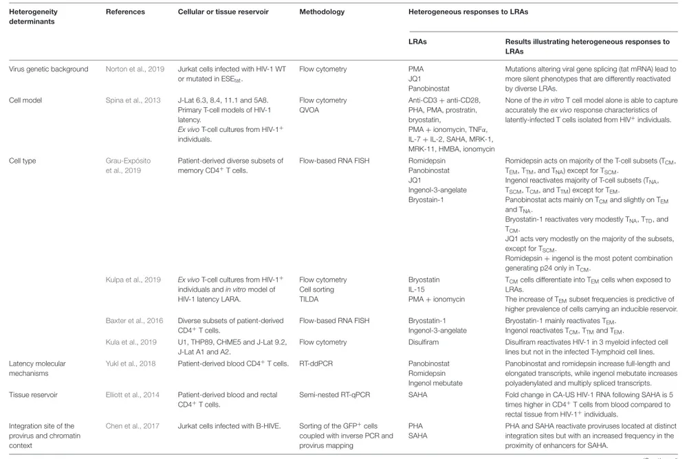
responses to LRAs. All these studies are summarized in Table 2.
HIV-1 Diversity Within the Latent and
Reactivated Reservoirs
Few studies so far have focused on investigating the contribution
of the viral diversity, the compartmentalization, the intact or
defective nature of the viral reservoir, and the origin of the
rebounding virus in latency reversal. A previous study in patients
who initiated ART during acute infection showed that proviral
sequences from PBMCs and GALT presented low level of
genomic diversity and divergence and remained unchanged after
treatment interruption (
Lerner et al., 2011
). Moreover, there
was no phylogenetic link between the rebounded plasma viral
sequences and those from the GALT proviral DNA, indicating
that HIV-1 cellular reservoirs in the GALT may be different from
those circulating in peripheral blood and might not contribute
to the rebounded plasma viremia (
Lerner et al., 2011
). Other
studies have supported the compartmentalization idea of the viral
population in the gut with divergent opinions (
van der Hoek
et al., 1998
;
Lewis et al., 2013
;
Rozera et al., 2014
). Depending
on the stage of HIV-1 infection, the diversity of HIV-1 RNA
appears lower in patients with early infection
versus chronic
infection, and thus, compartmentalization is lost during chronic
infection (
Rozera et al., 2014
). However, other works have
supported other conflicting ideas with findings showing absence
of compartmentalization of HIV-1 between the gut and blood
(
Avettand-Fenoel et al., 2011
;
Imamichi et al., 2011
;
Evering et al.,
2012
), providing evidence for cross infection between these two
compartments (
Chun et al., 2008
).
HIV-1 sequence diversity has been reported to be either higher
(
Klein et al., 2018
) or similar (
Piantadosi et al., 2019
) to genital
tract compared to blood. Viral compartmentalization between the
blood and the male genital tract has been reported by multiple
studies including SIV-infected macaques (
Delwart et al., 1998
;
Paranjpe et al., 2002
;
Pillai et al., 2005
;
Coombs et al., 2006
;
Diem
et al., 2008
;
Houzet et al., 2018
). More recently, patients under
suppressive ART exhibited a significant positive correlation
between viral diversity and genetic compartmentalization in the
blood and testes, but it was attributable to differential frequencies
of identical HIV-1 sequences between the two sites (
Miller et al.,
2019
). However, there was no evidence of compartmentalization
when only unique sequences per sites are considered, suggesting
that compartmentalization between blood and testes is linked to
clonal expansion (
Miller et al., 2019
).
HIV-1 phylogenetic analysis of
post-mortem CSF, brain,
and spleen from HIV-1 patients under ART and presenting
dementia symptoms showed that HIV-1 strains from the blood
fmic b-10-03060 January 23, 2020 T ime: 17:51 # 9 Ait-Ammar et al. LRAs Facing HIV -1 Reservoirs Heter ogeneity
TABLE 2 | The diverse responses of latently-infected cells to LRAs reflect the heterogeneity of the mechanisms driving HIV-1 latency.
Heterogeneity determinants
References Cellular or tissue reservoir Methodology Heterogeneous responses to LRAs
LRAs Results illustrating heterogeneous responses to LRAs
Virus genetic background Norton et al., 2019 Jurkat cells infected with HIV-1 WT or mutated in ESEtat.
Flow cytometry PMA
JQ1 Panobinostat
Mutations altering viral gene splicing (tat mRNA) lead to more silent phenotypes that are differently reactivated by diverse LRAs.
Cell model Spina et al., 2013 J-Lat 6.3, 8.4, 11.1 and 5A8. Primary T-cell models of HIV-1 latency.
Ex vivo T-cell cultures from HIV-1+
individuals.
Flow cytometry QVOA
Anti-CD3 + anti-CD28, PHA, PMA, prostratin, bryostatin,
PMA + ionomycin, TNFα, IL-7 + IL-2, SAHA, MRK-1, MRK-11, HMBA, ionomycin
None of the in vitro T cell model alone is able to capture accurately the ex vivo response characteristics of latently-infected T cells isolated from HIV+individuals.
Cell type Grau-Expósito
et al., 2019
Patient-derived diverse subsets of memory CD4+T cells.
Flow-based RNA FISH Romidepsin Panobinostat JQ1
Ingenol-3-angelate Bryostain-1
Romidepsin acts on majority of the T-cell subsets (TCM,
TEM, TTM, and TNA) except for TSCM.
Ingenol reactivates majority of T-cell subsets (TNA,
TSCM, TCM, and TTM) except for TEM.
Panobinostat acts mainly on TCMand slightly on TEM
and TNA.
Bryostatin-1 reactivates very modestly TNA, TTD, and
TCM.
JQ1 acts very modestly on the majority of the subsets, except for TSCM.
Romidepsin + ingenol is the most potent combination generating p24 only in TCM.
Kulpa et al., 2019 Ex vivo T-cell cultures from HIV-1+
individuals and in vitro model of HIV-1 latency LARA.
Flow cytometry Cell sorting TILDA Bryostatin IL-15 PMA + ionomycin
TCMcells differentiate into TEMcells when exposed to
LRAs.
The increase of TEMsubset frequencies is predictive of
higher prevalence of cells carrying an inducible reservoir.
Baxter et al., 2016 Diverse subsets of patient-derived CD4+T cells.
Flow-based RNA FISH Bryostatin-1 Ingenol-3-angelate
Bryostatin-1 mainly reactivates TEM.
Ingenol reactivates TCM, TTMand TEM. Kula et al., 2019 U1, THP89, CHME5 and J-Lat 9.2,
J-Lat A1 and A2.
Flow cytometry Disulfiram Disulfiram reactivates HIV-1 in 3 myeloid infected cell lines but not in the infected T-lymphoid cell lines. Latency molecular
mechanisms
Yukl et al., 2018 Patient-derived blood CD4+T cells. RT-ddPCR Panobinostat
Romidepsin Ingenol mebutate
Panobinostat and romidepsin increase full-length and elongated transcripts, while ingenol mebutate increases polyadenylated and multiply spliced transcripts. Tissue reservoir Elliott et al., 2014 Patient-derived blood and rectal
CD4+
T cells.
Semi-nested RT-qPCR SAHA Fold change in CA-US HIV-1 RNA following SAHA is 5 times higher in CD4+
T cells from blood compared to rectal tissue from HIV-1+individuals.
Integration site of the provirus and chromatin context
Chen et al., 2017 Jurkat cells infected with B-HIVE. Sorting of the GFP+cells
coupled with inverse PCR and provirus mapping
PHA SAHA
PHA and SAHA reactivate proviruses located at distinct integration sites but with an increased frequency in the proximity of enhancers for SAHA.
(Continued) Fr ontiers in Micr obiology | www .fr ontiersin.org 9 January 2020 | V olume 10 | Article 3060
fmic b-10-03060 January 23, 2020 T ime: 17:51 # 10 Ait-Ammar et al. LRAs Facing HIV -1 Reservoirs Heter ogeneity TABLE 2 | Continued Heterogeneity determinants
References Cellular or tissue reservoir Methodology Heterogeneous responses to LRAs
LRAs Results illustrating heterogeneous responses to LRAs
Abner et al., 2018 Jurkat cells infected with B-HIVE.
Sorting of the GFP+
cells coupled with RT-qPCR and provirus mapping
MMQO JQ1 SAHA Prostratin
BETi (MMQO and JQ1) target viruses integrated at distinct sites as compared to those targeted by SAHA and prostratin.
Battivelli et al., 2018
Primary CD4+
T cells infected with dual-labeled HIV-1.
Cells sorting coupled with semi-nested ligation-mediated PCR and provirus sequencing Panobinostat JQ1 Bryostatin-1 Anti-CD3 + anti-CD28
LRAs reactivate only 5% of latently-infected cells. The inducible and non-inducible populations exhibit distinct chromatin integration sites which were associated, respectively, with active chromatin and heterochromatin with non-accessible region. Patient to patient
and patient gender
Das et al., 2018 Patient-derived resting memory CD4+T cells.
EDITS Anti-CD3 + anti-CD28
SAHA
Women have reduced inducible RNA reservoirs compared to men following treatment with anti-CD3 + anti-CD28. ESR-1 antagonists potentiate HIV-1 reactivation by SAHA, however, females show higher reactivation than males HIV-1+
individuals.
Darcis et al., 2017 Patient-derived CD8+
-depleted PBMCs and resting CD4+T
cells.
Highly sensitive TaqMan based RT-qPCR
JQ1 + bryostatin JQ1 + ingenol-B 5-AzadC + panobinostat 5-AzadC + romidepsin
There is a positive correlation between the HIV-1 reservoir size and the ex vivo capacity of HIV-infected patient cell cultures to be reactivated by LRAs. However, some HIV-1+patients
deviate from this linearity (for example, patients who, despite a low reservoir, are more easily reactivated than many other patients who have a larger reservoir).
Yukl et al., 2018 Patient-derived CD4+
T cells. RT-ddPCR JQ1, Disulfiram
Chaetocin, Panobinostat Romidepsin, Ingenol mebutate, Ingenol 3,20-dibenzoate
All LRAs exhibit inter-patient variability to reverse the blocks to HIV-1 transcription with a very weak exception for romidepsin.
B-HIVE, barcoded HIV ensembles; CA-US RNA, cell-associated unspliced RNA; EDITS, envelope detection by induced transcription-based sequencing; LARA, latency and reversion assay; RNA FISH, RNA Fluorescence In Situ Hybridization; RT-ddPCR, reverse transcription droplet digital polymerase chain reaction; TILDA, Tat/rev-induced limiting dilution assay; QVOA, Quantitative Viral Outgrowth Assays.
Fr ontiers in Micr obiology | www .fr ontiersin.org 10 January 2020 | V olume 10 | Article 3060
fmicb-10-03060 January 23, 2020 Time: 17:51 # 11
Ait-Ammar et al. LRAs Facing HIV-1 Reservoirs Heterogeneity