Science Arts & Métiers (SAM)
is an open access repository that collects the work of Arts et Métiers Institute of Technology researchers and makes it freely available over the web where possible.
This is an author-deposited version published in: https://sam.ensam.eu
Handle ID: .http://hdl.handle.net/10985/19257
To cite this version :
Antoine NORDEZ, Gaël GUILHEM, Jennyfer LECOMPTE, Giuseppe RABITA - Interactions between fascicles and tendinous tissues in gastrocnemius medialis and vastus lateralis during drop landing Scandinavian Journal of Medicine and Science in Sports Vol. 29, n°1, p.5570 -2019
Any correspondence concerning this service should be sent to the repository
1
|
INTRODUCTION
The ability of muscle‐tendon units to dissipate energy is fun-damental in preventing muscle and bone injuries1 in various
daily‐life and sport tasks that involve braking actions and ec-centric muscle actions.2 Griffiths’ work on isolated cat
mus-cles 3 first suggested that tendons act as a mechanical buffer
during rapid stretching of the muscle‐tendon unit. This buf-fer mechanism was recently elucidated by a series of animal studies that elegantly analyzed fascicle‐tendon interactions involved in damping processes.4-6 These works appended
further evidence that tendons act as a shock absorber to avoid excessive fascicle lengthening and velocity to protect muscle fibers from subsequent damage.
Interactions between fascicles and tendinous tissues in
gastrocnemius medialis and vastus lateralis during drop landing
Enzo Hollville
1,2|
Antoine Nordez
3,4|
Gaël Guilhem
1|
Jennyfer Lecompte
2,5|
Giuseppe Rabita
11Laboratory Sport, Expertise and
Performance (EA 7370), Research Department, French Institute of Sport (INSEP), Paris, France
2NG lab, Natural Grass, Paris, France
3Laboratory ‘Movement, Interactions,
Performance’ (EA 4334), Faculty of Sport Sciences, University of Nantes, Nantes, France
4Faculty of Health and Environmental
Sciences, Health and Rehabilitation Research Institute, Auckland University of Technology, Auckland, New Zealand
5LBM ‐ Institut de Biomécanique Humaine
Georges Charpak, Arts et Métiers ParisTech, Paris, France Correspondence
Giuseppe Rabita, Laboratory Sport, Expertise and Performance (EA 7370), Research Department, French Institute of Sport (INSEP), Paris, France.
Email: [email protected]
Abstract
Animal tendons have been shown to act as shock absorbers to protect muscle fasci-cles from exercise‐induced damage during landing tasks. Meanwhile, the contribu-tion of tendinous tissues to damping activities such as landing has been less explored in humans. The aim of this study was to analyze in vivo fascicle‐tendon interactions during drop landing to better understand their role in energy dissipation. Ultrafast ultrasound images of the gastrocnemius medialis (GM) and vastus lateralis (VL), lower limb electromyographic activity, 2‐D kinematics, and ground reaction forces were collected from twelve participants during single‐ and double‐leg drop landings from various heights. For both muscles, length changes were higher in tendinous tis-sues than in fascicles, demonstrating their key role in protecting fascicles from rapid active lengthening. Increasing landing height increased lengthening and peak length-ening velocity of VL fascicle and GM architectural gear ratio, whereas GM fascicle displayed similar length and velocity patterns. Single‐leg landing lengthens the ten-dinous tissues of GM and, to a greater degree, VL muscles, without affecting the fascicles. These findings demonstrate the adjustment in fascicle‐tendon interactions to withstand mechanical demand through the tendon buffer action and fascicle rota-tion. The higher VL fascicle contribution to negative work as the drop height in-creases would suggest muscle‐specific damping responses during drop landing. This can originate from the distal‐to‐proximal sequence of joint kinetics, from differences in muscle and tendon functions (one‐ and two‐joint muscles), architectural and mor-phological properties (eg, tendon stiffness), as well as from the muscle activity of the GM and VL muscles.
K E Y W O R D S
In humans, in vivo fascicle‐tendon interactions of lower limb have been extensively studied using B‐mode ultrasound
7 during stretch‐shortening cycles8,9 and concentric tasks.10
These studies highlighted the role of tendons in enhancement of muscle fascicle’s performance through a catapult mecha-nism.11 In a study involving monoarticular eccentric exercises,
Hicks et al12 showed that the tendon partly accounts for the
amplitude of muscle‐tendon unit lengthening, suggesting its potential role in mechanical load modulation. Along this line, Guilhem et al 13 demonstrated that the amount of lengthening
and braking work withstood by muscle fascicles influences the amount of exercise‐induced muscle damage. These find-ings strongly suggest that the assessment of fascicle behavior during eccentric contractions is of primary interest to better understand the mechanisms involved in the shock absorption. Most of previous studies explored fascicle‐tendon interactions in eccentric conditions during monoarticular tasks 12-14 or
low‐intensity exercises.15,16 A recent study examined the
fas-cicle‐tendon interactions of the human triceps surae during a pure energy dissipation task of step landing.17 These authors
elegantly demonstrated that both gastrocnemius medialis and
soleus muscles displayed quite similar fascicle lengthening
during step landing irrespective of the loading condition (ie, body weight vs body weight with added mass) thanks to a buff-ering action of tendinous tissues. The greater stretch of tendi-nous tissues observed in the added mass condition underlines the key role of these structures to modulate energy dissipation.
Although the aforementioned study provides new insight into the role of fascicle and tendinous tissues when energy is dissipated,17 only the muscles that cross the ankle joint were
considered. However, with regard to the distal‐to‐proximal sequence of joint kinetics during landing tasks ,18 the analysis
of both ankle and knee extensor muscles might be required for a better understanding of the whole energy dissipation process throughout the lower limb. In addition, the load ap-plied in this previous study remained relatively low (ie, ~2 times body weight) compared to more intensive tasks such as jump or drop landing (ie, more than 10 times body weight,19)
that occurs in many sports. Indeed, regarding the high occur-rence of injuries resulting from jump landings ,20 these tasks
have been especially studied from both biomechanical 19,21
and neurophysiological 22,23 points of view. In addition, it has
been reported that the risk of sustaining a lower limb injury increased during a single‐leg landing.21 However, to date, the
role of tendons in an intensive and multiarticular eccentric task, such as drop landing, remains poorly understood.
The main purpose of the present study was to analyze
in vivo muscle fascicle and tendinous tissues interactions of
the gastrocnemius medialis (GM) and vastus lateralis (VL) muscles during drop landing. We examined the influence of various heights (ie, 25, 50, and 75 cm) on these interactions. In order to appraise the impact of the load on the behavior of the muscle‐tendon unit, we also analyzed a single‐leg landing
type that should be representative of the maximal load that could be applied during landing. Based on previous studies on the ankle extensors during landing 5,6,17 and considering
the differences between GM and VL function during loco-motion ,7-9,24 we hypothesized that: (a) Elastic structures
strongly contribute to the muscle‐tendon unit lengthening of GM and VL to reduce both fascicle lengthening ampli-tude and velocity thanks to a decoupling mechanism between the fascicles and the tendinous tissues; (b) increasing land-ing height would not impact fascicle lengthenland-ing of the GM muscle due to a greater tendinous tissues contribution to the elongation of the whole muscle‐tendon unit, while the VL fascicle would undergo higher lengthening and velocity; and (c) single‐leg landing would decrease the knee joint flexion reducing the muscle‐tendon unit elongation of the VL and in-creasing the one of the GM. This would result to an increase in the tendinous tissues lengthening of both muscles.
2
|
MATERIALS AND METHODS
2.1
|
Participants
Fifteen recreationally active men (age: 25.5 ± 3.8 years; height: 177.6 ± 5.8 cm; body mass: 72.2 ± 7.7 kg) volun-teered to participate in the study. None of them had suffered a previous lower limb injury. All participants were fully in-formed about the nature and aim of the study before they gave their written informed consent to participate. The study was approved by the local ethics committee (Ouest IV) and conducted in accordance with the Declaration of Helsinki.
2.2
|
Experimental design
2.2.1
|
Familiarization session
Participants attended a familiarization session one day before the measurement session. They were instructed to drop from a box with both legs extended and to keep both hands on their hips throughout the movement. The motion of arms and feet was restricted to the sagittal plane of mo-tion to ensure the two‐dimensional nature of the task.19 At
the beginning of each trial, subjects were standing on the box on both legs. To impose a standardized drop height, participants were asked to start the movement with both legs over the box, to move their body forward with the right leg straight into the void while keeping a horizontal trajec-tory of the hip. The free fall then started, and a quick hip extension was performed to bring back the left leg. For the single‐leg landing, participants did the same process with-out bringing back their left leg and landed on the right. To avoid extreme landing strategies (ie, too low or broad range of motion), we instructed to “land as naturally as possible by avoiding too low or high body deceleration at impact.”
Participants were also familiarized to maximal voluntary isometric contraction (MVC) before the test sessions. During familiarization and test sessions, each participant wore the same pair of shoes (Essential Star 2.0, Adidas, Herzogenaurach, Germany). The lengths of shank, thigh, and GM muscle were measured using a measuring tape.25,26
2.2.2
|
Test session
After a standardized warm‐up and the assessment of isomet-ric MVC peak torque, participants performed the same drop landing protocol twice to determine the behavior of both GM and VL muscle using one ultrasound scanner. Participants performed the vertical drop landing from a box onto the force platform. They executed double‐leg drop landing from 25, 50, and 75 cm and a single‐leg drop landing from 50 cm, with three trials for each condition. All participants were well fa-miliarized to these tasks and the technical aspects of single‐ and double‐leg drop landings thanks to a visual feedback during the familiarization session. Single‐leg landings were performed on the right leg which was also the leg chosen for all measures in every condition (single‐ and double‐leg drop landings). The order of ultrasound‐tracked muscle, landing height, and landing type were randomized. The experimenter visually checked the following criteria: (a) no jump‐off or step‐down at the start to ensure similar drop height; (b) the feet were placed entirely on the force platform throughout the landing phase; and (c) the participant self‐controlled his reception without loss of balance. Trials that did not meet one of these prerequisites were discarded and repeated.
2.3
|
Instrumentation and data collection
2.3.1
|
Force platform and motion analysis
Ground reaction force (GRF) data were recorded during landings by means of a force platform (60 × 40 cm, Kistler, Winterthur, Switzerland). The force signals were digitized at a sampling rate of 1000 Hz (DT 9804, Data Translation, Marlboro, MA). A motion capture system (Vicon Motion Systems Ltd., Oxford, UK), with six infrared cameras, recorded the three‐dimensional coordinates of eight reflective markers (12 mm diameter) located on the right part of the body at the following locations 27: on the 5th metatarsal,
lateral calcaneus, lateral malleolus, tibia head, lateral femoral epi-condyle, great trochanter, iliac crest, and on the bony prominence on top of the shoulder. The sampling frequency was set to 250 Hz.
2.3.2
|
Ultrasound
The fascicles of GM and VL were visualized using an ul-trasound scanner (Aixplorer, Supersonic Imagine, Aix en Provence, France) and a linear transducer (5‐12 MHz, 55 mm). The scanner was set at research mode to record raw
radio‐frequency signals during ultrafast acquisitions (1000 frames per second). For the GM, the transducer was placed on the skin surface over the muscle belly at different distances between the medial condyle and the musculo‐tendinous junc-tion depending on the subject’s GM morphology. For the VL, the transducer was placed at the midpoint of the muscle to avoid fascicle curvature existing close to insertions.28 For the
VL muscle, as fascicles are often too long for the transducer size,24 optimization of the transducer orientation was achieved
when most of the muscle fascicles could be traced between the aponeurosis to limit poor estimates in extrapolation of the visible part of the fascicle. We securely attached the trans-ducer with custom‐made equipment and carefully ensured that it was not displaced throughout the experimental protocol.
2.3.3
|
Surface electromyography
Surface electromyography (EMG) was recorded with a wireless device (ZeroWire, Aurion, Italy) from
tibi-alis anterior (TA), gastrocnemius meditibi-alis (GM), soleus
(SOL), vastus lateralis (VL), rectus femoris (RF), biceps
femoris (BF), and gluteus maximus (GMax) using
bipo-lar silver/silver chloride surface electrodes (Blue Sensor Q‐00‐S, Baltorpbakken, Denmark). The electrodes were placed longitudinally with respect to the fibers’ orienta-tions. The skin was shaved and cleaned with alcohol, and electrodes were placed over the skin with an interelectrode distance of 20 mm (center‐to‐center) according to the SENIAM (Surface Electromyography for the Non‐Invasive Assessment of Muscles) recommendations. Raw EMG sig-nals were pre‐amplified (input impedance; 20 MM, com-mon mode rejection ratio: 90 db; signal‐to‐noise ratio: >50 dB; gain: 1000), digitized at 2000 Hz, and then trans-mitted wirelessly to a remote unit.
2.3.4
|
Synchronization
Ultrasound and EMG measurements were synchronized with force platform signals thanks to external triggers sent by both devices to the digital converter used to record force platform signals (DT 9804, Data Translation, Marlboro, MA). Before the drop landing task, a reflective marker was launched on the force platform. The impact was detected on the vertical ground reaction force to synchronize kin-ematic measurements and force platform signals. The tem-poral delay between GRF and kinematics data was very consistent between trials (ranged from 0.5 ± 1.4 ms to 3.5 ± 1.4 ms) and between sessions (2.2 ± 2.0 ms).
2.4
|
Data processing
All data were analyzed using custom‐written MATLAB scripts (The MathWorks, Natick, MA). The onset and offset
of each landing trial were detected as the initial contact on the force platform and the peak knee flexion angle, respec-tively. For each trial, the processing was focused on this landing phase (landing duration ranged from 96 ms at 25 cm to 272 ms at 75 cm) and the pre‐activation phase starting 150 ms before the initial contact until initial contact. Due to the variability in landing duration, data were resampled and interpolated (ie, spline interpolation) such that they all have an equal number of points (ie, 101 points; time nor-malization) for both phases to allow between‐participants and between‐conditions comparisons. The trials that were the closest among the three GM and VL trials were selected for analysis. For that purpose, trials were compared in terms of joint angles, GRF, and center‐of‐mass displacement.
2.4.1
|
Kinematics and kinetics
Raw force platform signals were low‐pass filtered using a Butterworth second‐order and zero‐phase‐lag filter at a cutoff frequency of 125 Hz (Figure 1A). Peak vertical GRF was nor-malized to body weight. Vertical ground reaction forces were
used to calculate center‐of‐mass acceleration, velocity, and displacement using the second law of Newton. To compute the center‐of‐mass velocity, we firstly determined the initial verti-cal velocity from the vertiverti-cal coordinates of the hip marker over the last two video frames prior to foot‐ground contact.5
Center‐of‐mass displacement was obtained by integration of the center‐of‐mass velocity. The negative center‐of‐mass work was also calculated by taking the dot product of the ver-tical GRF and verver-tical center‐of‐mass displacement. We also computed the rate of force development (RFD) by dividing the peak vertical GRF by the GRF peak time. The reflective marker positions were low‐pass filtered using a Butterworth second‐order and zero‐phase‐lag filter at a cutoff frequency of 15 Hz.27 Given the 3‐D coordinates data from the reflective
markers, we first calculated the absolute 2‐D angles of lower limb segments to obtain relative ankle, knee, and hip angles in the sagittal plane (Figure 1B). By convention, the ankle angle was set to 0° when the foot is perpendicular to the shank. Knee and hip angles were set to 0° when fully extended.27 A
negative value of ankle angle corresponded to plantar flexion, while positive knee and hip angles were linked to flexion.
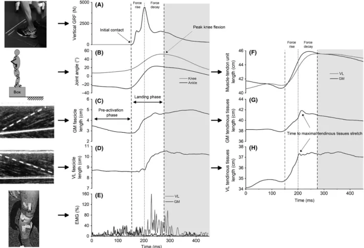
FIGURE 1 Individual examples of instrumentation and measurements from a double‐leg drop landing from 50 cm. A, vertical ground reaction forces (GRF); B, ankle and knee joint angles; C, gastrocnemius medialis (GM) fascicle length; D, vastus lateralis (VL) fascicle length; E, GM and VL muscle activity. Joint angle and fascicle length data allowed the computation of muscle‐tendon unit length (F) and tendinous tissue length (G and H)
2.4.2
|
EMG analyses
Raw EMG signals of lower limb muscles were band‐pass fil-tered (10‐500 Hz, second‐order zero‐phase‐lag Butterworth filter) and consistently analyzed with a 50 ms moving root mean square (RMS) window to produce a RMS envelope that was then normalized to the maximal corresponding muscle activity recorded during isometric MVC. The EMG RMS was averaged for pre‐activation and landing phases in order to compare the same muscle between heights and landing types.
2.4.3
|
Ultrasound analyses
Raw radio‐frequency signals were converted to B‐mode images through a classical beam‐forming procedure. The displacement of fascicles and aponeuroses was automatically tracked on B‐mode images using the method proposed by Cronin et al.29,30. When the fascicle was not fully visible, its
length was extrapolated as the length of the straight line be-tween the superficial and deep aponeurosis using a trigonom-etry method.9,24 The pennation angle was calculated as the
angle formed between the fascicle and the deep aponeurosis. Muscle fascicle length and pennation angle were low‐pass filtered using a Butterworth second‐order and zero‐lag filter at a cutoff frequency of 60 Hz. The fascicle lengths of GM and VL were derived to obtain fascicle velocities. Peak fas-cicle lengthening was calculated during the landing phase for each muscle and all drop landing conditions. According to the equations derived by Grieve et al 25 and Visser et al ,26 we
estimated the instantaneous length of the whole GM and VL muscle‐tendon units using ankle and knee angles (Figure 1F). The length of tendinous tissues was estimated as the differ-ence between muscle‐tendon unit length and the horizontal projection of fascicle length (ie, fascicle length multiplied by the cosine of pennation angle) 8,10 (Figure 1G,H). Tendinous
tissues length changes were computed during both pre‐ and landing phases, while tendinous tissues peak lengthening ve-locity was computed during the landing phase (Figure 1F‐H). The effect of fascicle rotation has been characterized as the muscle’s gear ratio 31,32 (AGR) calculated by the ratio
be-tween the horizontal fascicle length variation to the fascicle length variation during the landing phase (Δ horizontal fas-cicle length/Δ fasfas-cicle length 31,32). This ratio was used to
determine how the fascicle lengthening could be minimized by the fascicle rotation. Methodological differences existed to calculate the AGR between the original in vitro method of Brainerd & Azizi 32 and the in vivo adaptation in the current
study. The horizontal projection of fascicle length 9,13 was
considered rather than muscle length.
The moment of maximal stretch of the tendinous tis-sues was defined as the moment at which the tendinous tissues do not contribute any further to the muscle‐tendon
unit lengthening and expressed in ms (Figure 1G). In order to specify the muscle fascicle behavior, the landing phase was divided into two subphases based on GRF‐time course (Figure 1). First, we defined “force rise” as the phase between the ground contact (ie, 0% of landing phase) and the time of peak vertical GRF. “Force decay” corresponded to the phase between the peak vertical GRF and the end of the landing phase.17 It must be noted that the phases we used are based on
the vertical GRF 17 and differ from the method initially
pro-posed by Konow & Roberts 4,5 based on the muscle‐tendon
unit force of turkeys. Fascicle lengthening amplitudes and AGR of the GM and VL muscles were calculated for these two phases.
2.5
|
Statistical analyses
Statistical analyses were completed using Statistica (StatSoft, Tulsa, OK). Since the data consistently passed the normality test (Shapiro‐Wilk’s test), all data were expressed as mean ±standard deviation (SD). Statistical significance was set at
P < 0.05.
First, reliability assessments (within‐subject coeffi-cient of variation [CV], intraclass correlation coefficoeffi-cient [ICC], and standard error of measurement [SEM]) were performed for relative peak vertical GRF, negative center‐ of‐mass work, center‐of‐mass velocity, landing duration, and joint range of motion (ROM). Second, for double‐leg drop landing conditions, one‐way ANOVAs for repeated measures were used to assess the effects of landing height (25, 50, and 75 cm). These analyses were applied for the following: (a) kinetics variables (ie, relative peak vertical force; negative center‐of‐mass work); (b) kinematics vari-ables (ie, center‐of‐mass velocity; landing duration; ROM for ankle, knee, and hip; RFD; and time to peak vertical force); (c) length changes and peak lengthening velocity for the muscle‐tendon unit, fascicles, and tendinous tissues for both GM and VL; and (d) architectural gear ratio, the moment of maximal tendinous tissues stretch. All these parameters were tested only for the landing phase, except for the length changes in the muscle‐tendon unit, fascicles, and tendinous tissues that were analyzed for pre‐activation phase, landing phases, force rise, and force decay phases. Third, paired t tests were performed to determine the effects of landing type (single‐ vs double‐leg for the 50 cm landing height) on the same parameters. Fourth, repeated measures two‐way ANOVAs (2 phases × 3 heights) were performed to compare the activity of the seven muscles between phases (pre‐activation, landing) and across the three landing heights (25, 50, and 75 cm). Fifth, repeated measures two‐way ANOVAs (2 phases × 2 landing types) were performed to compare the activity of the seven mus-cles between phases (pre‐activation and landing) and land-ing type (sland-ingle‐ vs double‐leg landland-ing at 50 cm). When
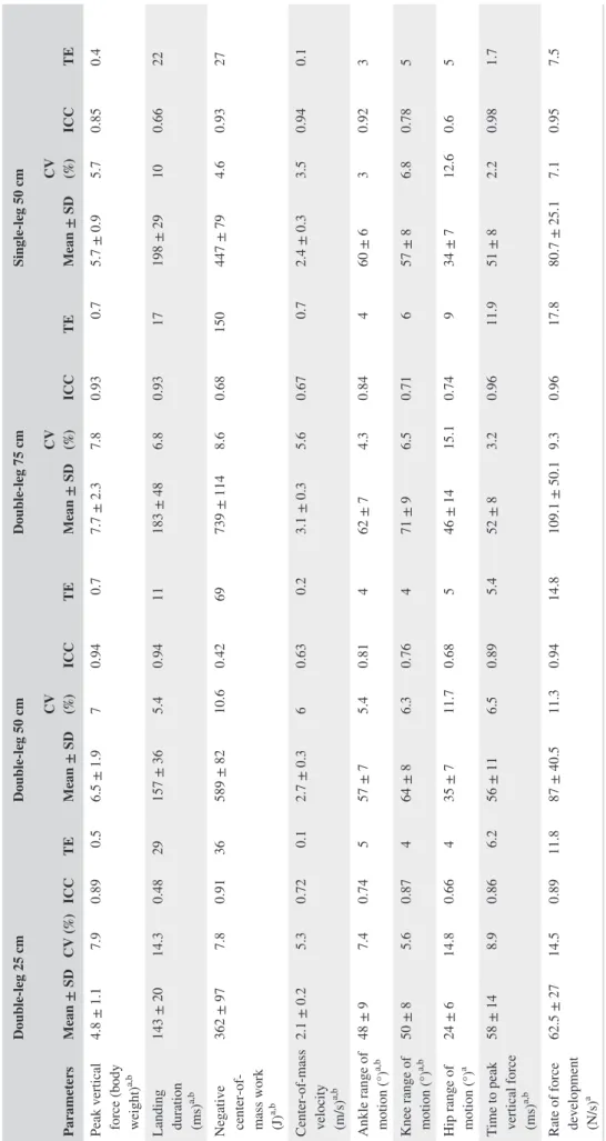
TABLE 1
Kinematics and kinetics parameters obtained during drop landing task in the four tested conditions
Parameters Double‐leg 25 cm Double‐leg 50 cm Double‐leg 75 cm Single‐leg 50 cm Mean ± SD CV (%) ICC TE Mean ± SD CV (%) ICC TE Mean ± SD CV (%) ICC TE Mean ± SD CV (%) ICC TE
Peak vertical force (body weight)
a,b 4.8 ± 1.1 7.9 0.89 0.5 6.5 ± 1.9 7 0.94 0.7 7.7 ± 2.3 7.8 0.93 0.7 5.7 ± 0.9 5.7 0.85 0.4 Landing duration (ms) a,b 143 ± 20 14.3 0.48 29 157 ± 36 5.4 0.94 11 183 ± 48 6.8 0.93 17 198 ± 29 10 0.66 22
Negative center‐of‐ mass work (J)
a,b 362 ± 97 7.8 0.91 36 589 ± 82 10.6 0.42 69 739 ± 114 8.6 0.68 150 447 ± 79 4.6 0.93 27 Center‐of‐mass velocity (m/s) a,b 2.1 ± 0.2 5.3 0.72 0.1 2.7 ± 0.3 6 0.63 0.2 3.1 ± 0.3 5.6 0.67 0.7 2.4 ± 0.3 3.5 0.94 0.1
Ankle range of motion (°)
a,b 48 ± 9 7.4 0.74 5 57 ± 7 5.4 0.81 4 62 ± 7 4.3 0.84 4 60 ± 6 3 0.92 3
Knee range of motion (°)
a,b 50 ± 8 5.6 0.87 4 64 ± 8 6.3 0.76 4 71 ± 9 6.5 0.71 6 57 ± 8 6.8 0.78 5
Hip range of motion (°)
a 24 ± 6 14.8 0.66 4 35 ± 7 11.7 0.68 5 46 ± 14 15.1 0.74 9 34 ± 7 12.6 0.6 5
Time to peak vertical force (ms)
a,b 58 ± 14 8.9 0.86 6.2 56 ± 11 6.5 0.89 5.4 52 ± 8 3.2 0.96 11.9 51 ± 8 2.2 0.98 1.7
Rate of force development (N/s)
a 62.5 ± 27 14.5 0.89 11.8 87 ± 40.5 11.3 0.94 14.8 109.1 ± 50.1 9.3 0.96 17.8 80.7 ± 25.1 7.1 0.95 7.5
Positive ankle joint angle corresponds to ankle plantar flexion. Positive knee and hip joint angle correspond to knee and hip f
lexion. Values are presented as mean
±
SD. Statistical significance was set at
P
<
0.05.
TABLE 2 Gastrocnemius medialis and vastus lateralis behavior during the landing phase of drop landing task in the four tested conditions
Parameters Double‐leg 25 cm Double‐leg 50 cm Double‐leg 75 cm Single‐leg 50 cm
Gastrocnemius medialis
Muscle‐tendon unit behavior Length changes
(pre‐activation phase,
cm)b
−1.1 ± 0.66 −1.37 ± 0.87 −1.17 ± 0.63 −1.82 ± 0.62
Lengthening amplitude
(landing phase, cm)a,b 2.61 ± 0.91 3.08 ± 0.6 3.38 ± 0.62 3.48 ± 0.62
Peak lengthening velocity (landing phase,
cm/s)a,b 44 ± 16 52.7 ± 12.1 59.7 ± 11.7 60.1 ± 11.1 Fascicle behavior Length changes (pre‐activation phase, cm)b −0.92 ± 0.52 −1.02 ± 0.66 −0.92 ± 0.41 −1.35 ± 0.51 Lengthening amplitude (landing phase, cm) 1.71 ± 0.81 1.71 ± 0.88 1.62 ± 0.81 1.64 ± 0.61 Lengthening amplitude (force rise, cm)a 0.93 ± 0.56 0.62 ± 0.37 0.6 ± 0.39 0.72 ± 0.48 Lengthening amplitude (force decay, cm) 0.78 ± 0.38 1.09 ± 0.78 1.02 ± 0.77 0.92 ± 0.58 Peak lengthening velocity (landing phase, cm/s)
42 ± 16.5 44.5 ± 18.4 43.1 ± 18.9 50.7 ± 15
Architectural gear ratio
(landing phase, %)a 1.09 ± 0.02 1.1 ± 0.04 1.14 ± 0.06 1.13 ± 0.06
Architectural gear ratio
(force rise, %)b 1.08 ± 0.08 1.09 ± 0.09 1.11 ± 0.07 1.14 ± 0.07
Architectural gear ratio
(force decay, %) 1.12 ± 0.05 1.12 ± 0.09 1.16 ± 0.15 1.14 ± 0.05
Tendinous tissues behavior Length changes (pre‐activation phase, cm) −0.12 ± 0.11 −0.21 ± 0.26 −0.21 ± 0.21 −0.16 ± 0.16 Lengthening amplitude (landing phase, cm)a 2.08 ± 0.75 2.68 ± 0.62 2.93 ± 0.73 2.91 ± 0.82 Peak lengthening velocity (landing phase,
cm/s)a 46.5 ± 28.7 63.1 ± 19.6 71.9 ± 19.5 65.2 ± 19.4 Time to maximal tendinous tissues lengthening (ms) 68 ± 12 66 ± 9 69 ± 13 66 ± 20 Vastus lateralis
Muscle‐tendon unit behavior Length changes
(pre‐activation phase,
cm)a,b
0.62 ± 0.44 1.08 ± 0.49 0.95 ± 0.39 0.56 ± 0.37
Lengthening amplitude
(landing phase, cm)a,b 4.38 ± 0.66 5.28 ± 0.67 5.43 ± 0.79 4.7 ± 0.69
the sphericity assumption was violated (Mauchly’s test), a Geisser‐Greenhouse correction was used. Bonferroni post hoc analyses (α = 0.05) were conducted when appropriate.
3
|
RESULTS
Due to the low quality of ultrasound data for three different trials (over 120 trials, representing 3.3%), two participants were discarded from the analysis. In addition, the kinematic data of another participant were erroneous. Therefore, results were obtained for 12 participants.
3.1
|
Landing kinetics and kinematics
All the kinematics and kinetics parameters reported in Table 1 were significantly altered by both landing height (all P val-ues <0.01) and landing type (all P valval-ues <0.009), except for hip ROM and RFD, which were affected by landing height (P < 0.001) but not by landing type (P values: 0.65 and 0.26). CV, ICC, and SEM are provided in Table 1. CVs for peak vertical force, center‐of‐mass velocity, negative center‐of‐ mass work, time to peak vertical force, and knee and ankle ROM ranged between 3.0% and 10.6%. Landing duration, hip ROM, and RFD presented higher CVs with almost all
Parameters Double‐leg 25 cm Double‐leg 50 cm Double‐leg 75 cm Single‐leg 50 cm
Peak lengthening velocity (landing phase,
cm/s)a 42.8 ± 6.8 50.1 ± 7.5 52.9 ± 7.8 47.3 ± 5.1 Fascicle behavior Length changes (pre‐activation phase, cm) 0.27 ± 0.29 0.27 ± 0.4 0.17 ± 0.23 0.16 ± 0.23 Lengthening amplitude (landing phase, cm)a 1.45 ± 0.73 1.78 ± 0.69 2.04 ± 0.82 1.37 ± 0.4 Lengthening amplitude (force rise, cm)b,c 0.5 ± 0.45 0.58 ± 0.55 0.45 ± 0.31 0.2 ± 0.11 Lengthening amplitude
(force decay, cm)a,c 0.92 ± 0.3 1.15 ± 0.31 1.56 ± 0.75 1.15 ± 0.28
Peak lengthening velocity (landing phase,
cm/s)a
23.2 ± 8.3 27.8 ± 6.4 37.7 ± 19.3 25.6 ± 11.4
Architectural gear ratio
(landing phase, %) 1.03 ± 0.05 1.03 ± 0.04 1.03 ± 0.03 1.05 ± 0.05
Architectural gear ratio
(force rise, %) 1.01 ± 0.04 1.01 ± 0.04 1 ± 0.06 1.01 ± 0.15
Architectural gear ratio
(force decay, %)c 1.03 ± 0.03 1.04 ± 0.03 1.05 ± 0.03 1.07 ± 0.07
Tendinous tissues behavior Length changes (pre‐activation phase, cm)a,b 0.33 ± 0.33 0.78 ± 0.29 0.76 ± 0.29 0.38 ± 0.22 Lengthening amplitude (landing phase, cm)b 2.48 ± 0.72 2.65 ± 0.75 2.8 ± 0.74 3.03 ± 0.82 Peak lengthening velocity (landing phase,
cm/s)a 38.8 ± 11.7 43.8 ± 13.9 50.9 ± 8.9 49.6 ± 10.6 Time to maximal tendinous tissues lengthening (ms)a 118 ± 27 119 ± 29 94 ± 31 127 ± 25
A negative value of length changes corresponds to shortening. Values are presented as mean ± SD. Statistical significance was set at P < 0.05.
aSignificant main effect of landing height
bSignificant effect of landing type (single‐ vs double‐leg landing)
cSignificant effect of phase (force rise vs force decay)
conditions higher than 10% with the maximal CV value of 15.2%. Overall, these results (ie, CV values of 20 conditions over 28 were under 10%) combined with those of ICC and SEM values demonstrated a satisfying reliability in the me-chanics of landing.
3.2
|
Gastrocnemius medialis behavior
Architectural measurements of gastrocnemius medialis are presented in Table 2 and patterns of muscle‐tendon unit, muscle fascicle, and tendinous tissue length in Figure 2A‐C. During the pre‐activation phase, the muscle‐tendon unit (Figure 2A, −1.10 ± 0.66 to −1.82 ± 0.62 cm) and muscle
fascicles (Figure 2B, −0.92 ± 0.52 to −1.35 ± 0.51 cm) short-ened, while tendinous tissue lengths remained almost con-stant (Figure 2C, −0.12 ± 0.11 to −0.21 ± 0.26 cm). In addition, there was no significant effect of the landing height for the muscle‐tendon unit (P = 0.19), fascicles (P = 0.27), and tendinous tissue length changes (P = 0.61). The shorten-ing was significantly larger for the sshorten-ingle‐leg condition com-pared to the double‐leg for both muscle‐tendon unit (+25%,
P = 0.004) and fascicles (+24%, P = 0.049), while it was not
significantly different for the tendinous tissues (P = 0.45). During the landing phase, the muscle‐tendon unit, fasci-cles, and tendinous tissues lengthened (Figure 2A‐C, land-ing phase). Lengthenland-ing and peak lengthenland-ing velocity of
FIGURE 2 Averaged patterns of gastrocnemius medialis (GM) muscle‐tendon unit (A), muscle fascicle (B), and tendinous tissue (C) length changes and averaged patterns of vastus lateralis (VL) muscle‐tendon unit (D), muscle fascicle (E), and tendinous tissue (F) length changes throughout pre‐activation and landing phases (in % of phase duration). Standard deviations are omitted for clarity. The vertical black dotted line between the two phases indicates the instant of foot‐ground contact. The time of peak vertical ground reaction forces is represented in the landing phase by vertical lines with corresponding colors to each of the four conditions
the muscle‐tendon unit increased with the landing height (lengthening: +23%, P < 0.001, and peak velocity: +24%,
P < 0.001 for the 25 cm vs the 75 cm landing) and was
higher in single‐leg compared to the double‐leg condition (lengthening: +11.5%, P = 0.006, and peak velocity: +12%,
P = 0.003). Lengthening and peak lengthening velocity of
tendinous tissues were also affected by increasing landing height (lengthening: +29%, P < 0.001, and peak velocity: +38%, P = 0.007 for 25 cm vs 75 cm landing), but not by the landing type (lengthening: P = 0.31 and peak velocity:
P = 0.32, respectively). However, lengthening and peak
lengthening velocity of fascicles were not significantly in-fluenced either by landing height (lengthening: P = 0.36 and peak velocity: P = 0.94, respectively) or by landing type (lengthening: P = 0.78, and peak velocity: P = 0.36, respectively). When looking at the force rise phase, the GM fascicle lengthening decreased as the dropping height increased (25 vs 50 and 75, P = 0.014) without any effect of landing type (P = 0.44). We found no effect in the force decay phase for both landing height and type (P = 0.72 and 0.57). Therefore, the effect of landing height was accounted for only by the tendinous tissues without effects for the fas-cicles (all P ≤ 0.007). Architectural gear ratio ranged be-tween 1.09 ± 0.02 and 1.14 ± 0.06 during the whole landing phase and was increased with landing height (P = 0.004 for 25 cm vs 75 cm landing), but no significant effect was found for landing type (P = 0.07). In addition, there was an effect of landing type on fascicle rotation during force rise (+4% during single‐leg landing, P = 0.007). We found no effect of landing height during force rise (P = 0.49) and landing height and type during force decay (P = 0.65 and 0.76). Finally, tendinous tissue maximal lengthening occurred al-most at the same time between the four conditions (66‐69 ms) without a significant effect of landing height (P = 0.63) or landing type (P = 0.93).
3.3
|
Vastus lateralis behavior
Architectural measurements of vastus lateralis are presented in Table 2 and patterns of instantaneous muscle‐tendon unit, muscle fascicle, and tendinous tissue length in Figure 2D‐F. During the pre‐activation phase, the muscle‐tendon unit (Figure 2D, 0.56 ± 0.37 to 1.08 ± 0.49 cm) and ten-dinous tissues (Figure 2F, 0.38 ± 0.22 to 0.78 ± 0.29 cm) were slowly stretched, while muscle fascicles exhibited a quasi‐isometric behavior (Figure 2E, 0.16 ± 0.23 to 0.27 ± 0.29 cm). In addition, lengthening of the mus-cle‐tendon unit and tendinous tissues was significantly influenced by both landing height (muscle‐tendon unit: +35%, P = 0.02, and tendinous tissues: +57%, P < 0.001 for 25 vs 75 cm) and type (muscle‐tendon unit: +48%,
P = 0.004 for the double‐leg vs single‐leg, and tendinous
tissues: +51%, P = 0.004 for the double‐leg vs single‐leg).
On the contrary, no significant effects of landing height (P = 0.31) and landing type (P = 0.32) were found for fas-cicle lengthening.
During the landing phase, the muscle‐tendon unit, fasci-cle, and tendinous tissues lengthened (Figure 2D‐F, landing phase). Lengthening (+16%, P < 0.001 for 25 cm compared to 75 cm landing) and peak lengthening velocity (+19%,
P < 0.001 for 25 cm vs 75 cm landing) of the muscle‐tendon
unit were significantly increased with landing height. A land-ing type effect was not significant for lengthenland-ing (P = 0.44) or peak lengthening velocity (P = 0.10) of the muscle‐tendon unit. For tendinous tissues, lengthening was not changed by landing height condition (P = 0.23), while peak lengthen-ing velocity (P = 0.002) increased when height increased. Differences in landing types were significant for lengthening of tendinous tissues (+12.5% for the single‐leg vs the dou-ble‐leg landing type, P = 0.036) and not significant for peak lengthening velocity (P = 0.10). VL fascicle lengthening am-plitude was affected by landing height (+29%, P = 0.025 for 25 cm vs 75 cm landing) but not by landing types (P = 0.06). Along the same line, the peak lengthening velocity of fascicles increased with landing height (+38%, from 25 to 75 cm land-ing; P = 0.02), while we observed no effect of landing type (P = 0.56). Fascicle lengthening was lower before than after the peak vertical GRF (phase effect, P < 0.001). During the force rise, the VL fascicle lengthening was significantly differ-ent between landing types (−34% of lengthening for single‐leg landing, P = 0.02) with no effect of landing height (P = 0.30) suggesting a large contribution of the elastic structures during this phase. The fascicle lengthening during the force decay was greater as the drop height increased (P = 0.006) with no significant effect of landing type (P = 0.97). No significant effects of landing height (P = 0.78) or landing type (P = 0.08) on AGR values (1.03 ± 0.03 and 1.05 ± 0.05) were reported. Fascicle rotation was greater during force decay compared to force rise (phase effect: P = 0.003) suggesting that this mech-anism helps the VL to dissipate the greater fascicle length-ening during the force decay. Finally, the time to maximal lengthening of tendinous tissues (ranging between 94 ms and 140 ms) displayed a landing height effect (−24 ms, P < 0.01 for 25 vs 75 cm landing), while there was no effect found for landing type (P = 0.34).
3.4
|
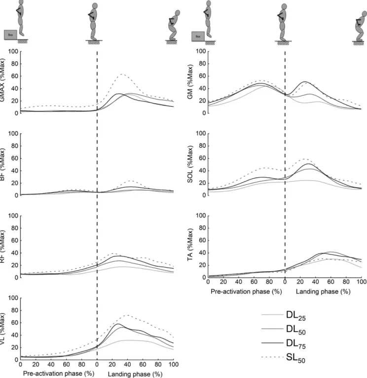
EMG activity
The time course of EMG patterns for the seven lower limb muscles is displayed in Figure 3, and the mean EMG activity (expressed in percentage of the maximal RMS value) during each phase is provided in Table 3. Larger values were almost always obtained in the single‐leg condition. In this condition, the highest values were reached by VL (72.7% ± 37.4% in the landing phase), GMax (53.5% ± 35.8% in the landing phase), and GM (51.9% ± 14.5% in the pre‐activation phase).
A significant main effect of phase was found for all the muscles (all P values <0.042). All the muscles displayed a higher muscle activity during the landing phase compared to the pre‐activation phase (all P values <0.043), except for the gastrocnemius medialis, which was significantly more acti-vated in the pre‐activation phase (P < 0.001). Indeed, GM muscle activity peaked in the pre‐activation phase before
decreasing slowly (Figure 3). Activities of BF, TA, and GMax remained relatively low until initial foot‐ground contact. VL, RF, and SOL muscle activities increased slowly from ~75 ms before the impact and continued to increase during the land-ing phase.
A significant increase in muscle activity as the landing height increased was found for all muscles (25 vs 75 cm
FIGURE 3 Averaged time course of normalized EMG activity patterns (%Max) from seven lower limb muscles during the different landing conditions. Standard deviations are omitted for clarity. The vertical dashed line indicates the instant of ground contact. The raw EMG data were full‐wave rectified and smoothed using a second‐order, low‐pass, Butterworth filter with a cutoff frequency of 10 Hz to obtain a linear envelope. TA: Tibialis anterior; GM: Gastrocnemius medialis; SOL: Soleus; VL: Vastus lateralis; RF: Rectus femoris; BF: Biceps femoris; GMax: Gluteus
landing; from +3.8% for the BF to +13.2% for SOL), except for GMax (P = 0.20). A significant main landing type effect was found for all muscles (all P values <0.05) with higher activity during the single‐leg drop landing for all muscles (from +4.5% for the BF to +17.6% for the GMax), except for the TA, which displayed higher activity during a double‐leg landing (P = 0.04, +4.6% of MVC).
4
|
DISCUSSION
The present study confirms the key role of tendinous tissues to buffer the work done by muscle fascicle during a pure energy‐dissipating task. Gastrocnemius medialis and vastus lateralis differently contributed to the dissipation of mechan-ical energy upon various landing conditions (height or type). The elastic structures of both muscles initially lengthened to account for the muscle‐tendon unit elongation, while muscle fascicles actively lengthened after the foot‐ground contact. In addition, the peak of fascicle lengthening was notably delayed according to the lengthening of tendinous tissues. This decoupling mechanism between the muscle‐tendon unit, muscle fascicle, and tendinous tissues is consistent with previous animal5,6 and human studies.17 In accordance
with our first hypothesis, GM tendinous tissues lengthened more when the loading increased allowing for a constant fas-cicle lengthening amplitude and peak velocity during land-ing. One could notice that GM muscle fascicle lengthening
was reduced as the mechanical demand increased during the force rise phase (ie, first 50‐60 ms after landing). This ob-servation is strengthened by a greater fascicle rotation and the muscle activity before landing. Conversely, VL fascicle lengthening amplitude increased concomitantly to the in-crease in VL muscle‐tendon unit, while the tendinous tissues only increased their lengthening velocity. As the height in-creased, VL fascicle maintained the same range of lengthen-ing durlengthen-ing the force rise phase resultlengthen-ing to a higher storage of elastic energy by tendinous tissues during this period. For both muscles, single‐leg landing resulted in tendinous tis-sues lengthening that preserves fascicles from high eccentric contractions.
The drop landing task used in the present study imposed mechanical loads that were in accordance with the literature (ie, exposing the body to forces ranging from 3.3 to 12.0 times body weight). Several previous studies reported increases in center‐of‐mass velocity and GRF as the drop landing height increased19,21 and during a double‐leg compared to single‐leg
drop landing type.21 The higher mechanical demand resulting
from a higher drop landing altered lower extremity kinemat-ics as reflected by larger ankle and knee joint ROM.19,21,33
Ankle ROM was higher during single‐leg compared to dou-ble‐leg drop landing due to higher plantar flexion in the pre‐ activation phase, while the opposite was found for the knee
ROM.19,21 Consequently, the adopted strategy during single‐
leg landing seemed to alleviate the knee extensors and over-load plantar flexors.33
TABLE 3 Averaged EMG from RMS during the pre‐activation and landing phases of seven lower limb muscles Muscle activity
(% RMSmax)
Double‐leg 25 cm Double‐leg 50 cm Double‐leg 75 cm Single‐leg 50 cm
Pre Land Pre Land Pre Land Pre Land
Tibialis anterior (TA)a,b,d 8.6 ± 8.6 31.3 ± 29.2 10.6 ± 14.4 40.2 ± 28.4 9.5 ± 7.4 40.8 ± 27.1 8.8 ± 5.3 32.7 ± 24.6 Gastrocnemius medialis (GM)a,b,d 39.5 ± 16.8 19.2 ± 18.8 47 ± 20.2 25.1 ± 20.2 47.2 ± 18.1 37.4 ± 41.4 51.9 ± 14.5 38.3 ± 36.8
Soleus (SOL)a,b,d 17.9 ± 9.6 23.5 ± 17.3 26.3 ± 18.7 36.5 ± 24 27.5 ± 11.3 40.2 ± 28.3 39 ± 17.8 48.7 ± 25.4 Vastus lateralis (VL)a,b,c,d,e 10.1 ± 6.2 34.4 ± 18.7 13.2 ± 12.7 49.3 ± 27.5 11.6 ± 6.3 54.1 ± 32.2 18.9 ± 8.7 72.7 ± 37.4 Rectus femoris (RF)a,b,c,d,e 9.6 ± 5 18.6 ± 10.6 11.6 ± 7.5 27.5 ± 13 12.7 ± 7.4 34.7 ± 21.6 17 ± 11.5 41.2 ± 17.6 Biceps femoris (BF)a,b,d,e 3.9 ± 3.2 8.4 ± 8 6.6 ± 6.5 9.3 ± 7.3 6.6 ± 6.3 13.3 ± 16.9 7.5 ± 5.8 17.5 ± 14.7 Gluteus maximus (GMax)b,d,e 5.3 ± 4 21.4 ± 28.3 8 ± 13.3 26.1 ± 39.4 5.6 ± 3.7 23.1 ± 26.2 16 ± 10.8 53.5 ± 35.8
TA: Tibialis anterior; GM: Gastrocnemius medialis; SOL: Soleus; VL: Vastus lateralis; RF: Rectus femoris; BF: Biceps femoris; GMax: Gluteus maximus. EMG data were normalized to the maximum isometric RMS and then averaged for each phase. Values are presented as mean ± SD. Statistical significance was set at P < 0.05.
aMain effect of landing height
bSignificant effect of phase (pre‐activation vs landing phase)
cSignificant interaction height × phase effect
dSignificant effect of landing type (single‐ vs double‐leg landing)
The behavior of GM fascicle during drop landing pre-sented a clear turning point (Figure 2B) from shortening to lengthening following the impact with a small delay of shortening between the foot‐ground contact and the onset of fascicle lengthening (ranged from 3 to 12 ms). Our pat-terns of fascicle lengthening were similar to those reported during the landing phase of a step‐down.17 In the current
study, although stretching of the GM muscle‐tendon unit was greater as landing height increased, muscle fascicle lengthening and peak lengthening velocity remained un-changed. This finding suggests that the increasing me-chanical demand resulting from landing was buffered by tendinous tissues due to a greater stretch of elastic tissues (from 2.08 at 25 cm to 2.93 cm at 75 cm) compared to fas-cicles in GM muscle. This is in accordance with the recent work of Werkhausen et al 17 for lower intensities. They
showed that the increase in muscle‐tendon elongation due to the increased mass results in greater tendinous tissues lengthening without any additional participation of the fascicles. The step landing task analyzed in this previous study 17 induced relatively low ground reaction forces up
to ~2 times body weight vs 12 times body weight in the present study. This suggests that, thanks to the buffering action of the tendon, there is a similar GM response in a continuum of lengthening demand from low to high load-ing constraint. While speculative, it is very likely that this limits the amount of exercise‐induced muscle damage and potential risk of injury at high intensity. Furthermore, the maximal lengthening of GM tendinous tissues occurred 66‐69 ms following ground contact, without significant differences between the four landing conditions (Figure 2F). Regardless of the drop height, the tendinous tissues reached their maximum length (ie, end of the stretch) at the same time (few milliseconds after the peak vertical GRF). This may be related to the buffering action of tendinous tissues enabled by the decoupling between the muscle‐ten-don unit, muscle fascicles, and tendinous tissues. The first milliseconds of muscle‐tendon unit lengthening was mainly withstood by the tendinous tissues before the fascicles ac-tively lengthened.5,6,17 The lengthening of the GM
tendi-nous tissues increased with the increase in height, while the global fascicle lengthening was unchanged. Moreover, the fascicle lengthening was even reduced for the higher height in the force rise phase. This can be seen as a protec-tive mechanism for the GM muscle fascicles. These results are in agreement with Konow & Roberts 5 who found that
tendinous tissues of turkeys reached their maximum length at the same time (~60 ms) during landing from various drop heights. However, contrary to animal studies ,5 the stretch
of fascicles started few milliseconds after the ground con-tact (on average after: 7 ms of GM shortening and 12 ms of VL isometric behavior) and muscle‐tendon unit elonga-tion was taken by both GM fascicle and tendinous tissues
for most of the ankle and knee joints’ excursion. Hence, in human GM, the stretch of tendinous tissues helps to reduce the lengthening velocity and amplitude of fascicles during landing with a smaller delay before the onset of fascicle lengthening as previously shown on turkeys.5 The GM
muscle‐tendon unit lengthening amplitude and peak veloc-ity were higher during a single‐leg landing than during a double‐leg landing. However, if the fascicles withstood the same level of lengthening at a similar rate, the tendinous tissues did not undergo additional lengthening. This may be due to the concomitant higher muscle activity and short-ening amplitude of the GM during the pre‐activation which stiffen the muscle to prepare the landing and allow a higher gearing potential thanks to a higher pennation during the force rise.
VL muscle fascicles contracted almost isometrically, while VL muscle‐tendon unit and tendinous tissues slightly length-ened during the pre‐activation phase. After the foot‐ground contact, while VL fascicle started to lengthen after ~12 ms, muscle‐tendon unit and tendinous tissues lengthened contin-uously until the maximum knee flexion similarly to that re-ported during a single support low‐intensity braking task.34
During a stair descent from 16 cm, larger VL fascicle length-ening amplitudes were reported (up to 3.2 cm) compared to a drop landing task (from 1.45 cm at 25 cm to 2.04 cm at 75 cm). These results confirm that modulation of fascicle length changes is highly dependent on the nature and intensity of the task.9 The VL fascicle lengthening and peak
lengthen-ing velocity increased with landlengthen-ing height, while tendinous tissues stretched over a similar amplitude. However, VL tendi-nous tissues reached their maximal lengthening earlier as the drop height increased. This induced an increase in tendinous tissues peak lengthening velocity, while the lengthening am-plitude was unchanged. Hence, VL fascicle lengthened ear-lier, over larger amplitude and upon higher velocity. This drop height effect occurred during the force decay with a similar lengthening amplitude of VL fascicle (~0.50 cm) during the first 50‐60 ms of landing (ie, force rise). This demonstrates the crucial contribution of the VL fascicle in energy dissipa-tion throughout landing. The VL muscle‐tendon unit length-ening was higher during the single‐leg in comparison with the double‐leg condition. The fascicles lengthened similarly in both landing types, while the tendinous tissues stretched more when using one versus two legs. This behavior results in a lower VL fascicle lengthening during the force rise phase.
While the time course of fascicle length changes was similar for the two muscles (progressive increase during the whole landing phase,Figure 2B,E), our results clearly showed different muscle‐tendon unit behaviors between GM and VL that could be mainly explained by five factors. First, GM is biarticular while VL is monoarticular. GM contributes both to extend the ankle and to flex the knee, while VL only ex-tends the knee joint. During the landing phase, the GRF ex-tends
to flex the ankle joint which is offset by the motion of the knee joint. Our results show that the biarticular GM muscle mitigates length changes and velocity associated with in-creases in landing height. We also observed that the inin-creases in drop height increased the amplitude and peak velocity in the monoarticular VL. Second, the maximal stretching of the muscle‐tendon unit and tendinous tissues occurred later for VL compared to GM. This can result from the joint sequence involved in landing 18,35 given that the ankle is the first joint
impacted by ground contact. There is evidence that the shock‐ absorbing phase of a jump (ie, landing) would require a dis-tal‐to‐proximal sequence of joint kinetics, with the proximal muscles helping the distal muscles to dissipate mechanical energy.18,33,35 In our study, the peak joint angular velocity of
the ankle, knee, and hip joint (ranging from the highest to the lowest) combined with the observed EMG patterns (Figure 3) support this previous finding of a distal‐to‐ proximal sequence during drop landing. Third, the intrinsic tissue properties of Achilles and patellar tendons, such as stiffness, may also af-fect the elongation capacity of the tendinous tissues.36,37 In
humans, the Achilles tendon is categorized as a high‐stress tendon that can sustain maximum stress about twofold greater than that of the patellar tendon.36,37 Moreover, it has recently
been shown in vivo that Achilles tendon stiffness is indepen-dent of loading rate 38 inversely to the patellar tendon.12 The
difference in viscosity between both tendons may explain why the GM tendinous tissues largely prevent the short GM muscle fascicles from rapid lengthening, while VL muscle fascicles stretched with increasing velocity when the landing height and velocity increased. Fourth, the difference in mus-cle activity in the pre‐activation phase could contribute to dif-ferentiate the fascicles behavior of VL and GM. In our study, increases in drop height increased the mean EMG amplitude of the GM over the pre‐activation phase, attesting of “prepara-tory” muscle activity before ground contact. This may stiffen the muscle 23 and adjust active muscle force before ground
contact to meet the demand resulting from the increased mechanical load.23 This process may in turn reduce both
fascicle lengthening amplitude and velocity during landing. Conversely, the low EMG activity of the VL in the pre‐acti-vation phase is reflective of a distal‐to‐proximal actipre‐acti-vation se-quence that provides more time for the quadriceps to activate its larger muscle mass. Together with its longer fascicles, the high force‐generating capacity of the VL then favors its con-tribution to braking work in the late phase of landing. Fifth, another important finding of the present study is related to the larger muscle fascicle rotation as the landing height increases for the GM (AGR from 1.09 to 1.14). This suggests that fasci-cle rotation substantially reduced the stretch sustained by GM fascicles throughout landing (between 9% and 14%) ,31 while
it remained constant for the VL (AGR from 1.03 to 1.05). This result may presumably be attributable to a larger length-ening potential allowed by longer fascicles.
Joint ROM data showed that the strategy adopted during single‐leg landing seemed to unload the knee extensors and overload plantar flexors.33 This is in agreement with
the decrease in VL fascicle lengthening for the single‐leg landing compared to double‐leg and the higher activity of proximal muscles during single‐leg landing compared to double‐leg (mean 1.5‐times higher in single‐leg vs double‐ leg). In addition, higher EMG activity in knee joint mus-cles enables adequate dampening of excessive loading. It is also interesting to note that, thanks to the buffering ac-tion of tendinous tissues, this overload did not induce any overstretching of GM fascicles. Thus, the adopted strategy during single‐leg landing seems efficient for limiting fasci-cle stretching of both musfasci-cles but overused both tendinous tissues which might lead to tendinopathy.20
Substantial methodological considerations should be kept in mind when interpreting the present data. Similar to all pre-vious 2D ultrasound studies,7 we assumed the linearity and
ho-mogeneity of fascicles’ line of action, whereas previous reports showed a fascicle curvature close to aponeuroses’ insertions resulting in slight length underestimation.28 In addition, when
the fascicle was not fully visible, mainly in VL, we used trig-onometry computations to estimate fascicle length. The error for estimating fascicle with the extrapolation method has been reported to be 2%‐7% 9,24 and could potentially be larger when
considering the high speed of drop and ground contact inten-sity (>3 m/s at 75 cm). A recent study suggests that using two synchronized in‐series transducers could overcome this limit and avoid absolute fascicle lengths misestimation compared to extrapolated method.39 As previously reported with the single
transducer method, the fascicle data set may not perfectly re-flect fascicle behavior; however, it could be assumed that this method does not alter the effects of landing height and type (ie, “similar differences in muscle contraction dynamics within participants.”39). In our study, we estimate tendinous tissues
length changes from fascicle length estimation and lower limb joint angles. This approach is imperfect and has recently been questioned due to potential incorrect interpretations regard-ing tendon behavior.40 While we acknowledge that tendinous
tissues length changes must be seen in light of this limit, the changes in fascicle length remain valid. Since our main results are directly inferred from these length changes, the potential bias in estimating tendinous tissues length would not influence the main conclusions of our study. Considering the difficulty of the drop landing task, we only included the best trial per condition for the data analysis. In order to compare GM and VL behavior, this trial was chosen considering the reliability in the landing mechanics and the quality of ultrasound data. This choice prevented us to assess the reliability of our fasci-cle length measurement. Finally, due to our single ultrasound scanner, we were not able to track both muscles at the same time, and the procedure involved repeating the protocol twice. However, we found good reliability in the mechanics of landing
measured at the global level, which is in line with a previous work on landing from a hang bar (ie, height standardization) that revealed ICC coefficients ranging from 0.79 to 0.93.41
5
|
CONCLUSION AND
PERSPECTIVES
The present study demonstrated that, during drop landing, tendinous tissues of both gastrocnemius medialis and
vas-tus lateralis act as shock absorbers by rapidly stretching and
storing elastic energy, which is then released to the fascicles and dissipated through active muscle lengthening. A previous study showed that mechanical loading up to ~2 times body weight did not influence GM fascicle stretch or lengthening velocity.17 The present study demonstrated that GM
behav-ior remains similar for landings with GRF up to ~12 times body weight indicating that the elastic structures absorb me-chanical energy to protect GM fascicles from potential dam-age and injury. In addition, novel evidence was provided that VL fascicle behavior differs by increasing its contribution to the muscle‐tendon unit elongation as the landing height increased. This study showed that simultaneous analyses of fascicle‐tendon interactions and muscle activity involved in multijoint braking actions, especially at high intensity, allow to improve our understanding of the energy dissipation pro-cess in humans.
Among different perspectives, further studies could focus on the effect of surface mechanical properties on the fascicle‐ tendon damping responses during landing. As human lands, energy is absorbed and released by the surface depending on its intrinsic mechanical properties and at the end dissipated by muscle fascicles. Specifically, energy storage is a function of both surface stiffness and deformation. Hence, the transfer of mechanical energy between the body and the surface can have a considerable influence on athletic performance. Such investigations would be paramount to determine whether these interfaces influence the risk of injury in sports tasks like tendinopathies.20
ACKNOWLEDGEMENTS
Enzo Hollville is funded by the Natural Grass company. We warmly thank Hugo Hauraix for his technical support.
CONFLICT OF INTEREST
No conflict of interest, financial or otherwise, is declared by the authors.
ORCID
Giuseppe Rabita http://orcid.org/0000-0002-0548-3019
REFERENCES
1. Proske U, Morgan DL. Muscle damage from eccentric exercise: mechanism, mechanical signs, adaptation and clinical applica-tions. J Physiol. 2001;537(Pt 2):333‐345.
2. Lindstedt SL, LaStayo PC, Reich TE. When active muscles lengthen: properties and consequences of eccentric contractions.
News Physiol Sci. 2001;16:256‐261.
3. Griffiths RI. Shortening of muscle fibres during stretch of the ac-tive cat medial gastrocnemius muscle: the role of tendon compli-ance. J Physiol. 1991;436:219‐236.
4. Konow N, Azizi E, Roberts TJ. Muscle power attenua-tion by tendon during energy dissipaattenua-tion. Proc Biol Sci. 2012;279(1731):1108‐1113.
5. Konow N, Roberts TJ. The series elastic shock absorber: tendon elasticity modulates energy dissipation by muscle during burst deceleration. Proc Biol Sci. 1804;2015(282):20142800. 6. Roberts TJ, Azizi E. The series‐elastic shock absorber: tendons
attenuate muscle power during eccentric actions. J Appl Physiol
(1985). 2010;109(2):396‐404.
7. Cronin NJ, Lichtwark G. The use of ultrasound to study muscle‐ tendon function in human posture and locomotion. Gait Posture. 2013;37(3):305‐312.
8. Fukunaga T, Kubo K, Kawakami Y, Fukashiro S, Kanehisa H, Maganaris CN. In vivo behaviour of human muscle tendon during walking. Proc Biol Sci. 2001;268(1464):229‐233.
9. Ishikawa M, Finni T, Komi PV. Behaviour of vastus lateralis muscle‐tendon during high intensity SSC exercises in vivo. Acta
Physiol Scand. 2003;178(3):205‐213.
10. Kurokawa S, Fukunaga T, Fukashiro S. Behavior of fascicles and tendinous structures of human gastrocnemius during vertical jumping. J Appl Physiol (1985). 2001;90(4):1349‐1358. 11. Alexander RM, Bennet‐Clark HC. Storage of elastic strain energy
in muscle and other tissues. Nature. 1977;265(5590):114‐117. 12. Hicks KM, Onambele‐Pearson GL, Winwood K, Morse CI.
Gender differences in fascicular lengthening during eccentric contractions: the role of the patella tendon stiffness. Acta Physiol
(Oxf). 2013;209(3):235‐244.
13. Guilhem G, Doguet V, Hauraix H, et al. Muscle force loss and soreness subsequent to maximal eccentric contractions depend on the amount of fascicle strain in vivo. Acta Physiol (Oxf). 2016;217(2):152‐163.
14. Reeves ND, Narici MV. Behavior of human muscle fascicles during shortening and lengthening contractions in vivo. J Appl
Physiol (1985). 2003;95(3):1090‐1096.
15. Penailillo L, Blazevich AJ, Nosaka K. Muscle fascicle behavior during eccentric cycling and its relation to muscle soreness. Med
Sci Sports Exerc. 2015;47(4):708‐717.
16. Spanjaard M, Reeves ND, van, . Dieen JH, Baltzopoulos V, Maganaris CN. Gastrocnemius muscle fascicle behavior during stair negotia-tion in humans. J Appl Physiol (1985). 2007;102(4):1618‐1623. 17. Werkhausen A, Albracht K, Cronin NJ, Meier R, Bojsen‐Moller
J, Seynnes OR. Modulation of muscle‐tendon interaction in the human triceps surae during an energy dissipation task. J Exp Biol. 2017;220(Pt 22):4141‐4149.
18. Iida Y, Kanehisa H, Inaba Y, Nakazawa K. Activity modulations of trunk and lower limb muscles during impact‐absorbing land-ing. J Electromyogr Kinesiol. 2011;21(4):602‐609.
19. McNitt‐Gray JL. Kinetics of the lower extremities during drop landings from three heights. J Biomech. 1993;26(9):1037‐1046.
20. Bisseling RW, Hof AL, Bredeweg SW, Zwerver J, Mulder T. Relationship between landing strategy and patellar tendinopathy in volleyball. Br J Sports Med. 2007;41(7):e8.
21. Yeow CH, Lee PV, Goh JC. Sagittal knee joint kinematics and energetics in response to different landing heights and techniques.
Knee. 2010;17(2):127‐131.
22. Galindo A, Barthelemy J, Ishikawa M, et al. Neuromuscular control in landing from supra‐maximal dropping height. J Appl
Physiol (1985). 2009;106(2):539‐547.
23. Santello M. Review of motor control mechanisms underlying im-pact absorption from falls. Gait Posture. 2005;21(1):85‐94. 24. Finni T, Ikegawa S, Lepola V, Komi PV. Comparison of force‐
velocity relationships of vastus lateralis muscle in isokinetic and in stretch‐shortening cycle exercises. Acta Physiol Scand. 2003;177(4):483‐491.
25. Grieve D, Pheasant S, Cavanagh PR. Prediction of gastrocne-mius length from knee and ankle joint posture. In: Asmussen E, Jorgensen K, eds. Biomechanics VI‐A. Baltimore, MA: University Park Press; 1978:405‐412.
26. Visser JJ, Hoogkamer JE, Bobbert MF, Huijing PA. Length and moment arm of human leg muscles as a function of knee and hip‐joint angles. Eur J Appl Physiol Occup Physiol. 1990;61(5–6):453‐460.
27. Winter DA. Biomechanics and motor control of human
move-ment, 4th edn. Hoboken, NJ: John Wiley & Sons; 2009.
28. Blazevich AJ, Gill ND, Zhou S. Intra‐ and intermuscular varia-tion in human quadriceps femoris architecture assessed in vivo. J
Anat. 2006;209(3):289‐310.
29. Cronin NJ, Carty CP, Barrett RS. Lichtwark G. Automatic track-ing of medial gastrocnemius fascicle length durtrack-ing human loco-motion. J Appl Physiol (1985). 2011;111(5):1491‐1496. 30. Gillett JG, Barrett RS, Lichtwark GA. Reliability and accuracy
of an automated tracking algorithm to measure controlled pas-sive and active muscle fascicle length changes from ultrasound.
Comput Methods Biomech Biomed Engin. 2013;16(6):678‐687.
31. Azizi E, Roberts TJ. Geared up to stretch: pennate muscle behav-ior during active lengthening. J Exp Biol. 2014;217(Pt 3):376‐381. 32. Brainerd EL, Azizi E. Muscle fiber angle, segment bulging and architectural gear ratio in segmented musculature. J Exp Biol. 2005;208(Pt 17):3249‐3261.
33. Zhang SN, Bates BT, Dufek JS. Contributions of lower extrem-ity joints to energy dissipation during landings. Med Sci Sports
Exerc. 2000;32(4):812‐819.
34. Chleboun GS, Harrigal ST, Odenthal JZ, Shula‐Blanchard LA, Steed JN. Vastus lateralis fascicle length changes during stair ascent and descent. J Orthop Sports Phys Ther. 2008;38(10):624‐631. 35. Prilutsky BI, Zatsiorsky VM. Tendon action of two‐joint muscles:
transfer of mechanical energy between joints during jumping, landing, and running. J Biomech. 1994;27(1):25‐34.
36. Hansen P, Bojsen‐Moller J, Aagaard P, Kjaer M, Magnusson SP. Mechanical properties of the human patellar tendon, in vivo. Clin
Biomech (Bristol, Avon). 2006;21(1):54‐58.
37. Lichtwark GA, Wilson AM. In vivo mechanical properties of the human Achilles tendon during one‐legged hopping. J Exp Biol. 2005;208(Pt 24):4715‐4725.
38. Peltonen J, Cronin NJ, Stenroth L, Finni T, Avela J. Viscoelastic properties of the Achilles tendon in vivo. Springerplus. 2013;2(1):212.
39. Brennan SF, Cresswell AG, Farris DJ, Lichtwark GA. In vivo fascicle length measurements via B‐mode ultrasound imag-ing with simag-ingle vs dual transducer arrangements. J Biomech. 2017;64:240‐244.
40. Zelik KE, Franz JR. It's positive to be negative: Achilles tendon work loops during human locomotion. PLoS One. 2017;12(7):e0179976.
41. Kernozek TW, Torry MR, VanHoof H, Cowley H, Tanner S. Gender differences in frontal and sagittal plane biomechanics during drop landings. Med Sci Sports Exerc. 2005;37(6):1003– 1012; discussion 1013.
View publication stats View publication stats