ARTICLE
Beta cells preferentially exchange cationic molecules
via connexin 36 gap junction channels
E. Charpantier&J. Cancela&P. Meda
Received: 11 May 2007 / Accepted: 19 July 2007 / Published online: 8 September 2007 # Springer-Verlag 2007
Abstract
Aims/hypothesis Pancreatic beta cells are connected by gap junction channels made of connexin 36 (Cx36), which permit intercellular exchanges of current-carrying ions (ionic coupling) and other molecules (metabolic coupling). Previ-ous studies have suggested that ionic coupling may extend to larger regions of pancreatic islets than metabolic coupling. The aim of the present study was to investigate whether this apparent discrepancy reflects a difference in the sensitivity of the techniques used to evaluate beta cell communication or a specific characteristic of the Cx36 channels themselves. Methods We microinjected several gap junction tracers, differing in size and charge, into individual insulin-producing cells and evaluated their intercellular exchange either within intact islets of control, knockout and transgenic mice featuring beta cells with various levels of Cx36, or in cultures of wild-type and Cx36-transfected MIN6 cells.
Results We found that (1) Cx36 channels favour the exchange of cations and larger positively charged mole-cules between beta cells at the expense of anionic molecules; (2) this exchange occurs across sizable portions of pancreatic islets; and (3) during glibenclamide (known as glyburide in the USA and Canada) stimulation beta cell coupling increases to an extent that varies for different gap junction-permeant molecules.
Conclusions/interpretation The data show that beta cells are extensively coupled within pancreatic islets via exchanges of mostly positively charged molecules across Cx36 channels. These exchanges selectively increase during stimulation of insulin secretion. The identification of this permselectivity is expected to facilitate the identification of endogenous permeant molecules and of the mechanism whereby Cx36 signalling significantly contributes to the modulation of insulin secretion.
Keywords Ca2+. Cell coupling . Connexins . Gap junctions . Islet cells . Ionic exchanges .
Intercellular communication . Molecular exchanges . Signalling . Synchronisation Abbreviations CF 6-carboxyfluorescein Cx32 connexin 32 Cx36 connexin 36 Cx43 connexin 43 EB ethidium bromide KO knockout LY Lucifer Yellow PI propidium iodide RIP rat insulin gene promoter
Introduction
Among the many membrane channels that contribute to control insulin secretion [1–3], those made by connexin proteins at gap junctions [4–6] are attracting increasing interest, since they are implicated in the ionic and metabolic coupling of beta cells, i.e. in the cell-to-cell exchanges of
Electronic supplementary material The online version of this article (doi:10.1007/s00125-007-0807-9) contains supplementary material, which is available to authorised users.
E. Charpantier
:
J. Cancela:
P. Meda (*) Department of Cell Physiology and Metabolism, University of Geneva, C.M.U.,1 rue Michel Servet, 1211 Geneva 4, Switzerland
current-carrying ions and other small cytosolic molecules [7–15]. Recent evidence has shown that these exchanges ensure the synchronisation and recruitment of beta cells during insulin secretion [9, 10, 15–23]. Thus, deletion of connexin 36 (Cx36), the only protein so far shown to form beta cell gap junctions in vivo [24,25], resulted in loss of these structures [21] and in the uncoupling of beta cells [21, 22]. In turn, these changes were associated with alterations in the synchronisation of glucose-induced calcium waves, basal insulin release, pulsatile pattern of insulin secretion, off response of glucose-stimulated islets and other mecha-nisms of beta cell to beta cell communication [21,22,26, 27]. To understand how loss of Cx36 leads to these changes, it is necessary to identify the signals that are transferred by Cx36 channels, and to evaluate the extent of such a transfer in the environment of intact pancreatic islets. Previous electrophysiological studies have shown that ionic coupling extends to large regions of the islets, as judged by both the electrical synchronisation of beta cells and the intercellular diffusion of electrotonic currents [5,7,10,22]. In contrast, experiments testing the cell exchange of radioactive precursors [11] and exogenous tracers [8, 12, 18] have suggested that metabolic coupling within pancre-atic islets may be restricted to territories comprising only a few beta cells [8,11,15]. This apparent discrepancy may reflect the different sensitivity of the techniques used to evaluate coupling [28,29], the specific characteristics of the Cx36 channels [4, 13, 30, 31] and/or an influence of the islet setting that could modulate these channels [5,10,13, 23,32].
We tackled this question by evaluating the intercellular exchange of various tracers of different size and charge following microinjection into individual beta cells of isolated mouse islets or cultures of MIN6 cells.
Methods
Cell lines Wild-type human cervix carcinoma HeLa cells (American Type Culture Collection-LGC Promochem, Teddington, Middlesex, UK) and stable clones of HeLa cells transfected with either Cx36 [31] or connexin 43 (Cx43) [33] were cultured in DMEM containing 25 mmol/l glucose and 10% calf serum. Wild-type mouse insulinoma MIN6 cells [34] and a stable clone of these cells that had been depleted of Cx36 [26, 35] were also cultured in DMEM medium containing 15% heat-inactivated fetal calf serum and 70μmol/l β-mercaptoethanol. Cells were kept at 37°C in a humidified incubator, gassed with air and 6% CO2, fed at 3 day intervals and passed once a week.
Mice and rats Adult mice of the C57/Bl6 (Charles River Laboratories, Lyon, France), KO-Cx36 [36] and rat insulin
gene promoter (RIP)-connexin 32 (Cx32) strains [17] were used to isolate islets of Langerhans. All transgenic mice were crossed with C57/Bl6 mice for a minimum of five generations. Normal adult rats were of the OFA strain (IFFA-Credo, Lyon, France). Maintenance and anaesthesia (5% isoflurane) of mice and rats were in accordance with the regulations of our institutional and state committees on animal welfare.
In some experiments, control mice received an i.p. injection of 2 mg/kg glibenclamide (known as glyburide in the USA and Canada; Sanofi-Aventis, Frankfurt, Ger-many) every 12 h [12, 37, 38]. Islets were isolated 24 h after the first injection.
Pancreatic islets Islets of Langerhans were isolated by collagenase digestion, as previously described [17]. For microinjection, the freshly isolated islets were attached to Sylgard- and poly-L-lysine-coated dishes [17] and kept in Hanks’ balanced salt solution (NaCl 138 mmol/l, KCl 5 mmol/l, CaCl2·2H2O 1.25 mmol/l, MgSO4·7H2O 0.8 mmol/l, Na2HPO4·2H2O 0.34 mmol/l, KH2PO4 0.35 mmol/l, NaHCO34.2 mmol/l, glucose 2.8 mmol/l and HEPES 10 mmol/l, pH 7.25) at room temperature and under a humidified atmosphere. All islets were tested within the next 8 h, during which time the extent of coupling remained stable.
Tracer microinjection Cell monolayers and isolated islets were transferred on to the heated (37°C) stage of either an inverted (cultures) or standard Zeiss microscope (islets). Individual cells were microinjected for 10 min, using glass pipettes pulled to a resistance of 50–60 MΩ, as assessed in 150 mmol/l LiCl. The tip of the pipette contained the tracer under study, whereas the shaft was filled with 3 mol/l LiCl [28]. The following tracers were compared: 4% Lucifer Yellow (LY; net charge −2; mol. wt 457; size 12.6×14× 5.5 Å); 4% ethidium bromide (EB; net charge +1; mol. wt 394; size 11.6×9.3×4.3 Å); 1% 6-carboxyfluorescein (CF; net charge −2; mol. wt 376; size 12.6×12.7×8.5 Å); 1% propidium iodide (PI; net charge +1; mol. wt 661; size 12.9×9.3×4.5 Å). All tracers were from Sigma-Aldrich (St Louis, MO, USA), except CF, which was from Eastman Kodak (Rochester, NY, USA), and were dissolved in 150 mmol/l LiCl buffered to pH 7.2 with 10 mmol/l HEPES. Following cell penetration, each tracer was iontophoretically injected for 10 min, by applying square pulses of 0.1 nA amplitude, 900 ms duration and 0.5 Hz frequency [12,28]. Pulses were positive for injection of EB and PI, and negative for injection of LY and CF. In some experiments, individual islet cells were simultaneously microinjected with LY and EB, by mixing the two tracers in the same micropipette and alternating the polarity of the pulses at each injection cycle. In all cases, the injected field
was recorded with either a 3CCD C5810 digital camera (Hamamatsu Photonics, Solothurn, Switzerland) or an Axiocam camera (Carl Zeiss SMT, Oberkochen, Germany), using fluorescence settings for FITC or rhodamine and an exposure time that was constant for each dye [28,39]. Fluorescence quenching Three-day cultures of MIN6 cells were loaded with 4μmol/l calcein-AM (net charge −4; mol. wt 623; size 13.88×13.79×4.29 Å) in DMEM for 20 min at room temperature [39]. After rinsing with DMEM, individ-ual cells were microinjected with 10 mmol/l MnCl2 in 150 mmol/l LiCl. The intercellular exchange of Mn2+ was monitored under epifluorescence, due to the calcein quenching it induced [40]. Each field was photographed before each microinjection and 10 min later.
Histology Some microinjected islets were fixed for 15 h in 4% (w/v) paraformaldehyde in 0.1 mol/l phosphate buffer, processed for either paraffin or epon embedding, sectioned at 1 to 5 μm thickness, immunostained for insulin and photographed as described [17,41].
Evaluation of coupling Cell coupling was defined as the tracer diffusion from the injected cell into at least one of its neighbour cells. Cell uncoupling was defined as the persist-ing presence of the tracer in only the microinjected cell. To evaluate coupling extent in cultures, the phase-contrast and fluorescence images of each injected field were superimposed using Adobe Photoshop software and the number of cells labelled by a tracer directly counted.
In the calcein quenching experiments, the same approach was used to score the number of cells that had lost fluorescence.
In the three dimensional islets, a direct counting of the coupled cells was rarely feasible. Thus, coupling was evaluated by measuring with Image J software (http://rsb. info.nih.gov/ij/, last accessed in August 2007) [42] the area labelled by each tracer, on photographs of the intact islets [12, 19, 21, 28]. The number of coupled cells was then estimated by dividing this area by that of individual beta cells, as determined in the uncoupled islets of KO-Cx36 mice. To validate this procedure and to identify the type of coupled cells by immunolabelling, some microinjected islets were fixed, plastic-embedded and serially sectioned [12,19,21,28], in order to reconstruct in three dimensions the territories made up of coupled islet cells [Electronic supplementary material (ESM) Video clips 1 and 2]. This approach is limited by the loss of some EB and LY from the fixed cells and is quite time-consuming. Therefore it could not be used to accurately compare the sizable number of islets that had to be microinjected.
In cultures and islets, the rank order of coupled cells was given by the position of the tracer-labelled (or fluorescence-quenched) cells, relative to that of the microinjected cell [17, 33, 39, 41]. Cells contacting the injected cells were referred to as first order. Cells at a one cell distance from the injected cell were referred to as second order and so on. Quantitative data were compared by analysis of variance and ad hoc t tests, as provided by SPSS software (SPSS, Chicago, IL, USA).
Fig. 1 Cx36 and Cx43 channels favour the exchange between HeLa cells of cationic and an-ionic molecules, respectively. a HeLa cells stably transfected for Cx36 exchanged the posi-tively charged EB more than the negatively charged LY and CF. b Cells expressing Cx43 showed an opposite pattern, i.e. ex-changed LY and CF to a much greater degree than either EB or PI. Bar 100μm. c Numbers of cells (wild-type, solid bars; Cx36-transfected, open bars; Cx43-transfected, grey bars) labelled by a gap junction tracer after microinjection into one cell. Data are presented as means±SEM; the values above the bars represent the number of microinjections. *p<0.05,
†p<0.005,‡p<0.0001 vs the
Results
Coupling of HeLa cells Since HeLa cells lack wild-type connexins and can be transfected to express individual isoforms of these proteins [31, 33], they were used to validate the tracer injection experiments to be performed with islets. Injection of each of four different tracers into wild-type HeLa cells consistently resulted in uncoupling (Fig. 1c, Table 1). In clones transfected for either Cx43 or Cx36, coupling was observed between HeLa cells in a sizable proportion of the injections (Fig. 1a–c, Table 1). The extent of this coupling varied according to the tracer used and the connexin expressed. Thus, coupling was minimal when assessed by PI injection and significantly larger when evaluated with LY, CF or EB (Fig. 1a–c, Table 1). The negatively charged LY and CF revealed a much greater coupling than the positively charged EB in HeLa cells expressing Cx43 (Fig. 1b,c, Table 1), whereas the reverse was observed in HeLa cells expressing Cx36 (Fig. 1a,c, Table 1). Strikingly, however, these cells markedly discriminate between the positively charged EB
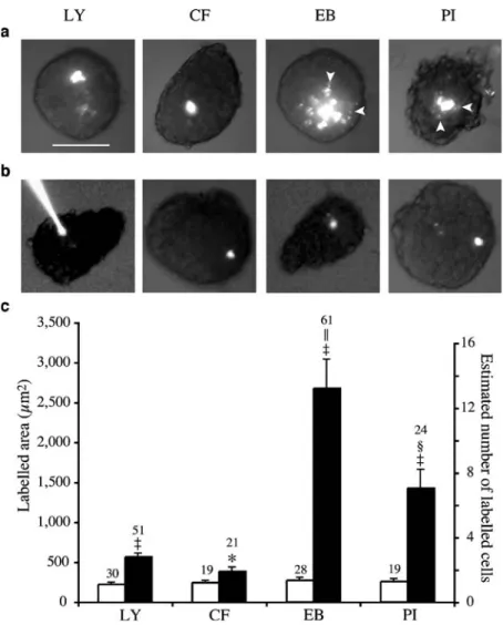
Fig. 2 Coupling of primary islet cells depends on Cx36 and is mostly due to cationic mole-cules. a In islets isolated from wild-type mice, cells poorly changed LY and CF, but ex-changed EB and PI to a greater degree. Note that LY and CF accumulate in the cytosol, whereas EB and PI mostly stain the nucleus, resulting in a spot-ted labelling of the most distant coupled cells (arrowheads). Bar, 100μm. b In islets of KO Cx36−/−mice, which lack Cx36, the four tracers revealed cell uncoupling. c Area labelled by each tracer in islets of wild-type (solid bars) and KO-Cx36−/− mice (open bars). Data are pre-sented as means±SEM; the values above the bars represent the number of microinjections. *p<0.05,‡p<0.0001 vs wild-type value for the same tracer;
§
p<0.05,||
p<0.005 vs the LY value in wild-type islets
Table 1 Percentage of injections resulting in coupling of HeLa cells and rank order of this event
Dye HeLa Coupling rank order
0 1 2 3 4 5 n LY wt 94.7 5.3 0 0 0 0 19 Cx36 7.1 92.9 7.1 0 0 0 14 Cx43 10.5 89.5 57.9 42.1 26.3 15.8 19 CF wt 100 0 0 0 0 0 22 Cx36 50 50 12.5 0 0 0 16 Cx43 13.3 86.7 66.7 33.3 20 6.7 15 EB wt 100 0 0 0 0 0 33 Cx36 27.5 72.5 52.5 32.5 17.5 12.5 40 Cx43 13.3 86.7 20 0 0 0 15 PI wt 100 0 0 0 0 0 37 Cx36 59.3 40.7 3.7 0 0 0 54 Cx43 46.7 53.3 6.7 0 0 0 15
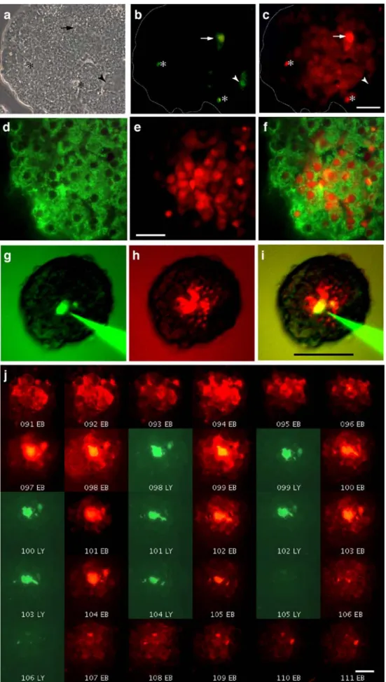
Fig. 3 Positively and negatively charged tracers reveal a different extent of beta cell coupling. a–c Individual cells of a control mouse islet (a shows phase-contrast view) were sequentially microinjected with LY (b) and EB (c). LY revealed either ap-parently uncoupled cells (*) or cells coupled to first-rank neighbours (arrowheads) (b). In contrast, after a cell was injected with EB (arrow), territories of coupled cells extending up to the fifth rank order were seen (c). The outline of the islet is shown by the dotted line. Bar (c), 20μm. d–f Immunostaining for insulin (d) of a control mouse islet microinjected with EB (e) revealed that most coupled cells were insulin-containing cells (f). f Panels d and e merged. Bar (e), 20μm. g–i Individual cells were simul-taneously microinjected with both LY (g) and EB (h). The latter tracer revealed a much larger extent of coupling than the former one. i Panels g and h merged, which are colour-coded versions of original black and white pictures. Bar (i), 100μm. j Consecutive sections (numbered) of a rat islet micro-injected with both EB and LY. A larger diffusion of the former tracer can be seen. Bar, 20μm
and PI, the latter, which easily permeates the Cx36 channels of beta (Figs.2,3, and4) and MIN6 cells (Fig. 5), being only minimally exchanged between Cx36-coupled HeLa cells (Fig.1). These findings document that the cell-to-cell transfer of the four tracers we selected depends on connexin expression and that Cx36 channels favour the intercellular exchange of selected cationic molecules.
Coupling of beta cells After microinjection into individual beta cells of islets isolated from control mice (C57/Bl6 and wild-type mice of the different knockout (KO) and transgenic lines mentioned below), the same four tracers showed frequent coupling (Fig. 2). The extension of this coupling was limited for LY (3±1 coupled cells in 82.4% of 51 injections) and CF (2±1 cells in 76.2% of 21 injections), sizable for PI (7±2 cells in 87.5% of 24 injections) and much larger for EB (14±2 cells in 90.2% of 61 injections;
Fig.2a). Histology showed that the number of coupled cells was much higher after injection of EB than LY (Fig.3b,c) and that these were mostly insulin-containing beta cells (Fig.3d–f). In islets that were simultaneously microinjected with LY and EB (Fig. 3g–i), we further observed that a number of cells that appeared to be coupled to only a few neighbours as indicated by LY transfer (2±1 cells in 89% of 18 injections) were actually coupled to more cells when assessed with EB (6±2 cells in 93% of 15 injections). Also, some of the cells that were scored as uncoupled with the former dye, were shown to be coupled when evaluated with the latter one. Some islet cells were found uncoupled with both LY and EB injection (data not shown). Comparable differences were seen in control rat islets (Fig. 3j), where the reconstruction of LY and EB labelling showed that the two tracers diffused in a non-radial manner to outline territories of complex geometry, which also comprised some uncoupled cells (Fig.3j, ESM Video clips1 and 2).
To determine whether the intercellular transfer of the four tracers mentioned above occurred via Cx36 channels, we repeated the experiments in islets of the KO Cx36 strain [21, 22,36]. Wild-type KO Cx36+/+mice, which express normal levels of Cx36 [21], showed limited islet cell coupling when tested with either LY or CF and a significantly larger coupling when tested with either EB or PI (Fig. 2a,c). In contrast, homozygous KO Cx36−/−littermates, which lack Cx36 [21], consistently showed uncoupling, irrespective of the tracer used (Fig.2b,c).
To assess whether the cationic selectivity of beta cell coupling was dependent on Cx36, we studied islets of the transgenic RIP-Cx32 mouse, whose beta cells ectopically express Cx32 in addition to native Cx36 [17]. These two connexins form junctional channels with rather different conductances and permeabilities [4, 33]. Islet cells of wild-type RIP-Cx32 mice, which express normal levels of Cx36 and no Cx32 [17], showed a much smaller coupling when tested with LY than when tested with either EB or PI (not shown), similar to results observed with other control mice. In contrast, islet cells of homozygous RIP-Cx32+/+ littermates, which co-express Cx32 and Cx36 [17], showed a significant (p<0.001) increase in coupling, as evaluated by LY microin-jection (3±1 coupled cells in 13 control inmicroin-jections vs 17±4 coupled cells in 22 injections in homozygous littermates). The intercellular transfer of PI was also larger (p<0.004) in mice expressing Cx32 (5,262±804μm2, n=12) than in their wild-type littermates (2,467±477μm2, n=13), whereas that of EB increased less (p<0.11) from 2,762±434μm2(n=15 controls) to 4,139±759 μm2(n=22) in mice co-expressing Cx32 and Cx36. These findings show that the selective cell-to-cell transfer of cationic tracers observed in control islets depends on the expression of Cx36 channels. They further indicate
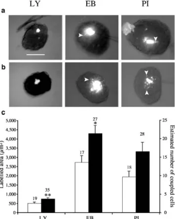
Fig. 4 Beta cell coupling changes selectively after glibenclamide stimulation. a In untreated islets of wild-type mice, cells poorly exchanged LY, but extensively exchanged EB and PI. Bar, 100μm. Note that LY accumulates in the cytosol, whereas EB and PI mostly stain the nucleus, resulting in spotted labelling of the most distant coupled cells (arrowheads). b After 24 h of glibenclamide treatment, the three tracers extended to larger areas of the microinjected islets. c Area labelled by each tracer in islets of control (open bars) and glibenclamide-stimulated (solid bars) mice. Data are presented as means±SEM; the values above the bars represent the number of microinjections. *p<0.05, **p<0.01 vs the value for the same tracer in non-stimulated islets
that, as in another model [17,39], this selectivity is altered in beta cells forced to express another connexin isoform. Beta cell coupling selectively increases after glibenclamide stimulation To assess whether beta cell coupling changes as a function of insulin secretion, we treated control mice with glibenclamide for 1 day, in conditions known to stimulate beta cells and to enhance the size of their gap junction plaques [12,37, 38]. During sulfonylurea stimulation, the size of the coupling territories delineated by EB and PI was much larger (p<0.001) than that delineated by LY (Fig.4), as seen in non-stimulated controls. However, relative to the size of the coupling territory evaluated in non-stimulated islets, that observed after glibenclamide treatment was differently modulated for different tracers, irrespective of their electrical charge. Thus, coupling increased on average by 46% (p=0.009), 55% (p=0.02) and 67% (p=0.06), as evaluated by LY, EB and PI injection, respectively (Fig.4). The data indicate that the number and/or function of Cx36 channels is increased during glibenclamide stimulation, resulting in a different increase in beta cell coupling for different cationic and anionic molecules.
Coupling of Min6 cells To further test the cationic permselectivity of Cx36 channels, we investigated MIN6 cells, which natively express Cx36 [26,35]. Wild-type cells featured a minimal coupling when assessed with LY and CF, but a larger and more frequent coupling when tested with EB and PI (Fig.5a,c, Table2). The dependence of this event on Cx36 was shown using a stable clone of MIN6
cells, which expresses minimal levels of Cx36 at the membrane, as a result of antisense transfection [26, 35]. In these cells, little coupling was observed, irrespective of the tracer used (Fig.5b,c, Table2).
To determine whether the intercellular exchange of cationic molecules was dependent on their size, we investigated MIN6 cells that had been loaded with calcein-AM, using a fluorescence quenching strategy [40]. We observed that wild-type cells featured a consistent progression of fluorescence quenching from the micro-injected cell into adjacent neighbours (Fig.6a,b,e) and that the extent of this coupling was intermediate between that
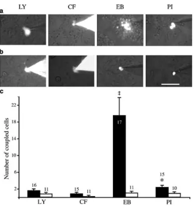
Fig. 5 MIN6 cells, like primary beta cells, are coupled by Cx36. a Wild-type MIN6 cells, which express Cx36, exchanged EB and PI to a greater degree than LY and CF. b In an antisense-transfected clone of MIN6 cells (Min6-AS-Cx36) that no longer expresses Cx36, limited inter-cellular exchange of these trac-ers was observed. Bar, 100μm. c Number of wild-type (solid bars) and Cx36-antisense cells (open bars) labelled by a tracer, after microinjection into one cell. Data are presented as means±SEM; the values above the bars represent the number of microinjections. *p<0.05,
‡p<0.0001 vs the value for the
same tracer in Cx36-antisense cells
Table 2 Percentage of injections resulting in coupling of MIN6 cells and rank order of this event
Dye MIN6 Coupling rank order
0 1 2 3 4 5 n LY wt 31.2 68.8 18.8 0 0 0 16 AS-Cx36 70 30 0 0 0 0 10 CF wt 53.3 46.7 6.7 0 0 0 15 AS-Cx36 72.7 27.3 0 0 0 0 11 EB wt 23.5 76.5 70.6 64.7 52.9 29.4 17 AS-Cx36 27.3 72.7 18.2 0 0 0 11 PI wt 20 80 26.7 6.7 0 0 15 AS-Cx36 70 30 0 0 0 0 10 Mn2+ wt 0 100 72.7 9.1 9.1 9.1 11 AS-Cx36 58.3 41.7 0 0 0 0 12
observed for PI and EB (compare Figs. 5c and 6e), two molecules significantly larger than the quenching-inducing Mn2+. MIN6 cells that had been transfected for an antisense Cx36 cDNA failed to show Mn2+-induced quenching outside the microinjected cell (Fig. 6c–e). These findings show that a comparable coupling of insulin-producing cells is revealed by cationic tracers of rather different size and is dependent on the presence of Cx36 channels.
Discussion
In pancreatic islets, beta cells are linked by Cx36 channels [14,20,24,25], which account for electrical coupling [5,7, 10, 13, 15, 22, 23], as well as for the intercellular synchronisation of glucose-induced electrical activity [5, 10,15,22] and Ca2+transients [15,21,23,35]. The same channels also allow beta cells to exchange cytosolic molecules [11, 15,23] and exogenous tracers [8,12,17– 19, 21,29]. Previous studies have shown that blockade or loss of Cx36 leads to altered function of beta cells, notably
with regard to basal and glucose-stimulated insulin secre-tion [19, 21, 27, 35, 43]. Alterations in insulin secretion have also been reported following either the overexpression of Cx36 [39,43] or the forced expression in beta cells of a connexin that is not natively expressed in the islets [17]. Together, these data imply a central role of beta cell coupling in the regulation of islet function and indicate that connexin-dependent signalling specifically requires Cx36.
Why this protein, which is only expressed in the nervous and endocrine systems [20], is essential for beta cells remains to be determined. Compared with the other 20 known connexins [4, 20], Cx36 confers upon homomeric channels a rather low voltage sensitivity, a small unitary conductance and a differential permeability to LY and neurobiotin [4, 30, 31, 39]. It typically forms gap junctions comprising only a few dozen connexons [37,38,42]. The minimal voltage sensitivity is advantageous to beta cells since it allows Cx36 channels to remain open under both basal and stimulated conditions. The modest number of low-conductance channels allows the fine-tuning of communication between beta cells, which have a high input resistance [5, 7, 10, 14,22, 23]. The differential permeability of Cx36 to molecules that have the size of endogenous metabolites and second messengers could explain the variable estimations of beta cell coupling so far reported. Thus, whereas electrophysiological data and Ca2+ measure-ments have indicated that ionic coupling may extend throughout entire islets [5,7,21,22,29,44,45], experiments evaluating the intercellular exchange of endogenous metabo-lites or exogenous tracers have indicated a more restricted coupling [8, 11, 12, 15, 17–19, 21, 23, 29]. However, the comparison of these findings is complicated, since previous studies have evaluated islet cell coupling with different techniques [28, 29], tested junctional conductance indirectly [5,7,10,22] and/or studied beta cells in a non-physiological environment [5,8,10,11,15,22,45]. Thus, the question of what is transferred between beta cells via the Cx36 channels of intact islets remains to be established.
To address these issues, we used novel animal models that allow unambiguous control of various connexins and analysis of coupling within the normal islet environment. Using a series of gap junction tracers selected for subtle differences in molecular weight and electrical charge, we showed that Cx36 channels favour the exchange between beta cells of selected cationic molecules, restraining that of anionic molecules. We showed that this exchange is drastically modified when beta cells are forced to express Cx32, a connexin that is not natively found in islets [14,20, 24, 25] and forms channels preferentially permeated by anionic molecules [4, 17, 33, 39]. Using a fluorescence quenching approach, we extended to Mn2+ the previous evidence that Cx36 channels are also permeated by current-carrying cations [21,22,26,35,44,45] and showed that the extent of this ionic coupling is similar to that observed with
Fig. 6 MIN6 cells exchange Mn2+in a similar way to larger cationic tracers. a, b Cultures of wild-type MIN6 cells were loaded with calcein-AM, before individual cells were microinjected with Mn2+. This ion induced the quenching of calcein fluorescence in the microinjected cell (asterisk) and then in all neighbours coupled to it (arrow heads). c, d In MIN6-AS-Cx36 cells, which express little Cx36, diffusion of the Mn2+-induced fluorescence quenching was observed in no more than one coupled cell (arrowhead). Bar, 100μm. e Number of wild-type (solid bar, 11 microinjections) and Cx36-antisense cells (open bar, 12 microinjections) featuring a quenching of calcein fluorescence after microinjection of Mn2+into one cell. Data are presented as means±SEM. ¶p<0.0005 vs the value in Cx36-antisense cells
much larger cationic molecules. By comparing normal and KO mice lacking Cx36, as well as wild-type and Cx36-antisense-transfected MIN6 cells, our data conclusively show that Cx36 is both necessary and sufficient to ensure the native, selective coupling of beta cells. Our data also indicate that Cx36 channels are not identical in insulin-producing cells and HeLa cells (e.g. the permeability to EB is similar in the two cell types, whereas that to PI is not), supporting the view that a cell-specific environment modulates their function [13, 23], as suggested for other connexin isoforms [46, 47]. While the underlying mechanism remains to be elucidated, plausible reasons are the interactions of connex-ins with different proteconnex-ins/lipids and/or a different regulation by cell-specific post-translational processes. In this respect, current evidence indicates that the level of Cx36 phosphor-ylation may vary in different cell types [13, 32]. Our data also show that after in vivo stimulation with sulfonylurea [12,37,38], beta cell coupling is enhanced. Together with the previous report that the same treatment enlarges islet gap junctions [37, 38], these findings indicate that increased number/function of Cx36 channels is required during stimulation of insulin secretion, possibly to recruit increasing numbers of secreting cells [14, 16, 19, 20]. Strikingly, the intercellular exchange of cationic and anionic molecules was not modified to the same extent, indicating that a subtle modulation of the resting Cx36 permselectivity occurs during stimulation of insulin secretion.
These data have three implications. First, they suggest that cationic molecules may be the main messengers of connexin signalling between beta cells. This knowledge should facili-tate the identification of these factors, in as far as most endogenous metabolites are negatively charged at cytosolic pH [4, 11, 23]. Second, our experiments indicate that the ionic and molecular coupling of beta cells may not be as different as previously thought [5,7,8,10,11,15,23,29]. The new data show that the extent of coupling provided by Cx36 channels is sufficient to involve sizable proportions of, though not all beta cells of an islet. Moreover, under all the conditions we tested, a number of beta cells appeared to be uncoupled from their neighbours, which is consistent with the previously reported distribution of gap junctions, the estimated number of functional junctional channels and their average open time probability [4,6,48]. Third, our experi-ments demonstrate that Cx36 is the only connexin that functionally couples pancreatic beta cells [21, 22, 24, 25], with specific deletion of this protein abolishing the intercel-lular exchange of the five probes we tested. These data extend recent freeze-fracture, dye injection and electrophys-iological observations [21, 22], indicating that if other connexins are expressed in beta cells [49,50], these proteins do not form functional cell-to-cell channels.
In summary, our data show that within pancreatic islets, beta cells featuring Cx36 channels extensively exchange
positively charged molecules of various size and exchange negatively charged molecules to a much smaller degree. The identification of such an extensive coupling resolves the long-standing controversy regarding a purported difference be-tween the extent of electrical and metabolic coupling of islet cells. Our data show that large numbers of beta cells, although not all of them, are coupled within primary islets. We also definitively show that this coupling is uniquely due to Cx36 channels. The identification of a permselectivity of the Cx36 channels is expected to facilitate the identification of the endogenous molecules that are exchanged between beta cells. Our findings also document that the beta cell exchanges of anionic and cationic molecules are differentially regulated during in vivo stimulation of insulin secretion. This finding opens new perspectives for the unravelling of the mechanism whereby Cx36 signalling contributes to the proper control of insulin biosynthesis and release.
Acknowledgements The authors thank A. Charollais, D. Caille and I. Borges for valuable assistance. The work of our team is supported by grants from the Swiss National Science Foundation (310000-109402), the Juvenile Diabetes Research Foundation (1-2005-46 and 1-2007-158), Novo Nordisk and the Geneva Program for Metabolic Disorders.
Duality of interest The authors declare that there is no duality of interest associated with this manuscript.
References
1. Ashcroft FM, Gribble FM (1999) ATP-sensitive K+channels and
insulin secretion: their role in health and disease. Diabetologia 42:903–919
2. MacDonald PE, Joseph JW, Rorsman P (2005) Glucose-sensing mechanisms in pancreatic beta-cells. Philos Trans R Soc Lond B Biol Sci 360:2211–2225
3. Rorsman P (1997) The pancreatic beta-cell as a fuel sensor: an electrophysiologist’s viewpoint. Diabetologia 40:487–495 4. Harris AL (2001) Emerging issues of connexin channels:
biophysics fills the gap. Q Rev Biophys 34:325–472
5. Kanno T, Gopel SO, Rorsman P, Wakui M (2002) Cellular function in multicellular system for hormone-secretion: electro-physiological aspect of studies on alpha-, beta- and delta-cells of the pancreatic islet. Neurosci Res 42:79–90
6. Verselis VK, Veenstra R (2000) Gap junction channels, perme-ability and voltage gating. Adv Mol Cell Biol 30:129–192 7. Andreu E, Soria B, Sanchez-Andres JV (1997) Oscillation of gap
junction electrical coupling in the mouse pancreatic islets of Langerhans. J Physiol 498:753–761
8. Kohen E, Kohen C, Thorell B, Mintz DH, Rabinovitch A (1979) Intercellular communication in pancreatic islet monolayer cul-tures: a microfluorometric study. Science 204:862–865
9. MacDonald PE, Rorsman P (2006) Oscillations, intercellular coupling, and insulin secretion in pancreatic beta cells. PLoS Biol 4:e49
10. Mears D, Sheppard NFJ, Atwater I, Rojas E (1995) Magnitude and modulation of pancreatic beta-cell gap junction electrical conductance in situ. J Membr Biol 146:163–176
11. Meda P, Amherdt M, Perrelet A, Orci L (1981) Metabolic coupling between cultured pancreatic b-cells. Exp Cell Res 133:421–430
12. Meda P, Michaels RL, Halban PA, Orci L, Sheridan JD (1983) In vivo modulation of gap junctions and dye coupling between B-cells of the intact pancreatic islet. Diabetes 32:858–868 13. Moreno AP, Berthoud VM, Perez-Palacios G, Perez-Armendariz
EM (2005) Biophysical evidence that connexin-36 forms func-tional gap junction channels between pancreatic mouse beta-cells. Am J Physiol Endocrinol Metab 288:E948–E956
14. Nlend RN, Michon L, Bavamian S et al (2006) Connexin36 and pancreatic beta-cell functions. Arch Physiol Biochem 112:74–81 15. Pedersen MG, Bertram R, Sherman A (2005) Intra- and inter-islet
synchronization of metabolically driven insulin secretion. Biophys J 89:107–119
16. Bosco D, Meda P (1997) Reconstructing islet function in vitro. Adv Exp Med Biol 426:285–298
17. Charollais A, Gjinovci A, Huarte J et al (2000) Junctional communication of pancreatic beta cells contributes to the control of insulin secretion and glucose tolerance. J Clin Invest 106:235–243 18. Kohen E, Kohen C, Rabinovitch A (1983) Cell-to-cell
communi-cation in rat pancreatic islet monolayer cultures is modulated by agents affecting islet-cell secretory activity. Diabetes 32:95–98 19. Meda P, Bosco D, Chanson M et al (1990) Rapid and reversible
secretion changes during uncoupling of rat insulin-producing cells. J Clin Invest 86:759–768
20. Michon L, Nlend Nlend R, Bavamian S et al (2005) Involvement of gap junctional communication in secretion. Biochim Biophys Acta 1719:82–101
21. Ravier MA, Guldenagel M, Charollais A et al (2005) Loss of connexin36 channels alters beta-cell coupling, islet synchroniza-tion of glucose-induced Ca2+and insulin oscillations, and basal insulin release. Diabetes 54:1798–1807
22. Speier S, Gjinovci A, Charollais A, Meda P, Rupnik M (2007) Cx36-mediated coupling reduces beta-cell heterogeneity, confines the stimulating glucose concentration range, and affects insulin release kinetics. Diabetes 56:1078–1086
23. Tsaneva-Atanasova K, Zimliki CL, Bertram R, Sherman A (2006) Diffusion of calcium and metabolites in pancreatic islets: killing oscillations with a pitchfork. Biophys J 90:3434–3446
24. Serre-Beinier V, Le Gurun S, Belluardo N et al (2000) Cx36 preferentially connects beta-cells within pancreatic islets. Diabetes 49:727–734
25. Theis M, Mas C, Doring B et al (2004) Replacement by a lacZ reporter gene assigns mouse connexin36, 45 and 43 to distinct cell types in pancreatic islets. Exp Cell Res 294:18–29
26. Calabrese A, Caton D, Meda P (2004) Differentiating the effects of Cx36 and E-cadherin for proper insulin secretion of MIN6 cells. Exp Cell Res 294:379–391
27. Konstantinova I, Nikolova G, Ohara-Imaizumi M et al (2007) EphA–ephrin-A mediated beta cell communication regulates insulin secretion from pancreatic islets. Cell 129:359–370 28. Meda P (2000) Probing the function of connexin channels in
primary tissues. Methods 20:232–244
29. Quesada I, Fuentes E, Andreu E, Meda P, Nadal A, Soria B (2003) On-line analysis of gap junctions reveals more efficient electrical than dye coupling between islet cells. Am J Physiol Endocrinol Metab 284:E980–E987
30. Srinivas M, Rozental R, Kojima T et al (1999) Functional properties of channels formed by the neuronal gap junction protein connexin36. J Neurosci 19:9848–9855
31. Teubner B, Degen J, Sohl G et al (2000) Functional expression of the murine connexin 36 gene coding for a neuron-specific gap junctional protein. J Membr Biol 176:249–262
32. Urschel S, Hoher T, Schubert T et al (2006) Protein kinase A-mediated phosphorylation of connexin36 in mouse retina results in decreased gap junctional communication between AII amacrine cells. J Biol Chem 281:33163–33171
33. Elfgang C, Eckert R, Lichtenberg−Frate H et al (1995) Specific permeability and selective formation of gap junction channels in connexin-transfected HeLa cells. J Cell Biol 129: 805–817
34. Miyazaki J, Araki K, Yamato E et al (1990) Establishment of a pancreatic beta cell line that retains glucose-inducible insulin secretion: special reference to expression of glucose transporter isoforms. Endocrinology 127:126–132
35. Calabrese A, Zhang M, Serre-Beinier V et al (2003) Connexin 36 controls synchronization of Ca2+oscillations and insulin secretion in MIN6 cells. Diabetes 52:417–424
36. Guldenagel M, Ammermuller J, Feigenspan A et al (2001) Visual transmission deficits in mice with targeted disruption of the gap junction gene connexin36. J Neurosci 21:6036–6044
37. Meda P, Halban P, Perrelet A, Renold AE, Orci L (1980) Gap junction development is correlated with insulin content in the pancreatic B cell. Science 209:1026–1028
38. Meda P, Perrelet A, Orci L (1979) Increase of gap junctions between pancreatic B-cells during stimulation of insulin secretion. J Cell Biol 82:441–448
39. Caton D, Calabrese A, Mas C et al (2003) Lentivirus-mediated transduction of connexin cDNAs shows level- and isoform-specific alterations in insulin secretion of primary pancreatic beta-cells. J Cell Sci 116:2285–2294
40. Sawahara H, Goto S, Kinoshita N (1991) Double fluorescent labeling method used for a study on liposomes. Chem Pharm Bull (Tokyo) 39:227–229
41. Meda P, Santos RM, Atwater I (1986) Direct identification of electrophysiologically monitored cells within intact mouse islets of Langerhans. Diabetes 35:232–236
42. Abramoff MD, Magelhaes PJ, Ram SJ (2004) Image processing with ImageJ. Biophoton Int 11:36–42
43. Le Gurun S, Martin D, Formenton A et al (2003) Connexin-36 contributes to control function of insulin-producing cells. J Biol Chem 278:37690–37697
44. Meda P, Atwater I, Goncalves A, Bangham A, Orci L, Rojas E (1984) The topography of electrical synchrony among beta-cells in the mouse islet of Langerhans. Q J Exp Physiol 69:719–735 45. Meissner HP (1976) Electrophysiological evidence for coupling
between beta cells of pancreatic islets. Nature 262:502–504 46. Eckert R (2006) Gap-junctional single-channel permeability for
fluorescent tracers in mammalian cell cultures. Biophys J 91:565–579 47. Koval M (2006) Pathways and control of connexin
oligomeriza-tion. Trends Cell Biol 16:159–166
48. Perez-Armendariz M, Roy C, Spray DC, Bennett MV (1991) Biophysical properties of gap junctions between freshly dispersed pairs of mouse pancreatic beta cells. Biophys J 59:76–92 49. Collares-Buzato CB, Leite AR, Boschero AC (2001) Modulation
of gap and adherens junctional proteins in cultured neonatal pancreatic islets. Pancreas 23:177–185
50. Leite AR, Carvalho CP, Furtado AG, Barbosa HC, Boschero AC, Collares-Buzato CB (2005) Co-expression and regulation of connexins 36 and 43 in cultured neonatal rat pancreatic islets. Can J Physiol Pharmacol 83:142–151