1
Revised version of Manuscript IAI00818-13
1 2
Capsular sialic acid of Streptococcus suis serotype 2 binds to swine
3influenza virus and enhances bacterial interactions with virus-infected
4tracheal epithelial cells
56
Yingchao Wanga,b, Carl A. Gagnona, Christian Savarda, Nedzad Musica, Mariela Srednika, 7
Mariela Seguraa, Claude Lachancea, Christian Bellehumeura and Marcelo Gottschalka# 8
9 a
Faculté de médecine vétérinaire, Université de Montréal, 3200 Sicotte, St-Hyacinthe, Québec, 10
Canada J2S 2M2. 11
b
College of Animal Science and Veterinary Medicine, Jilin University, Changchun, People's 12
Republic of China, Changchun, Jilin Province, 130062, China P.R. 13
14 15
#
Address correspondence to: Marcelo Gottschalk, Faculté de médecine vétérinaire, Université de 16
Montréal, 3200 Sicotte, St-Hyacinthe, Québec, Canada, J2S 2M2. Phone: 001-450-773-8521; 17
Fax: 001-450-778-8108; mail: marcelo.gottschalk@umontreal.ca 18
19
Running title: S. suis serotype 2 capsule binds to influenza virus 20
21 22
2 ABSTRACT
23
Streptococcus suis serotype 2 is an important swine bacterial pathogen and it is also an emerging 24
zoonotic agent. It is unknown how S. suis virulent strains, which are usually found in low 25
quantities in pig tonsils, manage to cross the first host defense lines to initiate systemic disease. 26
Influenza virus produces a contagious infection in pigs which is frequently complicated by 27
bacterial co-infections leading to significant economic impacts. In this study, the effect of a 28
preceding swine influenza H1N1 virus (swH1N1) infection of swine tracheal epithelial cells 29
(NTPr) on the ability of S. suis serotype 2 to adhere, invade and activate these cells was 30
evaluated. Cells pre-infected with swH1N1, showed bacterial adhesion and invasion levels 31
increased more than 100 fold when compared to normal cells. Inhibition studies confirmed that 32
the capsular sialic acid moiety is responsible for the binding to virus-infected cell surface. Also, 33
pre-incubation of S. suis with swH1N1 significantly increased bacterial adhesion/invasion to 34
epithelial cells, suggesting that S. suis may also use swH1N1 as a vehicle to invade epithelial 35
cells when the two infections occur simultaneously. Influenza infection may facilitate the 36
transient passage of S. suis at the respiratory tract to reach the bloodstream and cause bacteremia 37
and septicemia. S. suis may also increase the local inflammation at the respiratory tract during 38
influenza infection, as suggested by an exacerbated expression of pro-inflammatory mediators in 39
co-infected cells. These results give a new insight in the elucidation of complex interactions 40
between influenza virus and S. suis in a co-infection model. 41 42 43 44 45 46
3 INTRODUCTION
47 48
Streptococcus suis is one of the most important post-weaning bacterial pathogens in swine and it 49
is also an emerging zoonotic agent (1). Among the 35 S. suis described serotypes, type 2 is the 50
most virulent one for both pigs and humans (2), although differences in virulence have been 51
described for this serotype (3). Pigs may acquire S. suis very early in life and some colonized 52
animals may never develop disease (carrier animals); on the other hand, some carrier piglets will 53
eventually develop bacteremia, septicemia and meningitis following dissemination of S. suis in 54
the bloodstream (1). Human infections with S. suis manifest mainly as meningitis, septicemia and 55
septic shock (4). It is believed that people can become infected through skin lesions, surface 56
mucosa and/or the oral route (5). 57
It is still unknown how low quantities of S. suis virulent serotype 2 strains present in 58
tonsils of pigs manages to cross the first natural line of the host defense to initiate disease. It is 59
believed that the pathogen would breach the mucosal epithelium at the upper respiratory tract (6). 60
Bacterial adhesion and invasion of epithelial cells are usually associated with the first steps of 61
colonization by mucosal pathogens; however, few data are available concerning the interaction 62
between S. suis and swine respiratory epithelial cells. Ferrando and colleagues described for the 63
first time S. suis adhesion (but not invasion) to porcine tracheal epithelial cells (7). 64
The S. suis capsular polysaccharide (CPS), which defines the serotype, is essential for the 65
virulence of this pathogen mainly due to its antiphagocytic activity (6). The analysis of the 66
serotype 2 CPS revealed the presence of different sugars including Neu5Ac or sialic acid. 67
Interestingly, sialic acid was found to be terminal [(2→6)-β-D-Galactose], and the CPS can be 68
quantitatively desialylated by mild acid hydrolysis (8). It has been previously shown that 69
4 expression of CPS interferes with adhesion and (if any) invasion of S. suis to epithelial cells (9, 70
10). So, classically, the role of this virulence factor has been suggested to be crucial once bacteria 71
reach the bloodstream (6). Among other suggested S. suis virulence factor, secreted proteins, such 72
as the hemolysin (suilysin), surface proteins and other cell wall components have been reported 73
(11). 74
Secondary bacterial infections associated to influenza virus infection in humans are a 75
leading cause of human morbidity and mortality worldwide (12). Swine influenza virus infections 76
in pigs also cause serious respiratory disease (13). Although this infection is typically self-limited 77
with high-morbidity but low mortality, secondary complications substantially increase illness and 78
death (14). In fact, influenza is a key contributor to the porcine respiratory disease complex 79
(PRDC), a multifactorial syndrome characterized by severe respiratory disease after infection 80
with two or more agents (15). Pathogens associated with PRDC include (among others) 81
Actinobacillus pleuropneumoniae, Haemophilus parasuis, Mycoplasma hyopneumoniae, S. suis 82
and porcine reproductive and respiratory syndrome virus (15). Subtypes of swine influenza virus 83
that are most frequently identified in pigs include H1N1 (classical and pandemic), H1N2 and 84
H3N2 (13). Influenza virus strains uniformly recognize cell surface oligosaccharides with a 85
terminal sialic acid either 2,3 Neu5Ac–galactose or 2,6 Neu5Ac–galactose. However, their 86
receptor specificity varies according to host. Pigs are unique among influenza virus hosts in that 87
they are susceptible to infection with influenza viruses of human and avian origin as well as to 88
swine influenza virus, because their tracheal epithelium contains these two sialyloligosaccharides 89
(16). 90
In this study we demonstrated, for the first time, a novel mechanism used by a bacterial 91
species to facilitate the invasion of respiratory epithelial cells already infected with an influenza 92
virus. More specifically, we showed that the sialic acid moiety present in the CPS of S. suis 93
5 serotype 2 directly interacts with swine influenza virus leading to an increased bacterial adhesion, 94
invasion and activation of tracheal epithelial cells. This mechanism could explain, at least in part, 95
how secondary bacterial infection with virulent S. suis strain could be enhanced following 96 influenza infection. 97 98 99 100 101
6 MATERIALS AND METHODS
102 103
Bacterial strains, epithelial cells and influenza virus strain. S. suis strains used in this study are 104
listed in Table 1. The well characterized S. suis serotype 2 virulent strain 31533 (10, 17) was used 105
throughout this study. Other previously well characterized isogenic mutants derived from this 106
strain and devoid of either CPS or suilysin production, or modified at the either peptidoglycan 107
(PG) or lipoteichoic acid (LTA) levels were also included (18-21). In addition, serotype 2 field 108
strains with lower (Canadian strain) or higher (epidemic strain isolated from a deadly S. suis 109
human outbreak in China) virulence potential (3), as well as reference strains of serotypes 3 and 110
14 were also included for comparison purposes (Table 1). A swine influenza virus H1N1 111
(swH1N1, strain A/swine/St-Hyacinthe/148/1990) isolated from a case of swine flu in Canada 112
was used (22). 113
Bacterial were cultured as previously reported (17). The number of CFU/ml in the final 114
suspension before each experiment was determined by plating samples onto THA using 115
Autoplate® 4000 Automated Spiral Plater (Spiral Biotech, Norwood, MA). The pig trachea 116
epithelial cell line (NPTr) was used for virus growth and co-infection studies as described (23). 117
For assays, cells were treated with 0.05% trypsin in 0.03% EDTA solution and diluted in culture 118
medium to obtain a final concentration of 105 cells/ml. Then, the cell suspension was distributed 119
into tissue culture plates and incubated until cell confluence was reached. Twenty-four hours 120
before the assays, culture medium was removed from the wells and replaced by fresh complete 121
medium without antibiotics. Virus was produced by replication in NPTr cells as previously 122
described (23). The titer of the viral production was 107.25 TCID50/ml. 123
7 NPTr co-infection by swH1N1 and S. suis. swH1N1 (MOI: 1) was inoculated onto NPTr cell 125
monolayers in 24-well culture plates and incubated with 2% FBS (as standardized in preliminary 126
experiments) and antibiotic free MEM for 1 h at 37°C in 5% CO2. The virus-infected cells were 127
then washed twice with PBS and fresh media containing 10% FBS without antibiotic was added. 128
The increased serum concentration did not affect virus replication and kept cells healthy for the 129
whole experiment. Following a 12 h incubation time at 37°C in 5% CO2, cells were infected with 130
S. suis (106 CFU/well, MOI:10). Plates were centrifuged at 800 x g for 10 min in order to bring 131
bacteria in close contact with the cells (24). Bacterial infected cells were then incubated at 37°C in 132
5% CO2 for different incubation times (see below). Infectious viral load profile was determined in 133
cell cultures for virus infected cells and for virus-bacteria co-infected cells by virus titration 134
evaluation as described above. Cell cytotoxicity levels were determined using Cytotox 96 kit 135
(Promega, Madison, WI) from culture supernatants according to manufacturer’s instruction. In 136
selected experiments, swH1N1 and S. suis were pre-incubated for 1 h at 4°C (106 S. suis CFU and 137
106 TCID50 of swH1N1, respectively; final bacteria/virus ratio of 1). Afterwards, the virus-S. 138
suis mixture was washed twice with PBS and resuspended with complete medium, inoculated to 139
cells and incubated at 37°C in 5% CO2 for bacterial adhesion and invasion assays, as described 140
below. Mock-treated bacteria were used as control. 141
The invasion assay was performed as previously described (17), with some modifications. 142
After 2 or 4 h of incubation with S. suis, the NPTr cells monolayers were washed twice with PBS, 143
and 1 ml of cell culture medium containing 100 μg of gentamicin and 5 μg of penicillin G 144
(Invitrogen, Burlington, ON, Canada) was added to each well. The plates were then further 145
incubated for 1 h at 37°C with 5% CO2 to kill extracellular and surface-adherent bacteria. After 146
washing, cells were disrupted with sterile ice-cold deionized water followed by cell scrapping 147
from the bottom of the well in order to liberate intracellular bacteria. Bacterial CFU numbers were 148
8 determined by plating serial dilutions as described above. Levels of invasion were expressed as 149
the total number of CFU recovered per well. An “adhesion assay” which in fact quantifies total 150
cell-associated bacteria (intracellular bacteria and surface-adherent bacteria) was performed 151
similarly to the invasion assay. However, the cells were vigorously washed five times to eliminate 152
nonspecific bacterial attachment and no antibiotic treatment to kill the extracellular bacteria was 153
used. At different incubation times (see results), the levels of “adhesion” (total associated 154
bacteria) were expressed as the total number of CFU recovered per well. 155
For the inhibition studies, and after removing the cell supernatant and washing twice the 156
wells with fresh PBS, 100 µg of purified native CPS or desialylated CPS (prepared as described 157
below) resuspended in cell culture medium were added to swH1N1-infected cells. Control cells 158
were treated similarly but without addition of CPS. After 1 h of incubation, cells were washed 159
twice with PBS and infected with S. suis as previously described. Bacterial adhesion and invasion 160
studies were performed as described above and compared to non-treated cells. Results were 161
expressed in percentage when compared to bacterial adhesion and invasion of untreated swH1N1 162
pre-infected cells (considered as 100%). Swine polyclonal antibody serum against the whole 163
swH1N1 virus strain (serum from a convalescent animal) was used a positive inhibition control.
164
Supernatants of swH1N1-infected cells were removed and the serum (diluted 1/40 in cell culture 165
medium) was added to the wells. 166
167
Hemagglutination inhibition assay (HI). HI test was carried out as previously described (25) 168
with the modification that swine sera were replaced by different concentrations of S. suis. Serial 169
dilutions of S. suis strains (wild-type 31533 strain or non-encapsulated B218 mutant strain) were 170
used for the HI assay. Briefly, 50 μl of bacterial suspensions (grown as described above) were 171
dispensed at different concentrations in triplicate in a 96-well round bottom plate. Fifty μl of 172
9 swH1N1 (2 x 106.25 TCID50/ml) was then added to each well and incubated for 1 h at room 173
temperature. Different wells represented a 2-fold dilutions of S. suis/swH1N1virus ratios, 174
beginning at a ratio of 200 for the wild type encapsulated strain and 10 000 for the non-175
encapsulated B218 mutant. Afterward, 50 μl of a 0.5% suspension of whole rooster red blood 176
cells (RBC) in PBS were added to each well and gently mixed. The HI was evaluated after 177
incubating the plate at room temperature for 1 h. For this experiment, PBS was used as negative 178
RBC control and serial dilutions of reference heat-inactivated anti-swH1N1 serum was used as a 179
positive HI control. Under the conditions tested, capsulated and non-encapsulated S. suis strains 180
did not induce any hemagglutination (results not shown). 181
182
S. suis CPS purification and CPS desialylation. The CPS of S. suis serotype 2 reference strain
183
S735 was prepared and purified as previously described (8). For quality controls, CPS was 184
analyzed by nuclear magnetic resonance. Lack of protein and RNA/DNA contamination was 185
verified by Lowry method and by spectrophotometry, respectively. CPS was also desialylated by 186
mild acid hydrolysis. CPS (8 mg) was heated in 1 ml of HCl (70 mM) at 60 °C for 4 h, neutralized 187
with NH4OH (2 M), and purified on a Sephadex G10 column (1.5 x 10 cm). Presence (native 188
CPS) or absence (desialylated CPS) of sialic acid was verified by gas chromatography after 189
methanolysis and acetylation and by nuclear magnetic resonance as well as by a reaction with an 190
enzyme linked-lectin assay as previously described (26). 191
192
Confocal and electron microscopy. For confocal microscopy analysis, cells were placed on 193
coverslips and infected (or not) with swH1N1 and either S. suis strain 31533 or its non-194
encapsulated mutant strain (B218) as described above and further incubated for 2 h at 37°C in 5% 195
CO2. Coverslips were washed with PBS to remove non-associated bacteria and cells were fixed 196
10 with 4% paraformaldehyde solution for 10 min. Cells were then washed and permeabilized with 197
PBS containing 0.2% Triton X-100 (Thermo Hyclone, Burlington, ON, Canada) for 2 min. The 198
coverslips were blocked for 10 min with PBS containing 2% bovine serum albumin and 0.2% 199
gelatin (Sigma-Aldrich, Oakville, ON, Canada). Coverslips were then incubated for 1 h with a 200
mouse monoclonal antibody against an epitope within influenza virus A nucleoprotein H1N1 (US 201
Biologica, Swampscott, MA l; 1/500 dilution) and a rabbit anti-S. suis serum against either wild-202
type strain 31533 (1/5000) or its non-encapsulated B218 mutant strain (1/1000) (27). After 203
washing with PBS, coverslips were incubated with secondary antibodies Alex-Fluor 568 goat 204
anti-mouse IgG (for swH1N1) and Alex-Fluor 488 goat anti-rabbit IgG (for S. suis) (both from 205
Invitrogen) for 30 min. Coverslips were then washed and mounted on glass slides with moviol 206
containing DABCO. 207
For transmission electron microscopy (TEM) and scanning electron microscopy (SEM), 208
samples were fixed for 1 h at room temperature with 2% (vol/vol) glutaraldehyde in 0.1 M 209
cacodylate buffer (pH 7.3) and were then post-fixed for 45 min at room temperature with 2% 210
osmium tetroxide. Specimens for TEM were dehydrated in a graded series of ethanol solutions 211
and embedded with LR white resin. Thin sections were cut with a diamond knife and 212
werepoststained with uranyl acetate and lead citrate. Samples were observed with an electron 213
microscope model JEM-1230 (JEOL, Tokyo, Japan). Samples for SEM were dehydrated in a 214
graded series of ethanol solutions and covered with gold after critical point drying and were 215
examined with a HitachiS-3000N microscope. 216
217
Quantitative RT-PCR (qRT-PCR) for cytokine and chemokine expression. qRT-PCR assay 218
was performed as previously described (28). Primers (IDT DNA, Coralville, IA) used for 219
detection of genes were all verified to have PCR amplification efficiency ranked between 90-220
11 110% using a CFX96 rapid thermal cycler system (Bio-Rad, Hercules, CA) (Table 2). The 221
GeNorm applet v.3.5 (http://medgen.ugent.be/~jvdesomp/genorm/) was used to initially determine 222
the two most stable reference genes from a set of six reference genes using random samples from 223
the cDNA panel generated for the qPCR analysis of cytokine/chemokine gene expression. 224
Therefore, normalization of data was done using the reference genes hypoxanthine 225
phosphoribosyltransferase 1 (Hprt1) and Peptidylprolyl isomerase A (Ppia). Fold-change of gene 226
expression was calculated using the normalized gene expression (ΔΔCq) calculation method of the 227
CFX software manager v.2.1 (Bio-Rad). Mock-infected samples were used as calibrator and 228
consequently, relative fold-differences were calculated for the rest of the samples compared to the 229
mean of the calibrator samples. 230
231
Statistical analysis. All data are expressed as mean ± SEM. Prism statistical software v.5 232
(Graphpad, San Diego, CA) was employed for data analysis. Data from the adhesion and 233
invasion assays were analyzed for significance using Student’s unpaired t -test. Data from qPCR 234
assays were subjected to one way ANOVA analysis followed by Tukey’s post hoc test. A P value 235
< 0.01 was used as threshold for statistical significance. Results reflect mean values of at least 236
three independent experiments. 237
238 239
12 RESULTS
240 241
S. suis serotype 2 adhesion and invasion are significantly increased when cells are
242
previously infected by swH1N1, independently of the virulence of the S. suis strain. 243
The kinetics of adhesion of the highly virulent S. suis serotype 2 strain 31533 to NPTr 244
cells was studied. As shown in Fig.1A, in the absence of virus infection, adhesion was time 245
dependent, increasing from 30 min to 4 h of incubation. After 4 h of incubation, a plateau was 246
reached (data not shown). Results of the kinetics and levels of adhesion are similar to those 247
previously obtained with porcine endothelial and other epithelial cells (10, 17). However, when 248
cells were pre-infected with swH1N1 for 12 h, the adhesion levels increased more than 100 folds 249
compared to those observed in the absence of virus (Fig.1A). In addition, adhesion levels 250
immediately reached a plateau (Fi.g 1A), even after 5 min of incubation (data not shown). When 251
strains of serotype 2 with lower or higher virulence potential than that of 31533 strain were tested 252
(intermediate virulence Canadian strain 1591 or epidemic strain SC84 from a Chinese human 253
outbreak) (3), bacterial adhesion levels were statistically similar to those obtained with the 254
virulent strain 31533, either in the absence or presence of swH1N1 infection (Fig. 2A). 255
Surprisingly, and different from what has been previously reported with other epithelial 256
cells of swine origin (10), encapsulated S. suis serotype 2 was able to clearly invade NPTr cells 257
(Fig. 1B). However, when cells were pre-infected with the swH1N1 strain, invasion rates also 258
increased more than 100 folds at both, 2 h and 4 h incubation times (P < 0.01) (Fig. 1B). Similar 259
to the adhesion results, invasion rates of the two additional S. suis serotype 2 strains were 260
statistically similar to those obtained with strain 31533 in the presence or absence of swH1N1 261
pre-infection (Fig. 2B). For all adhesion and invasion experiments, cells presented cytotoxicity 262
13 levels lower than 20% (data not shown). Interestingly, virus replication levels in NPTr cells were 263
similar in the presence or absence of bacterial infection (Supplemental Fig. S1). 264
265
Critical role of the capsular polysaccharide (CPS) in the increased S. suis adhesion/invasion 266
to swH1N1 pre-infected NPTr cells. 267
Isogenic mutants defective in suilysin production, D-alanylation of LTA or N-268
deacetylation of PG behaved statistically similarly to the wild-type strain 31533 either in the 269
presence or absence of swH1N1 pre-infection. Only the non-encapsulated (CPS-) mutant 270
presented a different pattern. In the absence of virus infection, the adhesion and invasion levels of 271
the mutant strain were significantly higher (P < 0.01) than those of the wild-type strain (Fig. 2A 272
and 2B), confirming previous published results which indicated that the CPS interferes with S. 273
suis-host cell interactions (9, 10). However, these adhesion and invasion levels were unmodified 274
after a swH1N1 pre-infection. These data suggest that the CPS might play a role in the observed 275
increased levels of wild-type S. suis adhesion/invasion to virus infected cells (Fig. 2A and 2B). 276
Since the antigenic characteristics of the CPS define the serotype (1), two additional S. suis 277
serotypes (3 and 14) were tested. Although both strains are well encapsulated (29), only the 278
adhesion and invasion of S. suis serotype 14 reference strain (DAN13730), but not those of 279
serotype 3 (strain 4961), were significantly affected by a pre-infection with swH1N1 (Fig. 2A 280
and 2B). This would indicate that the CPS structure and/or composition directly influence the 281
interactions between S. suis and swH1N1 pre-infected cells. 282
Influence of epithelial cell swH1N1 pre-infection on adhesion/invasion abilities of S. suis 283
serotype 2 was confirmed by microscopy. First, confocal microscopy revealed that very few 284
encapsulated wild-type bacteria could be observed interacting with epithelial cells in the absence 285
of virus pre-infection (Fig. 3). However, after 12 h of swH1N1 pre-infection, levels of wild-type 286
14 encapsulated S. suis adhesion were clearly higher and grouped around the cells (in “grapes”), 287
especially where the red staining with anti-H1N1 monoclonal antibody was present, indicating a 288
possible co-localization of virus and bacteria. In the absence of virus infection, the non-289
encapsulated mutant showed a higher level of adhesion than the wild-type strain, although 290
bacteria were randomly distributed on the cell surface (diffuse adhesion). A similar adhesion 291
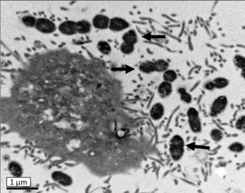
pattern of the mutant strain was observed when cells were pre-infected with swH1N1. Electron 292
microscopy (TEM and SEM) confirmed the influence of a pre-infection with influenza virus on S. 293
suis-cell interactions (Fig. 4). In the absence of virus infection, very few cocci (if any) could be 294
observed interacting with cells (Fig. 4A-I). In the presence of a virus pre-infection, cells were 295
highly activated (clearly showing cilia at their surface) and high numbers of cocci were at the cell 296
surface (closely interacting with cilia) (Fig. 4AII and 4B) and, sometimes, inside the cells (Fig. 4 297
AIII). 298
299
Bacterial capsular sialic acid is responsible for bacterial-virus interactions in infected cells. 300
Since the CPS of S. suis serotype 2 was shown to be implicated in the increased bacterial-301
cell interactions when cells were pre-infected with swH1N1, it was hypothesized that the sialic 302
acid moiety present in the CPS of this serotype may be involved through interactions with viral 303
hemagglutinin. In fact, the reference strain of serotype 14 CPS (which also interacted with 304
swH1N1 pre-infected cells) possesses an identical sialic acid-containing side chain (also with a 305
link 2,6 to the adjacent galactose) as serotype 2 CPS (30), whereas the reference strain of 306
serotype 3 lacks this sugar (31-33). To confirm such hypothesis, inhibition studies were 307
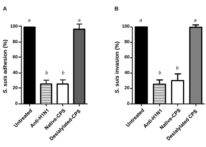
performed. Interestingly, when wells were simply washed before adding the bacterial suspension 308
and used as a control, no differences could be obtained with previous results with non-washed 309
wells, indicating that free virus were either not present at significant number or that they did not 310
15 significantly interfere with baceterial adhesion/invasion to epithelial cells. A pre-treatment of 311
swH1N1-NPTr pre-infected cells with purified native CPS inhibits >75% of adhesion and 312
invasion by S. suis serotype 2. This inhibition was similar to that obtained with a pre-treatment 313
with an anti-swH1N1 specific antibody (Fig. 5). When the same amount of desialylated CPS was 314
used, no inhibition of bacterial adhesion/invasion could be observed, confirming the involvement 315
of the CPS sialic acid in the interactions of S. suis with swH1N1 pre-infected cells (Fig. 5). 316
317
In vitro binding of swH1N1 to S. suis enhances bacterial adhesion and invasion to epithelial
318
cells. 319
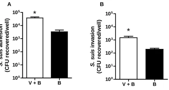
To investigate if well encapsulated S. suis may directly interact with the swH1N1 strain, a 320
test of hemagglutination inhibition was performed. Results showed that a 1 h pre-incubation of S. 321
suis serotype 2 strain 31533 and swH1N1 virus (in a bacteria/virus ratio >50) resulted in the
322
complete inhibition of RBC hemagglutination (Supplemental figure 2). Lower concentrations of 323
bacteria did not present any visual inhibition. Interestingly, no inhibition of RBC 324
hemagglutination was observed when the non-encapsulated mutant was used, even in a 325
bacteria/virus ratio of 10 000) (Supplemental figure 2). Finally, a pre-incubation of S. suis 326
serotype 2 strain 31533 with the swH1N1 strain significantly increase the interaction between S. 327
suis and NPTr cells, since bacterial adhesion and invasion to epithelial cells presented up to 10 328
fold increase values when compared to those infected with S. suis without a pre-incubation with 329
swH1N1 (Fig. 6). These results suggest that S. suis may also use swH1N1 virus as a vehicle to 330
adhere and invade epithelial cells. The fact that some bacteria may aggregate with virus (forming 331
micro-clumps) enhancing somehow the total number of bacterial adhesion to cells cannot be 332
ruled out. No increase in bacteria-cell interactions was observed when the non-encapsulated 333
mutant was used (data not shown). 334
16 335
Co-infected NPTr cells express higher levels of pro-inflammatory genes than single-infected 336
cells. 337
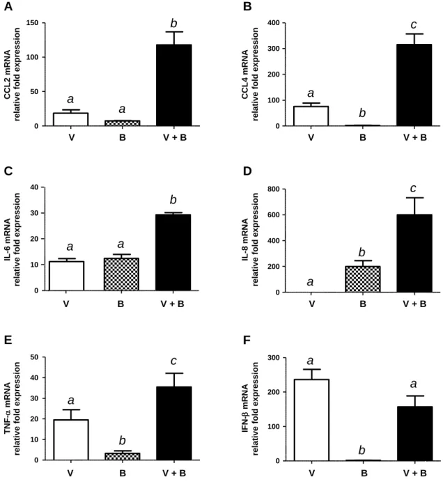
Although a complete kinetics was studied (results not shown), results showed that 24 h 338
post-bacterial infection (36 h post virus infection) reflected optimal differences among groups. 339
NPTr cells infected with bacteria alone showed absence or low expression levels of CCL2 (MCP-340
1), CCL4 (MIP-1β), IFN-β and TNF-α, intermediate expression levels of IL-6 and high levels of 341
IL-8 expression (Fig. 7). Virus-mediated NPTr cell activation at that incubation time showed 342
absence of IL-8 expression. On the other hand, the swH1N1 strain activated gene expression of 343
other mediators at similar levels (CCL2 and IL-6) or at significantly higher levels (CCL4, TNF-α 344
and IFN-β) than those obtained after activation with S. suis alone (Fig. 7). Interestingly, 345
swH1N1-S. suis co-infection significantly increased the expression of CCL2, CCL4, IL-6, IL-8 346
and TNF-α mRNA. In some cases, an additive effect seemed responsible for such differences (IL-347
6 and TNF-α). However, the increase of mRNA expression of CCL2, CCL4 and IL-8 mRNA 348
expression was clearly ahead of a simple additive effect. Expression of IFN-β mRNA was 349
probably attributed solely to the effect of swH1N1 (Fig. 7). 350
17 DISCUSSION
352
The pathogenesis of the infection caused by S. suis is far from being completely 353
understood (6). In swine, S. suis is mainly transmitted by aerosols, and airborne transmission 354
among pigs has been clearly demonstrated (34). S. suis play a certain role in mixed respiratory 355
infections, although it is not considered a primary cause of swine pneumonia (1), indicating that 356
it may also use the respiratory tract as a transient passage before reaching the bloodstream and 357
causing bacteremia, which is essential for the pathogen to cause meningitis (35). The actual early 358
mechanisms used by this pathogen to interact with epithelial cells to further invade the 359
bloodstream are, in fact, poorly known. 360
S. suis clinical association with virus infections have been largely reported (36, 37). More 361
recently, several outbreaks in swine due to swine influenza virus with a significant level of 362
systemic co-infection due to S. suis have been reported in England (38). In humans, it is well 363
known that influenza cases are heavily complicated by bacterial infections (12). In fact, it has 364
been previously reported that influenza as well as other respiratory virus increase the 365
adhesion/invasion capacities of bacterial pathogens (including streptococci) to epithelial cells, 366
although mechanisms have not been fully elucidated (39). The goal of the present work was to 367
study interactions between S. suis and tracheal epithelial cells either pre-infected or not with 368
swH1N1. 369
Results showed that S. suis is able to not only to adhere to but also invade swine tracheal 370
epithelial cells. In the absence of virus infections, adhesins involved in such interactions seem to 371
be located in the bacterial cell wall, since they are hindered by the presence of the CPS, as 372
previously suggested (6, 9, 10). Indeed, significant higher levels of adhesion and, most important, 373
invasion rates were observed with a non-encapsulated S. suis mutant. Interestingly, results 374
obtained with isogenic mutants showed that alteration at the LTA and PG as well as the lack of 375
18 suilysin production did not influence the adhesion/invasion capacities of S. suis. Different S. suis 376
surface-exposed proteins have been described as bacterial adhesins to extracellular matrix 377
proteins present in host cells (6, 11). In fact, ApuA, a surface protein with bifunctional 378
amylopullulanase activity, was described to play an important role in such adhesion to tracheal 379
epithelial cells (7). No differences could also be observed between strains of serotype 2 of 380
difference virulence potential or strains belonging to other serotypes showing that those adhesins 381
are probably common to most strains of S. suis, independently of their virulence/serotype. 382
In the presence of a prior swH1N1 infection, more than 100 fold increases in S. suis 383
adhesion and invasion could be observed. This increased interaction was confirmed by confocal, 384
TEM and SEM. Increased cell susceptibility to S. suis adhesion and invasion following a virus 385
infection may have different explanations. One of the most well-known interactions is that 386
between influenza virus and Streptococcus pneumoniae (40). In vivo increased susceptibility has 387
been attributed to an alteration of anti-bacterial phagocyte functions through a diminished 388
bactericidal activity and/or damage to the respiratory epithelium resulting in defective 389
mucociliary clearance mechanisms, which in turn leads to an increased numbers of bacteria that 390
remains in the respiratory tract (41). In vitro studies suggested damage to the respiratory 391
epithelium by exposing surface molecules and cell receptors to which pneumococci more readily 392
adhere and invade cells. This effect would be mainly done by the viral neuraminidase (42), 393
although a certain synergistic role of neuraminidase produced by S. pneumoniae cannot be rule 394
out (43). 395
Results from the present study indicate that interactions between influenza virus and S. 396
suis are clearly different from those with S. pneumoniae. In fact, no neuraminidase activities have 397
been so far demonstrated for S. suis. On the other hand, a clear role of the surface exposed CPS in 398
the S. suis interactions with swH1N1-infected cells could be established. Similar results were 399
19 previously obtained with Group A Streptococcus (GAS) and A549 epithelial cells (44). Although 400
a certain direct binding between GAS and influenza virus could be observed, molecules involved 401
in such interactions have so far not been elucidated (45). Interestingly, GAS lacks sialic-acid in 402
its surface. In the present study, the main serotypes of S. suis containing sialic acid (serotypes 2 403
and 14) clearly interact with swH1N1-infected cells, whereas interactions of a serotype lacking 404
this sugar (serotype 3) were not affected by a virus pre-infection. In addition, serotype 2 strains of 405
different virulence potential behaved similarly, due to the fact that capsular composition of the 406
three strains is most probably identical. In an inhibition assay using highly purified native and 407
desialylated CPS purified from the reference strain of serotype 2, it was clearly showed that the 408
bacterial sialic acid moiety was responsible for the virus-bacterial interactions. It was then 409
hypothesized that the S. suis sialic acid binds to the hemagglutinin of the swH1N1. This was 410
further demonstrated by the fact that well-encapsulated S. suis (but not its non-encapsulated 411
mutant) incubated with the swH1N1 strain was able to inhibit the RBC hemagglutination activity 412
of the virus. The binding of S. suis CPS to influenza hemagglutinin was not exclusive of the 413
H1N1 strain used. Another swine influenza field strain (H3N2) used in parallel studies offered 414
identical results than those obtained with the H1N1 strain (unpublished observations). 415
Interestingly, direct binding of Group B Streptococcus (GBS) to influenza virus has also been 416
described previously (46). It was hypothesized that the sialyl-galactose linkage in GBS was 417
responsible for binding to the virus (46). We suggest that GBS would behave similarly to S. suis 418
since the structures of the CPS of both pathogens are similar (8). Interestingly, not all bacterial 419
pathogens possessing capsular sialic acid use a similar mechanism. For example, it has been 420
proposed that a direct interaction between the neuraminidase of influenza virus and the CPS of 421
Neisseria meningitidis enhances bacterial adhesion to cultured epithelial cells, most likely 422
through cleavage of capsular sialic acid-containing bacterial polysaccharides (47). 423
20 Although a typical pre-infection with influenza virus is believed to be followed by a 424
bacterial complication, a simultaneous infection with both pathogens cannot be disregarded. In 425
pigs, for example, both pathogens may infect animals at the same age range (1). In this study, a 426
binding between free S. suis serotype 2 to free swH1N1 promotes enhanced bacterial adhesion 427
and invasion to swine epithelial cells, similarly to what has been shown for GAS (45). Similarly, 428
previous in vitro binding of non-identified surface exposed proteins of Staphylococcus aureus to 429
the viral hemoagglutinin enhances bacterial invasion to virus-uninfected cells (48). Hence, 430
influenza infection may promote adhesion and internalization of S. suis not only by binding of 431
bacteria to the membrane-associated hemagglutinin but also by binding of bacteria to free virions 432
followed by internalization of virus-coated bacteria into non-infected epithelial cells. Therefore, a 433
possible synergy between the two pathogens cannot be ruled out. However, further studies on the 434
exact mechanisms involved should be performed. 435
Influenza virus is able to stimulate epithelial cells and induce the over-production of 436
different inflammatory mediators. In addition, it may directly or indirectly interfere with the 437
balance of cytokine/chemokine production (49). In co-infection studies, activation of epithelial 438
cells by influenza virus enhances the induction of cytokine and chemokine gene transcripts by S. 439
pneumoniae (50). Inflammation has been reported to be highly important in S. suis infections 440
(51). So far, the inflammatory response of respiratory epithelial cells generated by S. suis has not 441
been addressed. In the present study, S. suis was shown to strongly up-regulate gene expression 442
of mainly IL-6 and IL-8, similar to that observed with epithelial cells of the choroid plexus (52). 443
Differently from what was described with these cells, relatively low levels of TNF-α expression 444
were observed with S. suis-activated NPTr cells, even at shorter incubation times (data not 445
shown), indicating some differences between the two cell types. When NPTr cells were pre-446
infected with swH1N1, the significant increase of IL-8 expression that was observed may be 447
21 explained by a higher number of bacteria interacting with influenza-infected cells. It has been 448
shown that IL-8 expression by S. suis-activated endothelial cells is bacterial-concentration 449
dependent (53). In the case of IL-6 and TNF-α, the increased expression observed under the co-450
infection conditions may be also explained by an additive effect of swH1N1 and S. suis. On the 451
other hand, S. suis alone did not produce significant levels of CCL2 and CCL4. However, when 452
cells were pre-infected with swH1N1, between 100 and 300 fold increases in mRNA expression 453
of these mediators were detected. Influenza virus replicates in the respiratory epithelium and 454
induces an inflammatory infiltrate comprised of mononuclear cells and neutrophils (54), to which 455
S. suis possesses anti-phagocytic capacities (6). Since S. suis is not a primary pulmonary 456
pathogen, an exacerbated production of pro-inflammatory mediators during a co-infection with 457
influenza virus may be important in the pathogenesis of the influenza infection. 458
In conclusion, a new role of S. suis CPS, other than that of anti-phagocytic factor (55), has 459
been demonstrated in the present study. Although it was previously reported that the presence of 460
sialic acid in S. suis could not be directly related to virulence (56), we demonstrated that its 461
presence plays a major role in the interactions with respiratory epithelial cells previously infected 462
by swine influenza virus, acting as a bacterial receptor for the virus. Simultaneous co-infections 463
with both pathogens may also be mutually beneficial due to direct bacterial-virus interaction. 464
Binding of bacteria to influenza virus-infected cells or directly to influenza virus could play an 465
important role allowing bacteria to move towards the lower airways, initiating the systemic 466
invasion that characterizes the pathogenesis of the infection caused by S. suis. The increased 467
production of local pro-inflammatory mediators in the presence of both pathogens may also play 468
an important role in the pathogenesis of the pneumonia caused by swine influenza. 469
22 ACKNOWLEDGEMENTS
471
We would like to thank Sonia Lacouture for technical assistance. Y. Wang was a recipient of 472
China Scholarship Council. This work was supported by Natural Sciences and Engineering 473
Research Council of Canada (NSERC) grant #154280 to M.Gottschalk. C. Savard was a recipient 474
of a Canadian Swine Health Board postdoctoral fellowship. 475
476
REFERENCES 477
478
1. Gottschalk M. 2012. Streptococcocis, p. 841-855. In B. E. Straw, J. J. Zimmerman, S. D’Allaire, and
479
D. J. Taylor (ed.), Diseases of Swine, 10th ed. Blackwell Publishing, Ames, Iowa, USA. 480
2. Gottschalk M, Xu J, Calzas C, Segura M. 2010. Streptococcus suis: a new emerging or an old
481
neglected zoonotic pathogen? Future Microbiol. 5:371-391. 482
3. Ye C, Zheng H, Zhang J, Jing H, Wang L, Xiong Y, Wang W, Zhou Z, Sun Q, Luo X, Du H,
483
Gottschalk M, Xu J. 2009. Clinical, experimental, and genomic differences between
484
intermediately pathogenic, highly pathogenic, and epidemic Streptococcus suis. J. Infect. Dis. 485
199:97-107.
486
4. Lun ZR, Wang QP, Chen XG, Li AX, Zhu XQ. 2007. Streptococcus suis: an emerging zoonotic
487
pathogen. The Lancet Infect. Dis. 7:201-209. 488
5. Wertheim HF, Nghia HD, Taylor W, Schultsz C. 2009. Streptococcus suis: an emerging human
489
pathogen. Clin. Infect. Dis. 48:617-625. 490
6. Fittipaldi N, Segura M, Grenier D, Gottschalk M. 2012. Virulence factors involved in the
491
pathogenesis of the infection caused by the swine pathogen and zoonotic agent Streptococcus 492
suis. Future Microbiol. 7:259-279. 493
7. Ferrando ML, Fuentes S, de Greeff A, Smith H, Wells JM. 2010. ApuA, a multifunctional
alpha-494
glucan-degrading enzyme of Streptococcus suis, mediates adhesion to porcine epithelium and 495
mucus. Microbiology. 156:2818-2828. 496
8. Van Calsteren MR, Gagnon F, Lacouture S, Fittipaldi N, Gottschalk M. 2010. Structure
497
determination of Streptococcus suis serotype 2 capsular polysaccharide. Biochem. Cell Biol. 498
88:513-525.
499
9. Benga L, Goethe R, Rohde M, Valentin-Weigand P. 2004. Non-encapsulated strains reveal novel
500
insights in invasion and survival of Streptococcus suis in epithelial cells. Cell. Microbiol. 6:867-501
881. 502
10. Lalonde M, Segura M, Lacouture S, Gottschalk M. 2000. Interactions between Streptococcus suis
503
serotype 2 and different epithelial cell lines. Microbiology. 146:1913-1921. 504
11. Baums CG, Valentin-Weigand P. 2009. Surface-associated and secreted factors of Streptococcus
505
suis in epidemiology, pathogenesis and vaccine development. Anim. Health Res. Rev. 10:65-83. 506
23 12. Smith AM, Adler FR, Ribeiro RM, Gutenkunst RN, McAuley JL, McCullers JA, Perelson AS. 2013.
507
Kinetics of Coinfection with Influenza A Virus and Streptococcus pneumoniae. PLoS pathogens 508
9:e1003238.
509
13. Jung K, Ha Y, Chae C. 2005. Pathogenesis of swine influenza virus subtype H1N2 infection in pigs.
510
J. Comp. Path. 132:179-184. 511
14. Loving CL, Brockmeier SL, Vincent AL, Palmer MV, Sacco RE, Nicholson TL. 2010. Influenza virus
512
coinfection with Bordetella bronchiseptica enhances bacterial colonization and host responses 513
exacerbating pulmonary lesions. Microb. Pathog. 49:237-245. 514
15. Opriessnig T, Gimenez-Lirola LG, Halbur PG. 2011. Polymicrobial respiratory disease in pigs.
515
Anim. Health Res. Rev. 12:133-148. 516
16. Ito T, Couceiro JN, Kelm S, Baum LG, Krauss S, Castrucci MR, Donatelli I, Kida H, Paulson JC,
517
Webster RG, Kawaoka Y. 1998. Molecular basis for the generation in pigs of influenza A viruses
518
with pandemic potential. J. Virol. 72:7367-7373. 519
17. Vanier G, Segura M, Friedl P, Lacouture S, Gottschalk M. 2004. Invasion of porcine brain
520
microvascular endothelial cells by Streptococcus suis serotype 2. Infect. Immun. 72:1441-1449. 521
18. Lun S, Perez-Casal J, Connor W, Willson PJ. 2003. Role of suilysin in pathogenesis of
522
Streptococcus suis capsular serotype 2. Microb. Pathog. 34:27-37. 523
19. Fittipaldi N, Harel J, D'Amours B, Lacouture S, Kobisch M, Gottschalk M. 2007. Potential use of
524
an unencapsulated and aromatic amino acid-auxotrophic Streptococcus suis mutant as a live 525
attenuated vaccine in swine. Vaccine 25:3524-3535. 526
20. Fittipaldi N, Sekizaki T, Takamatsu D, Harel J, Dominguez-Punaro Mde L, Von Aulock S, Draing
527
C, Marois C, Kobisch M, Gottschalk M. 2008. D-alanylation of lipoteichoic acid contributes to the
528
virulence of Streptococcus suis. Infect. Immun. 76:3587-3594. 529
21. Fittipaldi N, Sekizaki T, Takamatsu D, Dominguez-Punaro Mde L, Harel J, Bui NK, Vollmer W,
530
Gottschalk M. 2008. Significant contribution of the pgdA gene to the virulence of Streptococcus
531
suis. Mol. Microbiol. 70:1120-1135. 532
22. Bikour MH, Frost EH, Deslandes S, Talbot B, Elazhary Y. 1995. Persistence of a 1930 swine
533
influenza A (H1N1) virus in Quebec. J. Gen. Virol. 76:2539-2547. 534
23. Ferrari M, Scalvini A, Losio MN, Corradi A, Soncini M, Bignotti E, Milanesi E, Ajmone-Marsan P,
535
Barlati S, Bellotti D, Tonelli M. 2003. Establishment and characterization of two new pig cell
536
lines for use in virological diagnostic laboratories. J. Virol. Meth. 107:205-212. 537
24. Bouchet B, Vanier G, Jacques M, Auger E, Gottschalk M. 2009. Studies on the interactions of
538
Haemophilus parasuis with porcine epithelial tracheal cells: limited role of LOS in apoptosis and 539
pro-inflammatory cytokine release. Microb. Pathog. 46:108-113. 540
25. Tremblay D, Allard V, Doyon JF, Bellehumeur C, Spearman JG, Harel J, Gagnon CA. 2011.
541
Emergence of a new swine H3N2 and pandemic (H1N1) 2009 influenza A virus reassortant in two 542
Canadian animal populations, mink and swine. J. Clin. Microbiol. 49:4386-4390. 543
26. Lecours MP, Fittipaldi N, Takamatsu D, Okura M, Segura M, Goyette-Desjardins G, Van
544
Calsteren MR, Gottschalk M. 2012. Sialylation of Streptococcus suis serotype 2 is essential for
545
capsule expression but is not responsible for the main capsular epitope. Microbes Infect. 14:941-546
950. 547
27. Higgins R, Gottschalk M. 1990. An update on Streptococcus suis identification. J. Vet. Diagn.
548
Invest. 2:249-252. 549
28. Lecours MP, Segura M, Lachance C, Mussa T, Surprenant C, Montoya M, Gottschalk M. 2011.
550
Characterization of porcine dendritic cell response to Streptococcus suis. Vet. Res. 42:72. 551
29. Jacques M, Gottschalk M, Foiry B, Higgins R. 1990. Ultrastructural study of surface components
552
of Streptococcus suis. J. Bacteriol. 172:2833-2838. 553
24 30. Van Calsteren MR, Gagnon F, Calzas C, Goyette-Desjardins G, Okura M, Takamatsu D,
554
Gottschalk M, Segura M. 2013. Structure determination of Streptococcus suis serotype 14
555
capsular polysaccharide. Biochem. Cell Biol. 91:49-58. 556
31. Charland N, Kellens JT, Caya F, Gottschalk M. 1995. Agglutination of Streptococcus suis by sialic
557
acid-binding lectins. J. Clin. Microbiol. 33:2220-2221. 558
32. Smith HE, de Vries R, van't Slot R, Smits MA. 2000. The cps locus of Streptococcus suis serotype
559
2: genetic determinant for the synthesis of sialic acid. Microb. Pathog. 29:127-134. 560
33. Okura M, Takamatsu D, Maruyama F, Nozawa T, Nakagawa I, Osaki M, Sekizaki T, Gottschalk
561
M, Kumagai Y, Hamada S. 2013. Genetic analysis of capsular polysaccharide synthesis gene
562
clusters from all serotypes of Streptococcus suis: potential mechanisms for the generation of 563
capsular variation. Appl. Environ. Microbiol. 79:2796-2806. 564
34. Berthelot-Herault F, Gottschalk M, Labbe A, Cariolet R, Kobisch M. 2001. Experimental airborne
565
transmission of Streptococcus suis capsular type 2 in pigs. Vet. Microbiol. 82:69-80. 566
35. Dominguez-Punaro MC, Koedel U, Hoegen T, Demel C, Klein M, Gottschalk M. 2012. Severe
567
cochlear inflammation and vestibular syndrome in an experimental model of Streptococcus suis 568
infection in mice. Euro. J. Clin. Microbiol. Infect. Dis. 31:2391-2400. 569
36. Pallares FJ, Halbur PG, Opriessnig T, Sorden SD, Villar D, Janke BH, Yaeger MJ, Larson DJ,
570
Schwartz KJ, Yoon KJ, Hoffman LJ. 2002. Porcine circovirus type 2 (PCV-2) coinfections in US field
571
cases of postweaning multisystemic wasting syndrome (PMWS). J. Vet. Diagn. Invest. 14:515-572
519. 573
37. Schmitt CS, Halbur PG, Roth JA, Kinyon JM, Kasorndorkbua C, Thacker B. 2001. Influence of
574
ampicillin, ceftiofur, attenuated live PRRSV vaccine, and reduced dose Streptococcus suis 575
exposure on disease associated with PRRSV and S. suis coinfection. Vet. Microbiol. 78:29-37. 576
38. Williamson SM, Tucker AW, McCrone IS, Bidewell CA, Brons N, Habernoll H, Essen SC, Brown
577
IH, Cosi, Wood JL. 2012. Descriptive clinical and epidemiological characteristics of influenza A
578
H1N1 2009 virus infections in pigs in England. Vet. Rec. 171:271. 579
39. Avadhanula V, Rodriguez CA, Devincenzo JP, Wang Y, Webby RJ, Ulett GC, Adderson EE. 2006.
580
Respiratory viruses augment the adhesion of bacterial pathogens to respiratory epithelium in a 581
viral species- and cell type-dependent manner. J. Virol. 80:1629-1636. 582
40. Bosch AA, Biesbroek G, Trzcinski K, Sanders EA, Bogaert D. 2013. Viral and bacterial interactions
583
in the upper respiratory tract. PLoS pathogens 9:e1003057. 584
41. Pittet LA, Hall-Stoodley L, Rutkowski MR, Harmsen AG. 2010. Influenza virus infection decreases
585
tracheal mucociliary velocity and clearance of Streptococcus pneumoniae. Am. J. Resp. Cell Mol. 586
Biol. 42:450-460. 587
42. McCullers JA, Bartmess KC. 2003. Role of neuraminidase in lethal synergism between influenza
588
virus and Streptococcus pneumoniae. J. Infect. Dis. 187:1000-1009. 589
43. Nishikawa T, Shimizu K, Tanaka T, Kuroda K, Takayama T, Yamamoto T, Hanada N, Hamada Y.
590
2012. Bacterial neuraminidase rescues influenza virus replication from inhibition by a 591
neuraminidase inhibitor. PloS one 7:e45371. 592
44. Okamoto S, Kawabata S, Terao Y, Fujitaka H, Okuno Y, Hamada S. 2004. The Streptococcus
593
pyogenes capsule is required for adhesion of bacteria to virus-infected alveolar epithelial cells 594
and lethal bacterial-viral superinfection. Infect. Immun. 72:6068-6075. 595
45. Swildens B, Stockhofe-Zurwieden N, van der Meulen J, Wisselink HJ, Nielen M, Niewold TA.
596
2004. Intestinal translocation of Streptococcus suis type 2 EF+ in pigs. Vet. Microbiol. 103:29-33. 597
46. Hosaka Y, Kuroda K, Ikeura A, Iwamoto T, Suzuki Y. 1998. Binding of influenza and
598
paramyxoviruses to Group B Streptococcus with the terminal sialyl-galactose linkage. J. Electron 599
Micro. 47:169-174. 600
25 47. Rameix-Welti MA, Zarantonelli ML, Giorgini D, Ruckly C, Marasescu M, van der Werf S, Alonso
601
JM, Naffakh N, Taha MK. 2009. Influenza A virus neuraminidase enhances meningococcal
602
adhesion to epithelial cells through interaction with sialic acid-containing meningococcal 603
capsules. Infect. Immun. 77:3588-3595. 604
48. Passariello C, Nencioni L, Sgarbanti R, Ranieri D, Torrisi MR, Ripa S, Garaci E, Palamara AT.
605
2011. Viral hemagglutinin is involved in promoting the internalisation of Staphylococcus aureus 606
into human pneumocytes during influenza A H1N1 virus infection. Int. J. Med. Microbiol. 301:97-607
104. 608
49. Lam WY, Yeung AC, Chu IM, Chan PK. 2010. Profiles of cytokine and chemokine gene expression
609
in human pulmonary epithelial cells induced by human and avian influenza viruses. Virol. J. 610
7:344.
611
50. Tong HH, Long JP, Shannon PA, DeMaria TF. 2003. Expression of cytokine and chemokine genes
612
by human middle ear epithelial cells induced by influenza A virus and Streptococcus pneumoniae 613
opacity variants. Infect. Immun. 71:4289-4296. 614
51. Dominguez-Punaro MC, Segura M, Plante MM, Lacouture S, Rivest S, Gottschalk M. 2007.
615
Streptococcus suis serotype 2, an important swine and human pathogen, induces strong systemic 616
and cerebral inflammatory responses in a mouse model of infection. J Immunol. 179:1842-1854. 617
52. Schwerk C, Adam R, Borkowski J, Schneider H, Klenk M, Zink S, Quednau N, Schmidt N, Stump
618
C, Sagar A, Spellerberg B, Tenenbaum T, Koczan D, Klein-Hitpass L, Schroten H. 2011. In vitro
619
transcriptome analysis of porcine choroid plexus epithelial cells in response to Streptococcus 620
suis: release of pro-inflammatory cytokines and chemokines. Microb. Infect. 13:953-962. 621
53. Vadeboncoeur N, Segura M, Al-Numani D, Vanier G, Gottschalk M. 2003. Pro-inflammatory
622
cytokine and chemokine release by human brain microvascular endothelial cells stimulated by 623
Streptococcus suis serotype 2. FEMS Immun. Med. Microbiol. 35:49-58. 624
54. Wareing MD, Lyon AB, Lu B, Gerard C, Sarawar SR. 2004. Chemokine expression during the
625
development and resolution of a pulmonary leukocyte response to influenza A virus infection in 626
mice. J. Leuk. Biol. 76:886-895. 627
55. Smith HE, Damman M, van der Velde J, Wagenaar F, Wisselink HJ, Stockhofe-Zurwieden N,
628
Smits MA. 1999. Identification and characterization of the cps locus of Streptococcus suis
629
serotype 2: the capsule protects against phagocytosis and is an important virulence factor. 630
Infect. Immun. 67:1750-1756. 631
56. Charland N, Kobisch M, Martineau-Doize B, Jacques M, Gottschalk M. 1996. Role of capsular
632
sialic acid in virulence and resistance to phagocytosis of Streptococcus suis capsular type 2. FEMS 633
Immun. Med. Microbiol. 14:195-203. 634
57. Gottschalk M, Higgins R, Jacques M, Mittal KR, Henrichsen J. 1989. Description of 14 new
635
capsular types of Streptococcus suis. J. Clin. Micriobiol. 27:2633-2636. 636
637 638
26 TABLE 1 List of Streptococcus suis strains used in this study
Strains Relevant phenotype and/or description References 31533 Serotype 2highly pathogenic European strain isolated from a
disease pig (10)
SC84 Serotype 2 epidemic virulent strain isolated from a human
outbreak in China (3)
89-1591 Serotype 2 iintermediate virulent strain isolated from a disease pig
in Canada (3)
CPS- Non-encapsulated B218 mutant strain derived from strain 31533 (19) ∆Sly Suilysin negative SX911 mutant strain derived from strain 31533 (18) ∆dLTA D-alanylation of LTA mutant strain derived from strain 31533 (20)
∆pgdA N-deacetylation of peptidoglycan mutant strain derived from strain 31533 (21)
4961 Reference strain, serotype 3, isolated from a diseased pig (57) DAN13730 Reference strain, serotype 14 isolated from a diseased human (57) S735 Reference strain, serotype 2, isolated from a diseased pig (8)
TABLE 2 Sequences of porcine-specific real-time PCR primers
Gene Genebank ID Amplicon
size Forward Sequence Reverse Sequence
Efficiency (qPCR)
Hprt1 NM_001032376 142 bp GCAGCCCCAGCGTCGTGATT CGAGCAAGCCGTTCAGTCCTGT 99
Ppia NM_214353 133 bp TGCAGACAAAGTTCCAAAGACAG GCCACCAGTGCCATTATGG 97
Ccl2 NM_214214 169 bp CAGGTCCTTGCCCAGCCAGATG CACAGATCTCCTTGCCCGCGA 90
Ccl4 NM_213779 125 bp TCCCACCTCCTGCTGCTTCACAT GCCTGCCCTTTTTGGTCTGGAA 100
Il6 NM_214399 105 bp ACTCCCTCTCCACAAGCGCCTT TGGCATCTTCTTCCAGGCGTCCC 97
Il8 NW_003300390 80 bp TGTGAGGCTGCAGTTCTGGCAAG GGGTGGAAAGGTGTGGAATGCGT 95
Ifnβ NM_001003923.1 150 bp TGCAACCACCACAATTCCAGAAGG TCTGCCCATCAAGTTCCACAAGGA 96
27 FIGURE LEGENDS
FIG 1. Adhesion to and invasion of virus-free or swH1N1-infected NPTr cells by S. suis serotype 2 strain 31533. NPTr cells were either pre-infected with swH1N1 for 12 h (MOI: 1) or not, and subsequently infected with S. suis serotype 2 strain 31533 (MOI: 10). (A) Kinetics of adhesion of S. suis to virus-infected (“V+B”) or control (“B”) NPTr cells. After S. suis infection cells were extensively washed to remove non-adherent bacteria and then lysed to determine S. suis viable counts. (B) S. suis invasion of swH1N1-infected (“V+B”) or control (“B”) NPTr cells at 2 and 4 h bacterial incubation times. Results were determined as described above except that after washing, cells were exposed to antibiotics to kill extracellular bacteria. Data are expressed as mean ± SEM of at least four independent experiments, each done in triplicate. * indicates significant difference between samples infected with bacteria alone and those co-infected with virus and bacteria (P < 0.01).
FIG 2. Adhesion to and invasion of virus free or swH1N1-infected NPTr cells by different strains of S. suis. NPTr cells were either pre-infected with swH1N1 for 12 h (MOI: 1) or not, and subsequently infected with S. suis strains (MOI: 10). (A) Adhesion (incubation time of 2 h) of different S. suis strains to NPTr cells. Results were determined after exposure of swH1N1-infected (“V+B”) or control (“B”) NPTr cells to S. suis, followed by extensive washing of non-adherent bacteria and cell lysis to obtain S. suis viable counts. (B) Invasion (incubation time of 1 h) of swH1N1-infected (“V+B”) or control (“B”) NPTr cells by different S. suis strains. Results were determined as described above except that after washing, cells were exposed to antibiotics to kill extracellular bacteria. See Table 1 for strain description. Data are expressed as mean ± SEM of at least four independent experiments, each done in triplicate. * indicates significant difference between samples infected with bacteria alone and those co-infected with virus and bacteria (P < 0.01).
FIG 3. Confocal microscopy showing association of wild-type encapsulated S. suis serotype 2 strain 31533 and its non-encapsulated mutant (CPS-) with virus free cells (control) or swH1N1 pre-infected NPTr cells. Cells were non-infected (control) or virus-infected for 12 h
28 with swH1N1 (MOI: 1) and then infected with either S. suis wild-type strain or the CPS- mutant strain (MOI: 10) for 2 h. Samples were labeled using polyclonal antibodies conjugated with Alexa Fluor 488 against S. suis (green) and a mouse monoclonal antibody against influenza virus A nucleoprotein H1N1 conjugated with Alexa Fluor 568 against swH1N1 (red). (A) Wild type S.
suis strain 31533 shows high level of interactions with cells only when pre-infected with swH1N1.
Non-encapsulated S. suis/cell interaction is not altered by a pre-infection with influenza virus. (B) High adhesion/invasion of S. suis strain 31533 to swH1N1 pre-infected cells. Scale bar, 10 μm Original magnification 100X.
FIG 4. Transmission (TEM) and scanning (SEM) electron microscopy showing interactions between S. suis serotype 2 and NTPr cells. (A-I) TEM micrograph of S. suis serotype 2 strain 31533 infection of virus free (control) NPTr cells showing very few cocci at the cell surface. (A-II and A-III) TEM micrographs of S. suis strain 31533 infection of swH1N1 pre-infected NPTr cells showing high numbers of cocci interacting with epithelial cells (A-II) and also intracellular bacteria (A-III). Scale bar, 1 μm. Original magnification, 5000x. (B) SEM micrograph of S. suis serotype 2 strain 31533 infection of swH1N1 pre-infected NPTr cells showing high numbers of cocci intimately interacting with cell cilia. Scale bar, 1 μm. Original magnification, 10 000x. No bacteria could be found in all observed SEM fields of control NPTr cells infected with S. suis strain 31533 only (data not shown). Black arrows show bacterial cells and arrow heads show cilia. CM: cell membrane.
FIG 5. S. suis native, but not desialylated, capsular polysaccharide (CPS), inhibits S. suis adhesion to (A) and invasion of (B) of NPTr cells pre-infected with swH1N1. NPTr cells were infected with swH1N1 (MOI: 1) for 12 h and then incubated with the native CPS (100 µg/well) desialylated CPS (100 µg/well), or a polyclonal antibody serum against SIV H1N1 (1/40 dilution, positive control) for 1 h at 37°C. S. suis strain 31533 (MOI: 10) was then added to pre-treated NPTr cells. Two hours post-infection, adhesion and invasion of S. suis was assessed as described in Materials and Methods. Results are expressed in percentage when compared to bacterial adhesion and invasion of untreated swH1N1 pre-infected cells (considered as 100%). Data are
29 expressed as mean ± SEM of at least three independent experiments. Groups that are significantly different from each other are indicated by different letters (a and b), as determined by One-way ANOVA with P ≤ 0.01.
FIG 6. Pre-incubation of S. suis and swH1N1 significantly increases bacterial adhesion to and invasion of NPTr cells. swH1N1 and S. suis serotype 2 strain 31533 (1:1 ratio; TCID50: CFU) were pre-incubated for 1 h at 4° C. This mixture (“V+B”) was was then added to NPTr cells for an incubation time of 1 h or 2 h for adhesion/invasion assays, respectively, as described in Materials and Methods. Mock-treated bacteria were used as control (“B”). Data are expressed as mean ± SEM of at least three independent experiments. * indicates significant differences (P < 0.01).
FIG 7. Gene expression of pro-inflammatory mediators by NPTr cells. NPTr cells were either pre-infected with swH1N1 for 12 h (MOI: 1) or not, and subsequently infected with S.
suis serotype 2 strain 31533 (MOI: 10) for 24 h. Total RNA was extracted from S. suis and virus
co-infected cells (“V+B”), virus single-infected cells (“V”) or bacteria single-infected cells (“B”) and quantitative PCR analysis of selected genes was performed. Normalization of the data was done using the reference genes Hprt1 and Ppia. Mock non-infected samples were used as calibrator and consequently, relative fold-differences were calculated for the rest of the samples compared to the mean of the calibrator samples. Data represent mean values ± SEM of relative fold expression. Groups that are significantly different from each other are indicated by different letters (a, b, and c), as determined by One-way ANOVA with P ≤ 0.01.
B 30 60 90 120 150 180 210 240 102 103 104 105 106 V + B B
*
*
*
*
Time (min) S . s u is a d he s ion (C F U r e co v er ed /w el l) 2h 4h 100 101 102 103 104 105 106*
*
V+B B V+B B S . s u is i n vasi o n (C F U rec o ve red /w e ll )FIG 1 Adhesion to and invasion of virus-free or swH1N1-infected NPTr cells by S. suis serotype 2 strain 31533. NPTr cells were either pre-infected with swH1N1 for 12 h (MOI: 1) or not, and subsequently infected with S. suis serotype 2 strain 31533 (MOI: 10). (A) Kinetics of adhesion of S. suis to virus-infected (“V+B”) or control (“B”) NPTr cells. After S. suis infection cells were extensively washed to remove non-adherent bacteria and then lysed to determine S. suis viable counts. (B) S. suis invasion of swH1N1-infected (“V+B”) or control (“B”) NPTr cells at 2 and 4 h bacterial incubation times. Results were determined as described above except that after washing, cells were exposed to antibiotics to kill extracellular bacteria. Data are expressed as mean ± SEM of at least four independent experiments, each done in triplicate. * indicates significant difference between samples infected with bacteria alone and those co-infected with virus and bacteria (P < 0.01).
B
FIG 2 Adhesion to and invasion of virus free or swH1N1-infected NPTr cells by different strains of S. suis. NPTr cells were either pre-infected with swH1N1 for 12 h (MOI: 1) or not, and subsequently infected with S. suis strains (MOI: 10). (A) Adhesion (incubation time of 2 h) of different S. suis strains to NPTr cells. Results were determined after exposure of swH1N1-infected (“V+B”) or control (“B”) NPTr cells to S. suis, followed by extensive washing of non-adherent bacteria and cell lysis to obtain S. suis viable counts. (B) Invasion (incubation time of 1h) of swH1N1-infected (“V+B”) or control (“B”) NPTr cells by different S. suis strains. Results were determined as described above except that after washing, cells were exposed to antibiotics to kill extracellular bacteria. See Table 1 for strain description. Data are expressed as mean ± SEM of at least four independent experiments, each done in triplicate. * indicates significant difference between samples infected with bacteria alone and those co-infected with virus and bacteria (P < 0.01). 100 101 102 103 104 105 106
*
31533 CPS- ∆SLY ∆LTA ∆pgdA 89-1591 SC84 4961 DAN13730
*
*
*
*
*
*
S .s uis inv a s ion (C FU r e c ov e re d/we ll) 100 101 102 103 104 105 106 1031533 CPS- ∆SLY ∆LTA ∆pgdA 89-1591 SC84 4961 DAN13730