Université de Montréal
Bronchial angiogenesis
in asthmatic horses
par
Esther Millares Ramirez
Département de sciences cliniques Faculté de médecine vétérinaire
Mémoire présenté à la Faculté de médecine vétérinaire en vue de l’obtention du grade de Maîtrise ès sciences (M. Sc.)
en sciences vétérinaires, option sciences cliniques
Mars, 2020
Université de Montréal
Département de sciences cliniques, Faculté de médecine vétérinaire
Ce mémoire intitulé
Bronchial angiogenesis in asthmatic horses
Présenté par
Esther Millares Ramirez
A été évalué par un jury composé des personnes suivantes
Isabelle Masseau Présidente-rapporteuse
Jean Pierre Lavoie Directeur de recherche
Pierre Hélie Membre du jury
Résumé
L'asthme équin est une maladie inflammatoire chronique des voies respiratoires inférieures caractérisée principalement par des changements structuraux menant à un épaississement de la paroi des bronches et à l’obstruction du débit d'air. Le traitement de l'asthme équin inverse partiellement ce remodelage. Dans l’asthme, chez l’humain, la démonstration que l'angiogenèse contribue à l'épaississement de la paroi bronchique en augmentant la vascularisation de la muqueuse respiratoire ouvre une nouvelle fenêtre pour un traitement plus ciblé. Cependant, peu d'information est disponible sur le rôle potentiel exercé par l'angiogenèse dans l'asthme équin. L'objectif de cette étude est de documenter la présence d'angiogenèse dans les voies respiratoires inférieures des chevaux asthmatiques. Des échantillons bronchiques récoltés chez sept chevaux asthmatiques éprouvant une exacerbation de la maladie, sept chevaux asthmatiques en rémission clinique et chez sept chevaux sains du même âge ont été étudiés. L'analyse immunohistochimique a été réalisée en utilisant le collagène de type IV comme biomarqueur pour les membranes basales des vaisseaux sanguins. Le nombre de vaisseaux, la densité vasculaire, l'aire vasculaire et les valeurs moyennes de taille des vaisseaux ont été mesurés par histomorphométrie à l'aide d'un logiciel d'analyse d’images (Image J) et les valeurs provenant des trois groupes comparés à l'aide d'une ANOVA à une voie (p <0,05). Un test post hoc Benjamini-Hochberg par paire a été effectué pour corriger le niveau alpha pour les mesures répétées. Une augmentation significative du nombre de vaisseaux chez les chevaux asthmatiques en exacerbation (p = 0,007) et chez les chevaux en rémission (p = 0,02) a été observée par comparaison aux chevaux sains. De plus, l'aire vasculaire était augmentée chez les chevaux souffrant d'asthme en exacerbation comparativement aux chevaux sains (p = 0,02) et ceux en rémission (p = 0,04). Aucune autre différence significative n'a été observée. En conclusion, les voies respiratoires centrales des chevaux asthmatiques présentent des indices d'angiogenèse, ce qui suggère qu'elle puisse contribuer à l'épaississement de la paroi des bronches. D'autres études sont justifiées afin d'évaluer la réponse à un traitement ciblé.
4
Abstract
Equine asthma is a chronic inflammatory disease of the lower airways characterized by structural changes that lead to bronchial wall thickening and airflow obstruction. Treatment for equine asthma partially reverse these remodeling changes. Angiogenesis has been shown to increase vascularization of the bronchial mucosa, which contributes to the thickening of the bronchial wall in humans with asthma, opening a new window for a targeted treatment. However, little information is available related to the occurrence of angiogenesis in asthmatic horses. The objective of this study is to document the presence of angiogenesis in the bronchi of asthmatic horses. Bronchial samples from seven asthmatic horses collected during an episode of exacerbation of the disease, seven asthmatic horses in clinical remission, and seven age-matched healthy horses were studied. Immunohistochemistry analysis was performed with type IV collagen as a biomarker for basement membranes. The number of vessels, vascular density, vascular area and mean vessel size values were measured by histomorphometry using an image analysis software (Image J) and values from all three groups were compared using a one-way ANOVA (p <0.05). A Benjamini-Hochberg pairwise post hoc-test was performed to correct the alpha level for repeated measurements. A significant increase in the number of vessels in asthmatic horses in exacerbation (p = 0.007) and in horses in remission (p = 0.02) was observed in comparison to controls. Similarly, the vascular area was increased in horses with asthma in exacerbation when compared to controls (p = 0.02) and to horses in remission (p = 0.04). No other significant differences were observed. In conclusion, angiogenesis is present in the central airways of asthmatic horses, suggesting that it may contribute to the thickening of the airway wall. Further studies are warranted in order to assess the response to a targeted treatment. Keywords: Asthma, angiogenesis, horses, collagen IV
Table of Contents
Résumé ... 3 Abstract ... 4 Table of Contents ... 5 List of tables ... 8 List of figures ... 9 List of Abbreviations ... 11 Acknowledgments ... 14 INTRODUCTION ... 15 LITERATURE REVIEW ... 171. Respiratory system overview ... 17
1.1. Gross anatomy of the trachea and equine lung. ... 17
1.2. Subgross anatomy of the lower airways ... 20
1.3. Cytological features of the lower airways ... 22
1.4. Blood supply of the lower airways ... 23
1.5. Innervation of the lung ... 26
1.6. Function of the respiratory system ... 27
2. Extracellular matrix and components ... 28
2.1. Components ... 28
2.1.1. Basal membrane ... 28
2.1.2. Collagen ... 28
2.1.3. Elastin ... 29
2.1.4. Glycosaminoglycans and Proteoglycans ... 30
2.2. Functions and Importance ... 30
3. What is Equine Asthma? ... 31
3.1. Equine Asthma ... 31
3.1.1. Airway remodeling ... 31
3.1.1.1. Extracellular matrix remodeling ... 32
3.1.1.2. Smooth muscle hypertrophy/hyperplasia ... 33
6
3.1.1.4. Epithelial hyperplasia and denudation ... 34
4. Angiogenesis ... 35
4.1. Pathophysiology of angiogenesis ... 35
4.2. Angiogenesis in lung diseases ... 36
4.3. Angiogenic factors ... 38
4.3.1. Vascular endothelial growth factor (VEGF) ... 39
4.3.2. Angiopoetin-1 (Ang-1) and angiopoietin-2 (Ang-2) ... 40
5. Angiogenesis detection and quantification ... 41
5.1. Laboratory techniques ... 41
5.1.1. Antibody selection ... 42
5.1.2. Biomarkers ... 43
5.1.2.1. Platelet endothelial cell adhesion molecule (PECAM-1) ... 43
5.1.2.2. Collagen IV ... 44 5.2. Angiogenesis quantification ... 45 6. Purpose of study ... 47 6.1. Objectives ... 47 6.2. Hypothesis... 47 ARTICLE 1. ... 48 1. Abstract ... 50 2. Introduction ... 52
3. Material and Methods ... 53
Tissue sampling ... 54 Immunohistochemistry ... 54 Histomorphometric analysis ... 55 Statistical analysis ... 56 4. Results ... 56 5. Discussion ... 57 6. References ... 60 GENERAL DISCUSSION ... 72 CONCLUSION ... 79 Annex A ... 94
Annex B ... 96 Annex C ... 99
8
List of tables
Table 1. Comparison between Pulmonary and Bronchial Circulations. ... 25 Table 2. Factors involved in bronchial angiogenesis in humans with asthma and COPD ... 39 Table 3. Comparison between polyclonal and monoclonal antibodies . ... 43
List of figures
Figure 1. Representation of gross lung morphology . ... 18
Figure 2. Schematic representation of the bronchial tree of the horse. ... 19
Figure 3. Schematic representation of the equine bronchial structural components. ... 21
Figure 4. Epithelial composition in different parts of the lower airway ... 22
Figure 5. Cytology of bronchoalveolar lavage fluid in an asthmatic horse. ... 23
Figure 6. Identification of collagen type 1 (left) and type 3 (right) in the equine airways . .... 29
Figure 7. Non-asthmatic (a) and asthmatic (b) bronchial sections of two horses stained with modified Russel-Movat Pentachrome. ... 32
Figure 8. Smooth muscle hypertrophy in an asthmatic horse ... 33
Figure 9. Schematic representation of the angiogenic cascade. ... 36
Figure 10. Schematic picture of a normal (left) and asthmatic airway (right) . ... 38
Figure 11. Representation of the direct (left) and indirect (right) immunochemical staining methods . ... 42
Figure 12. IHC to outline blood vessels of a breast cancer xenograft with CD31. ... 44
Figure 13. Microphotographs showing bronchial mucosal staining with antibody directed against Collagen IV ... 45
Figure 14 (Figure 1 article). Representative image of a bronchus of an asthmatic horse ... 66
Figure 15 (Figure 2 article). Example of tracings used for the calculation of vascular area and the number of vessels. ... 67
Figure 16. (Figure 3 article) Lung function parameters. ... 68
Figure 17 (Figure 4 article). Total number of bronchial vessels per μm of ECM in control and asthmatic horses in exacerbation and remission. ... 69
Figure 18. (Figure 5 article). Vascular area of control and asthmatic horses in exacerbation and remission. ... 69
Figure 19. (Figure 6). Vascular density of control and asthmatic horses in exacerbation and remission. ... 70
Figure 20. (Figure 7 article). Total epithelial basement membrane length values of of control and asthmatic horses in exacerbation and remission. ... 70
10
Figure 21 (Figure 8 article). Mean bronchial vessel size of control and asthmatic horses in exacerbation and remission. ... 71 Figure 22. Lung dorsal view sections. ... 76 Figure 23. Illustration of the image in an airway. ... 77
List of Abbreviations
Adj.: Adjective
BAL: Bronchoalveolar lavage TTW: Transtracheal wash
CCSP: Clara cell secretory protein Etc.: Etcetera
NANC: Non-adrenergic-non-cholinergic ECM: Extracellular matrix
MMP: Matrix metalloproteases PG: Proteoglycans
GAGs: Glycosaminoglycans SM: Smooth muscle
EP: Epithelium
NBI: Narrow band imaging
COPD: Chronic obstructive pulmonary disease VEGF: Vascular endothelial growth factor HIF: Hypoxia-inducible factor
ANG-1: Angiopoietin-1 ANG-2: Angiopoietin-2 VVG: Verhoeff’s Van Gieson IHC: Immunohistochemistry
12 CD-31: Cluster of differentiation 31
CD-34: Cluster of differentiation 34 vWF: von Willebrand Factor
“No one who achieves success does so without acknowledging the help of others. The wise and confident acknowledge this help with gratitude”
14
Acknowledgments
To Dr. Jean-Pierre Lavoie, I would like to express my immense gratitude for welcoming me into the research world. Thank you so much for your guidance and support during this time, your great experience and immeasurable knowledge made me more analytical and thorough, a quality needed for conducting research. I could not imagine having a better mentor for my Master study.
To the Equine Asthma Research laboratory (Sheila Pourali, Sophie Manguy-Seers, Khristine Picotte, Selma Ben Hamouda) and Helene Richard. I could not have asked to have better coworkers and friends, your continuous support made me survive all the ups and downs of a research project, I will be eternally in debt with all of you.
To all my family, which are continuously supporting me, and more so to my parents. Muchas gracias papa y mama por todo vuestro apoyo, por soportar la distancia siempre con una sonrisa incluso en las situaciones más difíciles. No se que haría sin vosotros, sois un gran ejemplo a seguir.
And to my husband, Albert, who always supported me when I needed it. It has been a bumpy ride, and his continuous support helped me achieve the goals of this project. Muchas gracias por todo tu apoyo y por esos momentos de risas cuando mas lo necesitaba. No sé què faria sense tu. T'estimo molt.
INTRODUCTION
Disturbances of the respiratory system may lead to poor performance in horses and secondary worsening of their condition overtime. For this reason, respiratory abnormalities have been considered of great importance for several years. In 2016, facing the variety of processes inducing lower airway inflammation in horses, the consensus statement on Inflammatory airway disease of horses – Revised Consensus Statement from the American College of Veterinary Internal Medicine pooled together horses with mild (previously known as inflammatory airway disease, IAD) and severe (formerly recognized as recurrent airway obstruction, RAO) clinical signs of asthma under the term of “Equine asthma”.
Equine asthma syndrome is defined as a chronic inflammation of the lower airways characterized by structural changes that lead to remodeling of the airway wall contributing to impairment of the lung function [1]. Airway changes are only partially reversible with current conventional treatments, predisposing to the establishment of permanent baseline deficits in lung function of asthmatic horses.
Previous studies in asthmatic horses have demonstrated that structural changes, such as an increase in the bronchial smooth muscle mass, collagen deposition and epithelial hyperplasia contribute to the impairment of normal lung function in asthmatic horses. The vascular component associated with asthma in humans has been studied, suggesting that an increased angiogenesis is present in asthmatic patients when compared to non-asthmatic patients (controls). Several studies have proposed that angiogenesis in asthmatic humans is likely due to an increased number of vessels as well as an increase in vascular area. However, no association between airway wall vascularization and bronchial wall remodeling in equine asthma patients has been investigated to date. Based on similarities between equine and human asthma, an analogous phenomenon with increased angiogenesis may be expected. The objective of this study was to document the presence of angiogenesis in the bronchi of asthmatic and healthy control horses and determine whether asthmatic horses demonstrate greater indices of angiogenesis than healthy horses.
16
The results of this study may contribute to a better understanding of the pathophysiology of equine asthma, which may open a new window for treatment of this syndrome.
LITERATURE REVIEW
1. Respiratory system overview
The respiratory system of the horse is a unique and highly specialized system that can move large volumes of air in and out of the lungs each minute. It is composed by the upper (nares, nasal passages, paranasal sinuses, guttural pouches, nasopharynx and extrathoracic part of the trachea) and lower respiratory tract (intrathoracic trachea divides into bronchi, bronchioles and alveoli) [2]. The respiratory system can also be divided into a conducting and respiratory portion, according to the role played by its components [3]. The conducting portion, with the upper respiratory tract, allows transit of air to the pulmonary portion. It does not intervene in gas exchange. The respiratory portion is composed of respiratory bronchioles, alveolar ducts, alveolar sacs and pulmonary alveoli, main regions where gas exchange occurs. Lastly, the diaphragm serves as a pumping mechanism altering the negative pressure of the pleural cavities and promoting air distribution with each movement [3].
1.1. Gross anatomy of the trachea and equine lung.
The trachea is a tubular structure that connects the upper and lower airways and can be classified as extrathoracic or intrathoracic. The intrathoracic portion bifurcates at the level of the carina into the mainstem bronchi (left and right) and enters the hilum of the lung [4] (Figure 1). The equine lung along with cattle and pigs have a different morphology in comparison with other species such as companion animals [5; 6]. The separation of the right and left lungs into lobes is not visible externally as it is in dogs and cats, but their lungs are septate by elastic and collagenous tissue fibers into two lobes in both the right and left hemithorax. Furthermore, the right lung does have an accessory lobe [7; 8]. The absence of respiratory bronchioles and this characteristic morphology of the equine lung contributes to limit the collateral ventilation to a minimum (i.e. ventilation of alveolar structures through accessory pathways) [9], increasing the horse susceptibility to gas exchange problems, more pronounced in horses with airway obstruction [10]. With an incomplete septa, and a minimum collateral ventilation, the high resistance of the accessory pathways to airflow (e.g. pores of Kohn, the canals of Lambert, etc.) allows only an air maximum of 16% of its required volume [11]. The equine lung morphology has been classified as a type III,
18
characterized by incompletely developed secondary pulmonary lobules, thick vascular visceral pleura and well defined interlobular septae [12]. In addition, the lungs are covered by a thick serous membrane named pleura, divided into visceral and parietal pleurae. The visceral pleura covers the lung whilst the parietal pleura covers the rest of the thoracic cavity [3].
Figure 1. Representation of gross lung morphology 1. The equine lung is divided into left and right lobes in the corresponding hemithorax. The most cranial aspect is defined as the apex, and the most caudal part as the base.
Starting from the tracheal bifurcation, the mainstem bronchi will undergo multiple divisions, each daughter having a lumen of smaller diameter than the branching parent, giving rise to a multitude of branches. The smallest airways, the bronchioles are further differentiated into terminal bronchioles and poorly develop respiratory bronchioles, if present, which lead to the alveoli, where gas exchange occurs. The airway branching in the horse is pseudo-dichotomous or monopodial, meaning that it’s an asymmetrical branching (one branch is larger than the other) and all branches (daughter bronchi) arise from a main axis (parent bronchus) [13]. Branching begins at the level of the carina (tracheal bifurcation) and continues until the distal airways. The distance between the carina and the distal airways depends on the size of the horse, with small horses having the shortest distances [13]. The principal bronchi divide into lobar bronchi, which supply air to each lobe of the lung. Lobar bronchi are further divided in segmental and subsegmental bronchi.
The right mainstem bronchus yields air to the cranial, accessory and caudal lobar bronchi, which are subsequently divided into smaller segmental bronchi. Divisions of the left mainstem bronchus follows the same pattern, with the exception of the left accessory lobar bronchi that is inexistent [3; 14] (Figure 2). The diameter of the lung branches according to their location has previously been studied [13]. In brief, airways closer to the carina are larger than distal airways, located in the periphery of the lung. Due to the large inter-animal and intra-animal variations, there is no information on the exact number of branches [15], but some studies report that horses may undergo 38-43 divisions from the carina to the alveolar ducts [8].
Figure 2. Schematic representation of the bronchial tree of the horse. Left and right mainstem bronchi begin their division at the carina, followed by lobar and segmental bronchi.
20
Bronchi can be differentiated from other structures in most species, by the presence of cartilage in their wall and their caliber, which is usually larger than 2mm [15]. The bronchus further divides into small airways, called bronchioles, which are characterized by the absence of cartilage in their wall and by their small diameter (approximately <2mm) [16]. Furthermore, bronchi and bronchioles are characterized by the presence of a layer of smooth muscle in their wall, which plays a fundamental role in bronchoconstriction-bronchodilation. The bronchioles connect the small bronchi to the alveoli. In contrast to other species having well developed respiratory bronchioles, the horse has non-respiratory or poorly developed respiratory terminal bronchioles (most distal bronchiole) that connect directly to either the alveolar duct or the alveoli [12; 15]. Located distal to the bronchioles, the alveolar ducts and alveoli constitute the basic unit of ventilation, where the gas exchange occurs. The equine lung is formed at least by 107 alveoli, and 1000 times more capillary segments available for gas exchange. This surface is considerably larger than in other species [11; 17].
1.2. Subgross anatomy of the lower airways
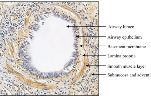
Tracheal and bronchial histology consists of ciliated and non-ciliated cells organized in a tall pseudostratified columnar epithelium overlying the basement membrane. The non-ciliated goblet cells, mainly produce mucus (mucin), a portion of the mucus layer of the airways, which is transported cranially by the ciliated cells [15]. The epithelium gradually changes from a pseudostratified to a simple columnar epithelium as bronchi become smaller with each division. Underneath, a connective tissue layer termed the lamina propria is observed. Different elements constitute this lamina, collagen being the main component, followed by elastic fibers, blood vessels and nerves among other structural components. Spiral bands of smooth muscle are also displayed beneath the lamina propria. The set of all the layers previously described are defined as the airway mucosa[15; 18].The submucosa is the layer observed underneath and is formed by a mix of connective tissue, bronchial glands (only in large bronchi) and blood vessels. Hyaline cartilage surrounds the submucosa, however this structure disappears in small bronchi, allowing differentiation between bronchi and bronchioles [16]. Finally, located at the most external region, is an adventitial layer formed by connective tissue that fuses with the lung parenchyma (Figure 3).
Figure 3. Schematic representation of the equine bronchial structural components. Lung tissue sample stained with type IV collagen.
The bronchioles have a different epithelium when compared to the bronchi. The epithelium consists of a layer of short ciliated cuboidal cells superficially, and non-ciliated secretory club cells (previously known as Clara cells) underneath, all surrounded by a layer of smooth muscle. The goblet cells present in healthy bronchi are absent in bronchioles, unless airway inflammation is present [15; 19]. The secretory club cells represent approximately 60% of the epithelium, each cell containing approximately 6 to 28 granules in the bronchi of a normal horse. These cells secrete Clara cell secretory protein (CCSP) and in a lesser degree, mucin. When airway inflammation is present, an increase in mucus production by the club cells occurs, leading to the “Clara-goblet cells” [20].
The most distal structure of the lung is the alveolus, which is formed by type I and II pneumocytes (Figure 4). Type I pneumocytes surround each alveolus and are characterized by a thin cytoplasmic extension. Type II pneumocytes are more numerous and metabolically active than type I pneumocytes [15; 21]. Type II cells are characterized by cytoplasmic inclusions that contain surfactant, contributing to maintain alveolar stability [22; 23]. Type II pneumocytes have an important role in repairing lung injuries, absorbing alveolar edema and differentiating into type I pneumocytes when needed during the repair process.
Smooth muscle layer Airway epithelium Basement membrane Airway lumen
Lamina propria
22
Figure 4. Epithelial composition in different parts of the lower airway: large bronchi, bronchioles and alveoli 2.
1.3. Cytological features of the lower airways
To evaluate cytological features of non-septic diseases of the lower airways, different diagnostic modalities are available. Bronchoalveolar lavage (BAL) is superior to trans-tracheal wash (TTW) due to the possible degeneration of the cells in TTW samples [24; 25]. BAL may serve to sample a specific portion of the lung, however it does not necessarily constitute a true representation of the cytologic features of the entire lung any more than an exact representation of the bacteria present in the entire lung [26]. The cellularity observed in BAL samples (Figure 5) is normally low and predominantly includes macrophages (50-70%), small lymphocytes (30-50%), and in a lesser extent, neutrophils (<5%), mast cells (<2%) and eosinophils (<1%). Epithelial cells may also be present [27; 28]. The BAL composition becomes important when identification of airway inflammation is warranted. Bronchoalveolar lavage features of airway inflammation include an increase in total cell count, increased percentage of neutrophils, some of which can show degenerative changes,
2Taken from and authorized by Art T. and Bayly W (2014). 27-Lower airway function: response to exercise and
training. Equine Sports Medicine and Surgery (Second Edition). K. W. Hinchcliff, A. J. Kaneps and R. J. Geor, W.B. Saunders: 587-603.
increased percentage of mast cells and eosinophils [27; 29]. Caution should be taken when performing a BAL since the cytological features, such as the percentage of neutrophils can be altered according to the volume of saline instilled [30].
Figure 5. Cytology of bronchoalveolar lavage fluid in an asthmatic horse. The predominant cell types are pulmonary alveolar macrophages (asterisk), neutrophils (black arrow) and lymphocytes (yellow arrow).
1.4. Blood supply of the lower airways
The equine lung is perfused from two different pathways, the pulmonary and the bronchial circulations. The bronchial circulation is a low flow, high pressure system [31] that originates from two different sources. The first source, the broncho-esophageal artery, supplies most of the airways, the interlobular septa and the sub-pleural connective tissue [12]. The second source, the bronchial artery (branch of the aorta), supplies the right lung and receives approximately between 1% and 2% of the cardiac output from the left ventricle in a horse at rest [22; 32]. This circulation is considered as a branch of the systemic circulation and supplies the intrapulmonary airways components with arterial blood. The more peripheral structures are supplied by the pulmonary artery [8]. The terminal
à
*
*
à à à24
bronchioles are vascularized by an anastomosis between the capillaries and the vein, arising from both types of circulation (pulmonary and bronchial). This phenomenon will contribute to maintain blood flow in situations where the pulmonary perfusion is impaired [22; 33]. The pulmonary arteries alongside the bronchi are elastic, whereas the arteries surrounding the bronchioles and alveolar ducts are muscular [31]. The hypoxia receptors of the smooth muscle are responsible of the response of vessels to hypoxia [11]. A difference in size between the two types of arteries is also observed, the pulmonary artery being larger than the bronchial artery. The venous drainage from the extra-pulmonary airways is carried by the azygos vein, which will drain into the cranial vena cava. The venous drainage from the intrapulmonary airways circulates into the pulmonary veins at the pulmonary capillary level. The outflow pressure of this venous drainage varies depending on the who is performing the drainage, either the azygos vein or the pulmonary circulation [31].
The pulmonary circulation is a high flow, low pressure system that holds the total cardiac output arising from the right side of the heart [32]. The deoxygenated blood is carried to the lungs via the pulmonary artery and its branches. Blood is then oxygenated (arterialized) at the level of the alveolar capillaries and returned by the pulmonary veins to the left atrium of the heart [11; 22]. Oxygenation of blood constitutes the principal function of the pulmonary circulation. However, it can also accomplish filtration and removal of chemical substances. Blood flow distribution in the equine lung is believed to be similar to that of the human lung [22], with the ventral region receiving more perfusion due to the influence of gravity and the generated vertical gradient. However, recent studies have shown the opposite, where blood flow was greater in the dorsal regions in comparison to the ventral regions, suggesting that vessel length and resistance may represent the major factors, and excluding gravity as previously thought [22; 33; 34]. Characteristics of both bronchial and pulmonary circulations are detailed in Table 1.
Pulmonary Bronchial Aims • • Gas exchange Venous blood filtration
• Blood reservoir
• Nutrition of airways, vessels, and visceral pleura. • Thermoregulation Structure Right ventricle ↓ Pulmonary artery ↓ Pulmonary arterioles ↓ Pulmonary capillaries ↓ Pulmonary veins Left ventricle ↓
Bronchial and bronchoesophageal arteries ↓ Peribronchial plexus ↓ Subepithelial plexus ↓
Pleural, vascular, and ganglia plexi
↓
Azygos vein
Blood flow (liters/min) ± 99 percent of the right ventricle: 30 (280) ± 2 percent of left ventricle: 0.6 (6)
Pressure (mmHg) Arterial: 30 (100) Capillary: 20 (80) Venous: 10 (60)
Arterial: 100 (200) Capillary: 20 (>60) Venous: 15 (60)
Capillary flow Pulsatile Constant
Vascular resistance (mmHg/liter/min) 0.7 (0.25) 140 (20)
Effect of hypoxia Vasoconstriction Vasodilation
Effect of hyperthermia - Vasodilation
Effect of pleural pressure changes +++ +
Table 1. Comparison between Pulmonary and Bronchial Circulations.3
A continuous production of lymph occurs in the lung, due to fluid transport between the pulmonary microvasculature and the interstitial tissue. The lymphatic vasculature is organized in a superficial and a deep layer. The superficial layer creates a network of vessels along the pleura (sub-pleural); while the deep layer follows the bronchi and pulmonary vessels [11]. These lymphatics, drain into the thoracic duct, and connect the hilum and mediastinal lymph-nodes [15]. All lymphatic vessels enclose the airways and non-capillary
3 Taken from and authorized by Lekeux, P., et al. (2014). Chapter 9 - The respiratory system: Anatomy, physiology,
and adaptations to exercise and training. The Athletic Horse (Second Edition). D. R. Hodgson, K. H. McKeever and C. M. McGowan, W.B. Saunders: 125-154
26
blood vessels, without penetrating the alveolar septa. During respiration, fluid transport is increased in the lymphatic vessels, since they can accommodate large quantities of fluid. Additionally, they have the capacity of removing fluid and other substances such as antigens and leukocytes from the different blood vessels (arterioles, capillaries, and veins). The lymphatic system is not only important for the movement of fluid, but also for alveolar hydration. The lack of permeability of the alveolar epithelium in comparison to the capillary endothelium, allows the alveoli to be exempt of interstitial fluid under normal conditions [31]. When the epithelium is present in the interstitium, fluid accumulates in the alveoli, a condition known as pulmonary edema [32]. Clearance of alveolar fluid accumulation by the lymphatic system is fundamental. However, an increase in the interstitial hydrostatic pressure associated with pulmonary edema, may lead to the compression of the lymphatic vessels, impeding their normal function [35; 36].
1.5. Innervation of the lung
The lung is innervated via different nervous systems: sympathetic, parasympathetic and non-adrenergic ± non-cholinergic (NANC) inhibitory and excitatory nervous systems [4; 22]. The sympathetic and parasympathetic systems form a pulmonary plexus from which they innervate the lung. The sympathetic motor fibers through the middle cervical and cervicothoracic ganglia, control bronchodilation of the airways; whereas the parasympathetic fibers through the vagus nerve control bronchoconstriction [37; 38]. The parasympathetic cholinergic nerves secrete acetylcholine to produce bronchoconstriction of the airway smooth muscle via muscarinic receptors, and to the submucosal glands via nicotinic receptors. The sympathetic system is smaller than the parasympathetic, and is believed to induce relaxation of the airway smooth muscle by activation of β-adrenoreceptors [39]. A dysregulation between both systems seems to play an important role on the pathophysiology of diseases like asthma [40], where inflammatory mediators can lead to an increased cholinergic neurotransmission contributing to the bronchospasm observed [41-43]. NANC excitatory system contribute to smooth muscle contraction, inflammatory cell activation and mucus secretion among others, by the release of neuropeptides such as tachykinins. In addition, NANC inhibitory system modulates the excitatory response of the
airways by the secretion of nitric oxide. This is important in the control of airway hyperresponsiveness in asthmatic horses [39].
1.6. Function of the respiratory system
The main function of the respiratory system is to oxygenate blood, and to remove carbon dioxide from the blood, a process defined as gas exchange [22]. The gas exchange occurs in the alveoli and the alveolar duct coated with a capillary structure, and then diffuses into the bloodstream. The oxygenated blood then returns to the heart who pumps it through the whole body. During exercise, the demand for oxygenated blood and disposal of carbon dioxide increases considerably [44]. There are multiple factors participating in the gas exchange, such as ventilation, perfusion, diffusion, and gas transport. Other non-pulmonary functions accomplished by the respiratory system include defense mechanisms, surfactant production, thermoregulation, filtration and humidification of the inhaled air, among others [11; 22].
2. Extracellular matrix and components
The extracellular matrix (ECM) is a dynamic three-dimensional fibrous network that provides physical scaffolding for the cellular components and is also fundamental for biomechanical and biochemical properties of the airways [45-47]. The ECM components can be primarily divided into two sections, the pericellular matrix or basement membrane, adjacent to epithelial cells, and the interstitial matrix, which surrounds the cells. The ECM is mainly composed of fibrous connective tissue proteins such as collagen, elastic fibers, laminin, structural glycoproteins and proteoglycans [48; 49]. The continuous remodeling of the ECM allows growth and regeneration; a regulation of the degradation is crucial. The ECM degradation is carried out by proteolytic enzymes such as proteases, especially the matrix metalloproteases (MMPs) [50; 51]. A dysregulation on the ECM has been associated with the development of pathologic conditions such as asthma [49].
2.1. Components
2.1.1. Basal membrane
The basal membrane is a type of pericellular matrix that serves as an anchor between the alveolar epithelium and the adjacent connective tissue, and contributes to the regulation of the epithelial cells’ differentiation [52; 53]. This membrane is composed of a multilayer structure that comprises the lamina densa, lamina lucida and lamina fibroreticularis. The lamina lucida being the closest to the epithelium, followed by the lamina densa. The last two layers (lucida and densa) are often grouped under the term of basal lamina [54]. Basal membranes are mainly composed of laminin, collagen, proteoglycans and entactin. Integrins connect the basal lamina to the epithelium, whilst collagen type IV connects it to the ECM [48; 49; 53].
2.1.2. Collagen
Collagen is the main protein component of the ECM in all animals. It is constituted by three α polypeptide chains that are intertwined with each other and form a right-handed triple helix that grants tensile strength to the ECM [48]. Collagen represents 60% of the lung connective tissue and comprises 30% of the total proteins in humans [49; 55]. The different collagen subtypes can be grouped into fibrillar or
non-fibrillar. The types more commonly found in the lung are collagen type I, II, III, IV, V and VI [48]. The distribution of collagen in the lung varies regionally in the ECM. Previous studies have shown type I, III and VI in large bronchi and blood vessels; collagen II in bronchial and tracheal cartilage, and type IV and V in alveolar and capillary basement membranes in humans [47; 56]. However, little is known about the distribution of collagen subtypes in the equine lung. Studies showed the presence of types I and III in the lamina propria and adventitia of small equine airways [46; 57](Figure 6).
Figure 6. Identification of collagen type 1 (left) and type 3 (right) in the equine airways 4. Collagen types 1 and 3 were present in the lamina propria and the adventitia of the airways, but not in the basement membrane.
2.1.3. Elastin
The intrinsic recoil properties of the lung depend on the presence of elastic fibers, which have two major components: elastin and microfibrils. Elastin is an insoluble matrix protein composed mainly of hydrophobic amino acids and glycine that can endure
4 Taken, modified and authorized from Setlakwe, E. L., et al. (2014). "Airway collagen and elastic fiber content
correlates with lung function in equine heaves." American Journal of Physiology: Lung Cellular and Molecular Physiology 307(3): L252-260. Doi: 10.1152/ajplung.00019.2014
30
high linear stress-strain, with a half-life similar to the lifespan of the organism [47; 53]. The distribution of elastin in the lung differs from the one observed with collagen, with the parenchyma having the highest elastin content. In 1955, elastin was considered fundamental during normal breathing, whereas collagen importance was thought to rely on total lung capacity. However, recent data propose an equal importance even at lower lung volumes [58; 59], which suggest that both component may modulate both bronchoconstriction and bronchodilation. In addition, elastin content in the airways has been positively correlated with pulmonary elastance (EL) in healthy horses [46].
2.1.4. Glycosaminoglycans and Proteoglycans
Proteoglycans (PGs) are a family of major core proteins synthetized by most cells. They have diverse functions such as permeability barriers and binder component to different molecules like growth factors, cytokines and proteases [53]. According to their location, PGs can be classified as intracellular, cell surface, pericellular-basement membrane, and extracellular [49]. These proteins are constituted of glycosaminoglycans (GAGs) covalently combined with a core protein, and together they control all cellular processes from signaling and proliferation, to apoptosis and adhesion [47; 49; 58].
2.2. Functions and Importance
The ECM functions in the lung are fundamental for tissue homeostasis and normal organ function. This includes tissue compliance, control of cell behavior and tissue repair and remodeling [48]. Disturbances of any of these components may lead to modifications of the environment and can lead to different pathologies. For example, the dysregulation of proteoglycans may jeopardize the protective duty of the ECM and lead to interstitial edema [48; 49].
3. What is Equine Asthma?
3.1. Equine Asthma
Equine asthma is the term used to describe a chronic inflammatory syndrome of the lower airways, which groups together horses with mild to moderate (previously known as inflammatory airway disease, IAD) and severe asthma (formerly recognized as recurrent airway obstruction, RAO). Rather than a single condition with different manifestations, equine asthma is a variety of conditions leading to similar clinical manifestation [1]. Clinical signs associated with this syndrome can vary in severity, but it is mainly characterized by inflammation of the lower airways resulting in airway obstruction, airway remodeling, excessive mucus accumulation and poor performance.
3.1.1. Airway remodeling
Airway remodeling develops as a consequence of the chronic inflammation occurring in asthmatic horses. Remodeling is characterized by histological changes including epithelial hyperplasia and denudation, subepithelial collagen deposition, smooth muscle hypertrophy/hyperplasia, leading to thickening of the airway wall. Furthermore, mucus accumulation results in luminal obstruction [41; 60; 61]. All these histological changes contribute to an impairment of the normal lung function leading to breathing dyspnea. Airway remodeling is reported to be only partially reversible with standard treatment [62; 63].
32
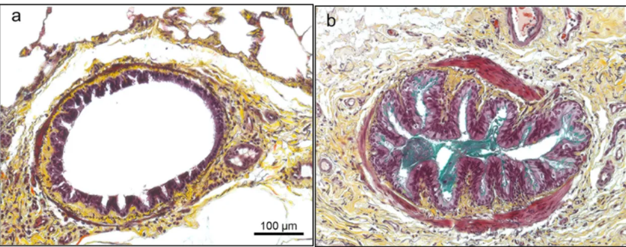
Figure 7. Non-asthmatic (a) and asthmatic (b) bronchial sections of two horses stained with modified Russel-Movat Pentachrome. Structural changes such as smooth muscle thickening (pink right image) are observed in conjunction with the presence of mucus in the lumen (green right image).5
3.1.1.1. Extracellular matrix remodeling
Subepithelial collagen deposition underneath the epithelium (lamina propria) is considered one of the main causes of the thickening of the airway wall, in conjunction with increased bronchial smooth muscle mass [62]. This deposition is most likely secondary to the activation of fibroblasts, key unit in the secretion of ECM components. Furthermore, this increased deposition has only a partial response to treatment [64]. A 2014 study showed an increase in elastic fibers and collagen deposition (collagen I and III) in horses with severe asthma when compared with non-asthmatic animals, and reported a positive correlation between collagen and pulmonary resistance (RL) in asthmatic horses in
5 Taken from and authorized by Bullone, M. and J.-P. Lavoie (2019). "The equine asthma model of airway
remodeling: from a veterinary to a human perspective." Cell and Tissue Research. Doi: 10.1007/S00441-019-03117-4.
remission, suggesting that collagen deposition may contribute to airway obstruction [46].
3.1.1.2. Smooth muscle hypertrophy/hyperplasia
Different techniques have been used to assess the characteristics of the bronchial smooth muscle in asthmatic horses. Studies showed a smooth muscle thickening in airways of different sizes, being more pronounced in peripheric than in the central airways [65; 66] (Figure 8). This increased mass contributes to the narrowing of the lumen of the airway. Furthermore, the bronchoconstriction observed in asthmatic horses contributes to an impairment of the normal lung function [62].
Smooth muscle hypertrophy and hyperplasia have been suggested to represent an early feature of asthma [67; 68]. However, more recent studies have pinpointed a dynamic process described as an increase in myocyte apoptosis occurring in conjunction with in situ proliferation [66; 69]. This hypertrophy is only 30% reversible with current conventional treatments (inhaled corticosteroids and bronchodilators) [62; 70].
Figure 8. Smooth muscle hypertrophy in an asthmatic horse stained with Russel-Movat Pentachrome. EP, Epithelium; SM, smooth muscle; ECM, extracellular matrix. Scale 100μm.
ECM
SM EP
34 3.1.1.3. Mucus accumulation
Mucus is a complex mixture of mucins (high weight molecular glycoproteins), water, enzymes, electrolytes, mixed with epithelial cells and leukocytes. The exact mechanism for mucus accumulation in airways of asthmatic horses, is not completely understood. Multiple theories exist such as, an increase in mucin due to the presence of hyperplasic secretory cells (goblet cells) [71]. Other theories include an excessive production of mucin being produced by normal cells, and without a change in their number, or by decreased clearance from a damaged ciliary apparatus [72].
3.1.1.4. Epithelial hyperplasia and denudation
Epithelial cells are fundamental for homeostasis of the equine lung. Multiple studies using electron and light microscopy have been conducted in order to describe the epithelial cell hyperplasia observed in asthmatic horses [73; 74]. These studies have shown a loss of ciliated cells in large diameter airways and goblet cell hyperplasia in airways of small diameter. Additionally, studies showed that asthmatic horses have epithelial damage secondary to inflammation which may lead to an impartment of the epithelial barrier function [20; 75].
4. Angiogenesis
Angiogenesis is defined as the formation of new vessels from pre-existing ones, an important difference with vasculogenesis, which is the formation of entirely new vessels (de novo). Vascular changes such as an increase in the number of vessels and vascular density, are among the structural modifications observed in humans with asthma [76]. In asthmatic horses, information regarding angiogenesis in airways is lacking.
4.1. Pathophysiology of angiogenesis
Angiogenesis is a fundamental process during embryonic development and physiological functions of the organism, including tissue repair and healing, chronic inflammation, etc. [77; 78]. In addition, angiogenesis is implicated in multiple pathologies, namely neoplasia, ocular disorders (e.g. neovascular glaucoma), and vascular malformations (e.g. cutaneous angiectasias and benign processes such as angiofibroma, hemangiomatosis) among others [77]. The formation of new vessels can be described according to their characteristics as sprouting or non-sprouting angiogenesis (i.e. intravascular subdivision).
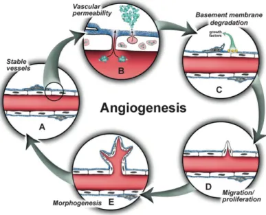
The first step starts with an angiogenic stimulus that contributes to the activation of endothelial cells. This stimulus play a fundamental role on physiological processes of the endothelial cells such as growth, proliferation and migration [78]. This activation leads to an increase in vascular permeability of the pre-existing vessels, and activation of growth factors of the endothelial cells. These latter will then release enzymes (proteases) triggering the proteolytic degradation of the basement membrane. This degradation allows endothelial cells to migrate from the vessel wall towards the angiogenic stimulus, where they proliferate in the surrounding matrix. Following their proliferation, endothelial cells undergo changes in shape through a process called morphogenesis leading to the development of a lumen. The newly formed solid sprouts connect adjacent vessels [77-79](Figure 9).
36
Figure 9. Schematic representation of the angiogenic cascade. Once angiogenesis is initiated, stable vessels (A) undergo an increase in vascular permeability, allowing extravasation of plasma proteins (B). Degradation of the ECM by MMPs relieves pericyte-EC contacts and ECM-sequestered growth factors are then released (C). ECs then proliferate and migrate to their final destination (D), where they assemble as lumen-bearing cords (E). ECM, extracellular matrix; MMPs, matrix-metalloproteases; EC, endothelial cell. 6
4.2. Angiogenesis in lung diseases
The exact mechanism for angiogenesis in diseases with complex pathophysiology such as asthma, has been the object of interest of multiple studies. [80; 81]. Chronic inflammation has been shown to have a codependent relation with angiogenesis [82]. The proliferation and migration of inflammatory cells observed will contribute to the release of pro-angiogenic factors. Additionally, hypoxic tissues damaged by chronic inflammation can also participate to the release of angiogenic factors [83]. An example is the hypoxia-inducible factor (Hif), a transcription factor
6 Taken from and authorized by Bryan, B. A. and P. A. D’Amore (2007). "What tangled webs they weave:
Rho-GTPase control of angiogenesis." Cellular and Molecular Life Sciences 64(16): 2053-2065. Doi: 10.1007/s00018-007-7008-z
stimulated when low oxygen levels are detected in the tissues. The Hif regulates pro-inflammatory genes and has been shown to increase expression of vascular endothelial growth factor (VEGF) in horses with asthma [84]. This growth factor, VEGF, participate in the migration of endothelial cells from pre-existing vessels to the surrounding extracellular matrix, where they elaborate solid sprouts. Furthermore, angiogenesis contributes to the maintenance of the chronic inflammation observed in pathologies such as asthma, by transporting cells and nutrients to the inflammation site [82]. Chronic inflammation and hypoxia may lead to the concentric thickening of the airway wall, and thus, leading to narrowing of the airway lumen [80].
Multiple studies have been conducted evaluating the presence of angiogenesis in asthma and other chronic lung diseases in humans [85-87]. A 2010 study [88] demonstrated an increased number of vessels in the small airways of patients with asthma and chronic obstructive pulmonary disease (COPD), when compared with healthy individuals. In addition, other studies have shown an increased vascular area and number of subepithelial vessels in bronchial biopsies from both adults and children with mild to moderate asthma when compared to controls [89-91] (Figure 10). In vivo studies have also shown an increased number of vessels in the trachea of asthmatic patients versus control groups [92]. Treatment in humans aims to decrease airway inflammation and hyperresponsiveness. A 2002 study [93], showed that inflammatory cell counts in BAL and basement membrane thickness were reduced after 3 and 12 months of inhaled fluticasone treatment, respectively. No difference was observed in collagen deposition with the same treatment. Even though a partial reversibility is seen, with a decrease in basement membrane and smooth muscle thickness, remodeling in asthma is considered to be poorly reversible [94].
38
Figure 10. Schematic picture of a normal (left) and asthmatic airway (right) 7. The different layers of the bronchi are outlined on the image. A larger number of vessels is seen on the asthmatic airway (right).
Due to the similarities in the immunopathology and clinical presentation between equine and human asthma, the angiogenic changes described in humans would be expected to occur in horses. However, the limited information currently available suggests otherwise. A 2012 study [95], evaluated the presence of angiogenesis in the large airways of horses with asthma using narrow band imaging (NBI). Superficial and deep vessels of three different sites along the airways were evaluated (trachea, carina and intermediate bronchi). The results obtained were not those expected since only a significant increase in vascular density of the superficial vessels of the trachea was observed. In face of these results, the authors concluded that the lack of significance at the level of the carina and bronchi, may suggest that the technique lacks sensitivity, or that angiogenesis only occurs in the trachea, contrary to the results obtained in human studies.
4.3. Angiogenic factors
Increased vascular permeability in humans with asthma has been related to the release of inflammatory mediators and growth factors, among others [88](Table 2).
7 Taken from open access article Zanini, A et al. (2010) The role of the bronchial microvasculature in the airway
Although in vitro studies have associated multiple factors with angiogenesis, vascular endothelial growth factor (VEGF) is considered to be the central protein leading to bronchial vascular remodeling [83; 96]. Furthermore, increased VEGF has been demonstrated in vitro in myeloid cells and in vivo in the BAL of asthmatic horses after exposure to hay dust [84]. Additionally, angiopoietins have been shown to play an important role in asthma due to their synergetic effect with VEGF on angiogenesis [97-100].
Table 2. Factors involved in bronchial angiogenesis in humans with asthma and COPD8
4.3.1. Vascular endothelial growth factor (VEGF)
VEGF, previously known as vascular permeability factor, is a potent proangiogenic factor that prompts endothelial cell migration and proliferation [101]. Studies have shown an increase of VEGF expression in tracheal epithelial
8 Modified from an open access article Zanini, A et al. (2010) The role of the bronchial microvasculature in the
airway remodeling in asthma and COPD. Respiratory Research 11, 132. Doi: 10.1186/1465-9921-11-132 Angiogenesis VEGF VIP FGF IL-8, IL-13 TGFβ TNF⍺ HGF NKA HIF Angiogenin Ang-1 MMPs Histamine IGF-1 PGD2 Chymase PGI2 VCAM-1 LTC2 E-selectin PAF ⍺vβ3 SP
40
cell culture of asthmatic specimens when compared with controls [96]. This element has also been directly related to an increased vascular permeability, contributing to the thickening of the airway in human asthmatic patients [102]. An increased expression of VEGF by the airway epithelium after exposure to allergens may trigger vascular remodeling [96]. In addition, a correlation between this factor and subepithelial fibrosis has been reported, suggesting an effect not only on the number of vessels, but also on basement membrane thickness [103]. Furthermore, a 2012 study [84] showed that Hif has a strong positive correlation with the expression of VEGF in horses with asthma.
4.3.2. Angiopoetin-1 (Ang-1) and angiopoietin-2 (Ang-2)
The angiopoietin family constitutes another important family of factors directly related to the angiogenic process. Ang-1 is considered a ligand for endothelial cell surface receptor. It stimulates sprouting and helps stabilize the new formed vessels [88; 101]. In contrast, Ang-2 acts as an antagonist of Ang-1, destabilizing and disrupting the formation of new vessels [88; 99].
5. Angiogenesis detection and quantification 5.1. Laboratory techniques
Multiple laboratory techniques are available for the detection and quantification of blood vessels in tissues including immunohistochemistry, immunofluorescence and standard histological stains [89; 104; 105].
Standard histological stains such as Hematoxylin & Eosin (H&E), Movat Pentachrome and Verhoeff’s Van Gieson (VVG) have been used to outline blood vessels. Movat Pentachrome stains red blood cells in yellow and elastic fibers in dark purple. Elastic lamina of large blood vessels adopts a dark brown/black color with VVG. However even though their sensitivity is high for large blood vessels, the specificity of these colorations for small vessels such as capillaries is low. Furthermore, regular stains such as H&E do not differentiate between blood and lymphatic vessels, which may lead to an increase in false positives [106-109].
Immunohistochemistry (IHC), is a laboratory technique used to identify a particular tissue component via specific antigen-antibody reactions in normal and pathological conditions [110; 111]. This technique uses an antibody (immunoglobulin) to react with a small area of the antigen of interest called epitope. These reactions are detected with fluorescent or enzymatic cell immunolabeling allowing detection of the precise location of the protein of interest [112]. Immunofluorescence (IF) is a specific type of IHC that uses fluorescent antibody labeling and can be further classified into direct or indirect according to their labeling technique [113]. Direct methods bind the primary antibody, which is immunolabeled, to the antigen, whereas the indirect process involves an additional step in which the secondary antibody binds to the primary, amplifying the signal (Figure 11). Indirect methods of immunostaining are more commonly used, due to their higher sensitivity [114; 115]. Although both techniques share a similar goal and both can outline blood vessels, IHC is considered the gold standard technique to quantify bronchial microvessels [88].
42
Figure 11. Representation of the direct (left) and indirect (right) immunochemical staining methods 9.
5.1.1. Antibody selection
Antibody selection is one of the key elements for successful IHC. These immunoglobulins are produced by B lymphocytes. According to their characteristics, antibodies can be classified into monoclonal and polyclonal. Monoclonal antibodies correspond to a homogenous group of molecules produced by the same clone of plasma B cells. Polyclonal antibodies refer to a variety of immunoglobulins which act against a specific antigen and are produced by different clones of plasma B cells [116](Table 3).
MONOCLONAL ANTIBODIES POLYCLONAL ANTIBODIES
Produced by the same clone of plasma B cells Produced by different clones of plasma B cells A homogeneous antibody population A heterogeneous antibody population
Interact with a particular epitope on the antigen Interact with different epitopes on the same antigen
Production is expensive Production is inexpensive
Possess low cross reactivity Possess comparatively high cross reactivity
9 Taken from and authorized by Katikireddy, K. R. and F. O'Sullivan (2011). "Immunohistochemical and
immunofluorescence procedures for protein analysis." Methods Mol Biol 784: 155-167. Doi: 10.1007/978-1-61779-289-2_11
Advantages include immortal supply, high specificity, and high reproducibility
Advantages include high affinity, tolerance of minor changes, and more robust detection
Table 3. Comparison between polyclonal and monoclonal antibodies 10.
5.1.2. Biomarkers
Numerous studies have evaluated the ability of certain antibodies such as collagen IV, PECAM-1 (CD31), von Willebrand factor (vWF) and cluster of differentiation 34 (CD-34) to outline blood vessels [117-120]. PECAM-1 has been recognized as a superior endothelial marker due to its strong and homogeneous expression when compared to others such as vWF and CD-34 [119]. Similarly, collagen IV allows a strong outlining of the basement membrane, which facilitates the recognition of blood vessels outlining the vascular basement membrane.
5.1.2.1. Platelet endothelial cell adhesion molecule (PECAM-1)
PECAM-1 or cluster of differentiation 31 (CD-31) is a transmembrane glycoprotein, found mainly at the surface of endothelial cells where they join each other (cell junctions). This glycoprotein is also present at the surface of platelets and macrophages among others. Its role in biological functions such as cell migration, adhesion and angiogenesis, is fundamental [121-123]. Multiple studies have been conducted with PECAM-1 as a biomarker for blood vessels in normal structures like human alveolar capillaries [117], and pathological processes such as wound repair in horses [124], canine tumors [118] among others. PECAM-1 is one of the most commonly used biomarkers for blood vessels [104; 118] (Figure 12).
10 Taken and modified from Panawala, L. (2017). "Difference Between Monoclonal and Polyclonal Antibodies."
44
Figure 12. IHC to outline blood vessels of a breast cancer xenograft with CD3111. Dark brown staining of endothelial cells highlights the localization of blood vessels on both images.
5.1.2.2. Collagen IV
Collagen IV is a major basement membrane protein found in the extracellular matrix. It is essential for preservation of the basement membrane integrity and is composed of six different α-chains (α1–α6)[119; 125; 126]. In addition, collagen IV is fundamental in biological processes such as cell adhesion and migration, among others [127]. Multiple studies have been conducted using collagen IV as a biomarker to determine the presence of blood vessels in different tissues [128; 129]. Furthermore, collagen IV has been used to outline the microvasculature of patients with asthma and COPD [88](Figure 13).
11 Taken and modified from Wang D. et al (2008) Immunohistochemistry in the evaluation of neovascularization
Figure 13. Microphotographs showing bronchial mucosal staining with antibody directed against Collagen IV to outline blood vessels from a normal subject (left image) and asthmatic patient (right image)12.
5.2. Angiogenesis quantification
Different morphometric methods are available to quantify vascularity in lung tissues including manual quantification, stereology or imaging techniques such as NBI or micro-computed tomography [130; 131].
Standard histomorphometric analysis consist in the evaluation of a digitalized image of a sample of interest using a dedicated computer software with manual counting or with a semiquantitative analysis [90; 132-134]. Different quantification methods are available such as the Weidner’s method for the measurement of microvascular density. This method consists in the evaluation of “hot spot” areas with a high magnification using one of the following: (1) manually on the digitalized image, (2) with computerized methods or (3) with the Chalkley method directly with the microscope [91; 104; 135; 136]. The Chalkley technique involves the quantification of vessels intersecting with the points of an array grid mounted into the microscopes’ eyepieces [91; 137].
Recently, other quantification techniques have gained popularity such as stereology and imaging evaluation based on an alleged unbiased quantification analysis [136; 138;
12 Taken and modified from open access article Zanini, A et al. (2010) The role of the bronchial microvasculature
46
139]. Stereology is a statistical technique that evaluates the properties of a 3D tissue with geometric tests based on quantification of a two dimensional histological image [130; 138]. Even though, stereology is considered to reduce the bias associated with this analysis, it is difficult to perform, especially in tissues stained by IHC, and therefore manual counting continues to be an accepted procedure [130].
The parameters used to quantify angiogenesis include the number of vessels per square millimeter of area, the percentage of vascular area in relation to the total ECM area, and the mean vessel size calculated when dividing the vascular area by the number of vessels [88; 120; 132]. Numerous studies suggested to analyze vessels located in the lamina propria at 100-160 μm from the epithelial basement membrane [90; 140; 141]. All the areas internal to the vascular endothelial basement membrane on each vessel were manually drawn (free hand tool) and the computer software was able to determine the area of these traces to estimate the vascular area. The total surface area of the airway and vessel wall can be calculated following the same process than for the vascular area, by drawing either manually or automatically the area to be measured, and automatically with the image software analysis estimate this parameter [142].
6. Purpose of study
Little information is known about angiogenesis in the lower airways of asthmatic horses. The inaccessibility of the bronchi in a clinical setting and the low sensitivity of the current diagnostic techniques available contribute to the lack of scientific studies evaluating angiogenesis in equine patients [95]. In humans with asthma, angiogenesis is observed has demonstrated by increases in vascular density and vessel size in lower airways? [83]. In this thesis, an equine asthma model has been used to investigate the presence of angiogenesis in the bronchi.
6.1. Objectives
The main objective of this project is to evaluate the presence of bronchial angiogenesis in bronchi of horses with asthma compared to control horses using immunohistochemistry.
6.2. Hypothesis
We hypothesized that an increase in bronchial angiogenesis occurs in the bronchi of severe asthmatic horses which is partially decreased by conventional treatment due to the decrease of inflammation.
ARTICLE 1.
In preparation for submission to Journal of Veterinary Internal Medicine (JVIM- Standard Article)
Bronchial angiogenesis in severely asthmatic horses
Esther Millares-Ramirez1, Jean-Pierre Lavoie11 Department of Clinical Sciences, Faculty of Veterinary Medicine, University of Montreal, Saint-Hyacinthe, Quebec, Canada.
Keywords: Asthma, angiogenesis, horses, collagen IV, immunohistochemistry, bronchi. Abbreviations:
ECM: Extracellular matrix RL: Pulmonary resistance EL: Pulmonary elastance
Corresponding author: Jean-Pierre Lavoie, Faculty of Veterinary Medicine, University of Montreal, 3200 rue Sicotte Saint-Hyacinthe, J2S 2M2 Qc, Canada; e-mail: [email protected]
Acknowledgements
Authors would like to thank Guy Beauchamp for his assistance with the statistical analysis, and Hélène Richard for her support with the design of the immunohistochemistry protocol.
This work was made possible with support from the Canadian Institutes of Health Research (CIHR). Authors declare no conflict of interest.
Off-label antimicrobial declaration
Authors declare no off‐label use of antimicrobials.
Institutional Animal Care and Use Committee (IACUC) or other approval declaration All experimental procedures were performed in accordance with the Canadian Council for Animal Care and the research protocol was approved by the University of Montreal Animal Care Committee (Rech-1578).
Human ethics approval
50
1. Abstract
Background: Equine asthma is a chronic inflammatory disease of the lower airways characterized by structural changes that contribute to airway thickening and airflow obstruction. Treatment for equine asthma partially reverses these remodeling changes. Angiogenesis has been shown to increase vascularization of the bronchial mucosa which contributes to the thickening of the bronchial wall in humans with asthma, opening a new window for a more targeted treatment. However, little information is available on angiogenesis in equine asthma. Objectives: To document the presence of indices of angiogenesis in the lower airways of severely asthmatic horses.
Animals: Bronchial samples from 7 asthmatic horses in exacerbation of the disease, 7 asthmatic horses in clinical remission, and 7 age matched healthy horses were studied.
Methods: Prospective, blinded, randomized controlled study. Immunohistochemistry analysis was performed with collagen IV as a biomarker for basement membranes. The number of blood vessels, vascular density, vascular area and mean vessel size values were measured by histomorphometry using an image software (Image J) and compared using a one-way ANOVA (p<0.05).
Results: A significant increase in the number of vessels was observed in asthmatic horses in exacerbation (p=0.007) and in horses in remission (p=0.02) in comparison to controls. Furthermore, the vascular area was increased in horses with asthma in exacerbation when comparted to controls (p=0.02) and to remission (p=0.04). No significant differences were observed in mean vessel size between the groups.
Conclusions and clinical importance: Angiogenesis is present in the bronchi of asthmatic horses, suggesting that it may contribute to the thickening of the airway wall. Furthermore, results of
the current study may offer a new avenue for targeted treatment. Further studies are warranted in order to assess response to those treatments.