HAL Id: dumas-02043038
https://dumas.ccsd.cnrs.fr/dumas-02043038
Submitted on 10 Apr 2019HAL is a multi-disciplinary open access archive for the deposit and dissemination of sci-entific research documents, whether they are pub-lished or not. The documents may come from teaching and research institutions in France or abroad, or from public or private research centers.
L’archive ouverte pluridisciplinaire HAL, est destinée au dépôt et à la diffusion de documents scientifiques de niveau recherche, publiés ou non, émanant des établissements d’enseignement et de recherche français ou étrangers, des laboratoires publics ou privés.
anévrysmale
Mariam Soumah
To cite this version:
Mariam Soumah. Évaluation quantitative du vasospasme cérébral à l’angioscanner dans l’hémorragie sous arachnoïdienne anévrysmale. Imagerie. 2018. �dumas-02043038�
UNIVERSITE DES ANTILLES FACULTE DE MEDECINE HYACINTHE BASTARAUD
********************** THESE
POUR LE DIPLOME D’ETAT DE DOCTEUR EN MEDECINE Discipline : RADIODIAGNOSTIC ET IMAGERIE MEDICALE *******
Présentée et soutenue publiquement le 25 / 09 / 2018 A Pointe-à-Pitre
*******
Par Mademoiselle SOUMAH Mariam
*******
EVALUATION QUANTITATIVE DU VASOSPASME CEREBRAL A L’ANGIOSCANNER DANS L’HEMORRAGIE SOUS ARACHNOIDIENNE
ANEVRYSMALE
JURY
Président du jury : Professeur MEJDOUBI Mehdi Membres du jury : Professeur HOUDART Emmanuel Professeur LANNUZEL Annie Professeur DEBANDT Michel Docteur POULLAIN Pascale Directeur de thèse : Docteur LABEYRIE Marc - Antoine
REMERCIEMENTS
Au jury
Mon président du jury, Monsieur le Professeur Mehdi Mejdoubi
Vous me faites l’honneur d’assurer la présidence de mon jury de thèse. Merci pour votre enseignement, votre présence et vos conseils au cours de ces années.
Monsieur le Professeur Emmanuel Houdart
Je vous remercie d’accepter de juger mon travail. Je vous remercie également pour ces 6 mois passés dans votre service à l’Hôpital Lariboisière à la fois pour l’accueil, l’ambiance, votre patience et vos enseignements. Je vous en suis très reconnaissante.
Madame le Professeur Annie Lannuzel
Vous me faites aujourd’hui l’honneur de juger cette thèse. Je vous en suis grandement reconnaissante.
Monsieur le Professeur Michel Debandt
Je vous remercie pour votre accueil lors de mon passage en stage clinique en rhumatologie au CHU de Martinique. Je suis très reconnaissante de l’honneur que vous me faîtes aujourd’hui de juger cette thèse.
Mon directeur de thèse, Monsieur le Docteur Marc-Antoine Labeyrie
Je te remercie de m’avoir proposé ce sujet, de m’avoir encadrée, de ta gentillesse et de t’être montré si disponible malgré tout ce que tu avais à faire et ce même pendant tes vacances. Merci d’avoir accepté d’être mon directeur de thèse.
Madame le Docteur Pascale Poullain
Je te remercie pour ton enseignement, tes staffs neurologues/ radiologues, ta gentillesse et ta grande disponibilité même en dehors des heures de travail. Sois assurée de mon profond respect et de mon admiration. Merci d’avoir accepté de faire partie de mon jury de thèse.
***********************************
A ma famille et amis
A mon quatuor, ma mère, ma grand-mère Mme Albertine Baclet, ma sœur Anne-Louisa et ma tante Maryla. Bien trop à remercier car sans chacune de vous, sans votre soutien et persévérance, je ne serais pas où j’en suis aujourd’hui. Merci de votre amour.
A mon oncle Alex et ma tante Lucette, je vous remercie de votre soutien, gentillesse et de l’accueil chez vous en Guadeloupe pendant 6 mois.
A ma tante Suzie, qui m’a si bien accueillie à mon arrivée en Martinique et s’est occupée de moi comme sa fille.
A mes cousins et cousine, Marc, Justine, Jérémy et Germain pour leur présence toutes ses années.
A ceux qui ne sont malheureusement plus là, mon grand-oncle Félix et Mamie Laporal qui auraient été si fiers de me voir arriver jusque-là. Merci pour leur amour.
Aux proches Marie-Galantais, Clémencia, Mr et Mme Hégesippe, Mme Comuce pour leur gentillesse et grande bonté d’âme qui me rendent si fiers d’être originaire de cette petite île.
A mes copines de toujours, Anaïs, Aurélie, Agathe, Olga, Stéphanie, Reenal. Merci pour votre amitié, aide et patience. Je vous remercie d’être toujours là pour moi après toutes ces années.
A Fanny VO DIEP, ma bonne amie de la Fac qui m’aura suivie jusqu’en Guadeloupe. Merci pour toutes ces années, ta constante bonne humeur, ton rire ravageur, nos discussions interminables, ta grande gentillesse et ton amitié.
A mes grandes copines d’internat, Vanessa Cingala, Adeline Piquot, Liza Valls et Pauline Simon, qui ont été ma famille aux Antilles. Je vous remercie pour votre bonne humeur, disponibilité à toute épreuve, votre sentiment fraternel et pour toutes nos aventures.
A Marie-Anne Berthelin, alias MAB, ma Cristina. Mon internat aurait été bien vide sans toi. Je suis bien triste de te quitter dans quelques mois mais je sais que nous resterons toujours aussi proches. Merci de ton amitié.
A mes co-internes de radiologie, alias la famille radio alias la “golden generation”, Mathieu (Monsieur Schertz), Sarah (Dhundass), JB et Mickael (Mima). Je vous remercie d’avoir été de si bons co-internes, amis, d’avoir eu une telle émulsion pour toujours nous pousser vers le haut, d’avoir su créer une telle cohésion entre nous. Un merci tout particulier à Mathieu qui a su trouver les mots pour me conforter vers mon choix d’internat aux Antilles et le tout pendant une garde un samedi.
A mes anciens co-internes et amis, Audrey Guillier et Dimitri Dinal. Je vous remercie pour ces 6 mois passés à vos côtés en stage clinique, pour votre bonne humeur et vos blagues. Merci pour votre amitié qui perdure malgré l’éloignement. A Jonathan Brami, pour sa relecture, son amitié et nos moments de folie à Larib.
A mes tout premiers chefs, Mehdi Lebbadi, Isaure Biette et Fanny Rouget qui m’ont si bien accueilli lors de mon arrivée en 1er semestre en Martinique, pour m’avoir transmis les fondements de la radiologie et inculqué ce qu’est d’être radiologue. Je vous en serais éternellement reconnaissante et suis honorée de vous avoir rencontrés sur mon chemin. Un merci particulier à Isaure qui s’extasiait tant devant les scanners abdominopelviens et m’a transmis cet amour.
Aux manipulateurs du CHU de Martinique, Sylvia, Ludivine et Jean-Michel, à la secrétaire du service de radiologie du CHU de Martinique, Nadine Granier, pour leur si bon accueil et gentillesse.
A mes anciens colocataires, Adrien Tolle et Lindou. Je vous remercie pour ces bons moments passés ensemble.
A Fabienne LOUIS-SIDNEY, ma colocataire actuelle et amie. Je te remercie de ton soutien, tes bons petits plats, tes supers tantes et d’avoir eu la patience de me relire.
Au Dr Damien BISSERET, ancien chef et ami. Je te remercie pour ton enseignement, notre présentation aux JFR, nos moments de rigolades et d’avoir cru en moi.
Au Dr Melinda MAJLATH, ancienne chef et amie. Merci pour ta douceur, ta gentillesse, tes bons conseils et ta précieuse relecture.
A mes futurs collègues de l’Hôpital St Antoine à Paris, je vous remercie pour votre si bon accueil lors de mon retour en Métropole.
A tous les médecins, co-internes, infirmières et brancardiers que j’ai eu le plaisir de côtoyer durant ces 5 années d’internat.
CT angiography for quantitative assessment of large vessel cerebral vasospasm in aneurysmal subarachnoid hemorrhage
Mariam Soumah, MD; Jonathan Brami, MD; Antonella Jean-Pierre ; Lama Hadid, MD; Samuel Gaugain, MD ; Vittorio Civelli, MD ; Jean-Pierre Saint-Maurice, MD ; Emmanuel Houdart, MD; Marc-Antoine Labeyrie, MD
ABSTRACT
Background and purpose—Assessment of large vessel cerebral vasospasm (CVS) with
CT angiography (CTA) is commonly used in aneurysmal subarachnoid hemorrhage (aSAH) to monitor the risk of delayed cerebral ischemia (DCI). However, its accuracy is still debated in clinical practice. We aim to assess sensitivity, specificity and radiation dose of CTA and digital subtraction angiography (DSA) to compute CVS.
Methods— Consecutive patients with aSAH who underwent both CTA and DSA within
less than 6 hours at admission / during CVS period (within the 3rd / 4th-21st days after aSAH onset) were included. CVS was retrospectively computed on CTA and DSA for each anterior intracranial artery segment until the end of their 2nd segments by 2 readers (reader 1 / reader 2; 5-year / 10-year experienced radiologist / interventional neuroradiologist) in blinded conditions. Radiation dose to the eye and scalp were also compared prospectively in 1 phantom study and in 5 patients.
Results—CVS was computed on a total of 210 arterial segments in 20 patients. The
correlation between CTA and DSA was better for the reader 2, CTA performed under our local protocol, CTA with great visualization of distal arteries and segments without metallic artefacts. The best sensitivity and specificity of CTA to predict CVS ≥ 50% on DSA was obtained for a threshold ≥ 30% for the reader 2 (86% and 86% respectively). Under this value, no patient had CVS ≥ 50% on DSA. Radiation dose to the scalp and eye were respectively lower and tends to be higher for CTA compared to DSA.
Conclusions—CTA in clinical practice may not be as excellent as reported to predict
CVS on DSA. Its accuracy highly depends on observers’ experience. A threshold value of ≤ 30% may rule out CVS ≥ 50% on DSA. Radiation dose limitation is not a valid argument to substitute DSA with CTA.
INTRODUCTION
Cerebral vasospasm (CVS) is a major provider of morbidity and mortality in patients hospitalized with aneurysmal subarachnoid hemorrhage (aSAH)1. The involvement of large vessel CVS in the occurrence of delayed cerebral ischemia (DCI) is debated2. Some authors propose the analysis of microcirculation by computed tomography perfusion (CTP) rather than the severity of CVS to determine the risk of DCI. However, no study concludes to the superiority of one or the other approaches3.The analysis of CVS therefore remains a recognized major marker of the DCI risk. In addition, the quantification of CVS is a key element for angioplasty decision making with a threshold usually of 50%.
The gold standard to confirm CVS remains digital subtraction angiography (DSA). But, it is costly, invasive, time-consuming and requires the displacement of fragile patients in the operating room. Neurologic complications are not uncommon (0.33 to 1.3% of cases including 0.5% of permanent neurological deficits)4–6. Transcranial Doppler (TCD) ultrasound is a non-invasive, non-ionising and inexpensive technique that can be performed at the bedside. Nevertheless, because of its limited acoustic window and several factors influencing blood velocity,7,8 many false negative results need to be considered, especially in the anterior brain territory or for CVS affecting distal segments of cerebral arteries9,10. As for it, CT angiography (CTA) has been studied in many articles11–19. A recent meta-analysis found out excellent sensitivity and specificity, comparable to CTP3. However, imaging protocols used in these studies were different. Analysis was based on qualitative or semi-quantitative criteria rather than quantitative ones, on examinations too widely spaced in time whereas CVS is a rapidly evolving phenomenon, or on too proximal arterial segments while more distal involvement seems
to also play a role in the occurrence of DCI. Therefore, the clinical interest of CTA seemed uncertain to us, mostly for angioplasty decision.
The aim of our study was to show a correlation between CTA and DSA in the determination of CVS and thus the angioplasty decision by a quantitative analysis of CVS till the 2nd segments of cerebral arteries in quasi-synchronous examinations. We also aimed to evaluate radiation implications in the angioplasty decision.
METHODS
Population
From a prospective data base of consecutive patients hospitalized in our center between January 2015 and December 2016 at acute phase of aneurysmal subarachnoid hemorrhage (aSAH), with a proven Fischer score ≥ 2, we included patients who underwent both CTA and DSA twice. The first set was performed prior aneurysm treatment and within the 3rd day of aSAH onset and the second one between the 4th and the 21th day from aSAH onset. Each CTA had to precede DSA within a delay of less than 6 hours and no curative medical or endovascular treatment was initiated or stopped during this period.
Non-invasive bedside monitoring of DCI was done using an every 2-hours neurological examination and measurement of middle cerebral artery velocity by TCD twice - a - day. CTA or directly DSA were performed when DCI was suspected on bedside monitoring. CTP was performed in rare cases when CTA was not interpretable. Intensive medical care and endovascular preventive treatment of DCI were systematically considered in patients with CVS on DSA (CVSDSA) ≥ 50% and if DCI was suspected. Patients or their relatives were informed and provided their consent for all the care concerning aSAH. The study
was approved by our local Ethics Committee.
Imaging protocol
We used a 64-section multidetector CT-scan, Philips Brillance (Philips Medical System, Amsterdam, Netherlands) and Siemens Definition Edge CT-scan (Siemens Healthineers, Germany) since September 2016. Acquisition parameters were as follows: 300mA; 120 kV; matrix, 512 x 512; field of view, 28-30 cm; section thickness, 1.5 mm on average; reconstruction interval, 1 mm; pitch, 0.55 mm. A biphasic intravenous brachial injection of 60 cc of contrast material (IOMERON 400, Iomeprol, Bracco Imaging, Milan, Italy) followed by 50 cc of saline was administrated with an injection speed of 4 mL/s. Helix acquisition was launched as soon as contrast enhancement was detected within skull base arteries, with a bolus-tracking method. Reconstructions of initial raw images were produced in 1 to 5 mm thin 3D and 3D MIP and co-registered with per-CVS CTA on processing console (PACS Carestream Health, France).
All patients had a DSA (biplane neuroangiography unit Artis Zee, Siemens Healthineers, France) under local or general anesthesia with standard frontal view angiograms of both internal carotid artery (ICA) and dominant vertebral artery (VA) territories using OMNIPAQUE 320 (Iohexol, GE Healthcare, Chicago, USA) as contrast material.
Imaging analysis
CVS was computed on 14 segments of bilateral anterior intracranial circulation including: cavernous internal carotid artery (CICA), terminal internal carotid artery (TICA), middle cerebral artery (MCA) with is first segment (M1) and second segment (M2) anterior and posterior, anterior cerebral artery (ACA) with its first (A1) and second segment (A2).
Percentage of narrowing of each intracranial artery was computed using measurements between the second and the first vascular imaging.
CVS on CTA (CVSCTA) was assessed independently by two observers: a 5-year of experience in diagnostic radiology with 1 year of neuroradiology training (reader 1) and a 10-year experienced in interventional neuroradiology (reader 2). Interpretation was blinded between readers and so were DSA data. A training period on analysis process was performed in consensus by the two readers with 10 patients. The different steps of analysis are shown in Figure 1. To limit partial volume artefacts on curved segments, each segment was analyzed in an oblique plane to unroll this segment. To limit cross-vessel sections in MIP reconstruction, thickness on MIP reconstruction may have been decreased from 5 to 2 mm. When “beads on a string” artefact was seen on a vessel segment, diameter was considered as mean value between the minimal and maximal diameter. In case of arterial trifurcation, the dominant artery was chosen. In case of metal-induced susceptibility artifacts, we aim to determine the diameter of the segment on its visible part. The presence of such artefacts was analyzed for each segment. CTA quality criterion was defined qualitatively such as the presence or absence of visibility of 3rd segments of cerebral arteries.
CVS on DSA (CVSDSA) was computed by 2 readers in consensus blinded to CTA data on frontal (M1, M2, A1) and lateral (TICA, A2) view angiograms (Figure 2).
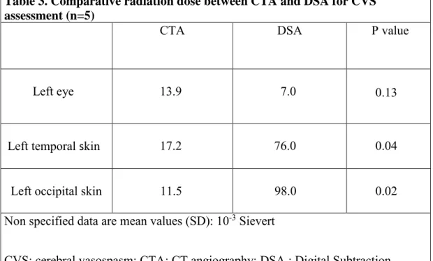
Radiation dose
Radiation effective dose was assessed prospectively on 1 acrylic head phantom (with RaySafe Xi CT detector) and 5 patients using 3 optically stimulated luminescence dosimeters (OSL) (Nanodot, Landauer, Glenwood, USA) put on the left eye, on the left
temporal region and on the occiput bone. CTA data was compared to DSA results (3 lateral and frontal series).
Statistical analysis
The interrater correlation between reader 1 and reader 2, the inter test correlation between CTA and DSA, the influence of CTA protocol, CTA quality and the location of arterial segments to assess CVS were analyzed for each arterial segment using linear regression, comparison of mean absolute differences of CVS values using a paired sample t-test, Pearson correlation, kappa correlation for dichotomized CVS with a threshold ≥ 50% and ROC analysis for a dichotomized CVSDSA with a threshold ≥ 50%. This final analysis was also done for each patient considering arterial segment with the most severe CVS. Radiation dose was described as mean value (SD) and compared using a paired sample t-test.
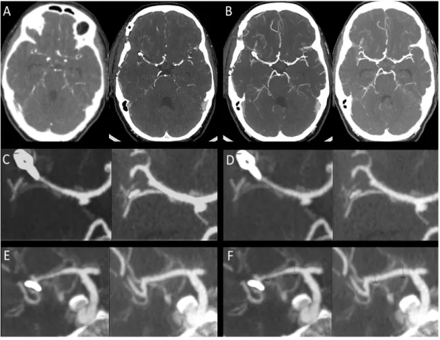
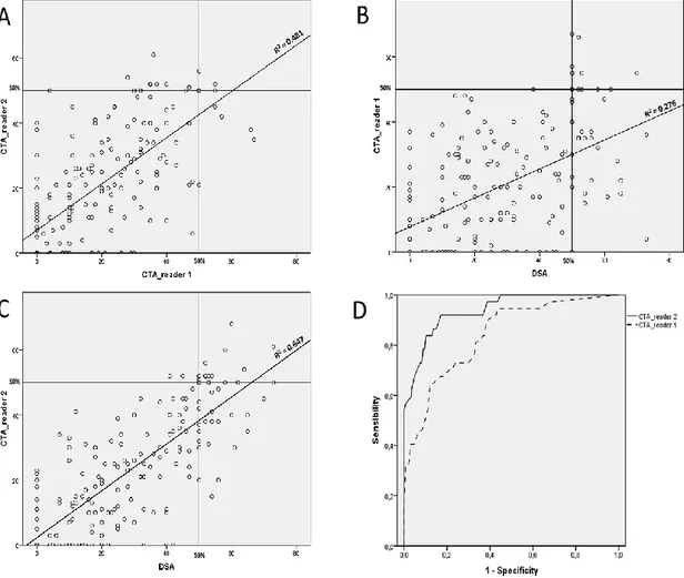
Figure 1. Cerebral vasospasm (CVS) computed on CTA. Initial (pre-CVS) and per-CVS CTA (A) were co-registered in a multiplanar reconstruction with 2 to 5 mm thin sections on maximal intensity projection (MIP) (B). Each arterial segment (here M1) was shown on a large view (C). Grayscale level was adapted to make sure all values from vessel’s center to perivascular space are well seen (D). Each arterial segment was analyzed in an oblique plane to unroll it (E). Vessel diameter was assessed on longitudinal reconstructions and included external border of the vessel (F).
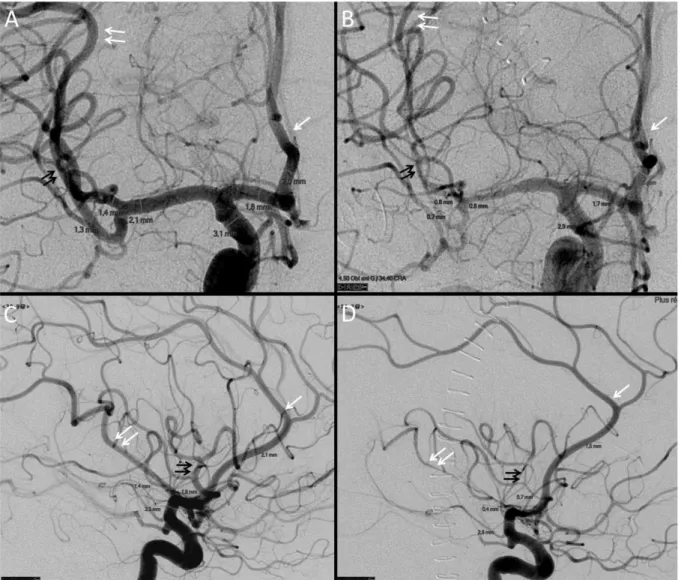
Figure 2. Cerebral vasospasm (CVS) computed on DSA. Initial frontal (A) and lateral (C) angiogram of internal carotid artery were compared to per-CVS angiogram (B, D). CVS was assessed on A2 (white arrow), on anterior (double black arrow) and posterior (double white arrow) M2.
RESULTS
Population
A total of 156 consecutive patients with aSAH were screened during this study period. CVS was computed in 20/156 patients (13%) following inclusion criteria. The main characteristics of these patients are presented in Table 1.
The median age was 47 years old. Patients were mostly women (65%). 85% of patients underwent aneurysmal treatment. They were in serious clinical condition, hence their hospitalization in intensive care with a WFNS scale > 4 in 40% of cases and a Fischer score = 4 in 80% of cases (Annex 1).
Exams
Initial CTA was performed on admission. CTA performed at the time of CVS was performed on the 6th day from the aSAH onset (median; IC 95% [5-8 days]). 95% of CTA during CVS were performed according to our local protocol versus 50% among initials CTAs.
Median delay between CTA and DSA were of 2 hours for initial exams and of 3 hours for those occuring during CVS.
Arterial analysis
210 arterial segments were finally analyzed. 30 segments were excluded due to absence of DSA.
Correlations
Correlation of CVS values for each arterial segment between CTA_reader 1, and CTA_reader 2 are presented in Figure 3 and Table 2. The correlation between CTA and DSA was better for reader 2 (Pearson; 0,805 vs 0,525 for reader 1), for CTA performed under our local protocol (Pearson; 0,847 vs 0,751), for CTA with great visualization of distal arteries and segments without metallic artefacts.
ROC curve analysis
With ROC curve analysis, the best sensitivity and specificity of CTA to predict a CVS≥50% on DSA was obtain for a threshold ≥ 30% (respectively 86% and 86% for reader 2 vs 73% and 71% for reader 1). In per-patient analysis, this threshold was CVS≥40% for reader 2 (respectively 87% and 100%) and ≥ 35% for reader 1 (respectively 78% and 50%). Maximal sensitivity was obtained for a threshold of CVS ≥ 30% for reader 2 (respectively 100% and 50%) and CVS≥20% for reader 1 (respectively 100% and 25%).
Radiation dose
Radiation dose is presented in Table 3. CTA was associated to a lower radiation dose for skin (11,5 and 17,2 versus respectively 98 and 76 x 10-3 Sv; p = 0,02 and 0,04) but tends to be associated to a higher radiation dose for eyes (13,9 versus 7 x 10 -3 Sv; p = 0,13) compared to DSA.
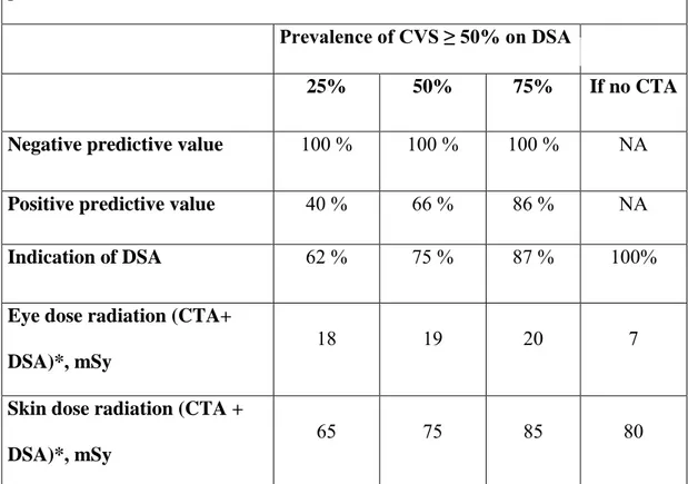
We also simulated 3 possible prevalence of CVS on DSA (CVSDSA) ≥ 50%: 25%, 50% and 75% on Table 4.
As prevalence of CVSDSA ≥ 50% increases, indication to perform DSA increases too but so does the radiation dose.
Table 1. Patients characteristics (n=20) Age, yr 47 (44-57) Female, n (%) 13 (65%) WFNS score >4, n (%) 8 (40%) Fischer score =4, n (%) 16 (80%) Aneurysm embolization, n (%) 17 (85%) Time to CTA1, dy 0 (0-1) Time to CTA2, dy 6 (5-8)
Time from CTA1 to DSA1, hr 2 (1-2)
Time from CTA2 to DSA2, hr 3 (2-4)
Local protocol for CTA1, n (%) 10 (50%)
Local protocol for CTA2, n (%) 19 (95%)
Local protocol for both CTA1 and CTA2, n (%)
10 (50%)
Quality criteria for CTA1, n (%) 10 (50%) Quality criteria for CTA2, n (%) 15 (75%) Quality criteria for both CTA1 and CTA2, n
(%)
10 (50%)
Patients with CVS on DSA ≥50%, n (%) 16 (80%)
WFNS: World Federation of Neurologic Surgeons score; CTA1 / DSA1: initial CTA / DSA before treatment; CTA2 / DSA2: CTA / DSA during CVS within the 3- 21st day; Time to CTA1/CTA2: time from subarachnoid hemorrhage onset to CTA1 or CTA2.
Table 2. Interrater and intertest correlation (210 arterial segments in 20 patients) Absolute difference Mean (SD) Pearson coefficient Kappa coefficient for CVS ≥50% Area under curve for CVS≥50% on DSA CTA_reader 1 / 2 10% (10) 0.694 0.437 NA CTA_reader 1 / DSA 15% (12) 0.525 0.420 0.839 CTA_reader 2 / DSA 10% (9) 0.805 0.606 0.938 Local CTA 8% (7) 0.847 0.751 0.965 Non-local CTA 11% (10) 0.778 0.507 0.912 Quality CTA 8% (8) 0.861 0.697 0.967 Non-quality CTA 11% (10) 0.739 0.548 0.899 Proximal segments 9% (9) 0.826 0.705 0.947 Distal segments 10% (9) 0.780 0.530 0.917 No metallic artefacts 10% (8) 0.843 0.611 0.971 Metallic artefacts 9% (10) 0.738 0.593 0.911 CVS: cerebral vasospasm; NA: non-applicable; CTA: computed tomodensitometry angiography; DSA: digital subtraction angiography; local CTA: local CTA protocol in our neuroradiology department; quality CTA: visibility of the 3rd segments of cerebral arteries; Distal / proximal segments: M2 and A2 / terminal internal carotid artery, M1 and A1.
Figure 3. Graphical representation of correlation between CTA and DSA. Blot dispersion between CTA for reader 1 and for reader 2 (A), CTA for reader 1 and DSA (B), CTA for reader 2 and DSA (C) and ROC curve of CTA for reader 1 and reader 2 to predict a cerebral vasospasm ≥ 50% on DSA.
Table 3. Comparative radiation dose between CTA and DSA for CVS assessment (n=5)
CTA DSA P value
Left eye 13.9 7.0 0.13
Left temporal skin 17.2 76.0 0.04
Left occipital skin 11.5 98.0 0.02 Non specified data are mean values (SD): 10-3 Sievert
CVS: cerebral vasospasm; CTA: CT angiography; DSA : Digital Subtraction Angiography
Table 4. Simulation of CTA predictive value and dose radiation for different prevalence of CVS ≥ 50% on DSA
Prevalence of CVS ≥ 50% on DSA
25% 50% 75% If no CTA
Negative predictive value 100 % 100 % 100 % NA
Positive predictive value 40 % 66 % 86 % NA Indication of DSA 62 % 75 % 87 % 100%
Eye dose radiation (CTA+ DSA)*, mSy
18 19 20 7
Skin dose radiation (CTA + DSA)*, mSy
65 75 85 80
* Mean probability of radiation dose for a patient with high suspicion or risk of delayed cerebral ischemia on clinical exam or TCD for whom CTA and potentially DSA are required. The threshold value for CVS on CTA ≥30% was considered as an indication to perform DSA to confirm CVS ≥50% and potentially indicate an angioplasty.
DISCUSSION
Our study is one of the first to provide an assessment of the correlation between DSA and CTA in clinical practice (namely comparing before and after CVS), to include an analysis till the 2nd segments of cerebral arteries and to evaluate CVS quantitatively.
First, it suggests that CTA interpretation is difficult and operator dependent. There is indeed a great variability in the correlation between CTA and DSA according to the experience of the observer. The correlation was better for the most experienced reader (0.805 versus 0.525). Interpretation can therefore improve with practice same as the average analysis time which was quite long for CTA in our study.
Then, our study also highlights the importance of image analysis strategies. A systematic analysis of each segment is essential because DCI can occur electively on distal CVS20. Therefore, it is important to compare the initial exam with the one during CVS to fully understand arterial anatomy. Morphology of arteries is usually easier to evaluate on initial examination blank of any treatment. Maximum image magnification is also needed to optimize measurements. If needed, oblique plane reconstructions of the analyzed artery helps to unroll the segment in MIP images, particularly for short stenosis. Window setting adaptation that does not dull the edge of vessels is essential in order not to underestimate the actual diameter of the vessel. Indeed, windowing (width, level) has a major effect. Hence, a change in the visualization of a vessel whose edges will be inadequately defined21,22. Adequate settings become even more relevant for low-contrast CTA or CTA with coils-related artifacts. Therefore, imaging should be interpreted by a skilled neuroradiologist with experience in this type of pathology.
Our study also suggests that reliability of CTA to determine CVS is not perfect even for an experienced observer. By privileging sensitivity (because it is a screening test), specificity in our study is relatively mediocre (in the order of 50%). There are several causes to explain certain limitations of CTA.
First, CTA quality is critical for its interpretation. This quality may depends on acquisition protocol, which is relatively standardized in the literature 15 but poorly respected in non-specialized centers. Indeed, CTA / DSA correlation was better when CTA was performed according to our standard protocol (0.847 vs 0.778). CTA therefore is more accurate if performed in a specialized center.
Secondly, artifacts related to aneurysm treatment (coils or clips) can also disturb imaging. In addition, a "cone beam artifact" has been described in some cases in multi-detector CTA23. Various solutions can overcome these artifacts such as metal artifact reduction (IMAR protocol not evaluated in our series due to a lack of patients) or coils subtraction in dual energy CT24.
Lastly, spatial resolution in CTA (0.7-0.8mm) remains lower than in DSA (0.4 mm)25,26. But standard measurements of normal intracranial arteries are well above these values27. CVS analysis will therefore be limited for arteries whose caliber approaches CTA resolution. Studies have also shown that measurement errors increase with decreasing arterial size, decreasing intraluminal contrast density and field of view size28,29.
Our results are hardly comparable to other series that use very different methodologies11– 19,30,31. Unlike most published studies14–17,32,33, we evaluated CVS quantitatively and not semi-quantitatively (various classifications used in literature) or even qualitatively. Although our methodology is limited by some degree of uncertainty in our measurements,
it is more likely to be reproducible than with a subjective approach. Furthermore, we calculated vasospasm up to the second segments of cerebral arteries while the only other study using quantitative approach like us studied it proximally11. Our methodology could be therefore a major contribution. Indeed, CVS seems important in the occurrence of DCI even if it’s distal. Santillan et al34 showed that new techniques make these segments accessible to angioplasty without increasing risk compared to proximal treatments. In addition, CVS is a dynamic phenomenon. It is therefore subject to variations. For this reason, we only included patients with a delay between CTA and DSA less than 6 hours (median 3h IC95% [2-4 h]) to be closest to reality. This is the shortest inclusion period, to our knowledge, in the literature15,16,33.
Our study suggests that CTA has an excellent NPV for CVS ≥ 50% on DSA for a threshold less than 30% and an excellent PPV above a threshold of 40%. For CVS below 30%, CTA eliminate any risk of CVS ≥ 50% on DSA and therefore any intracranial angioplasty in our center. CTA is superior compared to TCD which has many false negatives 9.It helps to avoid DSA to evaluate CVS and thus contributes to reduce neurological risk inherent to DSA but also to diminish the mobilization of fragile patients out of the intensive care unit. With CVS above 40%, CTA is a potent risk marker for CVS ≥50% in DSA and therefore of DCI. It helps to decide to intensify a preventive medical treatment of DCI and to discuss indication for angioplasty. However, the poor specificity of CTA between 30% and 40% invites us to consider other arguments as well to roll out CVS in order to avoid an abusive therapeutic intensification or unnecessary DSA before angioplasty decision.
Finally, our study emphasizes that CTA as a screening test has an impact on radiation on lens compared to DSA, superior compared to a CVS screening directly by DSA. Our results are obtained with recent dose reduction protocols. For radiation dose to the skin, the benefit of CTA disappears when the prevalence of CVS ≥ 50% in DSA becomes too high. In fact, even if in many cases CTA limits the need of DSA, it increases radiation dose received when it leads to perform DSA for angioplasty. Gain in terms of "DSA avoided" increases when prevalence of CVS ≥ 50% decreases. This test loses its interest for too high prevalence of CVS ≥ 50% both for "DSA avoided" but also radiation dose received on skin. Repetition of DSA (especially if associated with CTA or CTP) exposes to a large cumulative dose. Thus, 2 CTA and 2 DSA performed in the same patient exhibit a cumulative dose of about 74 mSv for the crystalline and 250 mSy for the skin which is far from the dose causing deterministic effects (2 and 3 Sy for the lens and the skin respectively) but already higher to recommended annual dose for lens for personnel exposed to ionizing radiation by EURATOM (20mSv)35. Note that in our structure, on average 4 CTA were performed for patients when CVS was suspected.
Our study has many limitations. CTA were assessed retrospectively. Our results need to be confirmed with a prospective study for a sufficient level of proof.
Our population sample included wasn’t representative of all patients who had a CTA. Only patients with very close delay between CTA and DSA and who underwent angioplasty were included limiting our data base. Therefore, we cannot determine the exact prevalence of CVS ≥ 50% in our study or calculate predictive values of CTA. To overcome this, we proposed a simulation of 3 possible prevalence. The exact prevalence of CVS≥ 50% is very difficult to analyze in the literature as this parameter has been
measured differently. It depends on pre-test risk assessed clinically and in relation to TCD (flow velocity > 120 cm/s in our structure). In Mascia et al 10, all patients had systematically DSA at day 1, day 7 and if neurologic deficit occurred up until day 8. CVS peak was at day 6.5. There was a high prevalence of CVSDSA ≥ 50 % (severe for the authors) if the patient presented a sudden neurological deficit associated with a flow velocity on TCD > 160 cm/s. TCD could therefore help screening patients.
DSA stays a technique with certain risks therefore CTA place is still to be defined particularly between day 5 and day 7 when assessment of DCI is difficult (sedated patients, lack of acoustic window in TCD) or when there is poorly insufficiently specified neurological degradation.
Finally, our study does not directly link CTA findings to DCI risk. This risk would be improperly appreciated in our study because it depends on therapeutics administrated after CTA. We know that not all severe CVS lead to DCI and conversely even if this is rare, some DCIs occur without CVS36.Therefore, it is likely that sensitivity and specificity of CTA to predict occurrence of DCI are lower than those to predict a significant CVS. Some authors argue that analysis of microcirculation by CTP would be better to predict DCI3. However, a recent metanalysis didn’t show any superiority of CTP compared to CTA on this criterion. They also found similar specificity and sensitivity with an overall sensitivity and specificity of 79.6% and 93.1% respectively for CTA and 74.1% and 93.0% respectively for CTP3. In our practice, CTP is more irradiant than CTA, and only indicated as second-line imaging when CTA remains inconclusive.
CONCLUSION
CTA is an interesting test in clinical practice to detect CVS and may replace DSA. Its challenging accurate interpretation must be reserved to specialized centers and expert neuroradiologists. Adequate selection of patients most likely to benefit from CTA before a possible DSA, is required due to radiation exposure, based on clinical criteria and TCD data.
REFERENCES
1. Rao, G. S. U. & Muthuchellappan, R. Cerebral vasospasm: current understanding.
Curr. Opin. Anaesthesiol. 29, 544–551 (2016).
2. Dankbaar, J. W. et al. Relationship between vasospasm, cerebral perfusion, and delayed cerebral ischemia after aneurysmal subarachnoid hemorrhage.
Neuroradiology 51, 813–819 (2009).
3. Greenberg, E. D. et al. Diagnostic Accuracy of CT Angiography and CT Perfusion for Cerebral Vasospasm: A Meta-Analysis. Am. J. Neuroradiol. 31, 1853–1860 (2010).
4. Leffers, A. M. & Wagner, A. Neurologic complications of cerebral angiography. A retrospective study of complication rate and patient risk factors. Acta Radiol. Stockh.
Swed. 1987 41, 204–210 (2000).
5. Heiserman, J. E. et al. Neurologic complications of cerebral angiography. AJNR Am.
J. Neuroradiol. 15, 1401-1407; discussion 1408-1411 (1994).
6. Earnest, F. et al. Complications of cerebral angiography: prospective assessment of risk. AJR Am. J. Roentgenol. 142, 247–253 (1984).
7. Krejza, J., Mariak, Z. & Babikian, V. L. Importance of Angle Correction in the Measurement of Blood Flow Velocity with Transcranial Doppler Sonography. Am. J.
Neuroradiol. 22, 1743–1747 (2001).
8. Klingelhöfer, J., Dander, D., Holzgraefe, M., Bischoff, C. & Conrad, B. Cerebral vasospasm evaluated by transcranial Doppler ultrasonography at different intracranial pressures. J. Neurosurg. 75, 752–758 (1991).
9. Lysakowski, C., Walder, B., Costanza, M. C. & Tramèr, M. R. Transcranial Doppler Versus Angiography in Patients With Vasospasm due to a Ruptured Cerebral
Aneurysm: A Systematic Review. Stroke 32, 2292–2298 (2001).
10. Mascia, L. et al. The accuracy of transcranial Doppler to detect vasospasm in patients with aneurysmal subarachnoid hemorrhage. Intensive Care Med. 29, 1088– 1094 (2003).
11. Hébert, J., Roncarolo, F., Tampieri, D. & Cortes, M. delPilar. 320-Row Multidetector CT Angiography in the Detection of Critical Cerebrovascular Anomalies. Can. J. Neurol. Sci. 43, 543–548 (2016).
12. Hébert, J., Roncarolo, F., Tampieri, D. & Cortes, M. delPilar. 320-Row Multidetector Computed Tomographic Angiogram in the Evaluation of Cerebral Vasospasm After Aneurysmal Subarachnoid Hemorrhage: A Pilot Study. J. Comput.
Assist. Tomogr. 39, 541–546 (2015).
13. Otawara, Y., Ogasawara, K., Ogawa, A., Sasaki, M. & Takahashi, K. Evaluation of vasospasm after subarachnoid hemorrhage by use of multislice computed
tomographic angiography. Neurosurgery 51, 939-942; discussion 942-943 (2002). 14. Anderson, G. B., Ashforth, R., Steinke, D. E. & Findlay, J. M. CT angiography
for the detection of cerebral vasospasm in patients with acute subarachnoid hemorrhage. AJNR Am. J. Neuroradiol. 21, 1011–1015 (2000).
15. Shankar, J. J. S., Tan, I. Y. L., Krings, T., Terbrugge, K. & Agid, R. CT angiography for evaluation of cerebral vasospasm following acute subarachnoid haemorrhage. Neuroradiology 54, 197–203 (2012).
16. Yoon, D. Y., Choi, C. S., Kim, K. H. & Cho, B.-M. Multidetector-row CT angiography of cerebral vasospasm after aneurysmal subarachnoid hemorrhage:
comparison of volume-rendered images and digital subtraction angiography. AJNR
Am. J. Neuroradiol. 27, 370–377 (2006).
17. Goldsher, D., Shreiber, R., Shik, V., Tavor, Y. & Soustiel, J. F. Role of Multisection CT Angiography in the Evaluation of Vertebrobasilar Vasospasm in Patients with Subarachnoid Hemorrhage. Am. J. Neuroradiol. 25, 1493–1498 (2004). 18. Chaudhary, S. R. et al. Prospective Evaluation of Multidetector-Row CT
Angiography for the Diagnosis of Vasospasm following Subarachnoid Hemorrhage: A Comparison with Digital Subtraction Angiography. Cerebrovasc. Dis. 25, 144–150 (2008).
19. Binaghi, S. et al. CT Angiography and Perfusion CT in Cerebral Vasospasm after Subarachnoid Hemorrhage. Am. J. Neuroradiol. 28, 750–758 (2007). 20. Brami, J. Delayed cerebral infarction is systematically associated to a severe
cerebral vasospasm of large intracranial vessels. Medicine Thesis (October 2018). 21. Bash, S. et al. Intracranial Vascular Stenosis and Occlusive Disease: Evaluation
with CT Angiography, MR Angiography, and Digital Subtraction Angiography. Am.
J. Neuroradiol. 26, 1012–1021 (2005).
22. Hoe, L. V. et al. Assessment of accuracy of renal artery stenosis grading in helical CT angiography using maximum intensity projections. Eur. Radiol. 6, 658– 664 (1996).
23. Barrett, J. F. & Keat, N. Artifacts in CT: Recognition and Avoidance.
RadioGraphics 24, 1679–1691 (2004).
24. Fitsiori, A. et al. Iterative Algorithms Applied to Treated Intracranial Aneurysms. Clin. Neuroradiol. 22, 1–9 (2018)
25. Villablanca, J. P. et al. MDCT Angiography for Detection and Quantification of Small Intracranial Arteries: Comparison with Conventional Catheter Angiography.
Am. J. Roentgenol. 188, 593–602 (2007).
26. Skutta, B., Fürst, G., Eilers, J., Ferbert, A. & Kuhn, F.-P. Intracranial
Stenoocclusive Disease: Double-Detector Helical CT Angiography versus Digital Subtraction Angiography. Am. J. Neuroradiol. 20, 791–799 (1999).
27. Kamath, S. Observations on the length and diameter of vessels forming the circle of Willis. J. Anat. 133, 419–423 (1981).
28. Suzuki, S., Furui, S., Kaminaga, T. & Yamauchi, T. Measurement of Vascular Diameter In Vitro by Automated Software for CT Angiography: Effects of Inner Diameter, Density of Contrast Medium, and Convolution Kernel. Am. J. Roentgenol. 182, 1313–1317 (2004).
29. Suzuki, S., Furui, S. & Kaminaga, T. Accuracy of Automated CT Angiography Measurement of Vascular Diameter in Phantoms: Effect of Size of Display Field of View, Density of Contrast Medium, and Wall Thickness. Am. J. Roentgenol. 184, 1940–1944 (2005).
30. Ochi, R. P., Vieco, P. T. & Gross, C. E. CT angiography of cerebral vasospasm with conventional angiographic comparison. Am. J. Neuroradiol. 18, 265–269 (1997).
31. Zouaoui, A. et al. Three-dimensional computed tomographic angiography in detection of cerebral aneurysms in acute subarachnoid hemorrhage. Neurosurgery 41, 125–130 (1997).
32. Otawara, Y., Ogasawara, K., Ogawa, A., Sasaki, M. & Takahashi, K. Evaluation of vasospasm after subarachnoid hemorrhage by use of multislice computed
tomographic angiography. Neurosurgery 51, 939-942; discussion 942-943 (2002). 33. Chaudhary, S. R. et al. Prospective evaluation of multidetector-row CT
angiography for the diagnosis of vasospasm following subarachnoid hemorrhage: a comparison with digital subtraction angiography. Cerebrovasc. Dis. Basel Switz. 25, 144–150 (2008).
34. Santillan, A., Knopman, J., Zink, W., Patsalides, A. & Gobin, Y. P.
Transluminal Balloon Angioplasty for Symptomatic Distal Vasospasm Refractory to Medical Therapy in Patients With Aneurysmal Subarachnoid Hemorrhage.
Neurosurgery 69, 95–102 (2011).
35. Council Directive 2013/59/Euratom of 5 December 2013 laying down basic
safety standards for protection against the dangers arising from exposure to ionising radiation, and repealing Directives 89/618/Euratom, 90/641/Euratom,
96/29/Euratom, 97/43/Euratom and 2003/122/Euratom. 013 OJ L, (2014).
36. Vergouwen, M. D. I. et al. Definition of Delayed Cerebral Ischemia After Aneurysmal Subarachnoid Hemorrhage as an Outcome Event in Clinical Trials and Observational Studies: Proposal of a Multidisciplinary Research Group. Stroke 41, 2391–2395 (2010).
BIBLIOGRAPHY
Annex 1 : WFNS Scale
Glasgow Scale Focal neurological deficit Grade
15 - 1
13 - 14 - 2
13 - 14 + 3
7 - 14 +/- 4
3 - 6 +/- 5
Modified Fisher Scale ASH IVH Grade No - 0
Minimal - 1
Minimal + 2
Important - 3
Important + 4
ASH: acute subarachnoid hemorrhage; IVH: intraventricular hemorrhage Beydon, L. Severe subarachnoid hemorrhage. Ann. Fr. Anesth. Reanim.2005
UFR SCIENCES MEDICALES HYACINTHE BASTARAUD
SERMENT D’HIPPOCRATE
Au moment d’être admis à exercer la médecine, en présence des maîtres de cette école et de mes condisciples, je promets et je jure d’être fidèle aux lois de l’honneur et de la probité qui la régissent.
Mon premier souci sera, de rétablir, de préserver ou de promouvoir la santé dans tous les éléments physiques et mentaux, individuels collectifs et sociaux. Je respecterai toutes les personnes, leur autonomie et leur volonté, sans aucune discrimination selon leur état ou leurs convictions.
J’interviendrai pour les protéger si elles sont affaiblies, vulnérables ou menacées dans leur intégrité ou dignité.
Même sous la contrainte, je ne ferai usage de mes connaissances contre les lois de l’humanité.
J’informerai les patients de décisions envisagées, de leurs raisons et de leurs conséquences.
Je ne tromperai jamais leur confiance et n’exploiterai pas le pouvoir hérité des circonstances pour forcer leurs consciences.
Je donnerai mes soins à l’indigent et à quiconque me les demandera.
Je ne me laisserai influencer ni par la recherche du gain ni par la recherche de la gloire. Admis dans l’intimité des personnes, je tairai les secrets qui me sont confiés.
Et ma conduite ne servira pas à corrompre les mœurs.
Je ferai tout pour soulager les souffrances, sans acharnement. Je ne provoquerai jamais la mort délibérément.
Je préserverai l’indépendance nécessaire à l’accomplissement de ma mission. Que je sois modéré en tout, mais insatiable de mon amour de la science.
Je n’entreprendrai rien qui ne dépasse mes compétences ; je les entretiendrai et les perfectionnerai pour assurer au mieux les services qui me seront demandés.
J’apporterai mon aide à mes confrères ainsi qu’à leurs familles dans l’adversité.
Que les hommes et mes confrères m’accordent leur estime si je suis fidèle à mes promesses,
NOM Prénom : SOUMAH Mariam
SUJET DE LA THESE : Evaluation quantitative du vasospasme cérébral à l’angioscanner dans l’hémorragie sous arachnoïdienne.
THESE D’EXERCISE EN MEDECINE SPECIALISE : Radiodiagnostic et imagerie médicale
ANNEE : 2018
NUMERO D’IDENTIFICATION :
MOTS CLEFS : vasospasme cérébral, artères intracrâniennes, proximal, distal, angioscanner cérébral, artériographie cérébrale, dose d’irradiation
RESUME:
Background and purpose— Assessment of large vessel cerebral vasospasm (CVS) with CT angiography (CTA) is commonly used in aneurysmal subarachnoid hemorrhage (aSAH) to monitor the risk of delayed cerebral ischemia (DCI). However, its accuracy is still debated in clinical practice. We aim to assess sensitivity, specificity and radiation dose of CTA and digital subtraction angiography (DSA) to compute CVS.
Methods— Consecutive patients with aSAH who underwent both CTA and DSA within less than 6 hours at admission / during CVS period (within the 3rd / 4th-21st days after aSAH onset) were included. CVS was retrospectively computed on CTA and DSA for each anterior intracranial artery segment until the end of their 2nd segments by 2 readers (reader 1/reader 2; 5-year/10-year experienced radiologist/interventional neuroradiologist) in blinded conditions. Radiation dose to the eye and scalp were also compared prospectively in 1 phantom study and in 5 patients.
Results— CVS was computed on a total of 210 arterial segments in 20 patients. The correlation between CTA and DSA was better for the reader 2, CTA performed under our local protocol, CTA with great visualization of distal arteries and segments without metallic artefacts. The best sensitivity and specificity of CTA to predict CVS≥50% on DSA was obtained for a threshold ≥30% for the reader 2 (86% and 86% respectively). Under this value, no patient had CVS≥50% on DSA. Radiation dose to the scalp and eye were respectively lower and tends to be higher for CTA compared to DSA.
Conclusions— CTA in clinical practice may not be as excellent as reported to predict CVS on DSA. Its accuracy highly depends on observers’ experience. A threshold value of ≤ 30% may rule out CVS≥50% on DSA. Radiation dose limitation is not a valid argument to substitute DSA with CTA.
JURY : Président – Professeur MEJDOUBI Mehdi Juge - Professeur HOUDART Emmanuel
Juge - Professeur LANNUZEL Annie Juge - Professeur DEBANDT Michel Juge - Docteur POULLAIN Pascale
Directeur de Thèse - Docteur LABEYRIE Marc - Antoine