Université de Montréal
Caractérisation des approches de stimulation tactile suite à une
lésion nerveuse périphérique avec allodynie à la main :
une étude de cas et une revue systématique
par Isabelle Quintal
Sciences de la réadaptation, École de réadaptation Faculté de Médecine
Mémoire présenté
en vue de l’obtention du grade de maîtrise en Sciences de la réadaptation
Mars 2020
Université de Montréal
Faculté des études supérieures et postdoctorales
Ce mémoire intitulé :
Caractérisation des approches de stimulation tactile suite à une lésion nerveuse périphérique avec allodynie à la main : une étude de cas et une revue systématique
Présenté par : Isabelle Quintal
A été évalué par un jury composé des personnes suivantes : Joseph-Omer Dyer, directeur de recherche
Daniel Bourbonnais, co-directeur Brigitte Vachon, présidente-rapporteur Yannick Tousignant-Laflamme, membre du jury
i Résumé
Problématique : Les lésions nerveuses périphériques peuvent entraîner une allodynie mécanique (AM) qui est une douleur neuropathique provoquée par le toucher. L’AM peut limiter les activités et les habitudes de vie des patients. Les approches de stimulation tactiles sont des interventions prometteuses pour traiter l’AM. Cependant, aucune étude n’a encore investigué l’intégration d’une telle approche dans un programme de réadaptation multimodal. De plus, il n’existe aucune synthèse des connaissances sur ces approches pour le traitement de l’AM.
Objectifs :
1- Décrire l’intégration d’une approche de stimulation tactile dans un programme de réadaptation multimodal;
2- Recenser les approches de stimulation tactiles et évaluer les évidences de ces approches pour traiter l’AM à la main suite à une lésion nerveuse périphérique.
Méthodologie : 1- Étude de cas. 2- Recension systématique sur les approches de stimulation tactile.
Résultats : L’étude de cas montre une diminution de l’AM et une amélioration des incapacités chez un patient présentant un syndrome de douleur régionale complexe qui a participé à un programme de réadaptation multimodal intégrant une approche de stimulation tactile. La recension systématique montre qu’il existe deux approches de stimulation tactiles (la désensibilisation et la rééducation sensitive de la douleur) pour traiter l’AM à la main. Ces approches ne se distinguent pas quant au niveau d’évidence de leur efficacité pour traiter l’AM suite à une lésion nerveuse périphérique.
ii
Conclusion : Les deux approches peuvent être utilisées par les cliniciens pour traiter l’AM en fonction de leur raisonnement clinique et des caractéristiques des patients.
Mots-clés : rééducation sensitive, désensibilisation, allodynie, thérapie de la main, sensibilité, douleur, douleur neuropathique, lésion nerveuse.
iii Abstract
Context: Peripheral nerve lesions can lead to mechanical allodynia (MA), that is a neuropathic pain provoked by touch. MA can limit patients’ activities and life habits. Tactile stimulations are promising approaches to treat MA. However, to our knowledge, there is no study that has investigated how such approaches can be integrated into a multimodal rehabilitation program. In addition, there is no synthesis of current knowledge on the tactile stimulation approaches for treating MA.
Objectives:
1- To describe the integration of a tactile stimulation approach in a multimodal rehabilitation program;
2- To identify existing tactile stimulation approaches and to assess evidences of the use of these approaches to MA in the hand following a peripheral nerve lesion.
Method: 1- Case report. 2- Systematic review on tactile stimulation approaches.
Results: The case report shows an abolition of MA and an improvement of incapacities in a patient with a complex regional pain syndrome who participated in a multimodal rehabilitation program including a tactile stimulation approach. The systematic review identified two tactile stimulation approaches (desensitization and somatosensory rehabilitation of pain). Those approaches do not differ in their level of evidence in the treatment of MA following a peripheral nerve lesion.
Conclusion: The two approaches can be used by clinicians to treat MA. The choice of these approaches should be based on clinical reasoning and patients’ characteristics.
iv
Keywords: somatosensory rehabilitation, desensitization, allodynia, hand therapy, sensibility, pain, neuropathic pain, nerve lesion.
v
Table des matières
RÉSUMÉ ... I ABSTRACT ... III TABLE DES MATIÈRES ...V LISTE DES TABLEAUX ... VIII LISTE DES FIGURES ... IX LISTE DES SIGLES ...X REMERCIEMENTS ... XII AVANT-PROPOS ... XIV
CHAPITRE 1 : INTRODUCTION ... 16
1.1NEUROPATHIES ... 16
1.2DOULEURS NEUROPATHIQUES ... 18
1.3SYNDROME DOULOUREUX RÉGIONAL COMPLEXE ... 20
1.4TRAITEMENT DES DOULEURS NEUROPATHIQUES ... 21
1.4.1 Désensibilisation ... 22
1.4.2 Méthode de rééducation sensitive de la douleur ... 23
1.5PROBLÉMATIQUE ... 24
1.6 OBJECTIFS ... 26
1.7ORGANISATION GÉNÉRALE DU MÉMOIRE ... 27
CHAPITRE 2 : MANUSCRIT, ARTICLE #1 ... 28
2.1MANAGEMENT OF LONG-TERM COMPLEX REGIONAL PAIN SYNDROME WITH ALLODYNIA:A CASE REPORT. ... 28
2.1.1 Apport de l’étudiante et de chacun des co-auteurs ... 28
2.2ABSTRACT ... 30
2.3INTRODUCTION ... 32
2.4PATIENT DESCRIPTION ... 36
2.4.1 Initial clinical examination ... 36
vi
2.5.1 General organization of patient’s care... 37
2.5.2 Clinical assessments ... 38
2.6RESULTS ... 47
2.6.1 Pain outcomes ... 47
2.6.2 Hypoesthesia outcomes ... 48
2.6.3 Active range of motion, strength, and functional outcomes ... 49
2.6.4 Motor imagery outcomes ... 50
2.6.5 Functional outcomes ... 51
2.6.6 Participation outcomes ... 52
2.7DISCUSSION ... 52
2.8CONCLUSION ... 57
CHAPITRE 3 : MANUSCRIT, ARTICLE #2 ... 66
3.1.TACTILE STIMULATION PROGRAMS IN PATIENTS WITH HAND DYSESTHESIA FOLLOWING A PERIPHERAL NERVE INJURY: A SYSTEMATIC REVIEW ... 66
3.1.1 Apport de l’étudiante et de chacun des co-auteurs ... 66
3.2ABSTRACT ... 68
3.3INTRODUCTION ... 69
3.4METHOD... 71
3.4.1 Eligibility criteria ... 71
3.4.2 Sources of information and search strategy ... 72
3.4.3 Selection of studies ... 73 3.5.RESULTS... 75 3.5.1 Study characteristics ... 76 3.5.2 Intervention characteristics ... 77 3.5.3 Outcome measures ... 78 3.5.4 Changes in pain/dysesthesia ... 79
3.5.5 Quality of selected studies ... 80
3.5.6 Risk of bias ... 81
3.6DISCUSSION ... 81
3.6.1 Heterogeneity of populations ... 81
3.6.2 Heterogeneity of interventions ... 82
3.6.3 Description of the interventions ... 83
vii
3.6.5 Methodological quality and risks of bias ... 84
3.6.6 Limitations ... 85
3.7CONCLUSION ... 87
CHAPITRE 4 : DISCUSSION ... 99
4.1DEUX APPROCHES DE STIMULATION TACTILE ... 100
4.1.1 Mécanismes neurophysiologiques ... 101
4.2SÉLECTION DES APPROCHES DE STIMULATION TACTILE ... 105
4.3APPROCHE MULTIMODALE ... 108
4.4CONTRIBUTIONS À L’AVANCEMENT DES CONNAISSANCES ... 112
CHAPITRE 5 : CONCLUSION ... 115
RÉFÉRENCES ... 117
viii
Liste des tableaux Article 1
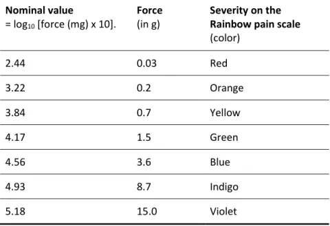
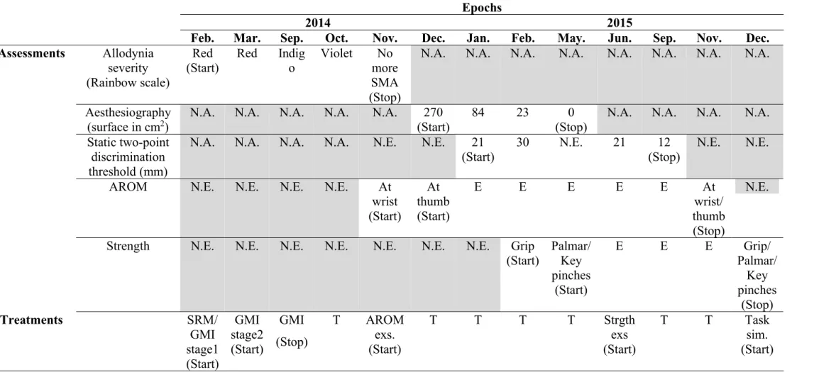
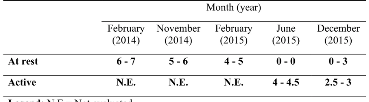
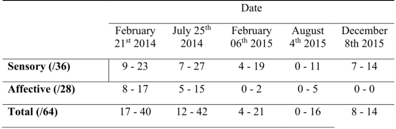
Table 1 : Characteristics of the Semmes-Weinstein monofilaments used to assess static mechanical allodynia (SMA) severity using the Rainbow pain scale assessment ... 59 Table 2 : Sequence of assessments and treatments performed during the tailored rehabilitation program. ... 60 Table 3 : Numeric pain scale scores on the right hand during the tailored rehabilitation program (minimal-maximal pain scores in the 24 hours preceding the assessment) ... 61 Table 4 : QDSA pain intensity scores on sensory, affective domains and total on the right hand during the tailored rehabilitation program (minimal-maximal pain scores in the 24 hours preceding the assessment) ... 62
Article 2
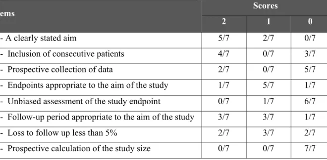
Table 1 : key characteristics of each study ... 90 Table 2 : Description of intervention according to study ... 93 Table 3 : Distribution of the seven articles included in the review according to the number of articles having obtained one of the three scores (0, 1, 2) for each item of the MINORS scale 97 Table 4 : risks of bias for each study ... 98
ix
Liste des figures Article 1
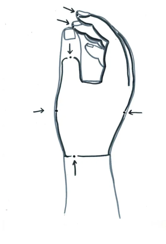
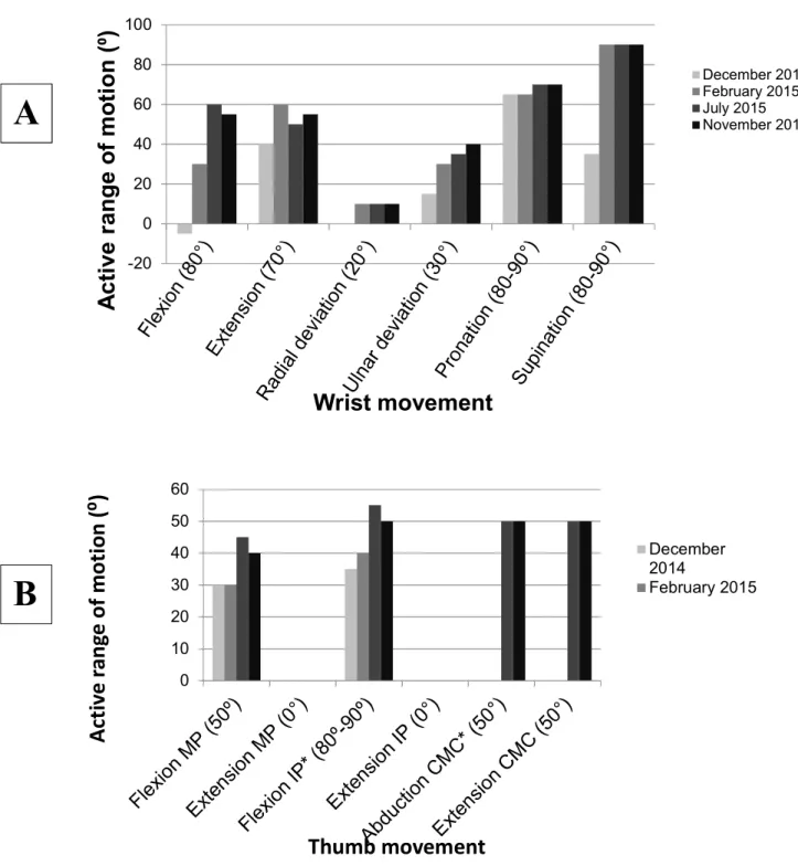
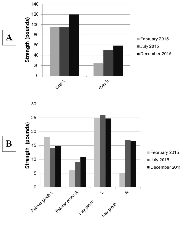
Figure 1 : Allodynography on the right hand performed on April 1st, 2014, showing the skin area innervated by the superficial branch of radial nerve on which static mechanical allodynia (SMA) was found. ... 63 Figure 2 : Active range of motion (AROM) of the right forearm wrist (A) and thumb (B) of the subject were measured at four different epochs during the tailored rehabilitation program. AROMs at the thumb are presented for flexion and extension of metacarpophalangeal (MP) and interphalangeal (IP) joints (MP); abduction and extension of carpometacarpal (CMC) joint. . 64 Figure 3 : Grip (A) and pinch (B) strengths (in pounds) on the left (L) and right (R) sides were measured at three different epochs during the treatment tailored rehabilitation program. ... 65
Article 2
x
Liste des sigles AM : Allodynie mécanique
AROM: Active Range of Motion
CINAHL: Cumulative Index to Nursing and Allied Health Literature CRPS: Complex Regional Pain Syndrome
DASH: Disabilities of the Arm, Shoulder and Hand Questionnaire DN : Douleur neuropathique
GMI: Graded Motor Imagery LNP : Lésion nerveuse périphérique
MINORS: Methodological Index for Non-Randomized Studies MRSD : Méthode de rééducation sensitive de la douleur NP: Neuropathic Pain
NSPA: Numeric Pain Scale Assessment NRS: Numerical Rating Scale
PNI: Peripheral Nerve Injury
PRISMA: Preferred Reporting Items for Systematic Reviews and Meta-Analyses QDSA: Questionnaire de la douleur Saint-Antoine
RPS: Rainbow Pain Scale
SDRC : Syndrome douloureux régional complexe SMA: Static Mechanical Allodynia
SRM: Somatosensory Rehabilitation Method
TENS: Transcutaneous Electrical Nerve Stimulation VAS: Visual Analog Scale
xi
Aux thérapeutes qui osent s’aventurer dans le défi de la réadaptation d’une clientèle présentant des douleurs neuropathiques, en espérant que ce travail vous soit utile.
xii
Remerciements
Ce mémoire de maîtrise représente l’achèvement d’une première grande étape dans le cadre de mon retour aux études. En effet, cela m’a permis de m’initier au monde de la recherche, qui me passionne toujours davantage, jour après jour. Tout au long de mon parcours, des gens près de moi m’ont supporté. Aussi, ce parcours m’a permis de rencontrer d’incroyables humains qui m’ont également aidé et motivé durant de nombreux mois.
J’aimerais d’abord remercier mes directeurs (Daniel et Joseph-Omer) pour votre grande disponibilité et le temps consacré à partager vos précieux conseils ainsi que le lot de connaissances nécessaires à mon cheminement aux études supérieures. Merci de m’avoir poussé à donner le meilleur de moi-même pendant ces études de maîtrise. Et enfin, merci d’accepter de poursuivre l’aventure en ma compagnie aux études supérieures.
Merci aussi à ma famille (Gilles, Carole, Annie, Jean-Philippe, Loulou, Claude, Danielle, Jocelyne, Paco) et à mes amis (Valérie, Éliane, Xavier, Steven, Julie, Marie-Ève) d’avoir cru en moi. Merci pour votre écoute, votre soutien émotionnel et surtout pour tous les fous rires qui m’ont tant fait de bien. Merci aussi à mes amis d’outre-mer (Karine, Emma, Manon, Enya, Nadège, Arnaud et vos amis) qui m’ont si chaleureusement accueilli et diverti pendant les vacances, afin de mieux reprendre mes travaux par la suite.
Merci à mes amis et collègues étudiants (Louis-Pier, Alicia, Maxime, Jacqueline, Alexandre, Christophe, Brigitte et Tokiko) pour votre soutien pendant les cours à la maîtrise, pour vos précieux conseils pendant la rédaction de ce mémoire et pour tous ces bons moments à caractère social.
xiii
Merci à mes collègues suisses, dont Claude, de m’avoir accueilli à votre clinique entre 2011-2012, période qui m’a permis de m’initier aux approches de stimulation tactile.
Merci aux étudiants à la maîtrise en projet d’intégration (Laurence et Alexis) qui ont accepté de plonger dans l’aventure de la recherche en ma compagnie.
Merci aux enseignants universitaires (Mélanie, Julie, Robert, Marc) pour avoir fait germer dans ma tête l’idée de retourner aux études, vos encouragements et votre implication.
Merci à mes collègues cliniciens du Centre professionnel d’ergothérapie (Julie, Laurent, Dominique, Thibault, Chloé, Justine, Anne-Sophie, Favio, Sorin, Camille, Camille), personnel de soutien (Isabel, Nathalie, Vanessa, Ludovic) et patrons en clinique (Sophie et Nathalie) pour votre soutien et pour votre implication dans les projets de recherche. Sans vous, équipe de travail formidable, ce projet n’aurait pas été possible.
Il ne faut pas oublier de mentionner l’apport important des obstacles placés sur mon chemin. Mon fort désir de les surmonter m’aura d’abord poussée à emboîter le pas dans les études supérieures, puis à me dépasser tout au long de mes études de maîtrise.
Finalement, vous aurez compris qu’une tribu entière a été présente tout au long de mon cheminement à la maîtrise et a contribué hors de tout doute à mes réussites.
xiv Avant-propos
En clinique, les douleurs neuropathiques, dont l’allodynie mécanique (hypersensibilité au toucher), sont fréquemment observées lors des blessures à la main. L’allodynie mécanique a un impact sur les capacités en interférant avec les activités qui demandent un contact cutané sur la main (ex. laver la vaisselle, utiliser un clavier, etc.). Elles sont également difficiles à traiter en réadaptation, mettant souvent en échec les modalités de traitement demandant un contact cutané (ex. massage, mobilisations, port de gant compressif, massage de cicatrice, etc.). C’est pourquoi cette problématique est importante à traiter. En réadaptation, l’approche conventionnelle par désensibilisation est utilisée depuis quelques dizaines d’années au Québec pour traiter l’allodynie mécanique. Tel qu’observé en clinique, cette approche parvient à diminuer les douleurs chez certains patients, mais il demeure qu’une bonne proportion de patients ne sera pas soulagée. C’est une des raisons pour lesquelles certains thérapeutes sont à l’affut de nouvelles approches et méthodes d’intervention innovantes pour le traitement de l’allodynie. Dans les dernières années, la méthode de rééducation sensitive de la douleur a été développée en Suisse. En raison de mon intérêt clinique pour le traitement de l’allodynie, j’ai été la première ergothérapeute du Québec à me former à cette méthode au Centre de rééducation sensitive du corps humain de Fribourg. Suite à cette formation, j’ai enseigné cette méthode à mes collègues dans le cadre de formations continues.
Les milieux cliniques reconnaissent que la méthode de rééducation sensitive de la douleur diminue la douleur de type allodynie en plus d’améliorer les capacités des patients qui en sont atteints. De plus, cette méthode est appliquée avec succès en combinaison avec d’autres modalités thérapeutiques. Malgré ces observations cliniques, cette combinaison de traitement qui semble prometteuse n’est pas illustrée dans la littérature. Cela a mené à ma première
xv
publication de ce mémoire soit une étude de cas présentant le raisonnement clinique qui sous-tend un programme multimodal incluant la méthode de rééducation sensitive, ainsi que ses effets potentiels sur la douleur et la performance motrice.
Cette étude de cas m’a amené à me questionner plus à fond sur les approches de stimulation tactiles. Quel est l’apport de la méthode de rééducation sensitive de la douleur, et plus encore des approches de stimulation tactiles, dans le traitement des douleurs de type allodynie ? Plus précisément, quel éventail de modalités de traitement est inclus dans ces approches de stimulation tactile Quel est le niveau de preuve de ces approches pour traiter l’allodynie ? Ces réflexions m’ont amené à faire une recension systématique des écrits comme première étape afin de tenter de répondre à ces questionnements.
En résumé, dans le cadre de ce mémoire, la publication d’une étude de cas a permis d’illustrer mon cheminement clinique pour le traitement de l’allodynie tout en assurant un transfert de connaissances aux cliniciens. Ce premier article permet de fournir des pistes de réflexion pour soutenir le raisonnement clinique dans le cadre de l’utilisation des approches de stimulation tactiles incluses dans un programme multimodal pour traiter l’allodynie. Quant à la recension des écrits, elle a permis d’explorer l’éventail des interventions par stimulation tactiles disponibles ainsi que les effets de ces approches et leurs niveaux d’évidence. Il est espéré que l’ensemble de cette démarche dans le cadre de ma maîtrise de recherche saura être utile aux cliniciens qui travaillent quotidiennement à améliorer les capacités et la vie quotidienne de ces patients avec allodynie.
16
Chapitre 1 : Introduction 1.1 Neuropathies
Les neuropathies sont définies comme des atteintes du système nerveux périphérique qui incluent, entre autres, les polyneuropathies et les mononeuropathies (Merskey & Bogduk, 1994). Les polyneuropathies consistent en des neuropathies diffuses et bilatérales (Merskey & Bogduk, 1994), c’est-à-dire qui affectent plusieurs nerfs dans l’ensemble du corps. Elles incluent un ensemble de conditions diverses dont la polyneuropathie diabétique, la polyneuropathie des soins intensifs et la maladie de Charcot-Marie-Tooth. Quant aux mononeuropathies, elles consistent en des atteintes focales, c’est-à-dire des atteintes qui touchent une seule branche nerveuse (Merskey & Bogduk, 1994). Les neuropathies sont souvent désignées par l’appellation de « lésions nerveuses périphériques (LNP) ». Leur prévalence est importante. Par exemple, en considérant le syndrome du tunnel carpien, qui est une atteinte focale par compression du nerf médian, cette mononeuropathie affecterait à elle seule 6,4% de la population néerlandaise (Atroshi et al., 1999; de Krom et al., 1992). D’ailleurs, les mononeuropathies sont le plus souvent causées par des atteintes mécaniques ou traumatiques (i.e., compression, étirement, lacération) qui affectent le système musculo-squelettique, mais également un nerf (Sunderland, 1951). Ainsi, dans un centre de trauma à Toronto (Canada), 2,8% des admissions pour trauma présentent des LNP sur une ou plusieurs branches nerveuses, dont plus de la moitié touche le membre supérieur (Noble, Munro, Prasad, & Midha, 1998). De ce fait, les LNP qui affectent les nerfs médian, radial et/ou ulnaire figurent parmi les diagnostics les plus fréquents en thérapie de la main (Keller et al., 2016).
17
Les mononeuropathies entraînent des coûts importants pour les systèmes de santé et d’indemnisation à travers le monde. À titre d’exemple, une étude suédoise réalisée en 2005 a estimé que les coûts des traitements et de la réadaptation de travailleurs ayant subi des mononeuropathies traumatiques nécessitant une intervention chirurgicale, était d’une médiane de 31 186 EUR et de 51 238 EUR par travailleur pour des lésions des nerfs ulnaire et médian respectivement (Rosberg et al., 2005). Plus de 87% de ces coûts résultaient du manque à gagner en raison des pertes de productivité chez les travailleurs (Rosberg et al., 2005). Ces données ne sont pas disponibles pour la population canadienne, mais il raisonnable de croire que ces lésions entraînent des coûts tout aussi importants au Canada.
Par ailleurs, des études ont démontré que les LNP, incluant les mononeuropathies, engendrent des incapacités importantes qui peuvent limiter les habitudes de vie, le retour en emploi et affecter la qualité de vie des personnes affectées (Hundepool et al., 2015; Novak, Anastakis, Beaton, Mackinnon, & Katz, 2011a). En effet, une étude a démontré qu’un an après une lésion ayant entraîné une neuropathie focale au membre supérieur nécessitant une chirurgie, 40% des patients ne sont toujours pas encore retournés en emploi (Bruyns et al., 2003). Chez ces patients, le niveau de capacités et le retour en emploi seraient liés, entre autres, à la sévérité de la lésion, la sensibilité à la main, la force de préhension et l’intensité de la douleur (Bruyns et al., 2003; Hundepool et al., 2015; Novak, Anastakis, Beaton, Mackinnon, & Katz, 2011b).
18 1.2 Douleurs neuropathiques
Les LNP peuvent entraîner des douleurs neuropathiques (DN) (Haanpää et al., 2011; Treede et al., 2008). La DN est définie comme une « douleur causée par une lésion ou une maladie du système nerveux somatosensoriel » (Merskey et al., 1994). Plusieurs études suggèrent que la présence de DN aurait un impact important sur les capacités des personnes ayant une LNP au membre supérieur. Par exemple, on a démontré une corrélation positive et significative entre la sévérité de la douleur et la perception des incapacités en lien avec les déficiences au membre supérieur évaluée à l’aide du questionnaire DASH («Disabilities of the Arm, Shoulder and Hand Questionnaire») chez des patients présentant des douleurs suite à une LNP (Novak et al., 2011b). D’autres études ont également mis en évidence un lien entre la douleur et les incapacités chez des personnes présentant des douleurs lors des traumas avec LNP affectant la main (Bailey, Kaskutas, Fox, Baum, & Mackinnon, 2009; Clement, Duckworth, Jenkins, & McEachan, 2016; Lozano Calderon, Paiva, & Ring, 2008; Souer, Lozano-Calderon, & Ring, 2008).
La DN peut se présenter sous plusieurs formes, dont la douleur spontanée (névralgie) ou provoquée (allodynie). La névralgie est une douleur neuropathique qui peut être perçue de manière incessante ou intermittente et qui apparaît spontanément, c’est-à-dire sans stimulation provoquante. Ce mémoire ne porte pas sur la névralgie, mais plutôt sur la DN de type allodynique. L’allodynie est définie comme une douleur provoquée par une stimulation normalement non-douloureuse (Merskey & Bogduk, 1994). Elle est précisée selon la modalité du stimulus provoquant la douleur, par exemple l’allodynie thermique provoquée la température
19
chaude et/ou froide et l’allodynie mécanique (AM) statique ou dynamique provoquée par un toucher immobile (statique) ou mobile (dynamique). L’AM n’est pas exclusivement neuropathique, car elle peut survenir par exemple dans des conditions inflammatoires (Merskey & Bogduk, 1994). Pour être considérée de type neuropathique, elle doit être corrélée avec des signes cliniques témoignant de l’atteinte neuropathique (symptômes, signes). Les mécanismes pathophysiologiques en cause dans l’AM ne sont pas précisés, mais il est possible que différents changements dans le système nerveux central au niveau spinal et supraspinal y contribuent, dont une neuromodulation et une neuroplasticité maladaptative. (Cervero & Laird, 1996; Cruciani, Stacy, & Knotkova, 2011; Finnerup, Otto, McQuay, Jensen & Sindrup, 2005; Latremoliere & Woolf, 2009; Osborne, Anastakis, & Davis, 2018; Wall, Xu, & Wang, 2002; Woolf, 2011).
À notre connaissance, les prévalences des DN et de l’AM ne sont pas connues au Canada. Cependant, dans la population générale du comté d’Olmsted (Minnesota, États-Unis), il est rapporté que la prévalence des DN varie entre 3,0 et 12,4% selon l’instrument de dépistage utilisé (Yawn et al., 2009). Également, dans la population générale en France, une étude a démontré une prévalence de 6,9% de personnes qui présentent de la douleur chronique avec des caractéristiques neuropathiques (Bouhassira, Lantéri-Minet, Attal, Laurent, & Touboul, 2008).
Quoiqu’il n’y ait pas, à notre connaissance, d’étude qui en fasse état, il est raisonnable de croire que l’AM à la main puisse avoir un impact important sur les habitudes de vie (activités courantes et rôles sociaux) en interférant avec les capacités spécifiques à l’utilisation de la main qui impliquent un contact cutané, telles que la dextérité et la force de préhension. Dans la
20
perspective où l’AM peut affecter la réalisation des activités, les interventions qui permettent de la réduire pourraient améliorer les capacités, ainsi que l’autonomie dans les habitudes de vie chez les personnes avec LNP affectant la main.
1.3 Syndrome douloureux régional complexe
L’AM est présente dans plusieurs conditions de santé. Par exemple, on peut l’observer chez certaines personnes atteintes de polyneuropathie diabétique (Scholz et al., 2016), chez certains grands brûlés (Nedelec et al., 2016; Schneider et al., 2006) et chez certains patients atteints de syndrome douloureux régional complexe (SDRC) (Harden et al., 2010; Packham, Spicher, MacDermid, Michlovitz, & Buckley, 2018). Le SDRC, qui est le diagnostic du patient présenté dans la première étude de ce mémoire, est caractérisé par une douleur continue disproportionnée par rapport à l’évolution habituelle après un trauma ou une autre lésion (Harden et al., 2010). La douleur, spontanée ou évoquée par un stimulus externe, est située dans une région du corps et non pas dans un territoire nerveux spécifique ou un dermatome. La douleur montre habituellement une prédominance distale d’anomalies sensitives, motrices, sudomotrices, vasomotrices (œdème) et/ou trophiques » (Harden et al., 2010). Deux sous-types de SDRC ont été définis : le SDRC-I et le SDRC-II. Le SDRC-I est associé à tout type de trauma, spécifiquement les fractures ou les lésions des tissus mous (Harden et al., 2010). Le SDRC-II est défini comme étant associé à une évidence physique ou électrodiagnostique de lésion nerveuse majeure (Harden et al., 2010). Bien que le SDRC-I n’est pas associé à une lésion nerveuse majeure, il demeure qu’il peut inclure des signes et symptômes caractéristiques des DN (ex : AM). Il n’est donc pas surprenant qu’il ait été démontré que certains SDRC-I soient
21
associés à certains types de LNP (Birklein & Schmelz, 2008; Oaklander & Fields, 2009), Une récente étude a trouvé que 54,3% de leurs participants avec SDRC-I rapportaient des symptômes d’AM, et que les signes cliniques confirmaient objectivement l’AM chez 27,6% de ces participants (Dietz et al., 2019). Par exemple, dans la première étude de ce mémoire, le patient présentait un SDRC-I avec AM. Chez ce patient, il n’y avait pas diagnostic de LNP majeure, mais la présence de LNP était objectivée, entre autres, par la diminution de la sensibilité tactile dans le territoire d’un nerf périphérique, la présence de paresthésies et d’AM à la main. Les hypothèses des mécanismes pathophysiologiques responsables des douleurs évoquées dans les cas de SDRC sont, comme pour les douleurs neuropathiques, liées à des modifications inadaptées du système nerveux central (Juottonen et al., 2002; Maihöfner, Handwerker, Neundörfer, & Birklein, 2003).
1.4 Traitement des douleurs neuropathiques
Plusieurs interventions ont été développées pour traiter les DN, dont l’approche médicamenteuse dont les effets sont limités (Attal et al., 2006; Dworkin et al., 2007; Mason, Moore, Derry, Edwards, & McQuay, 2004; Silver, Blum, Grainger, Hammer, & Quessy, 2007; Vranken, 2009). Les interventions non médicales incluent, entre autres, la psychothérapie (Moura et al., 2012), l’électrothérapie (TENS : Transcutaneous Electrical Nerve Stimulation ) (Gibson, Wand, & O'Connell, 2017), la désensibilisation (Yerxa, Barber, Diaz, Black, & Azen, 1983) ainsi que la méthode de rééducation sensitive de la douleur (MRSD) (Spicher & Quintal, 2013). Dans la pratique clinique, la psychothérapie (Evans, Fishman, Spielman, & Haley, 2003; Moura et al., 2012) est plutôt considérée comme un adjuvant et est combinée aux autres
22
modalités. Quant au TENS et à la désensibilisation, les protocoles d’utilisation ne font pas consensus compte tenu que les populations dans les études sont hétérogènes et que les évidences sur leur efficacité sont limitées (Gibson et al., 2017; Lewis, Coales, Hall, & McCabe, 2011; B. Pleger et al., 2005; Yerxa et al., 1983). La MRSD (Spicher, Quintal, & Vittaz, 2015) utilise un protocole d’utilisation qui est standardisé, mais une fois de plus, les populations étudiées sont hétérogènes (Nedelec et al., 2016; Packham et al., 2018; Spicher, Mathis, Degrange, Freund, & Rouiller, 2008). De plus, les devis des études qui ont porté sur la MRSD fournissent seulement des évidences limitées de son efficacité.
La littérature propose que les programmes multimodaux personnalisés sont à privilégier dans le traitement des DN (Deng, Luo, Hu, Fang, & Liu, 2016; Finnerup, Otto, McQuay, Jensen, & Sindrup, 2005) incluant celles associées au SDRC (Packham & Holly, 2018). Dans la perspective où les approches de stimulation tactiles pourraient induire une réorganisation du système nerveux central (Flor, 2002; Flor, Denke, Schaefer, & Grusser, 2001; Wand, Abbaszadeh, Smith, Catley, & Moseley, 2013) et qu’elles ne sont pas des approches adjuvantes puisqu’elles visent à traiter la condition, il y a donc lieu de s’intéresser à leur inclusion dans un programme multimodal. Il devient donc pertinent de vérifier leurs potentiels effets bénéfiques sur la DN.
1.4.1 Désensibilisation
Les approches de stimulation tactiles utilisées en clinique incluent la désensibilisation et la MRSD, qui sont deux approches différentes de traitement pour la DN évoquée par stimulation
23
mécanique, c’est-à-dire l’AM. Historiquement, la désensibilisation (Yerxa et al., 1983) a été la première méthode développée pour une population avec AM à la main et selon nos observations en clinique, elle semble encore à ce jour la plus utilisée. Cette approche de désensibilisation utilise l’application de textures directement sur la surface allodynique. Au fur et à mesure de l’amélioration de la condition du patient avec la diminution de l’intensité de l’AM, cette approche promeut d’adapter progressivement les textures plus douces vers des textures plus rugueuses dans le but de modifier à la hausse le seuil de perception de la douleur (Yerxa et al., 1983). Autrement dit, l’objectif est que ce soit des stimulations tactiles plus importantes qui soient nécessaires afin de déclencher la douleur. Jusqu’en 2003, la désensibilisation était la seule approche de stimulation tactile connue et utilisée en réadaptation pour traiter l’AM.
1.4.2 Méthode de rééducation sensitive de la douleur
Puis, les limites cliniques de la désensibilisation, on fait en sorte que des alternatives à cette approche ont été développées. Tel qu’observé en clinique et tel que discuté avec le premier auteur à avoir décrit la MRSD, une certaine proportion de patients avec AM ne répondent pas positivement à l’approche de désensibilisation, ou pire leur symptomatologie douloureuse augmente. C’est pourquoi la MRSD a été développée et son protocole d’utilisation a été publié dans un manuel en 2003 (Spicher, 2003). Cette méthode a été développée non seulement pour les atteintes sensitives et douloureuses à la main, mais également pour les autres territoires cutanés du corps humain (Spicher et al., 2015). La MRSD se différencie de la désensibilisation, entre autres en raison du fait qu’elle inclut des évaluations standardisées pour l’AM : l’allodynographie et l’arc-en-ciel des douleurs. La première évaluation est une cartographie de la surface allodynique évaluée avec un monofilament Semmes-Weinstein de 15.0g (#5.18), reportée sur papier millimétrée. La deuxième évaluation objective la sévérité de l’allodynie avec
24
une série de sept monofilaments (de 0.03g à 15.0g), correspondant à la plus petite force d’application causant de la douleur dans la surface allodynique. Elle se différencie aussi dans le protocole de traitement de l’AM qui comprend deux modalités, soit 1) l’enseignement thérapeutique et l’encadrement du patient pour diminuer voire éviter le toucher avec la surface allodynique et 2) des textures uniquement douces, et de la vibration mécanique de faible amplitude (fréquence de 100Hz, amplitude de 0.06mm) appliquées à distance, c’est-à-dire sur une zone anatomique proximale à la surface allodynique (Spicher et al., 2015). Elle vise à ce qu’une stimulation non-nociceptive soit perçue comme étant effectivement non-douloureuse (Spicher et al., 2015) et ainsi modifier à la hausse le seuil de perception de la douleur. Les deux approches (désensibilisation et MRSD) utilisent des textures afin de normaliser les perceptions sensitives. Cependant, elles se distinguent en ce qui a trait à la batterie d’évaluations, l’encadrement thérapeutique, le site d’application des stimulations tactiles et le type de textures employées.
1.5 Problématique
Plusieurs modalités d’interventions sont souvent combinées dans les programmes en réadaptation pour traiter l’AM. Cependant, force est de constater que les études portant sur les interventions pour traiter l’AM ne reflètent pas la pratique clinique réelle. En effet, ces études investiguent de manière isolée les effets d’une ou l’autre des modalités d’interventions sans toutefois considérer leur combinaison dans un programme d’interventions multimodal. Ceci limite l’applicabilité clinique des résultats des études portant sur des modalités isolées. À ce jour, aucune étude n’a encore décrit de quelle façon ces modalités d’interventions sont utilisées
25
en combinaison et en séquence dans le cadre d’un programme multimodal. Pourtant, plusieurs recommandations cliniques issues de la littérature, préconisent l’utilisation de programmes multimodaux (Deng et al., 2016; Finnerup et al., 2005), ce qui reflète la pratique clinique actuelle en réadaptation où la combinaison des interventions est préconisée.
Par ailleurs, il existe à l’heure actuelle des synthèses de connaissances de la littérature portant sur l’utilisation des approches de stimulation tactiles pour traiter l’hypoesthésie tactile à la main (Miller, Chester, & Jerosch-Herold, 2012; Oud, Beelen, Eijffinger, & Nollet, 2007). Cependant, il n’existe, jusqu’à présent, aucune synthèse des connaissances sur l’utilisation des approches de stimulation tactiles pour traiter l’AM. Cette lacune dans la littérature a des conséquences tant sur la pratique clinique que sur la recherche dans ce domaine. En effet, il n’est pas clair quelles sont toutes les interventions de stimulation tactiles existantes pour traiter l’AM qui ont effectivement fait l’objet d’études. De plus, on ne peut statuer parmi ces interventions lesquelles disposent des meilleures évidences concernant leurs effets bénéfiques sur l’AM. Par conséquent, il est difficile de comparer les interventions existantes et de sélectionner la ou les meilleures approches de traitement pour l’AM.
Les études qui sont présentées et discutées dans ce mémoire tentent de répondre à cette problématique. Elles portent plus précisément sur l’utilisation des approches de stimulation tactiles pour traiter l’AM à la main chez des patients présentant des mononeuropathies. Ce mémoire présente 1) de quelle manière une approche de stimulation tactile pour traiter l’AM peut être intégrée à un programme de réadaptation multimodal, et 2) une synthèse des
26
connaissances sur les différentes approches de stimulations tactiles pour traiter l’AM consécutive à une LNP qui ont fait l’objet d’études.
1.6 Objectifs
Le premier objectif de ce mémoire est de décrire l’intégration de la MRSD dans le cadre d’un programme de réadaptation multimodal chez un patient présentant un SDRC avec AM qui n’avait pas eu d’effets bénéfiques avec des interventions conventionnelles. Afin de répondre à cet objectif clinique, une étude de cas a été effectuée et publiée (Quintal, Poiré-Hamel, Bourbonnais, & Dyer, 2018). La MRSD a été utilisée dans un cadre de traitement multimodal puisque la désensibilisation classique n’avait pas eu les effets escomptés et que la MRSD possède un caractère innovant.
Comme le programme de réadaptation multimodal présenté dans l’étude de cas inclut une approche de stimulation tactile, cela a questionné l’impact relatif de ce type d’approche dans ce programme, et donc les niveaux d’évidences cliniques des approches de stimulation tactiles répertoriées dans la littérature. Ainsi, le deuxième objectif de ce mémoire est de recenser les approches de stimulation tactile étudiées dans la littérature et leurs niveaux d’évidences en lien avec le traitement de la douleur chez des personnes avec AM à la main. Afin de répondre à cet objectif de recherche, une recension systématique a été effectuée. Cette méthodologie a été utilisée pour ses capacités à identifier, évaluer et résumer les résultats d’études sur une problématique de santé précise (Gopalakrishnan & Ganeshkumar, 2013). De plus, ce type de méthodologie, qui résume les évidences scientifiques, améliore l’accessibilité aux données
27
probantes pour les utilisateurs de connaissances, ces derniers n’ayant pas toujours le temps de lire et d’analyser l’entièreté de la littérature disponible sur un sujet précis (Gopalakrishnan & Ganeshkumar, 2013).
1.7 Organisation générale du mémoire
Suite à l’introduction (Chapitre 1 : Introduction) présentée précédemment, ce mémoire ce compose de quatre autres chapitres. Ainsi, afin de répondre aux deux objectifs généraux de ce mémoire, deux articles rédigés en anglais sont présentés (Chapitre 2 : Manuscrit, article #1 et Chapitre 3 : manuscrit, Article #2). Suivent une discussion générale (Chapitre 4 : Discussion) présentant des éléments non discutés dans les articles et une conclusion (Chapitre 5 : Conclusion). Les tableaux et figures se retrouvent directement à la suite des articles correspondants. Toutes les Références bibliographiques associées aux cinq chapitres se trouvent à la fin de ce mémoire. À noter que les mots en bleu servent également de renvois à des parties spécifiques du mémoire (ctrl+clic pour suivre les liens).
28
Chapitre 2 : Manuscrit, article #1
Le chapitre 2 vise à répondre au premier objectif de ce mémoire : décrire l’intégration de la MRSD dans le cadre d’un programme de réadaptation multimodal chez un patient présentant un SDRC avec AM qui n’avait pas eu d’effets bénéfiques avec des interventions conventionnelles.
2.1 Management of long-term complex regional pain syndrome with allodynia: A case report.
Les résultats de cette étude de cas ont sous forme d’article a été soumis au Journal of Hand Therapy le 28 novembre 2017, a été révisé le 16 janvier 2018, puis accepté pour publication le 22 janvier 2018 (Quintal, Poire-Hamel, Bourbonnais, & Dyer, 2018). Le consentement du participant a été obtenu avant la soumission à l’éditeur et la publication de l’article.
2.1.1 Apport de l’étudiante et de chacun des co-auteurs
Isabelle Quintal a été la principale responsable de chacune des étapes menant à la publication de ce manuscrit, c’est-à-dire : l’élaboration du devis, la collecte de données, l’analyse de données, la rédaction du manuscrit, la soumission au journal ainsi que gérer les révisions demandées.
Laurent Poiré-Hamel ergothérapeute clinicien, a participé à la collecte de données, à l’analyse des données ainsi qu’à la rédaction du manuscrit.
Daniel Bourbonnais et Joseph-Omer Dyer ont dirigé la principale responsable de ce manuscrit, Mme Quintal, dans l’ensemble des étapes menant à la publication finale.
29
Management of long-term complex regional pain syndrome with allodynia: A case report
Auteurs : Isabelle Quintal OT a,b,c, Laurent Poiré-Hamel OTc, Daniel Bourbonnais OT, PhD a,b, Joseph-Omer Dyer PT, PhD a
Affiliations École de réadaptation, Université de Montréal, C.P. 6128, Succursale Centre-Ville, Montreal, Qc. H3J 3J7.
b. Centre de recherche interdisciplinaire en réadaptation du Montréal métropolitain (CRIR), IURDPM, site IRGLM, 6300 avenue Darlington, Montréal, Qc, H3S 2J4. c. Centre Professionnel d’Ergothérapie, 6960 Sherbrooke Est, Montréal, Qc, H1N 1E5.
30 2.2 Abstract Study design: Case report.
Introduction: Conventional rehabilitation alone may not be effective in reducing symptoms in some patients with complex regional pain syndrome.
Purpose of the study: This case report portrays the benefits of a new tailored rehabilitation program for a 39-year-old patient suffering from upper limb complex regional pain syndrome with severe touch- evoked pain (static mechanical allodynia).
Methods: This patient had previously received conventional rehabilitation for a year and a half including physical and nonsurgical medical interventions that did not improve symptoms or function. In the search for an alternative, this patient was referred to occupational therapy to try a tailored rehabilitation program, drawing on multiple strategies used sequentially according to the patient’s tolerance and symptom evolution. During this 22-month program, the following methods were added (listed chronologically): somatosensory rehabilitation of pain method, graded motor imagery, pain management modalities, active mobilizations, strengthening exercises, and task simulation. The patient successively showed resolution of mechanical allodynia, decreased pain, reduction of tactile hypesthesia and improvement in active range of motion, strength, and function. These improvements allowed him to return to work.
Discussion: This suggests that a tailored rehabilitation program combining somatosensory rehabilitation of pain method, graded motor imagery and more conventional approaches could improve symptoms and functional status in patients with upper limb complex regional pain syndrome, even with persistent refractory symptoms.
31
Conclusion: The addition of the somatosensory rehabilitation of pain method and the graded motor imagery approach to conventional therapy could be considered in cases of complex regional pain syndrome that do not respond to conventional rehabilitation alone.
32
2.3 IntroductionComplex regional pain syndrome (CRPS) incidence varies between 5 and 26 cases per 100,000 per year (Marinus et al., 2011), and is seen twice as often with the upper limb as with the lower limb (Sandroni, Benrud-Larson, McClelland, & Low, 2003). It is characterized by the presence of regional painful symptoms, seemingly
disproportionate, associated with sensory, motor, sudomotor, vasomotor edema, and/or trophic signs (Harden et al., 2010).Those impairments can severely affect the function of the upper limb (Galer, Henderson, Perander, & Jensen, 2000). Evidence suggests that people with upper limb CRPS suffers longer than those with lower limb CRPS (de Mos et al., 2009). About two-thirds of patients with CRPS continue to show substantial
limitations of their independence 1 year after the onset of symptoms (Bean, Johnson, & Kydd, 2014; Borchers & Gershwin, 2014; de Mos et al., 2009; Geertzen, de Bruijn-Kofman, de Bruijn, van de Wiel, & Dijkstra, 1998; Sharma, Agarwal, Broatch, & Raja, 2009).Patients with CRPS often have significant somatosensory symptoms (Gierthmuhlen et al., 2012; Rommel, Malin, Zenz, & Jänig, 2001).Among these symptoms, abnormal painful sensations such as hyperalgesia and allodynia, as well as skin sensibility disorders, are often the leading cause of complaints and decreased function (Huge et al., 2011; Savas, Baloglu, Ay, & Cerci, 2009). Hyperalgesia refers to increased pain due to a painful
stimulus (Huge et al., 2011; Merskey & Bogduk, 1994),whereas allodynia is pain evoked by a normally painless stimulation (Lolignier, Eijkelkamp, & Wood, 2015).The term “allodynia” encompasses several forms, including thermal allodynia evoked by heat or cold and mechanical allodynia evoked by static or dynamic touching. Hyperalgesia and allodynia are seen twice as frequently in CRPS than in other pathological conditions affecting the upper limb, such as neuropathic conditions and discrete musculoskeletal
33
entities (e.g. osteoarthritis, rotator cuff disease, frozen shoulder, or healing fracture) (Mailis-Gagnon, Lakha, Allen, Deshpande, & Harden, 2014; Merskey et al., 1994). Allodynia has been reported in 74% of patients presenting with CRPS (Harden et al., 1999).The neurophysiological mechanisms responsible for painful symptoms in CRPS are not fully understood. Evidence suggests that in both types of CRPS, without (CRPS type I) and with nerve damage (CRPS type II), it is possible to observe neuropathic pain that is attributable to somatosensory impairments (Bruehl, 2010; Packham, MacDermid, Henry, & Bain, 2012; Treede et al., 2008). There is also evidence that these
somatosensory impairments may contribute to the development of painful symptoms via peripheral and central sensitization mechanisms in CRPS (Goh, Chidambaram, & Ma, 2017; Smart, Wand, & O'Connell, 2016). For example, it is possible to observe
reorganization in the primary somatosensory cortex (S1) that would be associated with sensitization mechanisms contributing to painful symptoms in these patients (Maihöfner et al., 2003).
Since peripheral and central sensitization mechanisms (Campero, Bostock, Baumann, & Ochoa, 2010) might contribute to pain chronicization in CRPS (Borchers & Gershwin, 2014), interventions that seek to regulate these mechanisms may be helpful in preventing such chronicization. The somatosensory rehabilitation of pain method (SRM) described by Spicher (2008) and graded motor imagery (GMI) (Moseley, 2004) are two innovative approaches that could potentially target these mechanisms. SRM uses peripheral somatosensory stimulation that can potentially act on peripheral sensitization mechanisms. Moreover, this method does not require active movement, which can be an interesting asset
34
in individuals in whom active mobilization can exacerbate symptoms. SRM consists of avoiding or reducing any cutaneous stimulation as much as possible in the skin area where touch evokes pain (i.e. allodynic area), while stimulating the somatosensory system at a distant site (with a soft fabric or light mechanical vibration in a comfortable territory proximal to the allodynic area). The SRM approach contrasts with that of the conventional desensitization approach, which promotes stimulation of the allodynic area with stimulations that are initially mild and then stronger as the person becomes accustomed to them and feels less pain (Yerxa et al., 1983).Evidence shows that SRM can reduce static mechanical allodynia (SMA) in patients with neuropathic pain (Nedelec et al., 2016; Packham et al., 2018; C. Spicher et al., 2008; Spicher et al., 2015). A retrospective case series on SRM showed a significant decrease in pain among burn survivors with SMA (Nedelec et al., 2016). Recently, a retrospective case series showed a reduction in the severity of SMA following SRM in patients with upper limb CRPS (Packham et al., 2018).
Conversely, GMI uses sensorimotor integration processes to reduce central sensitization and integrates the progression of active movements in its advanced stages. GMI is a hierarchical rehabilitation method in which patients must perform increasingly demanding tasks to create new neural connections targeted at normalizing the representation of the affected body part in the primary somatosensory cortex. GMI involves three stages of rehabilitation progression: (1) left/right discrimination, (2) explicit motor imagery, and (3) mirror therapy (G Lorimer Moseley, Butler, Beames, & Giles, 2012). Evidence shows that GMI alone can have beneficial effects in chronic pain conditions (Bowering et al., 2013) and CRPS (Daly & Bialocerkowski, 2009; Moseley, 2006; Walz,
35
Usichenko, Moseley, & Lotze, 2013). Therapies that preserve the integrity of the cortical somatosensory representation of body parts affected by CRPS may reduce pain symptoms in these patients (Moseley & Flor, 2012). The beneficial effects of GMI in CRPS could be explained by its ability to regulate cortical reorganization mechanisms involved in CRPS painful symptoms.
Although CRPS can be treated with medication and conventional physical therapy, these therapeutic approaches do not always reduce pain and improve function satisfactorily (Daly & Bialocerkowski, 2009). Currently, more evidence is needed for existing CRPS clinical guidelines (Daly & Bialocerkowski, 2009; Perez et al., 2010).There is still a need to develop new mechanism-based treatment approaches to achieve better results in the treatment of pain and somatosensory symptoms in CRPS (Bharwani et al., 2017).SRM and GMI are two different mechanism-based intervention approaches that may be potentially used in combination or as a complement to conventional rehabilitation (pain management modalities, active mobilizations, strengthening exercises, and task simulation) to treat CRPS. In the present case, this combination was used for a patient who did not respond to conventional rehabilitation alone.
36
2.4 Patient descriptionMr. B, a left-handed 39-year-old sub-Saharan African living in Quebec, Canada, was diagnosed in October 2012 with CRPS affecting his right upper limb. This condition resulted from a work- related accident that occurred in August 2012 while he was a plant production worker. During this accident, Mr. B sustained a right wrist injury involving ligament tears (triangular fibrocartilage complex and scapholunate ligaments). A few days after the injury and until December 2013, Mr. B received
conventional treatments, including physical rehabilitation (conventional desensitization approach, contrast baths, passive mobilizations, active mobility, and strengthening exercises), prescribed medications (pregabalin and celecoxib), and pain management medical interventions (several stellate ganglion and venous blocks) without any subjective improvement. Due to the lack of improvement in his condition, he was referred by his plastic surgeon for occupational therapy at our private clinic to try a new rehabilitation approach.
2.4.1 Initial clinical examination
On his first visit to the occupational therapist in February 2014, Mr. B complained of intolerable pain in his entire right upper limb that was causing severe limitations and a fear of using his arm. He kept his hand held protectively against his trunk. On visual inspection, his affected hand looked waxy, swollen, and atrophied compared to his other hand. The pain was located on the dorsal side of his hand and thumb. It increased when he used his hand or when it was slightly touched (not able to tolerate any covering) or exposed to cold. He described his symptoms as follows: feeling of constant numbness, intermittent burning sensations, and shooting pain up to his right shoulder. With regard to hand function,
37
he reported being unable to move the entire arm from shoulder to fingers because of the pain. He could not use his right hand for any daily living activities. The only possible active limited motions were those of the index and thumb. He could use a pinch grasp with his index/thumb to hold light objects (eg, paper, fork, and so on) for no more than a few seconds because of the pain. He also complained of lack of strength and endurance in the affected hand. He was very emotional when he spoke about his accident or his condition. The mere act of talking about his arm was enough to trigger signs of emotional distress. Living with this pain was a great source of emotional burden to him. He felt unable to think of anything but this pain, unable to plan new projects. At the end of the first meeting with the occupational therapist, he reported being deeply discouraged, that he had no hope of healing to the point of wondering why he continued to seek treatment.
2.5 Methodology
2.5.1 General organization of patient’s care
The patient was invited to participate in a tailored rehabilitation program which consisted of two components: (1) rehabilitation sessions supervised by an occupational therapist at the clinic (30 to 60 minutes in duration) and (2) home sessions managed by the patient. Rehabilitation sessions at the clinic and at home included the same interventions, that is, SRM and GMI combined with conventional treatments. Conventional treatments consisted of follow-up and advice on pain management modalities (i.e. medication, transcutaneous nerve stimulation (TENS), cryotherapy), active mobilizations, strengthening exercises, and task simulation. The patient had his own TENS and could
38
apply ice at home. The sessions at the clinic were held twice a week for the first 15 weeks and once a week thereafter for the rest of the program, which lasted 22 months. During the sessions at the clinic, all assessments and treatments were performed by the same two occupational therapists, alternately, depending on the availability of Mr. B and the therapists from one week to the next. One of the two therapists had SRM certification (56 hours of training in this method). Clinical evaluations included pain, SMA, tactile hypoaesthesia, active range of motions (AROMs), hand muscle strength, upper limb function, and judgment of left-right discrimination assessments. Not all evaluations were performed at each session but periodically depending on condition evolution and the therapists’ judgment. Only one or two assessments were done at each rehabilitation session to assess pain severity and prevent any flare-ups.
2.5.2 Clinical assessmentsPain intensity
Pain was assessed by the means of three evaluations: the Numeric Scale Pain Assessment (NSPA), the Visual Analog Scale (VAS), and the French version of the McGill Pain Questionnaire (Questionnaire de la douleur de Saint-Antoine [QDSA]). The NPSA measures pain intensity on a scale from 0 to 10, where 0 represents no pain at all, and 10 the worst possible pain (Farrar, Young, LaMoreaux, Werth, & Poole, 2001). The VAS evaluates pain intensity on a 10-cm scale, where 0 cm represents no pain at all, and 10 the worst possible pain (Scott & Huskisson, 1976). It is a valid tool for measuring pain at a specific point in time (Kersten, White, & Tennant, 2014).
39
The QDSA assesses pain by the patient’s selection and rating of qualifiers that represent the sensory and emotional dimensions of pain (Boureau, Luu, & Doubrère, 1992).The QDSA has a maximum score of 64 points which includes 36 points for sensory and 28 points for affective qualifiers. When completing the QDSA, patients are asked to rate the minimum and maximum pain intensity they felt during the 24 hours preceding the assessment.
NSPA, VAS, and QDSA were used in this case because these assessments are complementary and allow for a thorough assessment and better understanding of the pain felt by the patient. The NSPA assessment is user-friendly for day-to-day assessment because it is short. The VAS was useful to help map the allodynic area by the allodynography technique which will be presented in the following section. As for the QDSA, it was used periodically to evaluate the evolution of CRPS pain phenomenon in its sensory and affective characteristics over several weeks.
Static mechanical allodyniaSkin surface with SMA and severity of allodynia were assessed using the allodynography method and the Rainbow Pain Scale (RPS), respectively. The allodynography method consists of mapping the skin area that exhibits SMA. The borders of the allodynic cutaneous area are determined by the skin points where the static touch with a Semmes-Weinstein monofilament of 15 g (No. 5.18) causes a pain increase of 1 cm on the VAS scale from the baseline pain (i.e. pain level at rest assessed just before the evaluation) or where the monofilament causes a pain of at least
40
3 cm if the baseline pain was below 2 cm. For example, if the patient has a baseline pain level of 4 cm on the VAS, allodynography will look for the skin point where the 15 g monofilament causes pain at an intensity of 5 cm. However, if the patient has baseline pain of less than 2 cm (e.g. 0 cm), the allodynography will look for a skin point that produces pain that reaches at least 3 cm when the patient is touched with the 15 g monofilament. The mapping is made along the longitudinal line of the damaged cutaneous nerve branch, finding the most proximal and distal points (i.e. borders) of the allodynic area. The same procedure is applied in a transverse direction to find the most medial and lateral points of the allodynic surface. A polygon is then drawn by joining the 4 points with a line to give an approximate representation of the allodynic area (Spicher et al., 2015).
The RPS assesses the severity of SMA in the allodynic area mapped by allodynography at the skin point where the patient indicates feeling the worst touch-evoked pain. RPS uses 7 monofilaments of different sizes (see Table 1, p.59) to determine the minimal application force required to increase the baseline pain by 1 cm on the VAS or to reach 3 cm if the baseline pain is below 2 cm. Each of these 7 monofilaments corresponds to a color, which is associated with an application force (g) and a number. The smaller the application force needed to evoke pain with the monofilament, the greater the SMA severity.
41
Tactile hypoesthesia Skin surface with tactile hypesthesia and severity of hypoesthesia were assessed by the esthesiography method and the static 2-point discrimination test, respectively. The esthesiography method consists of mapping the skin area with tactile hypoesthesia. On the dorsum of the hand, the borders of the hypoesthesic area are determined by the skin points where the patient does not detect the static touch with a Semmes-Weinstein monofilament of 0.4g (No. 3.61), which is the expected normal value for tactile sensibility on the dorsal surface of the hand (Bell-Krotoski, Fess, Figarola, & Hiltz, 1995; Spicher et al., 2015).The mapping is done in a longitudinal direction, finding the most proximal and distal points (i.e. borders) of the hypoesthesic area. The same procedure is applied in a transverse direction to find the most medial and lateral points of the hypoesthesic area. A polygon is then formed by joining the 4 points with a line to give an approximate representation of the hypoesthesic area (Spicher et al., 2015).
The static 2-point discrimination test uses a 2-point esthesiometer to vary the distance between 2 points of tactile stimulation. The test consists of evaluating the minimum distance between 2 points from which the person can distinguish between static touch with 1 or 2 points (Spicher, Hecker, Thommen, & Rouiller, 2005).
Active range of motion, strength, and function Hand and wrist active range of motion (AROM) were assessed by goniometry (Pendleton & Schultz-Krohn, 2013). Hand strength was assessed by grip strength, and by palmar and key pinch strength. Grip strength was
42
assessed with a standard, adjustable-handle JAMAR dynamometer (Mathiowetz et al., 1985).Palmar and key pinch strength was assessed with a B&L Engineering pinch gauge (Mathiowetz et al., 1985).
The patient’s perception of his arm function was assessed by means of the Disability of the Arm, Shoulder and Hand Questionnaire (DASH), which assesses the patient’s perception of his level of disability to use his upper limbs. The higher the score, the less functional the patient perceives himself (Angst, Schwyzer, Aeschlimann, Simmen, & Goldhahn, 2011; Dowrick, Gabbe, Williamson, & Cameron, 2005; Hudak, Amadio, & Bombardier, 1996).It has been demonstrated that the original DASH outcome measure has good construct validity, test-retest reliability, and responsiveness to change (Beaton et al., 2001). We used the Canadian French version of the DASH, which shows good acceptability and psychometric properties, comparable to those obtained with the original version (Durand, Vachon, Hong, & Loisel, 2005).The DASH score shows a high correlation with grip force (r=0.47), but the correlation is much weaker with range of motion (r= 0.24) (Beaton et al., 2001; De Smet, 2007).
Finally, about every month and sometimes more often if needed, semi-structured interviews were used to assess the patient’s perception of his level of functional independence.
43
All assessments, except RPS (i.e. severity of mechanical allodynia), were performed once a month, in order to avoid exacerbating the patient’s painful symptoms. RPS assessment was carried out more often, that is, every week, to ensure adequate monitoring of allodynia, and to adjust SRM and GMI activities accordingly.
Tailored rehabilitation programThe tailored rehabilitation program was designed to treat the patient by avoiding pain, respecting his tolerance and allowing gradual recovery of his upper limb active mobility and function. In order to achieve these objectives, the same rehabilitation program was performed at the clinic and at home: it combined conventional therapy with appropriate pain management, sensory and motor rehabilitation methods, as well as occupational therapy. As part of this program, the patient was required to go to therapy at the clinic once or twice a week for assessment of his condition, and also for the set-up, progression, and teaching of the daily exercise program (performed at the clinic and at home). Throughout the rehabilitation program, the patient continued to take the same medication as that used during the initial rehabilitation, before the tailored rehabilitation program (i.e. pregabalin and celecoxib). The program was reviewed weekly with the patient to optimize his functional activities and independence while minimizing evoked pain.
Table 2 (p.60) shows the sequence of assessment and treatment methods performed during the tailored rehabilitation program. The program started from the first visit on February 19, 2014 with the SRM described by Spicher et al. (2015),until the very
44
last session at the clinic in December 2015. In the presence of SMA, SRM involves an initial step of allodynia treatment and then a second step which is the treatment of the tactile hypoesthesia underlying allodynia (when SMA is resolved). SRM is initially aimed at reducing SMA in two ways: by encouraging the patient to adhere to the precaution of avoiding touching the allodynic area and by performing tactile stimulation away from the allodynic territory. More precisely, the tactile stimulation is performed at a distance on a proximal territory (working territory) with a comfortable light fabric or mechanical vibration, 8 times a day for 1 minute (or less long) without evoking pain. In this case, we used shaved beaver fur, with a progression in the location of the working territory, from proximal to distal. Since it was not possible to stimulate the right upper limb without pain, the right thoracic territory was first stimulated. The tactile stimulation at a distance from the allodynic area began on February 25, 2014, on the anterior branch of the 12th thoracic nerve until the following April 25, then progressed to the anterior branch of the third thoracic nerve. Once SMA was resolved, the underlying hypoesthesia was treated using the approach advocated in the second step of SRM. This step consists in using direct stimulation on the area with fabrics and light vibration in a progressive way. In the beginning of the approach, a tactile stimulation of 15-second duration is applied 12 times a day. This tactile stimulation is then gradually pursued by increasing its duration and decreasing its daily frequency in order to ultimately reach a stimulation of 5-minute duration applied 4 times a day.
The patient started the GMI method on February 25, 2014 (Table 2, p.60). GMI involves three stages of rehabilitation progression: (1) left/right discrimination, (2) explicit
45
motor imagery, and (3) mirror therapy (Moseley et al., 2012). The protocol used was from the GMI Handbook (Moseley et al., 2012). This protocol describes the progression of the exercises according to the different stages of the methods and the normal outcomes expected at the end of each stage. The protocol does not give details as to exercises frequency or duration but rather recommends that these parameters be adjusted according to each patient’s capability. Therefore, these parameters have been adjusted to minimize pain as much as possible, depending on the clinical reasoning and the information shared between the therapist and the patient. In order for him to do these activities at home, he received a package of 50 homemade pictures representing left and right hands in various positions. During the first stage of GMI, he was instructed to complete left/right discrimination sessions 4 to 6 times a day or more if he could tolerate it. During these sessions, he had to identify left/right laterality as fast as possible, without getting a conscious mental representation of the hand, as if he had to guess quickly. Each left/right discrimination session was timed with a digital stopwatch. The total duration of each session was divided by the number of pictures guessed to obtain the average time allocated to each picture. Average time by picture and percentage of correct answers were monitored until the patient reached the expected normal values for that activity, that is, a rate of correct answers of 80% or more, as well as an average time per image of 2.0 0.5 seconds (Moseley et al., 2012).During the second stage of GMI, Mr. B was asked to close his eyes and visualize his right hand in a static position.
Promoting self-efficacy through patient education was an important goal that the therapists considered during the course of rehabilitation. Notably, upon initiation of SRM,
46
the therapists provided the patient with tips to avoid as much as possible touching the allodynic area. This teaching was intended to allow the patient to better understand the evolution of his condition and how to manage the challenge of respecting the precaution of not touching the allodynic area. Moreover, the therapists suggested solutions and adaptations that the patient could integrate daily, like pain control modalities or assistive devices such as the use of a nonslip surface to help him open jars. Thus, the patient was trained to use TENS, as well as cryotherapy (cold) and superficial heat as pain control modalities during the entire program. TENS and cryotherapy were performed on the ulnar nerve palmar branch because this nerve branch is in the vicinity of the allodynic area but not directly on the painful territory. The site of TENS stimulation on a neighboring nerve branch was chosen according to the literature (Bouhassira & Attal, 2012). Conventional TENS was applied at least once every day, or more if needed, for 20 minutes per session, as recommended (Bouhassira & Attal, 2012).Ice was applied many times a day, from 1 to 10 times if needed, for only 1-2 minutes, so it did not exacerbate the pain. Pain management education included instructions to take breaks regularly during his daily tasks to prevent pain exacerbation. Heated gloves were provided to the patient so that he could protect his hand from the cold during the winter period.
The second step of the SRM (treatment of the underlying hypoesthesia) also started on October 31, 2014 and ended in December 2015. Rehabilitation of tactile hypoesthesia was per- formed using direct stimulation of the area for reduced tactile sensation with soft fabrics and light mechanical vibration provided by a vibration generator. During the last few months of rehabilitation, the reduction of pain led to the introduction of strengthening