doi: 10.3389/fgene.2018.00712
Edited by: Pascal Chartrand, Université de Montréal, Canada Reviewed by: Jean-Marc Gallo, King’s College London, United Kingdom Rita Sattler, Barrow Neurological Institute (BNI), United States *Correspondence: Shunmoogum A. Patten [email protected]
Specialty section: This article was submitted to Genetic Disorders, a section of the journal Frontiers in Genetics Received: 13 August 2018 Accepted: 20 December 2018 Published: 22 January 2019 Citation: Butti Z and Patten SA (2019) RNA Dysregulation in Amyotrophic Lateral Sclerosis. Front. Genet. 9:712. doi: 10.3389/fgene.2018.00712
RNA Dysregulation in Amyotrophic
Lateral Sclerosis
Zoe Butti and Shunmoogum A. Patten*
INRS-Institut Armand-Frappier, National Institute of Scientific Research, Laval, QC, Canada
Amyotrophic lateral sclerosis (ALS) is the most common adult-onset motor neuron
disease and is characterized by the degeneration of upper and lower motor neurons.
It has become increasingly clear that RNA dysregulation is a key contributor to ALS
pathogenesis. The major ALS genes SOD1, TARDBP, FUS, and C9orf72 are involved in
aspects of RNA metabolism processes such as mRNA transcription, alternative splicing,
RNA transport, mRNA stabilization, and miRNA biogenesis. In this review, we highlight
the current understanding of RNA dysregulation in ALS pathogenesis involving these
major ALS genes and discuss the potential of therapeutic strategies targeting disease
RNAs for treating ALS.
Keywords: ALS (amyotrophic lateral sclerosis), FUS, C9orf72, TDP-43, RNA processing, RNAi (RNA interference), antisense oligonucleotide-drug conjugates
INTRODUCTION
Amyotrophic lateral sclerosis (ALS) is a progressive and fatal neurodegenerative disorder of motor
function. It is characterized by the selective degeneration of the lower and upper motor neurons.
Among the symptoms of this disease are progressive muscle weakness and paralysis, swallowing
difficulties and breathing impairment due to respiratory muscle weakness that ultimately causes
death, usually within 2–5 years following clinical diagnosis (
Kiernan et al., 2011
). Though most
cases of ALS are sporadic, some families (10%) demonstrate a clinically indistinguishable form of
ALS with clear Mendelian inheritance and high penetrance (
Pasinelli and Brown, 2006
). Treatments
to slow the progression of ALS to date remains riluzole (
Bensimon et al., 1994
) and edaravone
(
Abe et al., 2014
) but they are only modestly effective. However, in the past couple years, there has
been a real encouragement in witnessing potentially efficacious treatments, such as Masitinib and
Pimozide (
Trias et al., 2016
;
Patten et al., 2017
;
Petrov et al., 2017
) claiming to demonstrate clinical
benefit. Furthermore, RNA-targeted therapies are currently intensively being evaluated as potential
strategies for treating this ALS (
Schoch and Miller, 2017
;
Mathis and Le Masson, 2018
). There is
indeed hope to have new and potentially more effective treatment options available for ALS in the
near future.
Mutations in over more than 20 genes contribute to the etiology of ALS (
Chia et al., 2018
)
(Table 1). Amongst these genes, the major established causal ALS genes are
SOD1 (Cu-Zn
superoxide dismutase 1),
TARDBP (transactive response DNA Binding protein 43kDa), FUS (fused
in sarcoma) and hexanucleotide expansion repeat in Chromosome 9 Open Reading Frame 72
(C9ORF72). These genetic discoveries have led to the development of animal models (
Julien and
Kriz, 2006
;
Kabashi et al., 2010
;
Patten et al., 2014
;
Picher-Martel et al., 2016
) that permitted
the identification of key pathobiological insights. Currently, RNA dysregulation appears to be
a major contributor to ALS pathogenesis. Indeed, TDP-43 and FUS are deeply involved in
RNA processing such as transcription, alternative splicing and microRNA (miRNA) biogenesis
(
Buratti et al., 2004, 2010
;
Polymenidou et al., 2012
). Mutations
in
C9ORF72, lead to a toxic mRNA gain of function through
RNA foci formation, and the subsequent sequestration in stress
granules and altered activity of RNA-binding proteins (
Barker
et al., 2017
). In addition to the major ALS genes, other ALS genes
including ataxin-2 (ATXN2) (
Ostrowski et al., 2017
), TATA-box
binding protein associated factor 15 (TAF15) (
Ibrahim et al.,
2013
), heterogeneous nuclear ribonucleoprotein A1 (hnRNPA1)
(
Dreyfuss et al., 1993
), heterogeneous nuclear ribonucleoprotein
A2 B1 (hnRNPA2 B1) (
Alarcon et al., 2015
), matrin 3 (MATR3)
(
Coelho et al., 2015
), Ewing’s sarcoma breakpoint region 1
(EWSR1) (
Duggimpudi et al., 2015
), T-cell-restricted intracellular
antigen-1 (TIA1) (
Forch et al., 2000
), senataxin (SETX) and
angiogenin (ANG) (
Yamasaki et al., 2009
), play critical role in
RNA processing (Table 1).
In this review, we focus on the four major ALS-associated
genes (SOD1, TARDBP, FUS, and C9orf72) and present how
they play critical roles in various RNA pathways. We particularly
highlight recent developments on the dysregulation of RNA
TABLE 1 | ALS genes and their involvement in RNA processing.
Gene Protein encoded Regulation of RNA
processing
SOD1 Superoxide dismutase 1 Yes
TARDBP Tar-DNA-binding protein-43 Yes
FUS Fused in sarcoma Yes
C9orf72 C9orf72 Yes
ATXN2 Ataxin-2 Yes
TAF15 TATA-box binding protein associated factor 15
Yes
UBQLN2 Ubiquilin 2 No
OPTN Optineurin No
KIF5A Kinesin family member 5A No
hnRNPA1 Heterogeneous nuclear ribonucleoprotein A1
Yes
hnRNPA2 B1 Heterogeneous nuclear ribonucleoprotein A2/B1
Yes
MATR3 Matrin 3 Yes
CHCHD10 Coiled-coil-helix-coiled-coil-helix domain containing 10
No
EWSR1 EWS RNA binding protein 1 Yes
TIA1 TIA1 cytotoxic granule associated RNA binding protein
Yes
SETX Senataxin Yes
ANG Angiogenin Yes
CCNF Cyclin F No
NEK1 NIMA related kinase 1 No
TBK1 TANK binding kinase 1 No
VCP Valosin containing protein No
SQSTM1 Sequestosome 1 No
PFN1 Profilin 1 No
TUBB4A Tubulin beta 4A class IVa No
CHMP2B Charged multivesicular body protein 2B No SPG11 Spatacsin vesicle trafficking associated No ALS2 Alsin Rho guanine nucleotide exchange
factor
No
pathways (Figure 1) as a major contributor to ALS pathogenesis
and discuss the potential of RNA-targeted therapies for ALS.
TAR DNA BINDING PROTEIN (TDP-43)
A major advance in our understanding of cellular mechanisms
in ALS came from the identification of causative mutations in
the
TARDBP gene (
Kabashi et al., 2008
;
Sreedharan et al., 2008
).
This gene encodes for the evolutionarily conserved RNA/DNA
binding protein, TDP-43. It is a protein that is normally nuclear,
however, in cases of
TARDBP mutations, it is mislocalized to
the cytoplasm and forms aggregates (
Van Deerlin et al., 2008
;
Winton et al., 2008b
). It is found in the pathological aggregates
in motor neurons in the majority of cases of ALS (
Neumann
et al., 2006
). It is believed that TDP-43 aggregation leads to a
gain of toxicity and its nuclear depletion results to a loss of
function of TDP-43. Indeed, several studies have demonstrated
that either overexpression or knockdown of TDP-43 causes
neurodegeneration and ALS phenotypes (
Kabashi et al., 2010
;
Stallings et al., 2010
;
Iguchi et al., 2013
;
Yang et al., 2014
).
For instance, the expression of the mutant TDP-43
A315Tin the
C. elegans’ GABAergic motor neurons results in age-dependent
motility defects and neurodegeneration (
Vaccaro et al., 2012
).
In drosophila, overexpression of TDP-43 in motor neurons was
found to cause cytoplasmic accumulation of TDP-43 aggregates,
neuromuscular junction (NMJ) morphological defects and cell
death (
Li et al., 2010
). Similarly, the loss of TDP-43 reduced
locomotion and lifespan (
Feiguin et al., 2009
;
Diaper et al.,
2013
). Implications of TDP-43 loss and toxic gain-of-function in
impaired motility, neurodegeneration and survival were further
confirmed in higher model systems such as the zebrafish (
Kabashi
et al., 2010
) and mice (
Wegorzewska et al., 2009
;
Iguchi et al.,
2013
). Altogether, these reports strongly suggest that alterations
in the level of TDP-43 are detrimental to neuronal function and
survival.
TDP-43 contains two RNA recognition motifs (RRM1-2), a
glycine rich domain in the C-terminus and nuclear localization
and export signals (NLS and NES) (
Buratti and Baralle, 2001
;
Winton et al., 2008a
). TDP-43 plays a major role in multiple
steps of RNA processing such as splicing, RNA stability and
mRNA transport (
Buratti and Baralle, 2008
). For instance,
TDP43 has been shown to bind to mRNA and regulate the
expression of other proteins implicated in ALS and other
neurodegenerative diseases such as FUS, Tau, ATXN 2 and
progranulin (
Polymenidou et al., 2011
;
Sephton et al., 2011
;
Tollervey et al., 2011
). This suggests that TDP-43 may be a central
component in the pathogenesis of several neurodegenerative
conditions (
Polymenidou et al., 2011
). By RNA-seq analysis,
Polymenidou et al. (2011)
reported that TDP-43 is required
for regulating the expression of 239 mRNAs, many of those
encoding synaptic proteins. Several independent studies have
corroborated that TDP-43 plays an important role in regulating
genes involved in synaptic formation and function and in the
regulation of neurotransmitter processes (
Godena et al., 2011
;
Sephton et al., 2011
;
Colombrita et al., 2012
;
Narayanan et al.,
2013
;
Chang et al., 2014
). Examples of such genes are neurexin
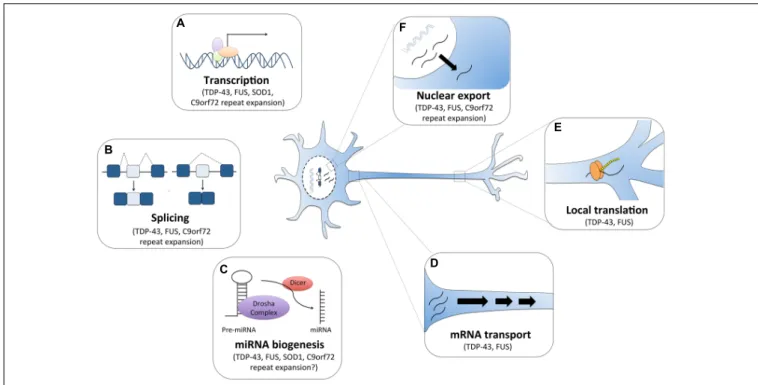
FIGURE 1 | RNA dysfunction in amyotrophic lateral sclerosis (ALS). Major ALS mutations may disrupt RNA processing by several mechanisms. For instance, (A) mutations in ALS genes SOD1, TDP-43, FUS and C9orf72 can alter gene expression. (B) The RNA binding proteins TDP-43 and FUS can affect global splicing machinery. Dipeptide repeat proteins from C9orf72 intronic expansion can also alter splicing patterns of specific RNAs. (C) TDP-43, FUS, and dipeptide proteins can also promote microRNA biogenesis as components of the Drosha and Dicer complexes. TDP-43 and FUS also alter mRNA transport (D) and local translation (E). (F) TDP-43 and FUS predominantly reside in the nucleus, but when mutated they are can mislocalization to the cytoplasm where they bind and regulate different sets of RNAs including the export and mislocalization of other transcripts to the cytoplasm. Poly-PR dipeptide can also bind nuclear pores channels blocking the import and export of molecules.
(NRXN1-3) (
Polymenidou et al., 2011
), neuroligin (NLGN1-2)
(
Polymenidou et al., 2011
), scaffolding protein Homer2 (
Sephton
et al., 2011
), microtubule-associated protein 1B (MAP1B) (
Coyne
et al., 2014
), GABA receptors subunits (GABRA2, GABRA3)
(
Narayanan et al., 2013
), AMPA receptor subunits (GRIA3,
GRIA4) (
Sephton et al., 2011
;
Narayanan et al., 2013
), syntaxin 1B
(
Narayanan et al., 2013
), and calcium channel cacophony (
Chang
et al., 2014
). The development of TDP-43 animal models has
offered the opportunity to explore synaptic alterations in ALS
(
Feiguin et al., 2009
;
Armstrong and Drapeau, 2013
;
Handley
et al., 2017
) and continuous efforts are being made to identify
compounds that can facilitate synaptic transmission in ALS
(
Patten et al., 2017
).
Armstrong and Drapeau (2013)
reported
that expression of mutant
TARDP
G348CmRNA in zebrafish
resulted in impaired synaptic transmission, reduced frequency
of miniature endplate currents (mEPCs) and reduced quantal
transmission. Remarkably, they also demonstrated that all these
synaptic dysfunction features in their zebrafish
TARDBP mutant
were stabilized by chronic treatment the L-type calcium channel
agonists (
Armstrong and Drapeau, 2013
). In drosophila neurons,
TDP-43 depletion was shown to reduce dendritic branching as
well as synaptic formation (
Feiguin et al., 2009
;
Lu Y. et al.,
2009
). Overexpression or knocking down TDP-43 in cultured
mammalian neurons also led to reduced dendritic branching
(
Herzog et al., 2017
). In TDP-43
A315Tmice,
Handley et al.
(2017)
showed that expression of mutant TDP-43 alters dendritic
spine development, spine morphology and neuronal synaptic
transmission. Collectively, these independent studies on several
model systems, suggest that TDP-43 may play an important role
in neuronal morphology, synaptic transmission and neuronal
plasticity likely via regulation of RNA processing of various
synaptic genes (
Godena et al., 2011
;
Sephton et al., 2011
;
Colombrita et al., 2012
;
Narayanan et al., 2013
;
Chang et al.,
2014
).
TDP-43 is also known to act as a splicing regulator to reduce
its own expression level by binding to the 3
0UTR of its own
pre-mRNA (
Ayala et al., 2011
). Additionally, it functions as
a splicing factor whose depletion or overexpression can affect
the alternative splicing of specific targets (
Polymenidou et al.,
2011
;
Tollervey et al., 2011
). Indeed, the alternative splicing
of several genes were reported to be altered in human CNS
tissues from TDP-43 ALS cases (
Shiga et al., 2012
;
Yang et al.,
2014
). For instance, the level of the polymerase delta interacting
protein 3 (POLDIP3) variant-2 mRNA (lacking exon 3) was
significantly increased in the CNS of ALS patients with ALS,
while that of variant-1 mRNA remained unchanged (
Shiga et al.,
2012
). This was consistent with findings that TDP-43 directly
regulates the inclusion of exon 3 of
POLDIP3 and that depletion
of TDP-43 in cell culture models increased variant-2 mRNA
(
Shiga et al., 2012
). TDP-43 has also been shown to regulate
splicing of the cystic fibrosis transmembrane regulator (CFTR)
gene and controls exon skipping by within the pre-mRNA
(
Buratti et al., 2004
). Importantly, it controls the alternative
splicing of apolipoprotein AII (APOAII) (
Mercado et al., 2005
)
and survival of motor neuron (SMN) transcripts (
Bose et al.,
2008
). Specifically, TDP-43 was shown to enhance the inclusion
of exon 7 during the maturation of human SMN2 pre-mRNA,
which results to an increase in full-length
SMN2 mRNA level
in neurons (
Bose et al., 2008
). Furthermore, recently TDP-43
was shown to bind to
HNRNPA1 pre-mRNA to modulate its
alternative splicing (
Deshaies et al., 2018
). TDP-43 depletion
resulted in exon7B inclusion, culminating in a longer hnRNAP
A1B isoform that is aggregation-prone and cytotoxic (
Deshaies
et al., 2018
). Collectively, these studies demonstrated that loss
of TDP-43 results to alterations in alternative splicing of many
genes and some of which, for example
HNRNPA1, can contribute
to cellular vulnerability. It would be interesting further to
investigate the contribution of the alteration of splicing of these
genes (POLDIP3, CFTR, APOAII, SMN2, HNRNPA1) to the
pathogenesis of ALS.
TDP-43 is actively transported along axons and co-localizes
with other well-known transport RNA binding proteins close to
synaptic terminals (
Wang I.F. et al., 2008
;
Narayanan et al., 2013
).
It was reported that TDP-43 mutations impair mRNA transport
function
in vivo and in vitro (
Alami et al., 2014
). In addition to
a role in mRNA transport, TDP-43 also acts as a regulator of
mRNA stability (
Strong et al., 2007
;
Fiesel and Kahle, 2011
). It
was shown to directly interacts with the 3
0UTR of neurofilament
light chain (NFL) mRNA to stabilize it (
Strong et al., 2007
) and
associates with
futsch/MAP1B mRNA in Drosophila to regulates
its localization and translation (
Coyne et al., 2014
). Particularly,
TDP-43 was found to interact with 14-3-3 protein subunits to
modulate the stability of the
NFL mRNA (
Volkening et al.,
2009
). Abnormal regulation of
NFL mRNA has been observed in
ALS patients (
Wong et al., 2000
) and disruption of
NFL mRNA
stoichiometry leads to motor neuron death and symptoms of
ALS in animal models (
Xu et al., 1993
;
Julien et al., 1995
). It is,
thus, very likely that TDP-43 mutations may cause motor neuron
degeneration by interfering with RNA processing of
NFL mRNA.
Other important identified targets regulated by
TDP-43 at mRNA level that may play a role in disease are
G3BP (
McDonald et al., 2011
) and
TBC1D1 (
Stallings et al.,
2013
). G3BP is an essential component of stress granules,
which are cytoplasmic non-membrane organelles that store
translationally arrested mRNAs that accumulate during cellular
stress (
Kedersha and Anderson, 2007
). Stress granules consists
of polyadenylated mRNAs, translation initiation factors (e.g.,
eIF3, eIF4E, and eIF4G), small ribosomal subunits and a
numerous RNA-binding proteins (
Protter and Parker, 2016
).
TDP-43 is recruited to stress granules in cellular models upon
exposure to different stressors (
Colombrita et al., 2009
;
Liu-Yesucevitz et al., 2010
;
Bentmann et al., 2012
). Importantly,
cytosolic TDP-43 mutants are more efficiently recruited to
stress granules upon cellular stress compared to nuclear
wild-type TDP-43 (
Liu-Yesucevitz et al., 2010
). Prolonged
stress is thought to promote sequestration of TDP-43 and
their mRNA targets in stress granules; thereby inhibiting
translation and potentially contributing to ALS progression
(
Ramaswami et al., 2013
).
FUSED IN SARCOMA (FUS)
Mutations in
FUS are detected in 4–5% of familial ALS
patients as well as in sporadic ALS (
Kwiatkowski et al., 2009
;
Vance et al., 2009
;
Corrado et al., 2010
;
DeJesus-Hernandez
et al., 2010
). FUS is an RNA/DNA-binding protein of 526
amino acids, consisting of an RNA-recognition motif, a SYGQ
(serine, tyrosine, glycine and glutamine)-rich region, several
RGG (arginine, glycine and glycine)-repeat regions, a C2C2
zinc finger motif and a nuclear localization signal (NLS)
(
Iko et al., 2004
). C-terminal ALS FUS mutations disrupt
the NLS region and the nuclear import of FUS; resulting in
cytoplasmic accumulation (
Kwiatkowski et al., 2009
;
Vance et al.,
2009
).
Similarly to TDP-43, FUS plays multiple roles in RNA
processing by directly binding to RNA. Using CLIP-based
methods, several groups have identified thousands of RNA
targets bound by FUS in various cell lines (
Hoell et al.,
2011
;
Colombrita et al., 2012
;
Ishigaki et al., 2012
), and brain
tissues (
Lagier-Tourenne et al., 2012
;
Rogelj et al., 2012
).
Interestingly, FUS was identified in spliceosomal complexes
(
Rappsilber et al., 2002
;
Zhou et al., 2002
) and interacting
with several key splicing factors (such as hnRNP A1, YB-1)
(
Rapp et al., 2002
;
Meissner et al., 2003
;
Kamelgarn et al.,
2016
) as well as with the U1 snRNP (
Yamazaki et al.,
2012
;
Yu et al., 2015
). FUS regulates splicing events for
neuronal maintenance and survival (
Lagier-Tourenne et al.,
2012
). Given that FUS plays an essential role in splicing
regulation, the consequence of its loss of function in ALS
on RNA splicing has been immensely investigated (
Lagier-Tourenne et al., 2012
;
Zhou Y. et al., 2013
;
Reber et al.,
2016
). For instance,
Reber et al. (2016)
showed by mass
spectrometric analysis that minor spliceosome components are
highly enriched among the FUS-interacting proteins. They
further reported that FUS interacts with the minor spliceosome
and directly regulates the removal of minor introns (
Reber
et al., 2016
). Moreover, the FUS
P525LALS mutation, which
destroys the NLS and results in cytoplasmic retention of FUS
(
Dormann et al., 2010
), inhibits splicing of minor introns and
causes mislocalization of the minor spliceosome components
U11 and U12 snRNA to the cytoplasm and inhibits splicing
of minor introns (
Reber et al., 2016
). Loss of function of
FUS led to splicing changes in more than 300 genes mice
brains (
Lagier-Tourenne et al., 2012
) and importantly a vast
majority minor intron containing mRNAs was altered (
Reber
et al., 2016
). Corroborating the results with mouse brain, many
minor intron-containing genes were found to be downregulated
in FUS-depleted SH-SY5Y cells (
Reber et al., 2016
). FUS
depletion has been shown to affect minor intron containing
genes that are important for neurogenesis (PPP2R2C), dendritic
development (ACTL6B) and action potential transmission in
skeletal muscles (SCN8A and SCN4A) (
Reber et al., 2016
)
and may contribute to ALS pathogenesis. FUS has also been
shown to regulate alternative splicing of genes related to
cytoskeletal organization, axonal growth and guidance such as
the microtubule-associated protein tau (MAPT) (
Ishigaki et al.,
2012
;
Orozco et al., 2012
;
Rogelj et al., 2012
), Netrin G1
(NTNG1) (
Rogelj et al., 2012
), neuronal cell adhesion molecule
(NRCAM) (
Rogelj et al., 2012
;
Nakaya et al., 2013
) and the
actin-binding
LIM (ABLIM1) (
Nakaya et al., 2013
). For example,
FUS knockdown has been shown to promote inclusion of
exon 10 in the MAPT/tau protein and to significantly cause
shortened axon length and growth cone enlargement (
Orozco
et al., 2012
). Loss of function of FUS altered MAPT/tau
isoform expression and likely disturbed cytoskeletal function
impairing axonal growth and maintenance. Interestingly, axon
retraction and denervation are early events in ALS (
Boillee
et al., 2006
;
Nijssen et al., 2017
). Disruption of cytoskeleton
function may thus play an important role in neurodegeneration
in ALS.
Besides its functions in splicing, FUS has been proposed to
regulate transcription by RNA polymerase II (RNAP2), RNA
polymerase III (RNAP3) or cyclin D1 (
Wang X. et al., 2008
;
Tan and Manley, 2010
;
Brooke et al., 2011
;
Schwartz et al.,
2012
;
Tan et al., 2012
). For instance, transcriptomic analyses
showed that knockdown of FUS results in differential expression
several genes (
Lagier-Tourenne et al., 2012
;
Nakaya et al.,
2013
) including many mRNAs encoding proteins important
for neuronal function. Transcriptome changes have also been
observed in human motoneurons obtained from FUS mutant
induced pluripotent stem cells (IPSCs) (
De Santis et al., 2017
)
and transgenic FUS knockin mice (
Scekic-Zahirovic et al.,
2016
). Alterations in the expression of several genes involved
in pathways related to cell adhesion, apoptosis, synaptogenesis
and other neurodegenerative diseases were reported in these
FUS models (
Fujioka et al., 2013
;
Scekic-Zahirovic et al.,
2016
;
De Santis et al., 2017
). Among these genes
TAF15,
which is mutated in some case of ALS (
Couthouis et al.,
2011
), has been found to be upregulated in several ALS FUS
models including human mutant IPSC derived motoneurons
(
De Santis et al., 2017
), FUS knockout and knockin mouse
(
Kino et al., 2015
;
Scekic-Zahirovic et al., 2016
). However,
it remains to be determined whether
TAF15 upregulation
upon FUS loss- or toxic gain- of function contributes to ALS
pathogenesis.
FUS is also incorporated into stress granules under cellular
stress conditions (
Sama et al., 2013
). Sequestration of FUS
and its protein partners into these cytoplasmic organelles
appears to contribute to ALS pathogenesis (
Yasuda et al.,
2013
). An example of such a protein partner is Pur-alpha,
which co-localizes with mutant FUS and becomes trapped
in stress granules in stress conditions, as reported in ALS
patient cells carrying FUS mutations (
Di Salvio et al., 2015
;
Daigle et al., 2016
). It has been shown that FUS physically
interacts with Pur-alpha.
In vivo expression of Pur-alpha
in Drosophila significantly exacerbates the neurodegeneration
caused by mutated FUS. Conversely,
Di Salvio et al. (2015)
showed that the downregulation of Pur-alpha in neurons
expressing mutated FUS significantly improves fly climbing
activity. It was subsequently demonstrated that overexpression
Pur-alpha inhibits cytoplasmic mislocalization of mutant FUS
and promotes neuroprotection (
Daigle et al., 2016
). However, the
function of Pur-alpha in regulating ALS pathogenesis remains
elusive.
SUPEROXIDE DISMUTASE-1 (SOD1)
Unlike TDP43 and FUS, SOD1 does not contain RNA-binding
motifs, however, several reports have demonstrated a potential
role of mutant SOD1 in regulating RNA metabolism (
Menzies
et al., 2002
;
Lu et al., 2007
;
Lu L. et al., 2009
;
Chen et al.,
2014
). Particularly, mutant SOD1 can bind mRNA species such
as vascular endothelial growth factor (VEGF) and NFL and
negatively affects their expression, stabilization and function
(
Menzies et al., 2002
;
Lu et al., 2007
;
Lu L. et al., 2009
;
Chen et al.,
2014
). More precisely, mutant SOD1 can directly bind to specific
adenylate- and uridylate-rich stability elements (AREs) located
in the 3
0UTR of transcripts of
VEGF (
Lu et al., 2007
) and
NFL
(
Chen et al., 2014
). It is believed that such a gain of abnormal
protein–RNA interactions can be caused by SOD1 misfolding
that results in the exposure of polypeptide portions with the
ability to bind nucleic acids (
Kenan et al., 1991
;
Tiwari et al.,
2005
).
Binding of mutant SOD1 to the 3
0UTR of the
VEGF mRNA
results in the sequestration of other ribonucleoproteins such as
TIAR and HuR into insoluble aggregates. These interactions,
which are specific to mutant SOD1, result in decline levels
of
VEGF mRNA, impairment of HuR function and ultimately
hampering their neuroprotective actions during stress responses
(
Lu et al., 2007
;
Lu L. et al., 2009
).
In motor neuron-like NSC34 cell lines expressing mutant
SOD1 (G37R or G93A), the level of
NFL mRNA is significantly
reduced (
Menzies et al., 2002
). Reduction in
NFL mRNA levels
has also been reported in G93A transgenic mice and human
spinal motor neurons from SOD1-ALS cases (
Menzies et al.,
2002
). It is proposed that destabilization
NFL mRNA by mutant
SOD1, result to altered stoichiometry of neurofilament (NF)
subunits and subsequent NF aggregation in motor neurons (
Chen
et al., 2014
). NF inclusion in the soma and proximal axons of
spinal motor neurons is a hallmark of ALS pathology (
Hirano
et al., 1984
). In IPSC-derived model of ALS, a reduction of
NFL
mRNA level has been reported to result in NF aggregation and
neurite degeneration (
Chen et al., 2014
). Altogether, these studies
support a pathogenic role for dysregulation of RNA processing in
SOD1-related ALS.
Interestingly, SOD1 has been shown to interact with TDP-43
to modulate
NFL mRNA stability (
Volkening et al., 2009
). As
mentioned above, TDP-43 was found to directly interact with
the 3
0UTR of
NFL mRNA to stabilize it (
Strong et al., 2007
).
Altogether, these studies suggest that SOD1 and TDP-43 may act
in a possible common action in regulating specific RNA stability.
In the case of
NFL mRNA, it would be interesting to investigate
whether mutant SOD1 dislodges TDP-43 from the
NFL mRNA in
a manner that would affect its mRNA metabolism and potentially
making NF prone to form aggregates.
Furthermore,
there
have
been
several
transcriptome
investigations in SOD1 human samples (
D’Erchia et al.,
2017
), motor neuron-like NSC34 cell culture model (
Kirby
et al., 2005
) and transgenic animals including mice (
Lincecum
et al., 2010
;
Bandyopadhyay et al., 2013
;
Sun et al., 2015
), rat
(
Hedlund et al., 2010
) and drosophila (
Kumimoto et al., 2013
).
These studies have reported dysregulation of genes involved
in pathways related to the neuroinflammatory and immune
response, oxidative stress, mitochondria, lipid metabolism,
synapse and neurodevelopment (
Hedlund et al., 2010
;
Lincecum
et al., 2010
;
Bandyopadhyay et al., 2013
;
Kumimoto et al.,
2013
;
Sun et al., 2015
;
D’Erchia et al., 2017
). However, in these
studies it is not clear whether SOD1 directly or indirectly
impact the regulation of the differentially expressed genes.
In a recent elegant study,
Rotem et al. (2017)
, compared
transcriptome changes in SOD1 and TDP-43 models. They
found that most genes that were altered in the SOD1
G93Amodel were not dysregulated in the TDP-43
A315Tmodel, and
vice versa (
Rotem et al., 2017
). There were, however, a few
genes whose expressions were altered in both ALS models
(
Rotem et al., 2017
). These findings are consistent with the ALS
pathology, which is distinguishable between the ALS-related
SOD1 phenotype and the TDP-43 phenotype. Although different
cellular pathways are likely activated by SOD1 versus TDP-43, it
is very plausible that they ultimately convergence onto common
targets to result in similar motor neuron toxicity and ALS
phenotype.
C9orf72 INTRONIC EXPANSION
In 2011, a large GGGGCC hexanucleotide repeat expansion in
the first intron or promoter region of the
C9orf72 gene has been
discovered as a new cause of ALS (
DeJesus-Hernandez et al.,
2011
;
Renton et al., 2011
).
C9orf72 repeat expansion mutations
account for about 50% of familial ALS and 5–10% of sporadic ALS
(
Majounie et al., 2012
). It remains a topic of debate whether the
repeat expansion in
C9orf72 causes neurodegeneration primarily
through a toxic gain of function, loss of function, or both.
The
C9orf72 repeat expansion is transcribed in both the sense
and antisense directions and leads to accumulations of
repeat-containing RNA foci in patient tissues (
Gendron et al., 2013
).
The formation of RNA foci facilitates the recruitment of
RNA-binding proteins, causes their mislocalization and interferes with
their normal functions (
Simon-Sanchez et al., 2012
;
Donnelly
et al., 2013
;
Lee et al., 2013
;
Gitler and Tsuiji, 2016
). Indeed, RNA
foci may bind RNA binding proteins and alter RNA metabolism
(
Donnelly et al., 2013
;
Lee et al., 2013
;
Mori et al., 2013a
).
For example,
Mori et al. (2013a)
and
Hutvagner et al. (2001)
showed that RNA foci can sequester hnRNP-A3 and repress its
RNA processing function. Aborted transcripts containing the
repeat can also disrupt nucleolar function (
Haeusler et al., 2014
).
Importantly, these foci can sequester nuclear proteins such as
TDP-43 and FUS, impacting expression of the their RNA targets
and culminating in a range of RNA misprocessing events. Other
RNA binding proteins binding to RNA foci include hnRNP A1,
hnRNP-H, ADARB2, Pur-
α, ASF/SF2, ALYREF and nucleolin
(
Donnelly et al., 2013
;
Lee et al., 2013
;
Sareen et al., 2013
;
Xu et al.,
2013
;
Cooper-Knock et al., 2014
;
Haeusler et al., 2014
). Antisense
oligonucleotides (ASOs) targeting the C9orf72 repeat expansion
suppress RNA foci formation, attenuate sequestration of specific
RNA-binding proteins and reverse gene expression alterations in
C9orf72 ALS motor neurons derived from IPSCs (
Donnelly et al.,
2013
;
Lagier-Tourenne et al., 2013
).
Additionally, simple dipeptide repeats (poly-GA, poly-GP,
poly-GR, poly-PA, and poly-PR) can be generated by
repeat-associated non-ATG-dependent (RAN) translation of both the
sense and antisense strands that have a variety of toxic effects (
Ash
et al., 2013
;
Mori et al., 2013b
). Poly-PR and poly-GR can alter the
splicing patterns of specific RNAs. For example, poly-PR has been
shown to cause exon-skipping in
RAN and PTX3 RNA (
Kwon
et al., 2014
). Dipeptides repeat proteins have also been found to
be toxic by creating aggregates sequestrating cytoplasmic proteins
(
Freibaum and Taylor, 2017
). Poly-GR dipeptide co-localizes with
several ribosomal subunits and with a transcription factor elF3
η
(
Zhang et al., 2018c
). This suggests a ribosomal dysfunction,
which implies a defect in RNA translation. In line with these
findings, a recent report demonstrated that poly-PR co-localizes
with the nucleolar protein, nucleophosmin, and reduces the
expression of several ribosomal RNA (
Suzuki et al., 2018
).
Suzuki
et al. (2018)
further showed that the reduction in the expression
of ribosomal RNA results in neuronal cell death and this could
be rescued by overexpression of an accelerator of ribosome
biogenesis, Myc (
Suzuki et al., 2018
). RNA sequencing reveals
that more than 6,000 genes are up or down regulated in mice
that express the dipeptide construct in the brain (
Zhang et al.,
2018c
). Other findings show that poly-PR dipeptide binds nuclear
pores channels blocking the import and export of molecules. The
dipeptide actually binds the nucleoporin proteins Nup54 and
Nup98 that rim the central channel of the pore (
Shi et al., 2017
).
The accumulation of poly-PR dipeptide at the nuclear pore was
found to correlate with defect in nuclear transport of RNA and
protein, which is consistent with previous findings (
Freibaum
et al., 2015
;
Zhang et al., 2015
).
The last proposed mechanism involved in ALS pathogenesis is
a haploinsufficiency due to the expansion of repetition leading
to a decreased transcription of the gene and consequently to
a decrease of its translation (
Ciura et al., 2013
). Studies have
demonstrated that C9orf72 expansion repeat can interfere with
transcription or splicing of
C9orf72 transcripts (
Mori et al.,
2013b
;
Haeusler et al., 2014
;
Highley et al., 2014
). It has also
been proposed that the C9orf72 expansion repeat could disrupt
the C9orf72 promoter activity thereby reducing its expression
(
Gijselinck et al., 2016
). Several studies have demonstrated
alterations in the C9orf72 ALS transcriptome (
Donnelly et al.,
2013
;
Prudencio et al., 2015
;
Selvaraj et al., 2018
). Interestingly,
a recent article reported an increased expression of the
calcium-permeable GluA1 AMPA receptor subunit in motoneurons
derived from IPSC of patients with
C9orf72 mutations (
Selvaraj
et al., 2018
). This alteration in AMPA receptor composition
led to an enhanced motoneuron vulnerability to AMPA-induced
excitotoxicity (
Selvaraj et al., 2018
). It remains to be determined
whether the increased expression of GluA1 AMPA subunit is
related to reduced levels of C9orf72, RNA foci and/or dipeptide
repeats.
C9orf72 has also been showed to be involved in the generation
of stress granules (
Maharjan et al., 2017
) and sequestering other
RNA binding proteins that are involved in nucleo-cytoplasmic
transport (
Zhang et al., 2015, 2018b
). It has been found that
stress granules observed in
C9orf72 mutants co-localizes with
Ran GAP (
Zhang et al., 2015, 2018b
); which is known to activate
Ran GTPase. This GTPase in involved in nucleo-cytoplasmic
transport. It has also been published that expressing Ran GAP
rescues the age-related motor defects in flies expressing the
GGGGCC repeats (
Zhang et al., 2018a
). Very recently, it has
also been reported that one of the dipeptide generated by the
expansion has a role in formation of these stress granules
(
Zhang et al., 2018c
). Moreover, importins and exportins are
sequestered in stress granules; which also implies that protein
transport in altered (
Zhang et al., 2018b
).
These toxic gain- or loss-of function mechanisms are thought
to be all involved in synergy in ALS pathogenesis and it can be
summed up that that altered RNA processing plays a key role in
C9orf72-mediated toxicity through two ways. The first is altered
processing of the expanded
C9orf72 transcript itself, in terms of
altered transcription, splicing defects, nuclear aggregation and
non-conventional translation (
Barker et al., 2017
). The second
involves downstream and indirect changes in RNA processing of
other transcripts. A thorough understanding of RNA metabolism
dysregulation could definitely bring a major enlightenment on
how
C9orf72 mutation leads to ALS and provide insights on
therapeutic targets.
DYSREGULATION OF MICRORNA
(miRNA) IN ALS
Multiple mechanisms control the proper levels of RNA and
subsequent protein expression; among these are microRNAs
(miRNAs) (
Catalanotto et al., 2016
). They are endogenous
small non-coding RNAs (approximately 22 nucleotides in
length) that are initially transcribed by the RNA polymerase
II as primary miRNA (miRNAs) transcripts. These
pri-miRNAs are processed into precursor pri-miRNAs (pre-pri-miRNAs)
by the nuclear ribonuclease III (RNase III), DROSHA, and the
double-stranded RNA-binding protein, DGCR8, which anchors
DROSHA to the pri-miRNA transcript (
Lee et al., 2003
;
Denli
et al., 2004
). Pre-miRNA is then exported into the cytoplasm
by exportin-5 (
Yi et al., 2003
), where it is processed into a
mature miRNA by the DICER enzyme (
Hutvagner et al., 2001
;
Ketting et al., 2001
). The mature miRNA is then incorporated
with a ribonucleoprotein (RNP) complex with argonaute (AGO)
proteins to form the RNA-induced silencing complex (RISC)
(
Hammond et al., 2001
;
Schwarz et al., 2003
;
Kawamata
and Tomari, 2010
), which mediates inhibition of translation
and/or mRNA degradation of targeted transcripts that are
complementary to the miRNA (
Hutvagner and Zamore, 2002
;
Yekta et al., 2004
). The recognition of mRNAs by miRNAs occurs
through base-pairing interactions within the 3
0-untranslated
region (UTR) of the targeted mRNAs. Besides their well-known
gene silencing functions, miRNAs can also induce up-regulation
of their targets (
Vasudevan et al., 2007
;
Lin et al., 2011
;
Truesdell
et al., 2012
;
Vasudevan, 2012
).
MiRNAs play important roles in several biological processes
such as cell proliferation (
Chen et al., 2006
), cell differentiation
(
Naguibneva et al., 2006
), apoptosis (
Matsushima et al., 2011
),
and patterning of the nervous system (
Johnston and Hobert,
2003
). Interestingly, several miRNAs have been particularly
shown to be essential for motor neuron development and
survival (see review,
Haramati et al., 2010
). For example, in
developing chick, it was demonstrated that the activation of
the miRNA miR9 is necessary to suppress the expression of
the transcription factor
onecut1, which in turn helps to drive
differentiation of neural progenitor cells into spinal motor
neurons (
Luxenhofer et al., 2014
). It is believed that several
miRNAs work in concert to establish motor neuron identity.
Indeed, in addition to miR9, other miRNAs such as
miR-128 (
Thiebes et al., 2015
), miR-196 (
Asli and Kessel, 2010
),
miR-375 (
Bhinge et al., 2016
) have been shown to play a
role in motor neuron differentiation and localization. Loss of
DICER function within progenitor cells results in aberrant motor
neuron development while its loss in motor neuron leads to
progressive motor neuron degeneration (
Haramati et al., 2010
;
Chen and Wichterle, 2012
). Furthermore, miRNAs are important
players for NMJ function, synaptic plasticity and for maintaining
cytoskeletal integrity (see review,
Hawley et al., 2017
).
The ALS genes, TDP-43 and FUS, were identified in a protein
complex with RNAse III DORSHA and shown to play a role
in miRNA biogenesis (
Freibaum et al., 2010
;
Da Cruz and
Cleveland, 2011
). TDP-43, in particular was shown to associate
with proteins involved in the cytoplasmic cleavage of pre-miRNA
mediated by the DICER enzyme (
Freibaum et al., 2010
). It is thus
to no surprise that dysregulation of miRNAs has been observed
in ALS (
Li et al., 2013
;
Zhang et al., 2013
;
Dini Modigliani et al.,
2014
;
Eitan and Hornstein, 2016
). Indeed, mutations in
TARDBP
result in differential expression of miRNAs – 9,
miR-132, miR-143, and miR-558 (
Kawahara and Mieda-Sato, 2012
;
Zhang et al., 2013
). Interestingly, the expression of several of
these miRNAs (miR-9, miR-132, miR-143) and including others
(such as miR-125, miR-192) are altered upon FUS depletion
(
Morlando et al., 2012
). MiR-9 expression is also found to
be upregulation in mutant SOD1 mice (
Zhou F. et al., 2013
).
These dysregulated miRNAs are essential for motor neuron
development and maintenance (
Otaegi et al., 2011
;
Luxenhofer
et al., 2014
), axonal growth (
Dajas-Bailador et al., 2012
;
Kawahara
and Mieda-Sato, 2012
) and synaptic transmission (
Edbauer et al.,
2010
;
Sun et al., 2012
). Thus, these miRNA alterations likely
contribute to the pathological phenotype observed in ALS.
Additionally, depletion of TDP-43 in cell culture systems
has also been shown to change the total miRNA expression
profile (
Buratti et al., 2010
). A similar observation was
recently observed in motoneurons progenitors derived from
human ALS IPSCs (
Rizzuti et al., 2018
). Particularly, it
was reported that 15 miRNAs were dysregulated including
disease-relevant miR-34a and miR504, which are known to
be, implicated synaptic vesicle regulation and cell survival
(
Rizzuti et al., 2018
). Additionally, another important miRNA,
namely microRNA-1825, was found to be downregulated in
CNS of both sporadic and familial ALS patients (
Helferich
et al., 2018
). Interestingly, reduced levels of
microRNA-1825 was demonstrated to cause a translational upregulation
of tubulin-folding cofactor b (TBCB) which consequently
to depolymerization and degradation of tubulin alpha-4A
(TUBA4A), which is encoded by a known ALS gene (
Helferich
et al., 2018
).
FIGURE 2 | RNA-based therapy approaches for potentially treating ALS. (A) SiRNAs operate through RNA interference pathway. After strand unwinding, one siRNA strand binds argonaute proteins as part of the RNA-induced silencing complex (RISC) and is recruited to a target mRNA which is then cleaved. Virus can provide a means of shRNA, which will be cleaved once in the cytoplasm by dicer enzyme into siRNA. This approach has been evaluated to reduce the level of mutant SOD1 protein. (B) Antisense oligonucleotide (ASO) binds to targeted mRNA and induces its degradation by endogenous RNase H or blocks the mRNA translation. This strategy is being exploited as a potential therapeutic avenue in ALS aiming principally to reduce the protein level of SOD1 protein or by targeting of C9orf72 RNA foci. (C) Small molecules can be designed to target and stabilize RNA structures. This approach was particularly tested to stabilize G-quadruplex of C9orf72 GGGGCC repeat RNA. Stabilization of G-quadruplex structure reduces RNA foci formation and blocks repeat translation.