HAL Id: hal-00093015
https://hal.archives-ouvertes.fr/hal-00093015
Submitted on 27 Jun 2018HAL is a multi-disciplinary open access archive for the deposit and dissemination of sci-entific research documents, whether they are pub-lished or not. The documents may come from teaching and research institutions in France or abroad, or from public or private research centers.
L’archive ouverte pluridisciplinaire HAL, est destinée au dépôt et à la diffusion de documents scientifiques de niveau recherche, publiés ou non, émanant des établissements d’enseignement et de recherche français ou étrangers, des laboratoires publics ou privés.
Selective estrogen receptor modulators in the
ruthenocene series. Synthesis and biological behavior
Pascal Pigeon, Siden Top, Anne Vessières, Michel Huché, Elisabeth A. Hillard,
Emmanuel Salomon, Gérard Jaouen
To cite this version:
Pascal Pigeon, Siden Top, Anne Vessières, Michel Huché, Elisabeth A. Hillard, et al.. Selective estrogen receptor modulators in the ruthenocene series. Synthesis and biological behavior. Journal of Medicinal Chemistry, American Chemical Society, 2005, 48 (8), pp.2814-2821. �10.1021/jm049268h�. �hal-00093015�
Selective Estrogen Receptor Modulators in the Ruthenocene Series.
Synthesis and Biological Behavior
Pascal Pigeon, Siden Top,* Anne Vessières,* Michel Huché, Elizabeth A. Hillard, Emmanuel Salomon, and Gérard Jaouen*
Laboratoire de Chimie et Biochimie des Complexes Moléculaires, Ecole Nationale Supérieure de Chimie de Paris, UMR CNRS 7576, 11, rue Pierre et Marie Curie, 75231 Paris Cedex 05, France
*
Corresponding authors. Phone: (+33)1-43-26-95-55. Fax (+33)1-43-26-00-61. E-mail: [email protected].
A series of ruthenocene derivatives, 1-[4-(O(CH2)nN(CH3)2
)phenyl]-1-(4-hydroxyphenyl)-2-ruthenocenylbut-1-ene, with n = 2-5, based on the structure of the breast cancer drug tamoxifen has been prepared. These compounds were obtained, via a McMurry cross-coupling reaction, as a mixture of Z and E isomers that could not be separated by HPLC. The relative binding affinity values for estrogen receptor α (ERα) for n = 2 and 3 were very high (85 and 53%) and surpassed even that of hydroxytamoxifen (38.5%), the active metabolite of tamoxifen. Ruthenocene derivatives act as anti-estrogens as effective (n = 2) or slightly more effective (n = 3-5) than hydroxytamoxifen on ERα-positive breast cancer cell lines but, unlike ferrocifens, do not show antiproliferative effects on ERα-negative breast cancer cell lines. Electrochemical studies showed that the ruthenocifen radical cations are unstable, which may account for this behavior. Some of these compounds could be useful as radiopharmaceuticals for ERα-positive breast cancer tumors.
1. Introduction
study in the fight against breast cancer, a disease that strikes about one in eight1 women in the Western world. Approximately two-thirds of these cases are classified as hormone-dependent, in which the estrogen receptor is present, (ER-positive). For these patients, an anti-estrogen treatment is prescribed, commonly the drug tamoxifen, the prodrug of the active metabolite hydroxytamoxifen, OH-Tam (Chart 1). However, tamoxifen is not effective against the remaining third of breast cancer cases classified as hormone-independent, where the estrogen receptor is not detected, (ER-negative). For these reasons, the discovery of new molecules that are active toward these tumors would supply a much-needed innovation in an area where the current therapeutic arsenal is deficient.2
The activity of OH-Tam in the breast has been illuminated by recent developments in the complex endocrinology of breast cancer.3 A second estrogen receptor, ERβ, was discovered in 1996.4 Tumors which had been classified as ER-negative due to the lack of ERα have been shown to contain ERβ,5 which may be important in the proliferation of tamoxifen-resistant tumors, although the role of this receptor is still poorly understood.6
Chart 1. Tamoxifen and Ferrocifen Derivatives
The conformational effect of the bioligand on the ER has been elucidated by the publication of the X-ray structure of the ligand binding domain crystallized with both
estrogens and anti-estrogens in the site.7 These studies underscore the importance of OH-Tam’s hydroxy group for receptor affinity, due to its interaction with Asp351,2b and of the
basic chain in changing the secondary structure of the protein, thus modifying transcriptional activity. Two different mechanisms for gene expression have been discovered. The estrogen receptor/bioligand complex can dimerize and interact directly with the DNA at the estrogen response element (ERE), or in the activated protein pathway (AP1), the monomer can interact with two proteins (Jun and Fos) to form a complex that binds to DNA.8 The recruitment of coactivators or corepressors that determine proliferative or antiproliferative effects seems to be dependent upon the pathway at the DNA level, the nature of the bioligand, and the organ tissue.
Because there is currently no satisfactory therapy for ER-negative breast tumors, and because these tumors are often heterogeneous, containing both ER-positive and ER-negative cells, it is currently of great interest to develop SERMs that are effective on both cell types. Our strategy has been to couple the vector, tamoxifen, with a potentially toxic organometallic moiety. In this way, the estrogen receptors (α and β) can be targeted by the shape and the binding properties of hydroxytamoxifen, which can simultaneously deliver a cytotoxic metal unit to the cancer cell nucleus.
Using these concepts, we have previously shown that, in the ferrocifen series (Chart 1), complexes such as OH[3]-ferrocifen and OH[4]-ferrocifen are anti-estrogenic in the hormone-dependent breast cancer cell line MCF-7 due to their conformational effects on ERα, even surpassing the activity of OH-Tam. More important, however, is their efficacy on the hormone-independent breast cancer cell line MDA-MB231,9 likely due to the advent of a cytotoxic moiety from the oxidation of Fe(II) in ferrocene,10 possibly via a pathway that involves ERβ.11 Although OH-Tam and its metabolites show cytotoxic behavior at high concentrations,12 the ferrocene compounds show a much greater activity. We are currently studying the role of the ferrocene moiety in modulating the cytotoxicity of these compounds via electrochemical means.
We now turn our attention to the ruthenocene series. Due to ruthenium’s position below iron in group 8, the ionization potential of such molecules should be considerably higher when iron is replaced by ruthenium. Thus, if oxidation is in fact the necessary precursor to the cytotoxic phenomena in ER-negative breast cancer cell lines, one would expect enhanced results when ruthenium is substituted for iron. Furthermore, anticancer properties have
already been shown for a number of rutheno-arene complexes, such as Ru(III) imidazole and indazole compounds, which are currently in clinical trials.13 Because the activity of the latter compounds is thought to arise from the in situ reduction of Ru(III) to Ru(II),14 Sadler and co-workers have focused on the anticancer properties of Ru(II) monoarene compounds,15 and a Ru(II)-cymene complex has been shown to damage DNA.16 Finally, ruthenium is of special interest due to the availability of two γ-emitting isotopes, 97Ru and 103Ru, which may be useful in the radioimaging of cancer tumors.17
In this work we present the synthesis and characterization of a series of hydroxytamoxifen-ruthenocene compounds, or “hydroxyruthenocifens”, 1-[4-(O(CH2)nN(CH3)2
)phenyl]-1-(4-hydroxyphenyl)-2-ruthenocenylbut-1-ene, with n = 2-5. We have also included the results from a series of biological tests to evaluate their antiproliferative effects against both hormone-dependent and -independent breast cancer cell lines and their suitability for use in radioimaging, as well as electrochemical and molecular mechanics studies.
2. Results and Discussion
2.1. Synthesis. The outline of the synthetic procedure is shown in Scheme 1. As the length of the basic side chain proved to be of importance on the antiproliferative effects of the ferrocifen series, we have varied the length of the side chain from n = 2-5 carbon atoms in the ruthenocene series. We have found that, for the rhenium18 and iron19 series of compounds, a McMurry coupling reaction is most effective for preparing the alkene derivatives.20 In this case, as with the rhenium series, we have chosen to attach the alkyl chain, in the form of a halogenated chain, to the dihydroxybenzophenones (2a-d) prior to the coupling reaction with the propionylruthenocene 5. The coupling reaction was then carried out with a 2:1 ratio of bromoalkylhydroxybenzophenone to propionylruthenocene in order to diminish the yield of the homogeneous propionyl-ruthenocene coupled compound. The halogen function was then converted to a dimethylamino function in the last step.
In the first step, 4,4’-dihydroxybenzophenone 1 was monoalkylated with the appropriate halogenoalkyl chain to prepare ketones 2a-d. This occurred by the combination of the monopotassium salt of 1, prepared with KH, with the dibromoalkyl derivative. McMurry coupling of the corresponding ketone 2a-d with the ruthenocene ketone 5, prepared by the
action of AlCl3 and propionyl chloride on ruthenocene, gave the alkenes 3a-d in good yields
(>70%). In the final step, the alkenes were converted to the amines 4a-d in an autoclave by the action of dimethyamine in methanol at 60 °C.
Scheme 1. Synthesis of Hydroxyruthenocifens 4a-d (n = 2-5)
The McMurry coupling reaction furnished mixtures of Z and E isomers of the bromo derivatives 3a-d, which were obtained as oils and converted to the amino compounds 4a-d. In both the rhenium18 and ferrocene9b series, these isomers could be separated by chromatography or fractional crystallization, and while the ferrocifens interconverted rapidly in chloroform, this tendency to isomerize was not observed in the case of rhenium. The ruthenocene compounds, however, could not be separated by chromatography, as only one broad peak was
seen in both semipreparative and analytical HPLC. This phenomenon can be explained by the rapid interconversion between Z and E isomers in the ruthenium series. We have suggested, in a previous study of the ferrocifens,9b that the intermediate species in the isomerization mechanism is the α-carbenium ion formed by protonation of the double bond, and thus, greater stability of this species would enhance isomerization. An evaluation of the pKR+
values for a series of organometallic compounds showed that the stability of [CpRu(η5 -C5H4CH2)]+ was 2 orders of magnitude higher than that of the corresponding iron cation.21
Presumably, the enhanced stability of this species derives from greater back-bonding from the more diffuse ruthenium orbitals as compared with iron, and DFT calculations on similar compounds have supported this interpretation.22 Prevention of isomerization in order to evaluate the isomers separately may be accomplished through the use of fixed ring systems in the future.23
Table 1. Relative Binding Affinities (RBAs) for ERα (cytosol) and ERβ (purified) and lipophilicy (log Po/w) of the Ruthenocene Derivatives of OH-Tamoxifen
compounda ERα ERβ log Po/w
E2 OH-Tam 100b 38.5c 100b 18.5c 3.5 Z = 3.2c E = 3.4 4a 85 ± 4 19 ± 0.4 4.5 4b 53 ± 6 13 ± 4 4.6 4c 10.6 ± 0.2 10.9 ± 0.1 4.7 4d 14.5 ± 0.3 13 ± 4 4.8 a
OH-Tam and the ruthenocifens 4a-d are the mixture of both Z and E isomers. Measurements were performed with stock solution in DMSO for 3 h at 0 °C. Mean of
two experiments ± range. b Value by definition. c Value from ref 9b.
2.2. Biochemical Studies. a. Measurement of the Relative Binding Affinities (RBAs) of the Complexes for the Estrogen Receptors ERα and ERβ. As the separation of the Z and
E isomers was impossible, all the biochemical studies were carried out on the mixture of
the two isomers. The RBA values obtained on the α form of the receptor were remarkably high for the complexes with the side chain with n = 2 and 3 (Table 1). These values are higher than the RBA value found for OH-Tam, which is unprecedented in our studies of other organometallic SERMs. The RBA values obtained for the β form were also high, and in this case, they are comparable to that found for OH-Tam.
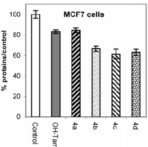
Figure 1. Antiproliferative effect of 1 µM OH-Tam and 4a-d on MCF7 cells (breast cancer cell line,
ERα-positive) after 5 days of culture. In this experiment E2 (1 nM) has a proliferative effect (176%; data
not shown). Representative data of one experiment performed twice with similar results (eight measurements ± limit of confidence; P = 0.1, t = 1.895).
b. Determination of log Po/w Values. The lipophilicity of the complexes was determined by reverse phase HPLC. Again, we were unable to separate the E and Z isomers in the ruthenocene derivatives; we always observed a broad peak in the HPLC, instead of the two distinct peaks observed for OH-Tam or the Re-Tam and Fc-Tam series. Thus, the log Po/w values correspond to the values of the mixtures, and they are given in Table 1. These values are higher than the value found for estradiol, and they increased slowly with the length of the side chain. These values are very similar to those found in the rhenium series.18
c. Study of the Effect of Ruthenocifen Complexes on the Proliferation of Hormone-Dependent (MCF7) and -Independent (MDA-MB231) Breast Cancer Cell Lines. On the MCF-7 (ERα-positive) breast cancer cell line the complexes have an antiproliferative
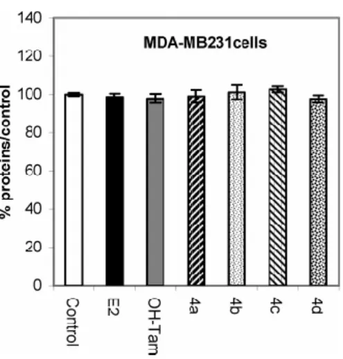
effect, as shown in Figure 1. The effect of 4a is similar to that observed with OH-Tam, while it is stronger for the three other ruthenium compounds 4b-d. On the MDA-MB231 (ERα-negative) breast cancer cell line, these complexes had no effect. E2 and OH-Tam also had no
effect (Figure 2). Therefore, the ruthenocene complexes of tamoxifen seem to act in an anti-estrogenic fashion similar to OH-Tam.
Figure 2. Effect of 1 µM OH-Tam and 4a-d and of E2 (1 nM) on MDA-MB231 cells (breast
cancer cell line, ERα-negative) after 6 days of culture. Representative data of one experiment performed twice with similar results (eight measurements ± limit of confidence; P = 0.1, t =
1.895).
2.3. Electrochemical Studies. Electrochemical studies were carried out upon ruthenocene and compounds 4a-d in MeOH at room temperature. The behaviors of compounds 4a-d were quite similar. A representative example, 4b, is shown in Figure 3. At low scan rates, these compounds all yielded a poorly formed oxidation wave, consisting of a lower potential shoulder and a sharper, irreversible higher potential wave. The shoulder featured more prominently with increasing n and may arise from slow electron transfer from the amino group; this feature diminished with scan rate. In general, the potential of the main peak, which we ascribe to the Ru(II) to Ru(III) oxidation, increased with scan rate, typical of an irreversible electrochemical process. The oxidation potentials of this peak varied from about 0.55 to 0.68 V, depending on the compound and scan rate. There was an uncoupled reduction wave that ranged in potential from 0.26 to 0.13 V, depending on the compound and scan rate. These data are indicative of a fast chemical reaction following the oxidation of the ruthenocene moiety. No coupled reverse wave was observed up to 20 V/s, and thus, the kinetics could not be studied in this scan rate range. The half-life of the radical cation can be estimated to be less than 1.3 ms.
We believe that the cytotoxicity of the ferrocifen SERMs toward the ERβ-positive breast cancer cell line is due to the production of radical species in situ, following the preliminary oxidation of Fe(II) to Fe(III). Genotoxicity and DNA cutting has been shown in the hydroxyferrocifen series and for other ferrocene-containing molecules.9a,10b,c The mechanism of radical production is not currently understood and may be associated with ERβ, the Fenton reaction, and/or the generation of reactive organic radicals or quinones via ferrocene-modulated phenol oxidation. The lack of cytotoxicity of the ruthenocene series can be rationalized along these lines. Although the oxidation potentials are reasonably accessible in vivo, the highly reactive cation likely transforms into another species before it has the opportunity to reach the target. On the other hand, the ferrocenium cation is stable in most media. It is worth noting that the ruthenocenium cation itself is generally unstable in all but the most innocent media. A reversible one-electron oxidation to [RuCp2]+ was obtained only in the
CH2Cl2/[NBu4][B(C6H3(CF3)2)4] solvent/electrolyte system.24
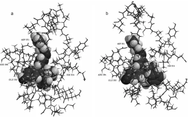
2.4. Molecular Modeling Studies. Molecular mechanics studies were performed on compound 4b, with an amino chain consisting of three carbon atoms, to determine the stability of the idealized orientation of both the E and Z isomers in the ligand binding domain of the estrogen receptor. We utilized the crystal structure of the ligand binding domain of
human ERα occupied by OH-Tam, published by Shiau et al.;7d the current resolution of the human ERβ structures does not permit these types of studies. Only the amino acids forming the wall of the cavity were retained, a total of 757 atoms. The hydroxytamoxifen was digitally removed and replaced with the E and Z isomers of compound 4b. With all the heavy atoms of the cavity immobilized, a position search was carried out to determine the energetically optimal location for the bioligand. Next, the lateral chain of the amino acid Met343, which represented a steric hindrance for the organometallic moiety, was liberated. This was justified by the fact that this part of the cavity has been shown to be flexible.25 An energy-minimization routine was then carried out with all of the heavy atoms immobilized except those of the bioligand and the Met343 side chain, using the Merck Molecular Force Field (MMFF). A conformational study in the cavity was also carried out to determine the best position for the organometallic moiety in relation to the rest of the molecule. This determined the ideal position for the bioligand. Next, the affinity of the bioligand for the cavity was also determined using MMFF molecular mechanics, with calculations for the bioligand-cavity combination and for the cavity and the bioligand done separately, with the latter two retaining the conformations they had in the molecular complex. This gave the ∆E energy for the reaction bioligand + cavity molecular complex. The result for the Z isomer of compound 4b was -52 kcal/mol, while that for the E isomer was -10 kcal/mol. The calculation shows that affinity for the cavity is considerably higher for the Z isomer than for the E isomer. These results are shown in Figure 4.
While the calculated values for all of the organometallic SERMs that we have examined so far show a greater cavity affinity for the Z isomer, the disparity between the values for the ruthenium compound is quite dramatic. For example, the Z:E ratio value for OH[3]-ferrocifen is -27:-21 kcal/mol, and that for OH[3]-rhenium is -42:-36 kcal/mol. Furthermore, the Z isomer of tamoxifen has been shown to be the most strongly anti-estrogenic, although, because the molecule isomerizes in solution, the mixture is administered. In the case of the ruthenium compound 4b, its rapid isomerization coupled with the results of the thermodynamic calculations suggest that the Z isomer is the active isomer in the biochemical studies. While RBA and enthalpy values cannot be simplistically correlated, the great stability of the Z isomer in the cavity supports the high observed RBA value for this compound. Our modeling studies have also shown that the hydrogen bond between the N atom of the side chain and Asp351 is 1.857 Å for (Z)-4b and 1.999 Å for (E)-4b. The
importance of this stabilizing effect has been previously recognized in related antagonist series.2b
Figure 4. Representations of the (a) E and (b) Z isomer of 4b docked in ERα.
3. Experimental Section
3.1. General Comments. Starting materials were synthesized using standard Schlenk techniques, under an argon atmosphere. Anhydrous THF and diethyl ether were distilled from sodium/benzophenone. Thin layer chromotography was performed on silica gel 60 GF254. FT-IR spectra were recorded on a BOMEM Michelson-100 spectrometer as a KBr plate. 1H and
13
C NMR spectra were acquired on Bruker 200 and 400 spectrometers by using CDCl3 as a
solvent. Mass spectrometry was performed on a Nermag R 10-10C spectrometer. High-resolution mass spectrometry (HRMS) was performed on a JEOL MS 700 instrument. Melting points were measured with a Kofler device. Cyclic voltamograms were obtained by utilizing an Autolab PGStat potentiostat driven by GPES software,26 a platinum wire counter electrode, a 500 µM platinum disk working electrode, and an aqueous standard calomel reference electrode. The electrolytic cell was connected to a Schlenk line, and all experiments
were performed under an inert atmosphere of argon gas. Analyte solutions were 1-2 mM in MeOH with 0.1 M Bu4NBF4 supporting electrolyte. Molecular modeling studies were carried
out utilizing Mac Spartan Pro.27
3.2 Synthesis. a. The formation of ketones 2a-d has been reported elsewhere.18
b. The Formation of Ethyl Ruthenocenyl Ketone 5. Ruthenocene (1.00 g, 4.32 mmol) was dissolved in dry dichloromethane (20 mL) while being stirred. Aluminum chloride (0.464 g, 3.45 mmol) was slowly added and then propionyl chloride (0.25 mL, 2.86 mmol) was added over 15 min with a syringe. The solution was stirred for 3 h and then poured in water. Dichloromethane (40 mL) was added and the solution was decanted. The aqueous layer was extracted with dichloromethane and the combination of organic layers was dried on magnesium sulfate and concentrated under reduced pressure. The residue was chromatographed on silica gel plates with dichloromethane as eluent to obtain the pure ketone 5 with a yield of 66% and a great amount of starting ruthenocene. The ketone 5 was recrystallized from ethanol. mp: 78 °C. IR: 2967 (CH3), 2932 (CH2), 1663 (C=O) cm-1; 1H
NMR: δ 1.07 (t, 3H, CH3, J = 7.4 Hz), 2.55 (q, 2H, CH2, J = 7.4 Hz), 4.52 (s, 5H, C5H5),
4.69 (t, 2H, C5H4, J = 1.8 Hz), 5.04 (t, 2H, C5H4, J = 1.8 Hz). 13C NMR: δ 8.9 (CH3), 31.9
(CH2), 70.5 (2CH), 71.7 (5CH), 73.2 (2CH), 83.6 (C), 203.0 (CO). MS (IE, 70 eV) m/e: 288
(M+•).
c. The Formation of Alkenes 3a-d. Titanium tetrachloride (1.2 mL, 11 mmol) was added dropwise to a suspension of zinc powder (1.40 g, 22 mmol) in 20 mL of THF at 0 °C. The mixture obtained was heated at reflux for 2 h. A second solution was prepared by dissolving ketone 5 (0.50 g, 1.74 mmol) and 4-bromoalkoxy-4’-hydroxybenzophenone 2a-d (3.47 mmol) in 30 mL of THF. This latter solution was added dropwise to the first solution and the resulting mixture was heated for 2 h. After cooling to room temperature, the mixture was stirred with water and dichloromethane. The mixture was acidified with dilute hydrochloric acid and decanted. The aqueous layer was extracted with dichloromethane and the combination of organic layers was dried over magnesium sulfate. After concentration under reduced pressure, the crude product was chromatographed on silica gel plates with dichloromethane as eluent to give a pure, oily mixture of Z + E 3a-d.
1-[4-(2-Bromoethoxy)phenyl]-1-(4-hydroxyphenyl)-2-ruthenocenylbut-1-ene, 3a. Yield: 76%. IR: 3420 (OH), 2964, 2925, 2861 (CH3) cm-1. MS (IE, 70 eV) m/e: 576 (M+•).
Z + E Isomers. 1H NMR: δ 0.91 (t, J = 7.6 Hz, 3H, CH3), 2.25 + 2.26 (q, J = 7.6 Hz, 2H, CH2), 3.53 + 3.56 (t, J = 6.0 Hz, 2H, CH2Br), 4.17 (t, J = 6.0 Hz, 2H, CH2O), 4.23 (d, J = 1.5 Hz, 2H, C5H4), 4.33 (d, J = 1.5 Hz, 2H, C5H4), 4.45 (s, 5H, Cp), 6.56-7.02 (m, 8H, 2C6H4). 13C NMR: δ 15.5 (CH3), 29.2 (CH2), 53.7 (CH2), 67.8 (CH2), 69.7 (2 CH, C5H4), 71.1 (5 CH, Cp), 72.0 (2 CH, C5H4), 92.7 (C, C5H4), 114.1 + 114.3 (2 CH, C6H4), 114.8 + 115.0 (2 CH, C6H4), 130.5 (2 CH, C6H4), 131.2 (2 CH, C6H4), 135.8 (C), 136.8 + 137.1 (C), 137.2 (C), 137.7 + 138.0 (C), 153.9 + 154.0 (C), 156.3 + 156.4 (C). 1-[4-(3-Bromopropoxy)phenyl]-1-(4-hydroxyphenyl)-2-ruthenocenylbut-1-ene, 3b. Yield: 91%. IR: 3423 (OH), 2964, 2924, 2856 (CH3) cm-1. MS (IE, 70 eV) m/e: 590
(M+•). Z + E Isomers. 1H NMR: δ 0.99 (t, J = 7.6 Hz, 3H, CH3), 1.45-2.15 (m, 2H, CH2), 2.20-2.45 (m, 2H, CH2), 3.59 + 3.61 (t, J = 6.4 Hz, 2H, CH2Br), 4.05 + 4.07 (t, J = 6.7 Hz, 2H, CH2O), 4.25-4.65 (m, 9H, C5H4 + Cp), 6.60-7.15 (m, 8H, 2C6H4). 13C NMR: δ 15.4 (CH3), 30.0 (CH2), 32.4 (CH2), 56.6 (CH2), 65.0 (CH2), 69.6 (2 CH, C5H4), 71.0 (5 CH, Cp), 71.9 (2 CH, C5H4), 92.6 (C, C5H4), 113.7 + 113.9 (2 CH, C6H4), 114.7 + 114.9 (2 CH, C6H4), 130.3 + 130.4 (2 CH, C6H4), 131.1 (2 CH, C6H4), 135.4 (C), 136.8 (C), 137.1 (C), 137.3 (C), 154.1 (C), 156.8 (C). 1-[4-(4-Bromobutoxy)phenyl]-1-(4-hydroxyphenyl)-2-ruthenocenylbut-1-ene, 3c. Yield:
83%. IR: 3414 (OH), 2958, 2927, 2869 (CH3) cm-1. MS (IE, 70 eV) m/e: 604 (M+•).
Z + E Isomers. 1H NMR: δ 1.01 (t, J = 7.5 Hz, 3H, CH3), 1.75-2.25 (m, 4H, CH2CH2), 2.35 (q, J = 7.5 Hz, 2H, CH2), 3.50 + 3.52 (t, J = 6.4 Hz, 2H, CH2Br), 3.95 + 3.99 (t, J = 6.7 Hz, 2H, CH2O), 4.30-4.38 (m, 2H, C5H4), 4.40 (t, J = 1.6 Hz, 2H, C5H4), 4.52 (s, 5H, Cp), 6.50-7.15 (m, 8H, 2C6H4). 13C NMR: δ 15.4 (CH3), 27.8 (CH2), 29.2 (CH2), 29.4 (CH2), 33.4 (CH2), 66.6 (CH2), 69.5 (2 CH, C5H4), 70.9 (5 CH, Cp), 71.8 (2 CH, C5H4), 92.6 (C, C5H4), 113.7 + 113.9 (2 CH, C6H4), 114.7 + 114.9 (2 CH, C6H4), 130.3 + 130.4 (2 CH, C6H4), 131.0 + 131.2 (2 CH, C6H4), 135.4 (C), 136.7 + 136.9 (C), 137.1 + 137.2 (C), 137.3 + 137.4 (C), 154.1 + 154.2 (C), 156.8 + 158.9 (C). 1-[4-(5-Bromopentoxy)phenyl]-1-(4-hydroxyphenyl)-2-ruthenocenylbut-1-ene, 3d. Yield: 94%. IR: 3422 (OH), 2954, 2931, 2868 (CH3) cm-1. MS (IE, 70 eV) m/e: 618 (M+•).
Z + E Isomers. 1H NMR: δ 0.91 (t, J = 7.5 Hz, 3H, CH3), 1.19-1.96 (m, 6H, -(CH2)3-), 2.25
(q, J = 7.5 Hz, 2H, CH2), 3.34 + 3.35 (t, J = 6.7 Hz, 2H, CH2Br), 3.83 + 3.87 (t, J = 6.9 Hz,
5.65 (s broad, 1H, OH), 6.51-7.04 (m, 8H, 2C6H4). 13C NMR: δ 15.5 (CH3), 24.9 (CH2), 28.5
(CH2), 29.2 + 29.3 (CH2), 32.5 (CH2), 33.6 (CH2), 67.5 (CH2), 69.7 (2 CH, C5H4), 71.1 (5
CH, Cp), 72.0 (2 CH, C5H4), 92.8 (C, C5H4), 113.8 + 114.0 (2 CH, C6H4), 114.8 + 115.0 (2
CH, C6H4), 130.4 + 130.5 (2 CH, C6H4), 131.1 + 131.3 (2 CH, C6H4), 135.5 (C), 136.8 +
136.9 (C), 137.1 + 137.2 (C), 137.4 (C), 154.0 + 154.1 (C), 157.2 + 157.3 (C).
d. The Formation of Amines 4a-d. Halides 3a-d (3.0 mmol) and a solution of dimethylamine in methanol (2 M, 15 mL, 30 mmol) were heated with stirring in an autoclave at 60 °C for 1 day. After cooling, the solution was concentrated under reduced pressure. The residue was chromatographed on silica gel plates (chloroform/triethylamine 80/20 as eluent). After filtration, the solution was concentrated under reduced pressure. The residue was dissolved in dichloromethane and the solution was washed three times with water. The organic layer was dried on magnesium sulfate and then the solution was concentrated under reduced pressure to furnish pure amines 4a-d as a mixture of Z and E isomers.
1-[4-(2-Dimethylaminoethoxy)phenyl]-1-(4-hydroxyphenyl)-2-ruthenocenylbut-1-ene, 4a. Yield: 81%. IR: 3430 (OH), 2925, 2856 (CH3) cm-1. HRMS: C30H34O2NRu calcd
542.1642, found 542.1630. Z + E isomers: 1H NMR: δ 0.89 + 0.90 (t, J = 7.5 Hz, 3H, CH3), 2.16-2.35 (m, 8H, N(CH3)2 + CH2), 2.70 + 2.73 (t, J = 5.2 Hz, 2H, CH2N), 3.91 + 3.96 (t, J = 5.2 Hz, 2H, CH2O), 4.22 + 4.26 (t, J = 1.6 Hz, 2H, C5H4), 4.30 (t, J = 1.6 Hz, 2H, C5H4), 4.43 (s, 5H, Cp), 6.29-6.97 (m, 8H, 2C6H4). 13C NMR: δ 14.1 (CH3), 27.8 + 28.1 (CH2), 43.8 + 43.9 (N(CH3)2), 56.7 (CH2), 63.0 + 63.2 (CH2), 68.2 (2 CH, C5H4), 69.6 (5 CH, Cp), 70.6 (2 CH, C5H4), 91.6 + 91.7 (C, C5H4), 112.1 + 112.4 (2 CH, C6H4), 113.9 + 114.1 (2 CH, C6H4), 129.0 + 129.1 (2 CH, C6H4), 129.8 + 129.9 (2 CH, C6H4), 133.5 + 133.6 (C), 134.3 + 134.8 (C), 135.8 + 136.0 (C), 136.3 + 136.4 (C), 153.8 + 154.0 (C), 155.4 + 155.5 (C). 1-[4-(3-Dimethylaminopropoxy)phenyl]-1-(4-hydroxyphenyl)-2-ruthenocenylbut-1-ene, 4b. Yield: 71%. IR: 3437 (OH), 2957, 2928, 2871 (CH3) cm-1. HRMS: C31H36O2NRu calcd
556.1798, found 556.1800.
Z + E isomers: 1H NMR: δ 1.02 + 1.03 (t, J = 7.4 Hz, 3H, CH3), 1.80-2.15 (m, 2H, CH2),
2.25-2.75 (m, 10H, CH2N(CH3)2 + CH2), 3.95 + 3.99 (t, J = 6.9 Hz, 2H, CH2O), 4.30-4.40 (m,
15.5 (CH3), 26.9 (CH2), 29.2 (CH2), 45.0 (N(CH3)2), 56.3 (CH2), 65.9 (CH2), 69.6 (2 CH,
C5H4), 71.0 (5 CH, Cp), 71.9 (2 CH, C5H4), 92.8 (C, C5H4), 113.7 + 113.9 (2 CH, C6H4),
115.1 + 115.2 (2 CH, C6H4), 130.3 + 130.4 (2 CH, C6H4), 131.0 + 131.2 (2 CH, C6H4), 135.0
(C), 135.7 + 136.0 (C), 137.0 + 137.3 (C), 137.7 (C), 155.1 + 155.2 (C), 157.0 + 157.1 (C).
1-[4-(4-Dimethylaminobutoxy)phenyl]-1-(4-hydroxyphenyl)-2-ruthenocenylbut-1-ene, 4c. Yield: 65%. IR: 3430 (OH), 2949, 2869 (CH3) cm-1. HRMS: C32H38O2NRu calcd
570.1955, found, 570.1942. Z + E isomers: 1H NMR: δ 0.99 (t, J = 7.0 Hz, 3H, CH3), 1.60-1.82 (m, 4H, -(CH2)2-), 2.20-2.52 (m, 10H, CH2N(CH3)2 + CH2), 3.88 + 3.92 (t, J = 6.5 Hz, 2H, CH2O), 4.25-4.35 (m, 2H, C5H4), 4.35-4.45 (m, 2H, C5H4), 4.51 (s, 5H, Cp), 5.35 (s broad, 1H, OH), 6.52-7.13 (m, 8H, 2C6H4). 13C NMR: δ 15.4 (CH3), 23.5 (CH2), 27.1 (CH2), 29.1 (CH2), 44.8 (N(CH3)2), 59.1 (CH2), 67.3 (CH2), 69.5 (2 CH, C5H4), 70.9 (5 CH, Cp), 71.9 (2 CH, C5H4), 92.7 (C, C5H4), 113.7 + 113.9 (2 CH, C6H4), 115.0 + 115.1 (2 CH, C6H4), 130.3 + 130.4 (2 CH, C6H4), 131.0 + 131.1 (2 CH, C6H4), 135.1 (C), 135.9 + 136.2 (C), 136.9 + 137.2 (C), 137.6 (C), 154.9 + 155.0 (C), 157.0 (C). 1-[4-(5-Dimethylaminopentoxy)phenyl]-1-(4-hydroxyphenyl)-2-ruthenocenylbut-1-ene, 4d. Yield: 72%. IR: 3434 (OH), 2939, 2867 (CH3) cm-1. HRMS: C33H40O2NRu calcd
584.2112, found 584.2101. Z + E isomers: 1H NMR: δ 0.90 + 0.91 (t, J = 7.5 Hz, 3H, CH3), 1.24-1.80 (m, 8H, (CH2)4N), 2.11-2.40 (m, 8H, N(CH3)2 + CH2), 3.77 + 3.81 (t, J = 7.0 Hz, 2H, CH2O), 4.18-4.27 (m, 2H, C5H4), 4.27-4.35 (m, 2H, C5H4), 4.42 (s, 5H, Cp), 6.42-7.07 (m, 8H, 2C6H4), 7.34 (s broad, 1H, OH). 13C NMR: δ 15.6 (CH3), 24.1 (CH2), 26.7 (CH2), 29.2 (CH2), 29.3 (CH2), 45.0 (N(CH3)2), 59.5 (CH2), 67.6 (CH2), 69.6 (2 CH, C5H4), 71.0 (5 CH, Cp), 72.0 (2 CH, C5H4), 92.9 (C, C5H4), 113.8 + 114.0 (2 CH, C6H4), 115.2 + 115.3 (2 CH, C6H4), 130.4 + 130.5 (2 CH, C6H4), 131.1 + 131.2 (2 CH, C6H4), 135.0 + 135.1 (C), 135.8 + 136.1 (C), 137.0 + 137.3 (C), 137.8 (C), 155.3 + 155.4 (C), 157.2 + 157.3 (C).
4.3 Biochemical Experiments. a. Materials. 17β-Estradiol and OH-Tam (Z + E) were obtained from Sigma-Aldrich (France). Stock solutions (1 × 10⁻³ M) of the compounds to be tested were prepared in DMSO and were kept at 4 °C in the dark; under these conditions they are stable at least 2 months. Serial dilutions in ethanol were prepared just prior to use.
Dulbecco’s modified eagle medium (DMEM) was purchased from Gibco BRL; fetal calf serum was from Dutscher, Brumath, France; glutamine, estradiol, and protamine sulfate were from Sigma. MCF7 and MDA-MB231 cells were from the Human Tumor Cell Bank. Sheep uteri weighing approximately 7 g were obtained from the slaughterhouse at Mantes-la-Jolie, France. They were immediately frozen and kept in liquid nitrogen prior to use.
b. Determination of the Relative Binding Affinity (RBA) of the Compounds for ERα and ERβ. RBA values were measured on ERα from lamb uterine cytosol and on ERβ purchased from Pan Vera (Madison, WI). Sheep uterine cytosol prepared in buffer A (0.05 M Tris-HCL, 0.25 M sucrose, 0.1% β-mercaptoethanol, pH 7.4 at 25 °C) as described in ref 28 was used as a source of ERα. For ERβ, 10 µL of the solution containing 3500 pmol/mL was added to 16 mL of buffer B (10% glycerol, 50 mM Bis-Tris-Propane pH = 9, 400 mM KCl, 2 mM DTT, 1 mM EDTA, 0.1% BSA) in a silanized flask. Aliquots (200 µL) of ERα in glass tubes or ERβ in polypropylene tubes were incubated for 3 h at 0 °C with [6,7-3H]estradiol (2 × 10⁻⁹ M, specific activity 1.62 TBq/mmol, NEN Life Science, Boston MA) in the presence of nine concentrations of the hormones to be tested. At the end of the incubation period, the free and bound fractions of the tracer were separated by protamine sulfate precipitation. The percentage reduction in binding of [3H]estradiol (Y) was calculated using the logit transformation of Y (logit Y: ln[y/1 - Y] versus the log of the mass of the competing steroid. The concentration of unlabeled steroid required to displace 50% of the bound [3H]estradiol was calculated for each steroid tested, and the results were expressed as RBA. The RBA value of estradiol is by definition equal to 100%.
Measurement of Octanol/Water Partition Coefficient (log Po/w) of the Compounds.
The log Po/w values of the compounds were determined by reverse-phase HPLC on a C-8
column (Nucleosil 5.C8, from Macherey Nagel, France) according to the method previously described by Minick29 and Pomper.30 Measurement of the chromatographic capacity factors (kN) for each compounds was done at various concentrations in the range 85%-60% methanol (containing 0.25% octanol) and an aqueous phase consisting of 0.15% n-decylamine in 0.02 M MOPS (3-morpholinopropanesulfonic acid) buffer pH 7.4 (prepared in 1-octanol-saturated water). These capacity factors (kN) are extrapolated to 100% of the aqueous component given the value of kNw. log Po/w (y) is then obtained by the formula: log Po/w = 0.13418 + 0.98452 log kNw.
Culture Conditions. Cells were maintained in monolayer in DMEM with phenol red (Gibco BRL) supplemented with 8-9% fetal calf serum (Gibco BRL) and 2 mM glutamine (Sigma) at 37 °C in a 5% CO2 air humidified incubator. For proliferation assays, cells were
plated in 1 mL of DMEM medium with phenol red (for MCF7 cells) and without phenol red (for MDA-MB231 cells), supplemented with 10% decomplemented and hormone-depleted fetal calf serum and 2 mM glutamine, and incubated. The following day (D0) 1 mL of the same medium containing the compounds to be tested was added to the plates (final volumes of alcohol 0.1%; four wells for each conditions, one plate per day). After 3 days (D3), the incubation medium was removed and fresh medium containing the compounds was added. After 5 or 6 days the total protein content of the plate was analyzed by methylene blue staining as follows. Cell monolayers were fixed for 1 h in methanol, stained for 1 h with methylene blue (1 mg/mL) in PBS, and then washed thorougly with water. One milliliter of HCl (0.1 M) was then added and the absorbance of each well was measured at 620 nm with a BioRad spectrophotometer. The results are expressed as the percentage of proteins versus the control.
4. Conclusions
We have synthesized a series of ruthenocene-substituted tamoxifen derivatives with varying carbonaceous chain lengths in high yield via the McMurry coupling reaction and tested their efficacy toward ER-positive and ER-negative breast cancer cell lines. These molecules act as anti-estrogens toward the ER-positive MCF7 breast cancer cell line and have no cytotoxic effect on the ER-negative MDA-MB231 breast cancer cell line. This result is surprising in that it contrasts with the ferrocene derivatives, which show a cytotoxic effect in the ER-negative breast cancer cell line. Electrochemical studies show that the ruthenium radical cation quickly decomposes after electron transfer, which is not the case for the stable ferrocenium radical. Molecular modeling and RBA studies, however, show that these compounds would be eminently suitable for isotopic radioimaging of tumor cells.
Acknowledgment. The authors thank A. Cordaville for technical assistance, C. Barbaroux for the gift of cells, and L. Thouin for helpful conversations about the electrochemical
experiments. E.A.H. thanks the National Science Foundation (USA) for a Postdoctoral Grant #0302042.
References
(1) (a) Feuer, E. J.; Wun, L. M.; Boring, C. C.; Flanders, W. D.; Timmel, M. J.; et al. The lifetime risk of developing breast cancer. J. Natl. Cancer Inst. 1993, 85, 892-897. (b) Gloeckler Ries, L. A.; Reichman, M. E.; Lewis, D. R.; Hankey, B. F.; Edwards, B. K. Cancer survival and incidence from the surveillance, epidemiology, and end results (SEER) program. Oncologist 2003, 8, 541-552.
(2) (a) Dhingra, K. Antiestrogens: Tamoxifen, SERMS and beyond. Invest. New Drugs 1999, 17, 285-311. (b) Meegan, M. J.; Lloyd, D. G. Advances in the science of estrogen receptor modulation. Curr. Med. Chem. 2003, 10, 181-210.
(3) For contemporary reviews on biological mechanisms of SERMs see (a) Jordan, V. C. Antiestrogens and selective estrogen receptor modulators as multifunctional medicines. 1. Receptor interactions. J. Med. Chem. 2003, 46, 883-908 and (b) Jordan, V. C. Antiestrogens and selective estrogen receptor modulators as multifunctional medicines. 2. Clinical considerations and new agents. J. Med. Chem. 2003, 46, 1081-1111.
(4) (a) Kuiper, G. G.; Enmark, E.; Pelto-Huikko, M.; Nilsson, S.; Gustafsson, J.-A. Cloning a novel estrogen receptor expressed in rat prostate and ovary. Proc. Natl.
Acad. Sci. U.S.A. 1996, 93, 5925-5930 (b) Mosselman, S.; Polman, J.; Dijkema, R.
Identification and characterization of a novel human estrogen receptor. FEBS Lett. 1996, 392, 49-53. (c) Tremblay, G. B.; Tremblay, A.; Copeland, N. G.; Gilbert, D. J.; Jenkins, N. A.; Labrie, F.; Giguere, V. Cloning, chromosomal localization, and functional analysis of the murine estrogen receptor β. Mol. Endocrinol. 1997, 11, 353-365.
(5) de Cremoux, P.; Tran-Perennou, C.; Elie, C.; Boudou, E.; Barbaroux, C.; et al. Quantitation of estradiol receptors alpha and beta and progesterone receptors in human breast tumors by real-time reverse transcription-polymerase chain reaction. Correlation with protein assays. Biochem. Pharmacol. 2002, 64, 507-515.
(6) (a) Gustafsson, J. A. What pharmacologists can learn from recent advances in estrogen signalling. Trends Pharmacol. Sci. 2003, 24, 479-485. (b) Speirs, V.; Kerin, M. J. Prognostic significance of estrogen receptor β in breast cancer. Br. J. Surgery 2000, 87, 405-409. (c) Speirs, V. Oestrogen receptor β in breast cancer: Good, bad or still too early to tell? J. Pathol. 2002, 197, 143-147.
(7) (a) Brzozowski, A. M.; Pike, A. C.; Dauter, Z.; Hubbard, R. E.; Bonn, T.; et al. Molecular basis of agonism and antagonism in the oestrogen receptor. Nature 1997,
389, 753-758. (b) Pike, A. C. W.; Brzozowski, A. M.; Hubbard, R. E.; Bonn, T.;
Thorsell, A. G.; et al. Structure of the ligand-binding domain of oestrogen receptor beta in the presence of a partial agonist and a full antagonist. EMBO J. 1999, 18, 4608-4618. (c) Pike, A. C.; Brzozowski, A. M.; Walton, J.; Hubbard, R. E.; Thorsell, A. G.; et al. Structural insights into the mode of action of a pure antiestrogen. Structure
(Camb) 2001, 9, 145-153. (d) Shiau, A. K.; Barstad, D.; Loria, P. M.; Cheng, L.;
Kushner, P. J.; et al. The structural basis of estrogen receptor/coactivator recognition and the antagonism of this interaction by tamoxifen. Cell 1998, 95, 927-937. (e) Tanenbaum, D. M.; Wang, Y.; Williams, S. P.; Sigler, P. B. Crystallographic comparison of the estrogen and progesterone receptor’s ligand binding domains. Proc. Natl. Acad. Sci.
U.S.A. 1998, 95, 5998-6003.
(8) Paech, K.; Webb, P.; Kuiper, G. G.; Nilsson, S.; Gustafsson, J.-A.; et al. Differential ligand activation of oestrogen receptors ERalpha and ERbeta at AP1 sites. Science 1997, 277, 1508-1510.
(9) (a) Top, S.; Vessières, A.; Cabestaing, C.; Laios, I.; Leclercq, G.; et al. Studies on organometallic selective receptor modulators (SERMs). Dual activity in the hydroxy ferrocifen series. J. Organomet. Chem. 2001, 637, 500-506. (b) Top, S.; Vessières, A.; Leclercq, G.; Quivy, J.; Tang, J.; et al. Synthesis, biochemical properties and molecular modelling studies of organometallic specific estrogen receptor modulators (SERMs), the ferrocifens and hydroxyferrocifens: Evidence for an antiproliferative effect of hydroxyferrocifens on both hormone-dependent and hormone-independent breast cancer cell lines. Chem. Eur. J. 2003, 9, 5223-5236.
(10) (a) Köpf-Maier, P. Complexes of metals other than platinum as antitumor agents. Eur. J.
Fontani, M.; et al. On the mechanism of the antitumor activity of ferrocenium derivatives. Inorg. Chim. Acta 2000, 306, 42-48. (c) Tamura, H.; Miwa, M. DNA cleaving activity and cytotoxic activity of ferricenium cations. Chem. Lett. 1997, 11, 1177-1178.
(11) Vladusic, E. A.; Hornby, A. E.; Guerra-Vladusic, F. K.; Lupu, R. Expression of estrogen receptor β messenger RNA variant in breast cancer. Cancer Res. 1998, 58, 210-214. (12) (a) Zhang, F.; Fan, P. W.; Liu, X.; Shen, L.; Van Breeman, R. J.; Bolton, J. L. Synthesis
and reactivity of a potential carcinogenic metabolite of tamoxifen: 3,4-Dihydroxytamoxifen-o-quinone. Chem. Res. Toxicol. 2000, 13, 53-62, and references therein. (b) Ferlini, C.; Scambia, G.; Marone, M.; Distefano, M.; Gaggini, C.; et al. Tamoxifen induces oxidative stress and apoptosis in oestrogen receptor-negative human cancer cell lines. Br. J. Cancer 1999, 79, 257-263.
(13) (a) Sava, G.; Alessio, E.; Bergamo, A.; Mestroni, G. In Topics in Biological Inorganic
Chemistry, Vol. 1; Clarke, M. J., Sadler, P. J., Eds.; Springer-Verlag: Berlin, 1999. (b)
Galanski, M.; Arion, V. B.; Jakupec, M. A.; Keppler, B. K. Recent developments in the field of tumor-inhibiting metal complexes. Curr. Pharm. Des. 2003, 9, 2078.
(14) Clarke, M. J.; Bitler, S.; Rennert, D.; Buchbinder, M.; Kelman, A. D. Reduction and subsequent binding of ruthenium ions catalyzed by subcellular components. J. Inorg.
Biochem. 1980, 12, 79-87.
(15) (a) Morris, R E.; Aird, R. E.; del S. Murdoch, P.; Chen, H.; Cummings, J.; Hughes, N. D.; Parsons, S.; Parkin, A.; Boyd, G.; Jodrell, D. I.; Sadler, P. J. Inhibition of Cancer Cell Growth by Ruthenium(II) Arene Complexes. J. Med. Chem. 2001, 44, 3616-3621. (b) Aird, R. E.; Cummings, J.; Ritchie, A. A.; Muir, M.; Morris, R. E.; Chen, H.; Sadler, P. J.; Jodrell, D. I. In vitro and in vivo activity and cross resistance profiles of novel ruthenium(II) organometallic arene complexes in human ovarian cancer. Br. J.
Cancer 2002, 86, 1652-1657.
(16) Allardyce, C. S.; Dyson, P. J.; Ellis, D. J.; Heath, S. L. Ru(6-p-cymene)Cl2(pta)decane:
A water soluble compound that exhibits pH dependent DNA binding providing selectivity for diseased cells. Chem. Commun. 2001, 1396-1397.
(17) (a) Taylor, A. J.; Wenzel, M. The fate of [103Ru]ruthenocene in mice and rats.
Xenobiotica 1978, 8, 107-112. (b) Wenzel, M.; Asindraza, P.; Schachschneider, G.
[103Ru]-markierte ruthenocen derivative biogener amine. J. Labelled Compd.
Radiopharm. 1983, 20, 1061-1071. (c) Stadlbauer, E.; Nipper, E.; Wenzel, M. Synthesis
of labeled metallocenes via double π-ligand transfer. J. Labelled Compd. Radiopharm. 1977, 13, 491-508. (d) Hoffmann, K.; Riesselmann, B.; Wenzel, M. Mit γ-emitter markierte steroide ester des östradiols und cholesterols mit [103 Ru]-ruthenocencarbonsäure. J. Labelled Compounds 1980, 17, 421-427.
(18) Top, S.; Vessières, A.; Pigeon, P.; Rager, M. N.; Huché, M.; et al. Selective estrogen receptor modulators (SERMs) in the cyclopentadienyl rhenium tricarbonyl series. Synthesis and biological behaviour. ChemBioChem 2004, 1104-1113.
(19) Top, S.; Dauer, B.; Vaissermann, J.; Jaouen, G. Facile route to ferrocifen, 1-[4-(2-dimethylaminoethoxy)]-1(phenyl-2-ferrocenyl-but-1-ene), first organometallic analogue of tamoxifen, by the McMurry reaction. J. Organomet. Chem. 1997, 541, 355-361. (20) McMurry, J. E. Titanium-induced dicarbonyl-coupling reactions. Acc. Chem. Res. 1983,
16, 405-411.
(21) Tang, J.; Top, S.; Vessières, A.; Sellier, N.; Vaissermann, J.; et al. Synthesis of 17alpha-ruthenocenyl-17beta-oestradiol and its potential as a radiopharmaceutical agent. Appl.
Organomet. Chem. 1997, 11, 771-781.
(22) Barlow, S.; Cowley, A.; Green, J. C.; Brunker, T. J.; Hascall, T. The ruthenocenylmethylium cation: Isolation and structures of η5-cyclopentadienyl-η6 -fulvene-ruthenium(II) salts. Organometallics 2001, 20, 5351-5359.
(23) Lloyd, D. G.; Hughes, R. B.; Zisterer, D. M.; Williams, D. C.; Fattorusso, C.; Catalanotti, B.; Campiani, G.; Meegan, M. J. Benzoxepin-derived estrogen receptor modulators: A novel molecular scaffold for the estrogen receptor. J. Med. Chem. 2004,
47, 5612-5615.
(24) Hill, M. G.; Lamanna, W. M.; Mann, K. R. Tetrabutylammonium tetrakis[3,5-bis(trifluoromethyl)phenyl]borate as a noncoordinating electrolyte: Reversible 1e -oxidations of ruthenocene, osmocene, and Rh2(TM4)42+(TM4 =
2,5-diisocyano-2,5-dimethylhexane). Inorg. Chem. 1991, 30, 4687-4690.
(25) Hanson, R. N.; Napolitano, E.; Fiaschi, R. Synthesis and evaluation of 11β-substituted, 21-chloro/iodo-(17α,20E/Z)-19-norpregna-1,3,5(10),20-tetraene-3,17β-diols: High-affinity ligands for the estrogen receptor. J. Med. Chem. 1998, 41, 4686-4692.
(26) General Purpose Electrochemical System, Version 4.8, EcoChemie B. V., Ultrect, The Netherlands.
(27) MacSpartan Pro, Wavefunction society, 18401 Von Karman Avenue, Irvine, CA 92612, USA.
(28) Vessières, A.; Top, S.; Ismail, A. A.; Butler, I. S.; Loüer, M.; et al. Organometallic estrogens: Synthesis, interaction with lamb uterine estrogen receptor, and detection by infrared spectroscopy. Biochemistry 1988, 27, 6659-6666.
(29) Minick, D. J.; Frenz, J. H.; Patrick, M. A.; Brent, D. A. A comprehensive method for determining hydrophobicity constants by reversed-phase high-performance liquid chromatography. J. Med. Chem. 1988, 31, 1923-1933.
(30) Pomper, M. G.; VanBrocklin, H.; Thieme, A. M.; Thomas, R. D.; Kiesewetter, D. O.; et al. 11β-Methoxy, 11β-Ethyl- and 17α-Ethynyl-substituted 16α-Fluoroestradiols: Receptor based imaging agents with enhanced uptake efficiency and selectivity. J. Med. Chem. 1990, 33, 3143-3155.