HAL Id: hal-02677676
https://hal.inrae.fr/hal-02677676
Submitted on 31 May 2020
HAL is a multi-disciplinary open access
archive for the deposit and dissemination of sci-entific research documents, whether they are pub-lished or not. The documents may come from teaching and research institutions in France or abroad, or from public or private research centers.
L’archive ouverte pluridisciplinaire HAL, est destinée au dépôt et à la diffusion de documents scientifiques de niveau recherche, publiés ou non, émanant des établissements d’enseignement et de recherche français ou étrangers, des laboratoires publics ou privés.
Distributed under a Creative Commons Attribution - ShareAlike| 4.0 International License
in mice
Laurent Ferrier, L. Mazelin, Nicolas Cenac, P. Desreumaux, A. Janin, D.
Emilie, J.F. Colombel, Rafael Garcia Villar, J. Fioramonti, Lionel Bueno
To cite this version:
Laurent Ferrier, L. Mazelin, Nicolas Cenac, P. Desreumaux, A. Janin, et al.. Stress-induced disrup-tion of colonic epithelial barrier: role of interferon-gamma and myosin light chain kinase in mice. Gastroenterology, WB Saunders, 2003, 125, pp.795-804. �hal-02677676�
Stress-Induced Disruption of Colonic Epithelial Barrier: Role of
Interferon-
␥ and Myosin Light Chain Kinase in Mice
LAURENT FERRIER,* LUDMILLA MAZELIN,* NICOLAS CENAC,* PIERRE DESREUMAUX,‡
ANNE JANIN,§ DOMINIQUE EMILIE,㛳 JEAN–FREDERIC COLOMBEL,‡ RAFAEL GARCIA–VILLAR,*
JEAN FIORAMONTI,* and LIONEL BUENO*
*Neuro-Gastroenterology and Nutrition Unit, Institut National de la Recherche Agronomique, Toulouse, France;‡Institut National de la Sante´
et de la Recherche Me´dicale EPI114, Centre Hospitalier Universitaire, Lille, France;§Institut National de la Sante´ et de la Recherche
Me´dicale ERM 0220, Universite´ Paris VII, Hoˆpital Saint Louis, Paris, France; and㛳Institut Paris Sud sur les Cytokines, INSERM U131, Clamart, France
Background & Aims: Stressful life events are supposed to
be involved in various diseases such as inflammatory bowel diseases and irritable bowel syndrome. Impairment of the intestinal epithelial barrier function is a suspected consequence of stress, but the underlying mechanisms remain unclear. This study aimed to determine the mech-anisms through which stress modulates the colonic epithe-lial barrier. Methods: Cytokine messenger RNA (mRNA) expression was evaluated in murine colon, liver, and spleen by competitive reverse-transcription polymerase chain reaction after 1–4 days of daily 2-hour stress ses-sions. Colonic paracellular permeability was measured as the in vivo lumen-to-blood ratio of 51
Cr–ethylenediami-netetraacetic acid. The effect of a myosin light chain (MLC) kinase inhibitor (ML-7) was assessed on stress-induced interferon (IFN)-␥ mRNA expression and colonic epithelial barrier impairment, and MLC phosphorylation was deter-mined by immunoblot. Finally, the incidence of repeated stress sessions on bacterial translocation was determined.
Results: Repeated stress induced an overexpression of
colonic IFN-␥. In the liver, higher levels of IFN-␥, interleukin (IL)-4, and IL-10 mRNAs were detected and were associ-ated with bacterial translocation, inflammation, and apo-ptosis. Stress increased colonic permeability of control mice, but not of SCID and IFN-␥–deficient mice. ML-7 inhibited the stress-induced increased permeability, bacte-rial translocation, and cytokine overexpression in the liver and restored a normal histology. Larger amounts of phos-phorylated MLC were detected in stressed animals.
Conclusions: Repeated stress sessions drive organ-specific
cytokine expression patterns and alter colonic mucosal barrier functions associated with bacterial translocation. This effect depends on the presence of CD4ⴙT cells and
requires IFN-␥ production and MLC phosphorylation.
C
hronic exposure to stressful events activates compen-satory mechanisms that attenuate or enhance the effects of acute stress. The major stress effectors involved in this adaptive process are the autonomic nervoussys-tem and the hypothalamo-pituitary-adrenal axis. Al-though the exact nature of the mechanisms involved still is unclear, a number of experimental studies have shown that corticotropin-releasing hormone (CRH) acts as a central mediator, initiating behavioral and physiologic responses.1Indeed, the main biologic responses to stress
are an enhanced sympathetic activity and the hypo-thalamo-pituitary-adrenal axis activation that respec-tively leads to a systemic secretion of catecholamines and glucocorticoids. This in turn strongly influences various physiologic and pathologic processes as immune and inflammatory reactions.2
Central stress hormones, such as CRH, can affect cytokine production and enhance humoral rather than cellular immunity by driving preferentially a Th2 re-sponse.3Direct effects of stress on the immune response
have been studied mainly with experimental models of acute stress and little is known about the effects of chronic stress, or at least repeated exposure to stressful situations. Nevertheless, a study by Millan et al.4 has
shown that long-lasting stress has immunosuppressive effects, whereas a stress of short duration stimulates the immune function.
The gastrointestinal tract is a well-known target of physiologic modifications that occur during stressful life events. In digestive effects stress is known to increase intestinal permeability, leading to an excessive uptake of luminal antigens and bacterial products that may initiate or modulate an inflammatory response.5Paracellular
per-meability across epithelial cells is regulated mainly by
Abbreviations used in this paper: CRH, corticotropin-releasing
hor-mone; IFN, interferon; IL, interleukin; MLC, myosin light chain; MLCK, myosin light-chain kinase; TJ, tight junction; TNF, tumor necrosis factor.
©2003 by the American Gastroenterological Association
0016-5085/03/$30.00 doi:10.1016/S0016-5085(03)01057-6
intercellular tight junctions (TJs). They are dynamic molecular structures, subjected to structural rearrange-ment under the influence of both physiologic and patho-logic stimuli. TJs also are related closely with the actin cytoskeleton, notably through the perijunctional actin ring. Myosin light chain (MLC) phosphorylation through MLC kinase (MLCK) is one of the regulators of TJ permeability.6 Stress-related factors such as CRH and
glucocorticoids have been shown to increase intestinal permeability, but the underlying mechanisms remain unclear.7,8Interestingly, a study performed on mast cell–
deficient rats pointed out that mast cells are required for stress-induced alteration of colonic permeability.9
More-over, stress is suspected to be an important environmen-tal factor in exacerbating inflammatory bowel disease. Indeed, an impaired intestinal barrier function has been evidenced in ulcerative colitis (UC).10In Crohn’s disease,
an increased TJ permeability in response to a luminal stimulus has been observed in the noninflamed part of the ileum.11 Furthermore, a recent study performed in
human tissue recovered from both Crohn’s disease and UC patients showed a significant down-regulation of TJ-associated proteins.12 Accordingly, animal studies
have revealed that stress could both enhance and facili-tate the reactivation of hapten-induced colitis.13,14Some
studies also pointed out that alcohol-related liver damage in humans was related to the so-called leaky gut (i.e., increased intestinal permeability).15
Several in vitro studies indicated that cytokines are powerful modulators of paracellular permeability. Inter-leukin (IL)-4 and IL-13 cause increased epithelial perme-ability.16 Similarly, interferon-␥ (IFN-␥) increases
para-cellular permeability on cultured cell monolayers by reducing the messenger RNA (mRNA) levels of ZO-1 and occludin, 2 proteins of the TJ complex.17,18
None-theless, in cell line models increased permeability occurs days after IFN-␥ exposure and, thus, probably requires protein synthesis and remodeling of the cytoskeleton.19
The aims of the present study were (1) to investigate the relationship between increased permeability related to stress exposure and the cytokine profile in colonic and hepatic tissues compared with the spleen; (2) to elucidate the role of IFN-␥ on stress-induced changes in TJ per-meability; and (3) to evaluate the effects of a MLCK inhibitor on the liver injury observed under stressful conditions.
Materials and Methods
Animals
All experiments were approved by the local institu-tional Animal Care and Use Committee. Seven-week-old
fe-male Swiss 3T3 mice (Harlan, Gannat, France) and B6-SCID mice (Iffa-Credo, Saint Germain sur l’Arbresle, France) were used. Female B6-Ifng⫺/⫺ mice20were obtained from Jackson
Laboratory (Bar Harbor, ME), bred, and maintained in the animal facility of the Institut Fe´de´ratif de Recherche Claude De Preval (INSERM IFR 30, Toulouse, France). Female C57BL/6J (Elevage Janvier, Le Genest St-Isle, France) were used as a control background strain. SCID and B6-Ifng⫺/⫺ mice received sterilized tap water and were maintained under a filter cover. No spontaneous mortality or behavioral changes were observed.
Stress Sessions
Stress was performed as repeated mixed restraint and acoustic stress sessions. For restraint stress, we placed the animals in individual cylinders (length, 11 cm; 2.7 cm for Swiss mice, diameter, 2.1 cm for SCID and IFN-␥ knockout mice) 2 hours per day for 1– 4 days. During these sessions, mice also were submitted to acoustic stress using an ultrasound bath (operating frequency, 46 kHz), placed 20 cm away from the animals (sound level, 82 dB). Control mice (sham-stress) were left undisturbed in their cages (ambient sound level, 65 dB).
Permeability Measurements
Mice were anesthetized with a subcutaneous injection of 2 mg xylazine/2 mg ketamine in 0.1 mL. For stressed groups, experiments were performed just after the last 2-hour stress session. To measure colonic paracellular permeability, a catheter (OD 1 mm; Portex, Hythe, UK) was inserted rectally at 4 cm from the anus. We slowly perfused 0.7 Ci 51
Cr-ethylenediaminetetraacetic acid (EDTA) (Perkin Elmer Life Sciences, Paris, France) in 0.5 mL NaCl 0.9% into the colon (0.25 mL/h). No significant leakage of the probe was detected, and similar counts of total radioactivity were obtained. After 2 hours, mice were killed by cervical dislocation and colons were removed. Then the colon and the rest of the body were placed separately in a gamma-counter (Packard Cobra II; Packard Bioscience, Rungis, France). Permeability was expressed as the ratio between the rest of the body and total (body plus colon) radioactivities.
Histologic Studies and Myeloperoxidase Activity Determination
After cervical dislocation, we collected hepatic, co-lonic, and splenic tissue samples with the same conditions in control and stressed mice just after the last 2-hour stress session. All tissue specimens were fixed overnight in 4% paraformaldehyde acid and embedded in paraffin. Sections stained with H&E were examined blindly. A quantitative analysis was performed on 2 criteria: number of apoptotic hepatocytes, and number of inflammatory cells in portal tracts where bile duct sections were located. Counts were performed blindly by 2 different pathologists on 3 microscopic fields on an Olympus AX 70 microscope (Rungis, France) with wide-field eyepiece number 26.5, providing a wide-field size of 0.344 mm2at a magnification of 400⫻.
Myeloperoxidase activity was determined in samples as de-scribed.21
Competitive Reverse-Transcription Polymerase Chain Reaction
Mice were killed by cervical dislocation. Total RNA was extracted with Tri-Reagent (Molecular Research Center, Mundolsheim, France) after collection of colon, spleen, and liver samples, following the manufacturer’s instructions. We conducted reverse transcription with 5 g of total RNA using 200 U of Superscript II RNase H⫺ RT (Invitrogen, Cergy-Pontoise, France) in a final reaction volume of 20 L. The complementary DNA (cDNA) (1 L) there-after was amplified by competitive polymerase chain reac-tion with 5 L of various concentrations of competitors (linearized plasmids pQB and pMus3), 50 pmol of each primer, and 1.25 U of AmpliTaq Gold (Applied Biosystems, Courtaboeuf, France) in a final reaction volume of 50 L. We used primers for: IFN-␥: 5⬘-GCTCTGAGACAATGA-ACGCT-3⬘ and 5⬘-AAAGAGATAATCTGGCTCTGC-3⬘; tu-mor necrosis factor␣ (TNF-␣): 5⬘-TCTCATCAGTTCTATG-GCCC-3⬘ and 5⬘-GGGAGTAGACAAGGTACAAC-3⬘; IL-4: 5⬘-TCGACATTTTGAACGAGGTC-3⬘ and 5⬘-GAAAAGC-CCGAAAGAGTCTC-3⬘; IL-5: 5⬘-TCACCGAGCTCTGTTG-ACAA-3⬘ and 5⬘-CCACACTTCTCTTTTTGGCG-3⬘; IL-10: 5⬘-ATGCAGGACTTTAAGGGTTACTTG-3⬘ and 5⬘-AGA-CACCTTGGTCTTGGAGCTTA-3⬘; -actin: 5⬘-GGGTCA-GAAGGACTCCTATG-3⬘ and 5⬘-GGTCTCAAACATGAT-CTGGG-3⬘.
Amplification was performed by 40 cycles consisting of: 94°C for 30 seconds, 52°C to 55°C for 30 seconds, and 72°C for 40 seconds. Polymerase chain reaction products were sep-arated on a 3% agarose gel stained with SYBR Gold (Molec-ular Probes, Leiden, The Netherlands).
The ratio between the amount of amplified molecules for the target cDNA and for the competitor was calculated for each graded concentration of the competitor using the image ana-lyzer QuantityOne software (Bio-Rad, Marnes La Coquette, France). To quantify cytokine cDNA in samples more accu-rately, we expressed the number of cytokine cDNA molecules as compared with the number of cDNA molecules of the internal control,-actin, in the same sample. The expression of -actin remained constant under the experimental stress con-ditions (data not shown).
IFN-␥ Protein Levels Assessment by Enzyme-Linked Immunosorbent Assay
Samples of total colon were homogenized in 1 mL of ice-cold phosphate buffer saline. After centrifugation, super-natants were analyzed by enzyme-linked immunosorbent assay following the manufacturer’s instructions (R&D Systems, Abingdon, UK). Results are expressed as picograms of IFN-␥ per milligram of protein.
Western Blotting Analysis for MLCK, MLC, and phospho-MLC
Mice were killed by cervical dislocation. After laparot-omy, colons were excised, washed with saline, and the mucosa was scraped off and recovered. Proteins were extracted with Tri-reagent and solubilized with 1% sodium dodecyl sulfate. Protein concentration was assessed using the DC protein assay kit (Bio-Rad). Equal amounts of each extract were then sub-jected to sodium dodecyl sulfate–polyacrylamide gel electro-phoresis in 12% (MLC and phospho-MLC determination) or 7.5% (MLCK) slabs, then electrotransferred onto 0.45-m nitrocellulose membranes. Membrane was blocked with Tris-buffered saline/milk 6%, then incubated overnight at 4°C with the primary antibody. We used a 1/1000 dilution for all primary antibodies, that is, anti-MLC, anti–phospho-MLC (Santa Cruz Biotechnology, Le Perray en Yvelines, France), and anti-MLCK (Sigma, Saint-Quentin Fallavier, France). Sodium fluoride 10 mmol/L was added for phospho-MLC determina-tion. After washing, peroxidase-labeled G-protein (Sigma) was added for 1 hour at room temperature. The membrane was incubated for 1–5 minutes with SuperSignal Reagent (Pierce, Bezons, France).
Bacterial Translocation
After death, mesenteric lymph nodes, liver, and spleen were removed under sterile conditions. After weighing and homogenization, portions of tissues were plated for aerobe and anaerobe bacteria determination. We examined the dishes after 24 hours of incubation at 37°C. Results are presented as the percentage of positive organs (i.e., when a significant number of colony-forming units [⬎2 log] was detected).
Statistical Analysis
Data were analyzed by one-way analysis of variance followed by post hoc Tukey test or nonparametric Mann-Whitney test where appropriate. The incidence of bacterial translocation was analyzed by Fisher exact test.
Experimental Design
Two groups of 8 Swiss mice were used for histologic studies and cytokine profile determination. One group was exposed to stress sessions for 4 days, the other group was left undisturbed (sham-stressed).
Five groups of 16 Swiss mice were used to assess time-dependent changes in colonic permeability (8 mice/group) and colonic IFN-␥ mRNA and protein expression (8 mice/group). One group was sham-stressed, whereas the other 4 groups were submitted to stress for 1– 4 days.
Three groups of 24 Swiss mice were used to evaluate the effects of ML-7 (Sigma), an inhibitor of MLCK,22on colonic
permeability, colonic IFN-␥ mRNA expression, bacterial translocation, liver histology, and liver cytokine mRNA ex-pression. One group was exposed to stress for 4 days, the other group was sham-stressed. In each group, we injected 8 mice twice daily with ML-7 (2 mg/kg intraperitoneally) in 0.1 mL
of vehicle (ethanol 2%) for 4 days and 8 mice with the ML-7 vehicle. The other 8 mice served as controls. The stressed group received ML-7 or vehicle injection before the first stress session.
Liver histology was studied in 1 group of 8 animals treated for 2 weeks with ampicillin and neomycin (Euromedex, Mun-dolsheim, France) in drinking water (0.5 and 1.0 g/L, respec-tively), to decrease colonic bacterial load. Stress sessions, per-formed as described later, were started at the beginning of the second week.
To investigate the contribution of CD4⫹/CD8⫹ T cells in stress-induced permeability increase, we measured colonic per-meability in 2 groups (one stressed group, one sham-stressed group) of 8 B6-SCID mice.
Finally, to reinforce that IFN-␥ is strongly involved in the stress-related impairment of colonic barrier function, 2 groups (one stressed group, one sham-stressed group) of B6-Ifng⫺/⫺ mice were used for colonic permeability determination. We also measured paracellular permeability in the control back-ground strain C57BL/6J mice.
Results
Colon, Liver, and Spleen Histologic Alterations
No histologic alteration of colon and splenic tis-sues of control and stressed mice was observed. Similarly,
no myeloperoxidase activity increase was noted, showing the absence of granulocyte infiltrate (data not shown). In contrast, microscopic hepatic lesions were found in stressed mice compared with controls (Figure 1). Lesions observed in stressed mice were characterized by apoptotic hepatocytes with picnotic nuclei reaching about 15% of all hepatocytes and a thickening of bile ducts associated with an inflammatory infiltrate (Figure 1). In livers of mice treated with ML-7 (Table 1), the percentage of apoptotic hepatocytes was diminished (5.4 ⫾ 0.4 vs. 15.0⫾ 0.5 in stressed mice; P ⬍ 0.001), as well as the number of inflammatory cells per field (4.2 ⫾ 0.4 vs. 20.4 ⫾ 0.8; P ⬍ 0.001). Similarly, antibiotic-treated animals presented a normal liver (Table 1). No steatosis or necrosis was found in any liver sample.
Tissue-Specific Changes in Cytokine mRNA Expression
At day 4 (i.e., after 4 daily 2-hour stress or sham-stress sessions), all mRNA encoding the cytokine panel tested (i.e., IFN-␥, IL-4, IL-5, IL-10, and TNF-␣) were detected in the colon, spleen, and liver of both sham-stressed and stressed mice (n⫽ 8 per group; Table 2). We observed a significantly enhanced expression of IFN-␥ mRNA in the colon of stressed mice (P ⬍ 0.05).
Figure 1. Effects of repeated stress on colon and liver histol-ogy. No modification of the ar-chitectural structure of the co-lonic wall was observed on transverse sections of colon from (A ) sham-stressed mice and (B) mice submitted to 4 days of daily 2-hour stress ses-sions. In contrast, inflamma-tory cell infiltration associated with duct wall thickening was observed in hepatic samples from (D) stressed mice, com-pared with (C ) sham-stressed mice. Finally, hepatocellular DNA fragmentation, indicating hepatocyte apoptosis, was de-tected in the liver from (F ) stressed mice, but not in (E ) sham-stressed mice.
Table 1. Effect of ML-7 and Nonabsorbable Antibiotic Treatment on Stress-Induced Liver Injury
Stress Stress⫹ ML-7 Stress⫹ antibiotics Apoptotic hepatocytes (%) 15.0⫾ 0.6 5.4⫾ 0.4a 1.4⫾ 0.3a
Inflammatory cells in portal tract (no. per field) 20.4⫾ 0.8 4.2⫾ 0.4a 1.2⫾ 0.2a aP⬍ 0.001 vs. stressed group (Mann–Whitney test).
TNF-␣, IL-4, and IL-10 mRNA levels were unchanged between control and stressed mice.
In the liver, the expression of mRNA encoding IFN-␥ (P⬍ 0.01), TNF-␣ (P ⬍ 0.01), IL-4 (P ⬍ 0.001), and IL-10 (P⬍ 0.01) was increased in stressed mice, whereas IL-5 mRNA levels remained unchanged (P⫽ 0.25). This increase appeared specifically on day 4 because cytokine mRNA levels were unchanged on day 3 (Table 2). More-over, 4 days of stress did not modify the splenic expres-sion of mRNA encoding the cytokines tested, indicating the absence of a systemic activation of the immune system.
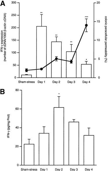
Time-Dependent Changes in Colonic Permeability, IFN-␥ mRNA Expression, and Protein Level After Stress Exposure
After 4 days of daily stress sessions, paracellular permeability, expressed as the percentage of51Cr-EDTA
that crosses the colonic barrier, was significantly higher in stressed mice compared with control mice (14.0⫾ 1.7 vs. 2.0 ⫾ 0.4; P ⬍ 0.001; n ⫽ 8). However, such a change was not observed at days 1, 2, and 3 (i.e., at earlier stages of stress) (Figure 2A).
We also observed an increase in IFN-␥ mRNA ex-pression on day 1 (P ⬍ 0.01), and the mRNA levels decreased with time until day 4 (Figure 2A), but still remained significantly higher than in sham-stressed an-imals (P⬍ 0.05). Concerning the protein, we observed an increase on day 2 (61.54⫾ 11.65 pg/mg of protein vs. 22.45⫾ 5.23 for controls; P ⬍ 0.05; n ⫽ 8 each group). Then, IFN-␥ levels decreased with time, as observed for the messengers (Figure 2B).
Effects of Daily Repeated Stress on Colonic Permeability in SCID Mice
In SCID mice, deficient for the zap70 protein and lacking mature CD4⫹/CD8⫹ T cells, 2-hour repeated
Figure 2. (A ) Time-dependent effects of repeated daily 2-hour stress sessions on colonic paracellular permeability (line) and IFN-␥ mRNA expression (bars). IFN-␥ mRNA expression increased after one stress session, whereas permeability increased only after 4 days of re-peated stress. (B) Time-dependent effects of rere-peated daily 2-hour stress sessions on colonic IFN-␥ protein level. IFN-␥ increased 1 day after mRNA, then slowly decreased. *P⬍ 0.05, **P ⬍ 0.01, ***P ⬍ 0.001 vs. sham-stress group.
Table 2. Effect of Repeated Stress on the Cytokine Profile of Mouse Colon, Liver, and Spleen Samples
Colon Control Stress (day 4) Stress⫹ ML-7 IFN-␥ 12.6⫾ 2.6 44.5⫾ 11.4a 41.8⫾ 3.2a TNF-␣ 170.7⫾ 46.6 193.7⫾ 61.9 ND IL-4 45.4⫾ 5.7 43.1⫾ 18.3 ND IL-5 39.3⫾ 13.6 39.0⫾ 15.9 ND IL-10 219.7⫾ 152.0 438.5⫾ 60.9 ND Liver Control Stress (day 3) Stress (day 6) Stress⫹ ML-7 IFN-␥ 22.3 ⫾ 7.7 25.5 ⫾ 10.75 56.6 ⫾ 17.6a 2.32⫾ 0.5a,b TNF-␣ 3.2⫾ 1.6 3.2⫾ 0.9 34.0⫾ 9.9a 8.0⫾ 1.4a,b IL-4 7.6⫾ 1.7 2.5⫾ 0.4 43.9⫾ 9.1a 2.4⫾ 1.0a,b IL-5 1.9⫾ 0.5 ND 3.0⫾ 0.7 ND IL-10 3.4⫾ 1.6 ND 33.8⫾ 11.4a Undetectable
Spleen Control Stress (day 4) IFN-␥ 3.6⫾ 0.8 2.9⫾ 0.7 TNF-␣ 9.0⫾ 4.8 5.9⫾ 2.0 IL-4 4.7⫾ 2.7 7.1⫾ 1.6 IL-5 1.9⫾ 0.5 3.0⫾ 0.7 NOTE. In the colon, 4 days of stress induced an overexpression of IFN-␥ that was maintained with ML-7 treatment. In the liver, modifica-tions occurred on day 4, but cytokine mRNA levels were unchanged on day 3. ML-7 inhibited the effect of stress. No modifications were noted in the spleen under stress exposure. Samples were recovered 2 hours after the last stress session. Data (mean⫾ SEM) from at least 8 different samples are expressed as the number of cDNA molecules/1000-actin cDNA molecules.
ND, not determined.
aP⬍ 0.05 vs. control group; bP⬍ 0.05 vs. stress (day 4) group
(Mann–Whitney test).
daily stress sessions for 4 days did not modify colonic permeability (3.0 ⫾ 0.7 vs. 3.4 ⫾ 0.9). In control conditions (sham-stress), colonic permeability was not statistically different between SCID and Swiss mice (3.0⫾ 0.7 vs. 2.0 ⫾ 0.6, respectively; Figure 3).
Effects of Daily Repeated Stress on Colonic Permeability in IFN-␥ Knockout Mice
Colonic permeability did not vary between stressed and sham-stressed IFN-␥ knockout mice (1.7 ⫾ 0.4 vs. 2.5⫾ 0.4, respectively; Figure 3). We observed no difference between sham-stressed IFN-␥ knockout mice and C57BL/6J mice (1.7 ⫾ 0.4 vs. 2.0 ⫾ 0.5, respectively; Figure 3). Under stress conditions, C57BL/6J mice presented an increased colonic perme-ability compared with sham-stressed animals (18.4 ⫾ 3.2 vs. 2.0⫾ 0.5, respectively; P ⬍ 0.001; Figure 3).
Effects of Daily Repeated Stress on MLCK, MLC, and Phospho-MLC Protein Levels MLCK levels were not affected by repeated expo-sure to stress (Figure 4). Conversely, we observed higher levels of phospho-MLC and decreased levels of MLC in stressed mice compared with sham-stressed mice (Figure 4). This may indicate that stress, likely through IFN-␥, activates the MLCK activation pathway without
affect-ing the expression level of the protein. Effects of MLCK Inhibition on
Stress-Induced Permeability Increase and Cytokine mRNA Levels
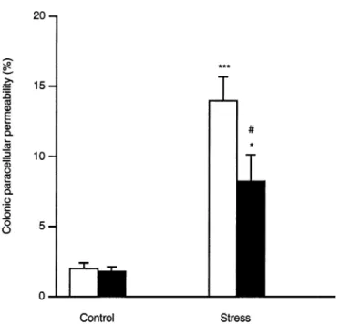
The stress-induced increase of51Cr-EDTA
perme-ability was inhibited significantly in Swiss mice chroni-cally treated with the MLCK inhibitor ML-7 (8.2⫾ 1.9 vs. 14.0⫾ 1.7; P ⬍ 0.05; Figure 5). ML-7 treatment did not affect per se colonic permeability in sham-stressed mice, and its vehicle (ethanol 2%) also did not produce significant changes in colonic permeability in both stressed and sham-stressed groups. ML-7 treatment did not affect the stress-induced changes in IFN-␥ mRNA expression in the colon (Table 2). In the liver, IFN-␥ and IL-4 mRNA levels were strongly decreased (Table 2), whereas IL-10 mRNA was undetectable. TNF-␣ mRNA levels, however, were diminished significantly but re-mained higher than in controls (P⬍ 0.05).
Effects of Repeated Stress and MLCK Inhibition on Bacterial Translocation to the Liver, Spleen, and Mesenteric Lymph Nodes A significant incidence of stress on bacterial trans-location occurred into the liver, but not into the spleen or mesenteric lymph nodes (P⬍ 0.05; n ⫽ 8 each group; Table 3). ML-7 treatment significantly reduced the in-cidence of stress on bacterial translocation (P⬍ 0.05).
Figure 3. Effects of (■) 4 days of daily 2-hour stress sessions, compared with (䊐) sham-stress, on colonic paracellular permeability in Swiss mice, SCID mice, C57BL/6J, and IFN-␥ knockout mice. Stress significantly increased permeability in Swiss and C57BL/6J mice, but not in SCID and IFN-␥ knockout mice. ***P ⬍ 0.001 vs. sham-stress group.
Figure 4. Western blot for MLC, phospho-MLC (p-MLC), and MLCK on colons from sham-stressed and stressed mice. MLCK did not vary between samples. Conversely, MLC levels were lower and p-MLC levels were higher in stressed animals, showing that MLC phosphor-ylation is associated with the increased colonic permeability observed after stress exposure.
Discussion
This study brings novel data regarding the mech-anisms through which stress disrupts the colonic barrier function, and the pathophysiologic consequences of this alteration. We first observed that, in the colon, an en-hanced expression of IFN-␥ occurred early (day 1), whereas increased colonic paracellular permeability was observed later (day 4). These alterations were not associ-ated with modifications of the architectural structure of the colonic mucosa of stressed Swiss mice. We also showed that CD4⫹/CD8⫹T cells are involved in stress-induced impairment of the colonic mucosal barrier be-cause stress did not induce any modification of colonic permeability in SCID mice. Moreover, the lack of effect of repeated stress sessions on colonic permeability in IFN-␥ knockout mice strongly suggests an important role for IFN-␥ in triggering stress-induced alterations of the colonic epithelial barrier. It is noteworthy that re-peated stress-induced breakdown of the colonic barrier is inhibited partly by the MLCK inhibitor, ML-7. Mean-while, in the liver, the increased expression of mRNAs encoding IFN-␥, IL-4, and IL-10 that occurred specifi-cally on day 4, was associated with bacterial translocation and histologic alterations such as apoptotic hepatocytes, inflammatory cell infiltration, and thickening of the bile duct wall. Treatment by an MLCK inhibitor abolished
the incidence of stress on bacterial translocation into the liver, dramatically reduced the clinical signs of hepatic injury, and restored basal levels of cytokine expression. Antibiotic treatment also inhibited stress-induced liver injury, confirming that bacteria from the colon lumen may translocate when the colon epithelium is leaky. No modification of cytokine expression and histologic struc-ture was detected in the spleen, confirming the fact that all the changes observed in cytokine expression in the colon and the liver were not caused by a systemic acti-vation of the immune system.
Thus, we show herein that repeated stress exposure leads to an increase in colonic paracellular permeability and identified IFN-␥ as a major mediator of this effect. Previous studies have evoked the role of CRH in mod-ulating both immune system and intestinal barrier func-tion. With regard to immunity, the central release of CRH, in response to an acute stress situation, stimulates preferentially a Th2 response as opposed to a Th1-type response through the activation of the hypothalamo-pituitary-adrenal axis and the release of glucocorticoids and catecholamines.3Indeed, intracellular glucocorticoid
receptors have been detected in several immune cells, such as lymphocytes or macrophages. Their activation inhibits IL-2, IL-12, and IFN-␥ expression, at the levels of transcription, translation, and mRNA stability, or their secretion.23,24A study has identified glucocorticoids
as possible mediators of stress-induced alterations of the intestinal barrier function because adrenalectomy and pharmacologic blockade of glucocorticoid receptors by RU486 inhibited the effects of stress.8
At the colonic level, few studies have evaluated the effect of repeated stress on the local inflammatory re-sponse. In an experimental model of trinitrobenzenesul-fonic acid–induced colitis, it has been shown that
re-Figure 5. Effect of the MLCK inhibitor, ML-7 (■), or its vehicle (䊐), injected twice daily, on colonic paracellular permeability in Swiss mice. ML-7 treatment significantly reduced the stress-induced perme-ability increase, which remained, however, significantly higher than in the sham-stress group. No modification of basal colonic permeability was observed after ML-7 treatment. *P⬍ 0.05, ***P ⬍ 0.001 vs. sham-stress group; #P⬍ 0.05 vs. stress group.
Table 3. Effect of 4 Days of Daily Repeated Stress and MLCK Inhibition on Bacterial Translocation Into the Liver, Spleen, and Mesenteric Lymph Nodes
Without ML-7 With ML-7 Number of positive organs P value vs. control groupa Number of positive organs P value vs. stress groupb Liver 5/8 P⬍ 0.05 1/8 P⬍ 0.05 Spleen 3/8 NS 0/8 NS MLN 3/8 NS 0/8 NS
NOTE. Expressed as the number of positive organs. Experiments were conducted 2 hours after the last stress session. Aerobes and anaer-obes were detected in the same samples. No bacterial translocation occurred under basal conditions.
NS, not significant; MLN, mesenteric lymph nodes.
aFisher exact test vs. control group.
bFisher exact test vs. stress group not receiving ML-7.
peated restraint stress sessions enhance the severity of inflammation, and that the activity of the different com-ponents of the hypothalamo-pituitary-adrenal axis are strongly implicated in this effect.13,25These results have
been confirmed with an experimental model of reacti-vated colitis. Indeed, Qiu et al.14 observed that chronic
stress reactivates colitis in mice 8 weeks after dinitro-benzenesulfonic acid–induction of colitis, and that this reactivation was CD4⫹ T-cell dependent. Interestingly, we show here that impairment of the colonic barrier function induced by repeated stress sessions required CD4⫹/CD8⫹T cells. Because T cells are potent IFN-␥– secreting cells, and because we showed that IFN-␥ is a prerequisite for stress-induced impairment of the colonic barrier, we can hypothesize that colitis reactivation under stress conditions is mediated by IFN-␥, which thereafter opens the TJ. Then an increased exposure to luminal antigens may occur as a consequence of TJ opening, which could facilitate the reactivation of colitis. In hu-mans, a prospective study on UC patients clearly has shown that short-term stress does not trigger exacerba-tion of UC but, in contrast, long-term perceived stress increases the risk for UC exacerbation over a period of months to years.26Moreover, an impaired intestinal
bar-rier function has been reported in patients with UC and Crohn’s disease.27 In patients with UC, altered
perme-ability seems to be limited to inflammatory loci, whereas in patients with Crohn’s disease, it occurs both in in-flamed and noninin-flamed tissues. All these studies on colonic inflammation support the hypothesis that stress has a local effect on intestinal immunity. Accordingly, our study suggests that this effect could be owing to a background effect promoting the local expression of Th1-type cytokines, and more particularly IFN-␥, which may in turn induce TJ opening.
We report here that increased colonic permeability occurred after 4 days of stress, whereas IFN-␥ mRNA levels were highest at day 1. This agrees with in vitro studies showing a deleterious effect of IFN-␥ on barrier integrity several days after exposure to the cytokine.19
This offers some indications about the mechanisms through which IFN-␥ may act to open TJs. First, the delay between IFN-␥ exposure, that arose after a single 2-hour stress session, and permeability alteration ob-served 4 days later, may indicate transcriptional effects. This is likely because it was shown that IFN-␥ decreased mRNAs encoding ZO-1 and occludin, 2 proteins in-volved in the TJ complex.17,18 Second, the inhibition
provoked by ML-7 treatment indicates that MLCK, and thus the cytoskeleton, is a potential link between IFN-␥ signaling and epithelial barrier impairment. Indeed,
Everding et al.28 have shown in a cell line derived from
lung carcinoma that IFN-␥ exposure for 2 days modified microtubule organization and ␣-tubulin expression. Moreover, in T84 monolayers IFN-␥ impaired the bar-rier function by disrupting apical actin and activating MLC phosphorylation.17,29 We also describe that IFN-␥
mRNA levels were unchanged in stressed mice treated or not with ML-7, which indicates that MLCK activation is downstream of IFN-␥ secretion.
In the liver, changes observed in cytokine expression after 4 days of repeated stress are associated with an inflammatory process. The enhanced expression of IFN-␥ being observed at both intestinal and liver tissues, we can hypothesize that a high level of hepatic IFN-␥ mRNA is a direct response to stress, and that this IFN-␥ release may be in part responsible for the inflammatory response. On the other hand, according to our results, stress could lead to an excessive uptake of colonic luminal content and thus enhance bacterial translocation through the opening of TJs, a consequence of the increased intestinal secretion of IFN-␥. Consistent with this hypothesis, studies performed in rats and alcoholic patients sug-gested that ethanol, by affecting colonic TJ permeability, may be responsible for liver bacterial translocation and injury.22,30 This hypothesis is corroborated by our
present findings because we show that the treatment by the MLCK inhibitor decreased the stress-induced in-crease of colonic permeability, and abolished the subse-quent bacterial translocation and cytokine overexpression in the liver. We also observed that ML-7 treatment decreased the number of apoptotic hepatocytes and the inflammatory infiltrate in the liver, indicating that a normal histology was restored. Bacteria that translocated into the liver were endogenous because stress-induced liver injury was diminished in animals given nonabsorb-able antibiotics.
IFN-␥ is involved in several hepatic inflammatory disorders, such as alcohol-induced liver disease, graft-versus-host disease, primary biliary cirrhosis, or chronic hepatitis B.31,32 Effects of IFN-␥ such as macrophage
activation, up-regulation of class I and II major histo-compatibility complex molecules necessary for antigen presentation, induction of expression of TNF receptors, induction of adhesion molecules such as intercellular adhesion molecule 1 and vascular cell adhesion molecules involved in the homing of inflammatory cells, may pro-mote local inflammation.33However, these inflammatory
effects, probably caused by the enhanced level of IFN-␥, have not been observed in the gut. At the hepatic level, the enhanced expression of the Th2-type cytokines may result from infiltrated inflammatory cells and activated
Kupffer cells, these cells being the main source of IL-4 and IL-10. In the liver, their enhanced expression in many inflammatory disorders has been described. In an experimental model of Schistosoma mansoni infection in baboons, an increased expression of IL-4, IL-10, and IFN-␥ in the liver of infected animals has been ob-served.34,35Moreover, IL-4 and/or IL-10 also are involved
in alcohol-induced hepatitis, lipopolysaccharide-induced hepatic inflammation, and chronic hepatitis B.32,36,37
In addition to the histologic signs of inflammation, we also have observed an increased number of apoptotic hepatocytes in the liver of chronically stressed animals when compared with controls. Apoptosis plays a pivotal role in liver development and homeostasis, but also in the pathogenesis of several liver diseases.38To our
knowl-edge, the mechanism by which repeated stress may mod-ulate apoptosis in the liver has never been investigated before, and only a limited hypothesis can be made. It has been shown that IFN-␥ alone or in combination with TNF-␣ and IL-1, increases the mRNA of inducible nitric oxide synthase in rat hepatocytes.39 So a direct
modulatory effect of IFN-␥ on nitric oxide–induced he-patocyte death could be suspected.
In conclusion, our results show that repeated stress modifies intestinal barrier function and causes bacterial translocation and liver injury. Stress-induced opening of colonic epithelial TJ requires CD4⫹/CD8⫹ T cells and IFN-␥, and involves MLCK activation. Inhibiting MLCK in vivo resulted in a decreased incidence of bac-terial translocation by stress, and restored a normal liver histology and cytokine mRNA expression. Conse-quently, epithelial MLCK may represent a target for therapeutic approaches to stress-associated intestinal and liver disorders.
References
1. Chrousos GP. The hypothalamic-pituitary-adrenal axis and im-mune-mediated inflammation. N Engl J Med 1995;332:1351– 1362.
2. Ottaviani E, Franceschi C. The neuroimmunology of stress from invertebrates to man. Prog Neurobiol 1996;48:421– 440. 3. Webster EL, Torpy DJ, Elenkov IJ, Chrousos GP.
Corticotropin-releasing hormone and inflammation. Ann N Y Acad Sci 1998; 840:21–32.
4. Millan S, Gonzalez-Quijano MI, Giordano M, Soto L, Martin AI, Lopez-Calderon A. Short and long restraint differentially affect humoral and cellular immune functions. Life Sci 1996;59:1431– 1442.
5. Perdue MH. Mucosal immunity and inflammation. III. The mucosal antigen barrier: cross talk with mucosal cytokines. Am J Physiol 1999;277:G1–G5.
6. Turner JR, Rill BK, Carlson SL, Carnes D, Kerner R, Mrsny RJ, Madara JL. Physiological regulation of epithelial tight junctions is associated with myosin light-chain phosphorylation. Am J Physiol 1997;273:C1378 –C1385.
7. Saunders PR, Santos J, Hanssen NP, Yates D, Groot JA, Perdue
MH. Physical and psychological stress in rats enhances colonic epithelial permeability via peripheral CRH. Dig Dis Sci 2002;47: 208 –215.
8. Meddings JB, Swain MG. Environmental stress-induced gastroin-testinal permeability is mediated by endogenous glucocorticoids in the rat. Gastroenterology 2000;119:1019 –1028.
9. Santos J, Saunders PR, Hanssen NP, Yang PC, Yates D, Groot JA, Perdue MH. Corticotropin-releasing hormone mimics stress-in-duced colonic epithelial pathophysiology in the rat. Am J Physiol 1999;277:G391–G399.
10. Schmitz H, Barmeyer C, Gitter AH, Wullstein F, Bentzel CJ, Fromm M, Riecken EO, Schulzke JD. Epithelial barrier and transport function of the colon in ulcerative colitis. Ann N Y Acad Sci 2000;915:312–326.
11. Soderholm JD, Olaison G, Peterson KH, Franzen LE, Lindmark T, Wiren M, Tagesson C, Sjodahl R. Augmented increase in tight junction permeability by luminal stimuli in the non-inflamed ileum of Crohn’s disease. Gut 2002;50:307–313.
12. Gassler N, Rohr C, Schneider A, Kartenbeck J, Bach A, Obermul-ler N, Otto HF, Autschbach F. Inflammatory bowel disease is associated with changes of enterocytic junctions. Am J Physiol 2001;281:G216 –G228.
13. Gue M, Bonbonne C, Fioramonti J, More J, Del Rio-Lacheze C, Comera C, Bueno L. Stress-induced enhancement of colitis in rats: CRF and arginine vasopressin are not involved. Am J Physiol 1997;272:G84 –G91.
14. Qiu BS, Vallance BA, Blennerhassett PA, Collins SM. The role of CD4⫹ lymphocytes in the susceptibility of mice to stress-induced reactivation of experimental colitis. Nat Med 1999;5: 1178 –1182.
15. Keshavarzian A, Holmes EW, Patel M, Iber F, Fields JZ, Pethkar S. Leaky gut in alcoholic cirrhosis: a possible mechanism for alco-hol-induced liver damage. Am J Gastroenterol 1999;94:200 – 207.
16. Ceponis PJ, Botelho F, Richards CD, McKay DM. Interleukins 4 and 13 increase intestinal epithelial permeability by a phospha-tidylinositol 3-kinase pathway: Lack of evidence for STAT 6 in-volvement. J Biol Chem 2000;275:29132–29137.
17. Youakim A, Ahdieh M. Interferon-gamma decreases barrier func-tion in T84 cells by reducing ZO-1 levels and disrupting apical actin. Am J Physiol 1999;276:G1279 –G1288.
18. Mankertz J, Tavalali S, Schmitz H, Mankertz A, Riecken EO, Fromm M, Schulzke JD. Expression from the human occludin promoter is affected by tumor necrosis factor alpha and inter-feron gamma. J Cell Sci 2000;113:2085–2090.
19. Nusrat A, Turner JR, Madara JL. Molecular physiology and patho-physiology of tight junctions. IV. Regulation of tight junctions by extracellular stimuli: nutrients, cytokines, and immune cells. Am J Physiol 2000;279:G851–G857.
20. Dalton DK, Pitts-Meek S, Keshav S, Figari IS, Bradley A, Stewart TA. Multiple defects of immune cell function in mice with dis-rupted interferon-gamma genes. Science 1993;259:1739 – 1742.
21. Bradley PP, Priebat DA, Christensen RD, Rothstein G. Measure-ment of cutaneous inflammation: estimation of neutrophil con-tent with an enzyme marker. J Invest Dermatol 1982;78:206 – 209.
22. Ma TY, Nguyen D, Bui V, Nguyen H, Hoa N. Ethanol modulation of intestinal epithelial tight junction barrier. Am J Physiol 1999;276: G965–G974.
23. Northrop JP, Crabtree GR, Mattila PS. Negative regulation of interleukin 2 transcription by the glucocorticoid receptor. J Exp Med 1992;175:1235–1245.
24. DeKruyff RH, Fang Y, Umetsu DT. Corticosteroids enhance the capacity of macrophages to induce Th2 cytokine synthesis in CD4⫹ lymphocytes by inhibiting IL-12 production. J Immunol 1998;160:2231–2237.
25. Million M, Tache Y, Anton P. Susceptibility of Lewis and Fischer rats to stress-induced worsening of TNB-colitis: protective role of brain CRF. Am J Physiol 1999;276:G1027–G1036.
26. Levenstein S, Prantera C, Varvo V, Scribano ML, Andreoli A, Luzi C, Arca M, Berto E, Milite G, Marcheggiano A. Stress and exac-erbation in ulcerative colitis: a prospective study of patients enrolled in remission. Am J Gastroenterol 2000;95:1213–1220. 27. Peeters M, Ghoos Y, Maes B, Hiele M, Geboes K, Vantrappen G, Rutgeerts P. Increased permeability of macroscopically normal small bowel in Crohn’s disease. Dig Dis Sci 1994;39:2170 – 2176.
28. Everding B, Wilhelm S, Averesch S, Scherdin U, Holzel F, Steffen M. IFN-gamma-induced change in microtubule organization and alpha-tubulin expression during growth inhibition of lung squa-mous carcinoma cells. J Interferon Cytokine Res 2000;20:983– 990.
29. Zolotarevsky Y, Hecht G, Koutsouris A, Gonzalez DE, Quan C, Tom J, Mrsny RJ, Turner JR. A membrane-permeant peptide that inhibits MLC kinase restores barrier function in in vitro models of intestinal disease. Gastroenterology 2002;123:163–172. 30. Keshavarzian A, Choudhary S, Holmes EW, Yong S, Banan A,
Jakate S, Fields JZ. Preventing gut leakiness by oats supplemen-tation ameliorates alcohol-induced liver damage in rats. J Phar-macol Exp Ther 2001;299:442– 448.
31. Howell CD, Li J, Chen W. Role of intercellular adhesion mole-cule-1 and lymphocyte function-associated antigen-1 during non-suppurative destructive cholangitis in a mouse graft-versus-host disease model. Hepatology 1999;29:766 –776.
32. Shindo M, Mullin GE, Braun-Elwert L, Bergasa NV, Jones EA, James SP. Cytokine mRNA expression in the liver of patients with primary biliary cirrhosis (PBC) and chronic hepatitis B (CHB). Clin Exp Immunol 1996;105:254 –259.
33. Olsson T. Critical influences of the cytokine orchestration on the outcome of myelin antigen-specific T-cell autoimmunity in exper-imental autoimmune encephalomyelitis and multiple sclerosis. Immunol Rev 1995;144:245–268.
34. Farah IO, Mola PW, Kariuki TM, Nyindo M, Blanton RE, King CL. Repeated exposure induces periportal fibrosis in Schistosoma
mansoni-infected baboons: role of TGF-beta and IL-4. J Immunol 2000;164:5337–5343.
35. Mola PW, Farah IO, Kariuki TM, Nyindo M, Blanton RE, King CL. Cytokine control of the granulomatous response in Schistosoma mansoni-infected baboons: role of exposure and treatment. In-fect Immun 1999;67:6565– 6571.
36. Jarvelainen HA, Fang C, Ingelman-Sundberg M, Lindros KO. Effect of chronic coadministration of endotoxin and ethanol on rat liver pathology and proinflammatory and anti-inflammatory cytokines. Hepatology 1999;29:1503–1510.
37. Thompson K, Maltby J, Fallowfield J, McAulay M, Millward-Sadler H, Sheron N. Interleukin-10 expression and function in experi-mental murine liver inflammation and fibrosis. Hepatology 1998; 28:1597–1606.
38. Benedetti A, Marucci L. The significance of apoptosis in the liver. Liver 1999;19:453– 463.
39. Geller DA, Nussler AK, Di Silvio M, Lowenstein CJ, Shapiro RA, Wang SC, Simmons RL, Billiar TR. Cytokines, endotoxin, and glucocorticoids regulate the expression of inducible nitric oxide synthase in hepatocytes. Proc Natl Acad Sci U S A 1993;90:522– 526.
Received December 19, 2002. Accepted June 12, 2003.
Address requests for reprints to: Lionel Bueno, Ph.D., Neuro-Gastro-enterology and Nutrition Unit, Institut National de la Recherche Agronomique, 180 chemin de Tournefeuille, BP 3, 31931 Toulouse Cedex 9, France. e-mail: [email protected]; fax: (33) 5-61-28-53-97.
Supported by Institut de Recherche sur les Maladies de l’Affareil Digestif (IRMAD), Fondation pour la Recherche Me´dicale, and an institutional grant of Institut National de la Recherche Agronomique. L.M. is a recipient of the Ministe`re de l’Education Nationale, de l’Enseignement Supe´rieur, de la Recherche, et de la Technologie.
The authors thank Valerie Geronimo and Caroline Bisiaux for their skillful technical assistance, and Dr. Neil Ledger for critical reading of the manuscript.