HAL Id: hal-00734811
https://hal.archives-ouvertes.fr/hal-00734811
Submitted on 24 Sep 2012HAL is a multi-disciplinary open access archive for the deposit and dissemination of sci-entific research documents, whether they are pub-lished or not. The documents may come from teaching and research institutions in France or abroad, or from public or private research centers.
L’archive ouverte pluridisciplinaire HAL, est destinée au dépôt et à la diffusion de documents scientifiques de niveau recherche, publiés ou non, émanant des établissements d’enseignement et de recherche français ou étrangers, des laboratoires publics ou privés.
Synthesis and characterization of Cx-Siy-HA for bone
tissue engineering application
Antoine Boyer, David Marchat, Didier Bernache-Assollant
To cite this version:
Antoine Boyer, David Marchat, Didier Bernache-Assollant. Synthesis and characterization of Cx-Siy-HA for bone tissue engineering application. Key Engineering Materials, Trans Tech Publications, 2013, 529-530, pp.100-104. �10.4028/www.scientific.net/KEM.529-530.100�. �hal-00734811�
Synthesis And Characterization Of C
x-Si
y-HA For Bone Tissue
Engineering Application
Boyer Antoine
1,a, Marchat David
1,bBernache-Assollant Didier
1,c1 Ecole Nationale Supérieure des Mines de Saint-Etienne, CIS-EMSE, CNRS : FRE3312, F-42023
158 Cours Fauriel, Saint-Etienne, France
a [email protected], b [email protected], c [email protected]
Keywords: co-substituted hydroxyapatite, carbonate, silicate, bioceramics, pellets, aqueous precipitation.
Abstract. The main goal of this work is to prepare carbon and silicon co-substituted calcium
hydroxyapatite (Cx-Siy-HA) for bone tissue engineering application. This study includes the synthesis
of pure powders with a controlled amount of carbonate (x) and silicate (y) ions within the apatite structure, their characterization with the establishment of database for different compositions, and the
manufacture of dense bioceramics. Carbon-silicon co-substituted hydroxyapatite (C0.5-Si0.5-HA)
powders are synthesized by aqueous precipitation. According to structural, spectroscopic and elemental characterizations, silicate and carbonate are included in the apatite lattice and their
stoichiometries are controlled. The heat treatments under CO2 atmosphere allow the sintering of
pellets without decomposition of the apatite structure.
Introduction
Hydroxyapatite (HA), Ca10(PO4)6(OH)2 is the most commonly used bioceramics for dental and
orthopedic implants due to its chemical and crystallographic similarities with bone’s mineral part. Natural bone is comparable to a badly crystallized apatitic calcium phosphate with substitutions in its lattice. The apatite lattice is very tolerant with substitutions and vacancies [1]. All biological apatites contain carbonate ions in erratic quantities (between 3-8wt %) [2], filling preferentially in phosphate (B-type) compared to hydroxyl (A-type) positions in the apatite structure [3,4]. The composition depends on bone location, age, sex, etc. [3]. Carbonate substitution in apatite decrease the lattice cristallinity, as well as the temperature of the maximum rate of sintering [5,6], and increase its solubility in biological conditions (in vitro and in vivo) [4]. To improve bioactivity of synthetic grafts as resorption or osteogenesis, the literature talks over incorporation of carbonate or silicate groups in HA. Among trace elements existing in natural bone, silicon show essential characters for bone growth and development [7]. Even if the benefits of soluble silicon have been proved, the bioactivity of silicon substituted hydroxyapatite ceramics was not well defined. Results heterogeneity could be due to uncontrolled composition of Si-HA bioceramics [8]. Adequate solubility of carbonated hydroxyapatite and benefits of soluble silicon could be associated to improve bioactivity of apatite bioceramics. Some studies about carbon-silicon substituted hydroxyapatite were recently published [9–13]. Unfortunately, phase purity was not evidently proved. The main idea of this work was the
preparation of a SiO4 and CO3 co-substituted hydroxyapatite bioceramics with controlled
stoichiometry for bone tissue engineering. The study was first dedicated to the synthesis of Cx-Siy-HA
powders by aqueous precipitation, and second to the preparation of dense pellets.
Materials and methods
Synthesis of apatite powders. Cx-Siy-HA powders were prepared by aqueous precipitation based
on the method of Marchat et al [8]. This synthesis method allowed preparing monophasic silicon
substituted hydroxyapatite ceramics with controlled composition (e.g. Si0,5-HA). The amount of
reagents was calculated assuming silicate and carbonate substitution for phosphate ions according to the following theoretical formula:
Ca10-x+y 2+ (PO43-)6-y-x (SiO44-)y (CO32-)x (OH-)2-x+y with x = y = 0.5
A reagent molar ratio Ca/(P+Si+C) of 10/6 was used. Besides, materials of reference were
prepared like a pure HA, Cx-HA, Siy-HA or different Cx-Siy-HA to establish databases.
A diammonium hydrogen phosphate aqueous solution ((NH4)2HPO4, 99%, Merck, Germany), an
ammonium hydrogen carbonate ((NH4)HCO3, 99%, Merck, Germany) and an alkaline silicate
solution [8] were added to a calcium nitrate solution (Ca(NO3)2,4H2O, 99%, Merck, Germany) by
means of peristaltic pumps.Deionized water was used to dissolve the reagents. An argon flow (Air
Liquide) was maintained on the reactor to prevent any atmospheric uncontrolled carbonation. The pH was kept at 10.8 by addition of a 28% ammonia solution (Merck, Germany) using a pH stat (Hanna instrument) and the temperature was regulated automatically at 50°C with an external cryothermostat. The suspension was continuously stirred. After complete introduction of reagents, suspension was matured for 48h, and then filtered on Whatmann filter (Ø> 6 µm). Finally, precipitates were dried at 100°C overnight.
Pellets preparation. Protocol consists to precalcine the powders to get a specific surface area
around 30 m²/g. 160 mg of powder were first uniaxially pressed at 48 MPa in 8 mm diameter stainless steel die, then isostatically pressed at 300 MPa. Lafon et al. studied the synthesis of carbonated hydroxyapatite and their thermal stability under different atmospheres (PCO2, PH2O, PN2) [14,15].
According to their work and a complementary thermal study, it happens a decarbonation of carbonated and silico-carbonated hydroxyapatite powders beyond 800°C under air atmosphere
[5,6,14]. To avoid this decomposition, heat-treatments and sintering of C0.5-Si0.5-HA samples were
made under CO2 atmosphere (PCO2 = 1 atm). Heating rates were fixed at ±5°C/min.
Characterizations.
The crystalline phases of powders were studied using X-ray diffraction (XRD). The device used is a θ/2θ X-ray diffractometer, Siemens D5000, using CuKα radiation. The crystalline phases were evaluated on calcined powders over the range 27-38° with a step size of 0.02° and a count time of 9s by step. Phase identification was performed by comparison to standard patterns from International Center for Diffraction Data – Powder Diffraction Files (ICDD-PDF).
Fourier Transformed Infrared (FT-IR) absorption spectra of powders were performed using a
Spectrometer MIR TF VERTEX 70. They were recorded over the range 400-4000 cm-1 with a
resolution of 2 cm-1 and with an iteration of 64 successive scans. All spectra were normalized from
the ν4 band of the phosphate group at 600 cm-1 as usual [4].
Calcium, phosphorous and silicon contents of powders were determined by elemental analysis via Inductively Coupled Plasma Atomic Emission Spectrometry (ICP/AES) (Spectrometer HORIBA, Jobin-Yvon, with Activa model). Powders samples were dissolved in nitric acid solution (pH < 2). Likewise, carbon content of powders was determined by an elemental analyzer using an infrared detector (LECO CS-444 carbon and sulfur analyzer).
Results and discussions
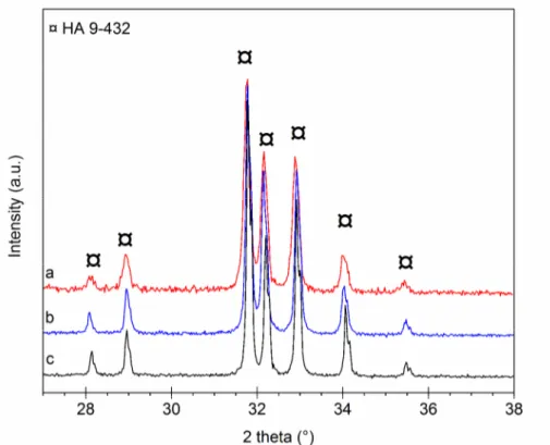
Structural analysis. The XRD patterns of heat-treated (under CO2) C0.5-Si0.5-HA powders and
C0.5-Si0.5-HA pellets (Fig. 1) show an apatitic well crystallized structure (PDF 9-432). No secondary
crystallized phase is detected, as the main characteristic line of CaO at 2θ = 37.347° (PDF 37-1497).
Thus, the decarbonation of B-site, according to the following reaction (Eq. 1) is not observed [14]: Ca10(PO4)5(CO3)0.5(SiO4)0.5(OH)2 →
(11/12) Ca10(PO4)(60/11)(SiO4)(6/11)(OH)(16/11) + (10/12) CaO + (1/2) CO2 + (1/3) H2O (1)
The CO2 atmosphere keeps carbonate in apatite lattice and avoids phase decomposition. Thereby,
the results indicate that silicon and carbon can be incorporated in the apatitic structure, or in an amorphous phosphate, or both.
Fig. 1: XRD patterns: (a) C0.5-Si0.5-HA pellets after sintering under CO2 ; (b) C0.5-Si0.5-HA powders after calcination under CO2 ; (c) HA powders in reference after calcination 1000°C-15h
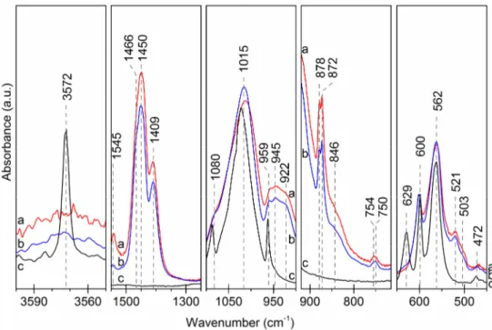
Spectrometric analysis. The FTIR patterns of C0.5-Si0.5-HA powders and C0.5-Si0.5-HA pellets
after heat-treatment under CO2 (Fig. 2) reveal the same main phosphate bands as spectra of heat
treated HA under air atmosphere : 472 cm-1 (ν2), 562 and 600 cm-1 (ν4), 959 cm-1 (ν1), 1015 and 1080
cm-1 (ν3). Characteristics bands of carbonate groups are shown on spectra of C-Si-HA samples: from
B site (872 cm-1, 1409 cm-1, 1450 cm-1, 1466 cm-1), and A site (754 cm-1, 878 cm-1, 1545 cm-1). The
CO2 atmosphere seems enriched the A sites along the channels in CO3. Indeed, the νS and νL modes of
hydroxyl group (629 cm-1 and 3572 cm-1) are not observed anymore. More, spectra of C0.5-Si0.5-HA
display the specific bands attributed to SiO4 substitution in hydroxyapatite structure: 503 cm-1
(ν2 : SiO4), 521 cm-1 (ν4 : SiO4), 750 cm-1 (ν1 : SiO4), 846 cm-1 (ν3 : SiO4), and 922 cm-1 (Si-OH) [8].
Besides, an extra band appeared at 945 cm-1. This large band results from the degeneration of ν1 PO4
3-domain at 960 cm-1 due to the presence of OH- vacancies and CO32- ions along the channel in the
environment of the phosphate ions. Finally, no evidence of secondary compounds as CaO or TCP is
detected. Likewise, the SiO2 specific bands (e.g. ~680 cm-1, ~792 cm-1 and ~870 cm-1 [8]) are not
observed. To conclude, results prove that C0.5-Si0.5-HA samples (powders and pellets) are
Fig. 2: FTIR patterns: (a) C0.5-Si0.5-HA pellets after sintering under CO2 ; (b) C0.5-Si0.5-HA powders after calcination under CO2 ; (c) HA powders in reference after calcination 1000°C-15h
Elemental analysis. C0.5-Si0.5-HA powder and C0.5-Si0.5-HA pellet elemental compositions were
measured after heat treatment at 1000°C for 1h under CO2 atmosphere (Table 1). The Ca/P molar
ratios are equal to 1.961 and 2.042 for C0.5-Si0.5-HA powder and pellet respectively. These results are
in coherence with the theoretical ratio Ca/P of reagent introduced equal to 2. For the powders and
pellets heat treated under CO2 atmosphere, samples present a Ca/(P+Si+C) molar ratios inferior than
estimated, respectively 1.546 and 1.567. In the ideal case of total co-substitution of carbonate and silicate in B anionic site, the Ca/(P+Si+C) molar ratios for a C0.5-Si0.5-HA composition should be
equal to 10/6 (=1.667). Difference can be explained by an increase of carbon amount in compounds,
i.e., in A site, due to calcination under CO2 atmosphere. The substitution of OH- by CO32- along the
channels throughout this heat treatment is confirmed FTIR spectrometry.
Table 1 : Results from elemental analysis
Compound Heat treatment Sintering Ca/P
measured Ca/(P+Si+C) measured C0.5-Si0.5-HA powder 1000°C-1h-CO2 / 1.961 1.546 C0.5-Si0.5-HA
pellet 835°C-1h-CO2 1000°C-1h-CO2 2.042 1.567
Conclusion
Carbon and silicon co-substituted calcium hydroxyapatite (C-Si-HA) powders were synthesis by aqueous precipitation. Silicate and carbonate were incorporated in the apatite lattice and their
stoichiometry was controlled. The heat treatments under CO2 atmosphere allowed the sintering of
pellets without decomposition of the apatite structure. Powders and pellets obtained were monophasic apatite polysubstituted with silicate and carbonate ions and free of secondary phase. Studies about the improvement of the sintering conditions are in progress, as well as in vitro and in vivo biological evaluation of dense pellets and scaffolds.
References
[1] J.C. Elliott, Structure and chemistry of the apatites and other calcium orthophosphates,
Studies in Organic Chemistry, (1994).
[2] F. Driessens, H. Schaeken, R. Verbeeck, On the mechanism of subsitution in carbonated
apatites, Journal of Dental Research, 62 (1983) 455.
[3] G. Montel, G. Bonel, J.C. Heughebaert, J.C. Trombe, C. Rey, New concepts in the
composition, crystallization and growth of the mineral component of calcified tissues, Journal of Crystal Growth, 53 (1981) 74–99.
[4] C. Rey, B. Collins, T. Goehl, I.R. Dickson, M.J. Glimcher, The carbonate environment in
bone mineral: A resolution-enhanced fourier transform infrared spectroscopy study, Calcified Tissue International, 45 (1989) 157–164.
[5] J. Barralet, S. Best, W. Bonfield, Effect of sintering parameters on the density and
microstructure of carbonate hydroxyapatite, J. Mater. Sci.-Mater. Med., 11 (2000) 719–724.
[6] Z. Zyman, M. Tkachenko, CO2 gas-activated sintering of carbonated hydroxyapatites, Journal
of the European Ceramic Society, 31 (2011) 241–248.
[7] E.M. Carlisle, Silicon: A Possible Factor in Bone Calcification, Science, 167 (1970) 279–280.
[8] D. Marchat, M. Zymelka, L. Gremillard, C. Coelho, L. Joly-Pottuz, F. Babonneau, C. Esnouf,
J. Chevalier, D. Bernache-Assollant, Accurate characterization of silicon-substituted hydroxyapatites
powders synthesized by a new precipitation route, Acta Biomaterialia, 2012,Submitted.
[9] E. Landi, J. Uggeri, S. Sprio, A. Tampieri, S. Guizzardi, Human osteoblast behavior on
as-synthesized SiO4 and B-CO3 co-substituted apatite, Journal of Biomedical Materials Research Part
A, 94A (2010) 59–70.
[10] T. Huang, Y. Xiao, S. Wang, Y. Huang, X. Liu, F. Wu, Z. Gu, Nanostructured Si, Mg, CO3
2-Substituted Hydroxyapatite Coatings Deposited by Liquid Precursor Plasma Spraying: Synthesis and Characterization, Journal of Thermal Spray Technology, 20 (2011) 829–836.
[11] D.M. Ibrahim, A.A. Mostafa, S.I. Korowash, Chemical characterization of some substituted
hydroxyapatites, Chem Cent J, 5 (2011) 74.
[12] N.Y. Mostafa, H.M. Hassan, O.H. Abd Elkader, Preparation and Characterization of Na+,
SiO44-, and CO32- Co-Substituted Hydroxyapatite, Journal of the American Ceramic Society, 94
(2011) 1584–1590.
[13] N.Y. Mostafa, H.M. Hassan, F.H. Mohamed, Sintering behavior and thermal stability of Na+,
SiO44- and CO32- co-substituted hydroxyapatites, Journal of Alloys and Compounds, 479 (2009)
692–698.
[14] J. Lafon, E. Champion, D. Bernache-Assollant, R. Gibert, A. Danna, Thermal decomposition
of carbonated calcium phosphate apatites, Journal of Thermal Analysis and Calorimetry, 72 (2003) 1127–1134.
[15] J.P. Lafon, E. Champion, D. Bernache-Assollant, Processing of AB-type carbonated
hydroxyapatite Ca10-x(PO4)6-x(CO3)x(OH)2-x-2y(CO3)y ceramics with controlled composition, Journal
of the European Ceramic Society, 28 (2008) 139–147.
[16] J. Barralet, J. Knowles, S. Best, W. Bonfield, Thermal decomposition of synthesised