Case 3
Diarrhoea and vomiting in a Yorkshire Terrier
Eve Ramery1, Manuel Pinilla2, Hester McAllister2, Hanne Jahns3, Barbara Gallagher4, Bernadette Legget5,Stratos Papakonstantinou1, Peter J O’Brien1
University College of Dublin, Veterinary Hospital, 1Department of Clinical Pathology, 2Department of Diagnostic Imaging, 3Department of Pathology, 4
Department of Small Animal Medicine, 5Department of Microbiology
Case Presentation
An entire female Yorkshire terrier, 9 years old was presented with a history of a 2 weeks episode of vomiting and diarrhoea. The report of the referring veterinarian revealed fever (39,8°C), anorexia, loss of weight and dullness at clinical examination. Laboratory Results
The data from haematology and biochemistry are presented in Table 1 and Table 2 respectively. There were no abnormal findings on urinalysis. Clotting times were also normal (PT: 13, N:12-17; APTT: 89, N: 71-102).
Table 1: Haematology data for the patient
Table 2: Biochemistry data for the patient
Parameter Value Unit Reference range
Tot Protein 76 g/L (54 - 71) Albumin 30 g/L (31 - 40) Globulin 46 g/L (28 - 42) ALT 429 U/L (0 - 50) GLDH 94 U/L (0 - 10) ALP 4336 U/L (0 - 140) GGT 27 U/L (0 - 8)
Parameter Value Unit Reference range
Hct 0.45 L/L (0.37 - 0.55) Hgb 157 g/L (120 - 180) RBC 6.4 x10¹²/L (5.5 - 8.5) MCHC 348 g/L (310 - 362) MCV 71 fL (60 - 77) MCH 25 pg (19.5 - 25) Plt 300 x109/L (150 - 500) MPV 12 fL (7 - 11) WBC 20 x109/L (6 - 17) Neut 13.5 x10⁹/L (3 - 11.5) Lym 2.9 x10⁹/L (1 - 3.6) Mono 1.55 x10⁹/L (0 - 1.35) Eos 0.98 x10⁹/L (0 - 1.47)
Bile Acids 37 umol/L (0 - 15) Post prandial bile
acids 13.7 umol/L.
Total Bili 10.6 umol/L (0.9 - 10)
Amylase 876 U/L (0 - 730) LIPASE 51 U/L (0 - 130) CK 47 U/L (0 - 50) Cholesterol 5.8 mmol/L (3.2 - 6.5) Glucose 3.8 mmol/L (3 - 6.5) Urea 10 mmol/L (3.6 - 8.6) Creatinine 68 umol/L (20 - 120) Sodium 152 mmol/L (137 - 151) Chloride 113 mmol/L (99 - 110) Calcium 2.7 mmol/L (2.3 - 3) Phosphorus 1.8 mmol/L (0.8 - 1.8) Potassium 3.9 mmol/L (3.7 - 5.8) Ultrasonography
Figure 1: Ultrasonography of the gallbladder
Ultrasonography of the abdomen was performed in order to precise the suspected liver damages (Figure 1). Ultrasonography indicated severe thickening of the gallbladder wall measuring up to 4 mm in some areas, with normal reference range of 2-3 mm (Spaulding 1993). The common bile duct was marginally distended measuring up to 3 mm in the vicinity of the duodenal papilla. The liver was otherwise normal. There was a focal area of gastric wall thickening at the level of the lesser curvature of the pyloric canal measuring up to 1 cm. In the mucosal surface of this area there was a persistent gas accumulation generating multiple comet tail artefacts. These findings suggest distal gastric ulceration.
Cytology
Direct smears of bile obtained by gallbladder lumen aspiration were stained with Wright’s-Giemsa for cytologic examination (Figures 2 and 3).
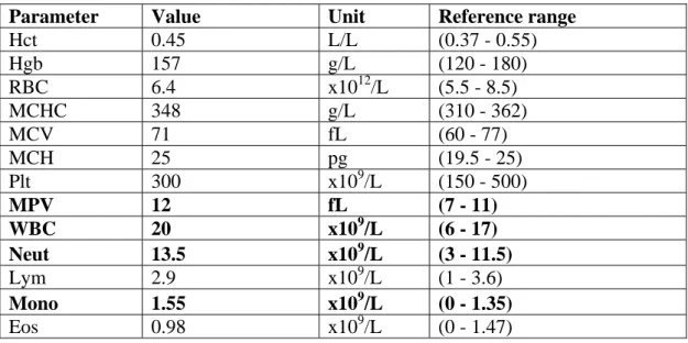
Figure 2: bile aspirate (Wright Giemsa, x1000 magnification)
Figure 3: bile aspirate (Wright Giemsa, x1000 magnification)
Large numbers (up to 100 / 100 x objective field) of uniform, large bacterial rods in pairs and small chains typical of clostridial morphology were observed in an abundant bright-pink, fibrillar background (possibly degenerate nuclear material). No intact cells were retrieved.
Bacterial Culture
The bacterial culture resulted in a pure culture of Clostridium sp. Histology
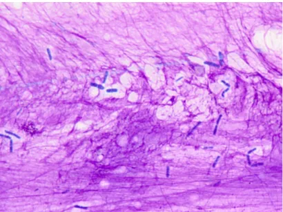
Figure 4: Liver biopsy
The microscopic examination of a core biopsy from the liver (Figure 4) revealed an expansion of the periportal areas by macrophages, plasma cells, neutrophils and occasional lymphocytes. The bile ducts were very prominent and there was low grade bile duct hyperplasia present. The hepatocytes were mildly swollen and contained occasionally dark yellow granules (bile). There were multifocal small foci of macrophages containing dark brown granular pigment (haemosiderin). The Kupffer cells were prominent and the sinusoids contain moderate numbers of neutrophils. Mild multifocal hepatocyte necrosis with neutrophilic and histiocytic infiltration was present.
The above histology findings are characteristic of a mild to moderate subacute hepato-pericholangitis.
Description of Results
Cytological examination of bile obtained from gallbladder lumen aspiration, showed bacterial contamination and infectious agents were further identified as Clostridium
sp. Reports of bacterial infections that are confined to the hepatobiliary system
include diffuse cholangitis/cholangiohepatitis, cholecystitis, choledochitis, focal suppurative lesions and multiple hepatic microabscesses (Center, 1998). Along with the laboratory results indicating cholestasis, severe hepatocellular injury, decreased hepatic function, inflammation and with the gallbladder wall thickening observed on ultrasonography, a diagnosis of cholangiohepatitis was made. Moreover, a distal gastric ulceration was observed on ultrasonography. Concurrent subacute hepato-pericholangitis was noticed on histopathology.
The dog was successfully treated with clindamycin (40 mg p.o BID) and amoxycillin clavulanate (50 mg p.o BID). It also received sucralfate (0.5 g p.o TID) and ursodeoxycholic acid (50 mg p.o SID) and lansoprazole (3.75 mg p.o SID ). After 2 months, all haematology and biochemistry parameters were returned to normal.
Discussion
Cholangitis is the inflammation of the intrahepatic biliary ducts and leads to cholangiohepatitis when it is associated with secondary inflammation of the surrounding hepatic parenchyma (O’Neill et al., 2006). It is a rare disorder not only in dogs (O’Neill et al., 2006) but also in cattle (Coombs et al., 2002; Whitlock and Brown, 1969) and humans (Helmberger et al., 2000). In humans, it occurs mainly in Asia (oriental cholangiohepatitis) and usually results from hepatolithiasis (Mori et al, 2006). Rare other human-cases report cholangiohepatitis as a consequence of the congenital Caroli’s disease (Madjov et al, 2005) or of cholangiocarcinoma (Suzuki et al, 2006). Cholangiohepatitis is more commonly documented in the cat (Day, 1995; Zawie and Garvey, 1984). The difference between species has been suggested to result from differences in pancreatic and bile duct anatomy (Center, 1996; Zawie and Garvey, 1984).
Only 4 published papers have previously reported suppurative cholangiohepatitis in dogs (Forrester et al., 1992; Rivers et al., 1997; Neel et al., 2006; O'Neill et al., 2006), although occasionnal, very limited descriptions do exist within articles on other topics (Uno et al, 2009; Morrison et al, 2008). Culture of liver tissue and bile from these cases yielded 4 dogs with Escherichia coli (Neel et al., 2006; O'Neill et al., 2006), 3 dogs with Clostridium sp. (Rivers et al., 1997; O'Neill et al., 2006) and one dog with
Klebsiella sp. (Forrester et al., 1992).
Commonly associated clinical signs are fever, anorexia, vomiting, weight loss, dullness and icterus. Laboratory abnormalities usually include neutrophilia with left shift; non-regenerative anemia; hyperbilirubinemia; moderate to marked increases in ALT, AST, and ALP; and a mild to moderate increase in GGT. Serum bile acids are usually increased, especially postprandially. Histopathologic changes include neutrophilic inflammatory infiltrates in the hepatic parenchyma and bile ducts, periportal necrosis, bile duct hyperplasia, and fibrosis. Biopsy samples should be cultured for both aerobic and anaerobic bacteria. (Kearns, 2009, O’Neill et al., 2006) Acute cholangiohepatitis can progress into chronic cholangiohepatitis. In that case, ascites and icterus are the most frequently reported clinical signs, and lymphadenopathy may be present. In advanced disease, hypergammaglobulinemia, hypoalbuminemia, low BUN, and coagulopathies due to vitamin K deficiency can be seen. Histopathologic changes include portal and biliary infiltration with lymphocytes and neutrophils; marked bile duct proliferation, degeneration, and fibrosis; and periportal necrosis and fibrosis. Progression to cirrhosis or end-stage liver disease is possible. If the disease has progressed to cirrhosis, clinical signs of hepatic encephalopathy may also be seen (Fuentabela et al., 1997).
In the present case the neutrophilia with a slight left shift was indicative of an acute process. However, hypoalbuminemia and hyperglobulinemia can be interpreted as being caused by the inflammatory process or as a progression towards a more chronic
form. Along with the histological findings, these results indicated that a subacute process was more likely. Marginal hyperbilirubinemia was in agreement with previous reports stating that bilirubin increase is not a constant finding in biliary system disorders (Center, 2009). Mild GGT increase together with marked ALP increase were in accordance with previous results reporting ALP increase up to 100 fold higher than GGT increase (Center, 2007). Lack of inflammatory cells in bile cytology was in agreement with reports from human literature according to which inflammatory cells were not always observed in bile from patients with clinically significant cholecystitis (Neel et al., 2006). Preprandial serum bile acids exceeding postprandial serum bile acids have been previously reported in 30 out of 170 (~20%) dogs (Center et al., 1991) and in 6 out of 108 (~6%) cats (Center, 1995) with suspected liver disease. Similar findings were attributed to differences in the optimal time of post-prandial sample collection in humans (Beckett et al., 1981) and to spontaneous gallbladder contraction in humans (Shaffer et al., 1980; Shaffer, 2000) and in dogs (Keane et al., 1980).
It is suspected that, in cholangiohepatitis, bacteria originate from the gut and ascend via the bile duct due to some predisposing condition, such as inflammatory bowel disease, cholestasis, choleliths, chronic pancreatitis, immunosuppression, altered gut motility or anatomic abnormalities of the gallbladder (Kearns, 2009). Accordingly,
Clostridium sp. is normal component of the flora of the intestines and the gallbladder
infection thus likely originates from the gut.
In the present case, cholangiohepatitis was concurrent with distal gastric ulceration. Concurrent gastric ulceration was previously reported with cholangiohepatitis in a dog (Neel et al., 2006) and in a horse (Buote et al., 2003). It is hypothesised that it might be linked to the liver dysfunction.
References
Beckett GJ, Douglas JG, Finlayson ND, Percy-Robb IW. Differential timing of maximal postprandial concentrations of plasma chenodeoxycholate and cholate: its variability and implications. Digestion. 1981;22(5):248-54.
Buote M. Cholangiohepatitis and pancreatitis secondary to severe gastroduodenal ulceration in a foal. Can Vet J. 2003 Sep; 44(9):746-8.
Center SA, ManWarren T, Slater MR, Wilentz E. Evaluation of twelve-hour preprandial and two-hour postprandial serum bile acids concentrations for diagnosis of hepatobiliary disease in dogs. J Am Vet Med Assoc. 1991 Jul 15;199(2):217-26. Center SA, Erb HN, Joseph SA. Measurement of serum bile acids concentrations for diagnosis of hepatobiliary disease in cats. J Am Vet Med Assoc. 1995 Oct 15;207(8):1048-54.
Center SA, Diseases of the gall bladder and biliary tree. In: Strombeck’s Small Animal Gastroenterology. 3rd edn. Eds W. G. Guildford, S. A. Center, D. R. Strombeck, D. A. Williams and D. J. Meyer. W. B. Saunders, Philadelphia, PA, USA, 1996: 860-877
Center SA, Hepatobiliary infections. In: Infectious Diseases of the Dog and Cat. 2nd edn. Ed C. E. Greene. W. B. Saunders, Philadelphia, PA, USA, 1998: 616-625
Center SA.Interpretation of liver enzymes. Vet Clin North Am Small Anim Pract. 2007 Mar;37(2):297-333, vii. Review.
Center SA, Diseases of the Gallbladder and Biliary Tree. Vet clinics of north America. 2009 May; 39(3): 543-598
Coombs DK, MacWilliams PS, Phillips LA, Nelson KM, Darien BJ. Cholangiohepatitis in a calf. Vet Rec. 2002 Apr 27;150(17):551-2.
Day DG. Feline cholangiohepatitis complex. Vet Clin North Am Small Anim Pract. 1995 Mar;25(2):375-85. Review.
Fuentealba C, Guest S, Haywood S, Horney B. Chronic hepatitis: a retrospective study in 34 dogs. Can Vet J. 1997 Jun;38(6):365-73.
Forrester SD, Rogers KS, Relford RL. Cholangiohepatitis in a dog. J Am Vet Med Assoc. 1992 Jun; 200(11):1704-6.
Helmberger H, Hellerhoff K, Rüll T, Rösch T. Chronic infections of the biliary system. Radiologe. 2000 Jun;40(6):530-6. Review.
Keane FB, DiMagno EP, Dozois RR, Go VL. Relationships among canine interdigestive exocrine pancreatic and biliary flow, duodenal motor activity, plasma pancreatic polypeptide, and motilin. Gastroenterology. 1980 Feb;78(2):310-6.
Kearns S. Infectious hepatopathies in dogs and cats. Top Companion Anim Med. 2009 Nov; 24(4):189-98.
Madjov R, Chervenkov P, Madjova V, Balev B. Caroli's disease. Report of 5 cases and review of literature. Hepatogastroenterology. 2005 Mar-Apr;52(62):606-9. Review.
Morrison S, Prostredny J, Roa D. Retrospective study of 28 cases of cholecystoduodenostomy performed using endoscopic gastrointestinal anastomosis stapling equipment. J Am Anim Hosp Assoc. 2008 Jan-Feb;44(1):10-8.
Mori T, Sugiyama M, Atomi Y. Gallstone disease: Management of intrahepatic stones. Best Pract Res Clin Gastroenterol. 2006;20(6):1117-37.
Neel JA, Tarigo J, Grindem CB. Gallbladder aspirate from a dog. Vet Clin Pathol. 2006 Dec; 35(4):467-70.
O'Neill EJ, Day MJ, Hall EJ, Holden DJ, Murphy KF, Barr FJ, Pearson GR. Bacterial cholangitis/cholangiohepatitis with or without concurrent cholecystitis in four dogs. J Small Anim Pract. 2006 Jun; 47(6):325-35.
Rivers BJ, Walters PA, Johnston GR, Merkel LK, Hardy RM. Acalculous cholecystitis in four canine cases: ultrasonographic findings and use of ultrasonographic-guided, percutaneous cholecystocentesis in diagnosis. Journal of the American Animal Hospital Association 1997; 33: 207-214
Shaffer EA, McOrmond P, Duggan H. Quantitative cholescintigraphy: assessment of gallbladder filling and emptying and duodenogastric reflux. Gastroenterology. 1980 Nov;79 (5 Pt 1):899-906.
Shaffer EA. Review article: control of gall-bladder motor function. Aliment Pharmacol Ther. 2000 May;14 Suppl 2:2-8. Review.
Spaulding KA. Gall bladder wall thickness.Veterinary Radiology & Ultrasound 1993; 34: 270-272
Suzuki S, Mori J, Yamazaki M, Sato A, Hosoda W, Hashizume K. Beneficial effects of pioglitazone on cholangiohepatitis induced by bile duct carcinoma. Intern Med. 2007;46(20):1723-7. Epub 2007 Oct 15.
Uno T, Okamoto K, Onaka T, Fujita K, Yamamura H, Sakai T. Correlation between ultrasonographic imaging of the gallbladder and gallbladder content in eleven cholecystectomised dogs and their prognoses. J Vet Med Sci. 2009 Oct;71(10):1295-300.
Whitlock RH, Brown WR. Chronic cholangiohepatitis in a dairy cow. A case report. Cornell Vet. 1969 Oct;59(4):515-24.
Zawie DA, Garvey MS. Feline hepatic disease. Vet Clin North Am Small Anim Pract. 1984 Nov;14(6):1201-30. Review.