Université de Montréal
The Role of GAPDH in maintaining the functional state of the
DNA repair enzyme APE1
par
Emily Ayoub
Biochimie
Faculté de Médecine
Mémoire présenté à la Faculté de Médecine
en vue de l’obtention du grade de M.Sc. en Biochimie
Août 2010
Faculté des études supérieures et postdoctorales
Ce mémoire intitulé :
The Role of GAPDH in maintaining the functional state of the
DNA repair enzyme APE1
Présenté par :
Emily Ayoub
a été évalué par un jury composé des personnes suivantes :
Président-raporteur : Martine Raymond
Directeur de recherche : Pascal Chartrand
Co-directeur de recherche : Dindial Ramotar
Résumé
Les sites apuriniques/apyrimidiniques (AP) sont des sites de l’ADN hautement mutagène. Les dommages au niveau de ces sites peuvent survenir spontanément ou être induits par une variété d’agents. Chez l’humain, les sites AP sont réparés principalement par APE1, une enzyme de réparation de l’ADN qui fait partie de la voie de réparation par excision de base (BER). APE1 est une enzyme multifonctionnelle; c’est une AP endonucléase, 3’-diestérase et un facteur redox impliqué dans l’activation des facteurs de transcription. Récemment, il a été démontré qu’APE1 interagit avec l’enzyme glycolytique GAPDH. Cette interaction induit l’activation d’APE1 par réduction. En outre, la délétion du gène GAPDH sensibilise les cellules aux agents endommageant l’ADN, induit une
augmentation de formation spontanée des sites AP et réduit la prolifération cellulaire. A partir de toutes ces données, il était donc intéressant d’étudier l’effet de la délétion de GAPDH sur la progression du cycle cellulaire, sur la distribution cellulaire d’APE1 et d’identifier la cystéine(s) d’APE1 cible(s) de la réduction par GAPDH. Nos travaux de recherche ont montré que la déficience en GAPDH cause un arrêt du cycle cellulaire en phase G1. Cet arrêt est probablement dû à l’accumulation des dommages engendrant un retard au cours duquel la cellule pourra réparer son ADN. De plus, nous avons observé des foci nucléaires dans les cellules déficientes en GAPDH qui peuvent représenter des agrégats d’APE1 sous sa forme oxydée ou bien des focis de la protéine inactive au niveau des lésions d’ADN. Nous avons utilisé la mutagénèse dirigée pour créer des mutants (Cys en Ala) des sept cystéines d’APE1 qui ont été cloné dans un vecteur d’expression dans les cellules de mammifères. Nous émettons l’hypothèse qu’au moins un mutant ou plus va être résistant à l’inactivation par oxydation puisque l’alanine ne peut pas s’engager dans la formation des ponts disulfures. Par conséquent, on anticipe que l’expression de ce mutant dans les cellules déficientes en GAPDH pourrait restaurer une distribution
cellulaire normale de APE1, libérerait les cellules de l’arrêt en phase G1 et diminuerait la sensibilité aux agents endommageant l’ADN. En conclusion, il semble que GAPDH, en préservant l’activité d’APE1, joue un nouveau rôle pour maintenir l’intégrité génomique
des cellules aussi bien dans les conditions normales qu’en réponse au stress oxydatif.
Apurinic/apyrimidinic (AP) sites are highly mutagenic DNA lesions occurring either spontaneously or by the action of DNA damaging agents. In human cells, AP sites are processed by the major DNA repair enzyme APE1 through the base excision repair (BER) pathway. APE1 is a multifunctional protein that has AP endonuclease/3’-diesterase activities in addition to its role as a redox factor in activating many
transcription factor. Recently, it has been shown that APE1 interacts with the glycolytic enzyme glyceraldehyde-3-phosphate dehydrogenase (GAPDH), an interaction that results in the activation of APE1 by reduction. Interestingly, depletion of GAPDH sensitized the cells to DNA damaging agents and induced an increase in spontaneous AP sites
frequency. Moreover, cells knocked-down for GAPDH showed defects in proliferation. Here we set up to investigate the effects of GAPDH knockdown on cell cycle
progression, APE1 subcellular localization and to identify the cysteine residue(s) of APE1, target(s) of GAPDH reduction. Our studies showed that GAPDH deficient cells arrested in G1 phase of the cell cycle. The defect in cell cycle progression is most probably due to accumulation of DNA damage which activates checkpoints leading to a delay in the cell cycle to allow DNA repair. Furthermore, in GAPDH deficient cells, APE1 formed nuclear foci-like structures that could represent aggregates of the oxidized form of APE1 or inactive APE1 foci on DNA lesions. Using site-directed mutagenesis, we created seven APE1 cysteine to alanine mutants which were cloned into a mammalian expression vector. We expect that at least one of these mutants is likely to resist the inactivation by oxidation as it cannot engage in disulfide bridge formation. Therefore, the expression of this mutant(s) in GAPDH knockdown cells is expected to restore a normal APE1 cellular distribution, rescue the cell cycle defects, and render the cells less sensitive to DNA damaging agents. In conclusion, our results show a new role of GAPDH in maintaining genomic stability under oxidative stress by maintaining APE1 in its functional state.
Table of contents
Résumé……… i
Abstract………... ii
Table of contents... iii
List of figures... viii
List of tables... ix
List of Abbreviations... x
Acknowledgment... xiii
Dedication... xiv
Chapter 1: Introduction... 1
1. General Overview... 1
2. DNA damage……….... 2
2.1. Sources of DNA damage... 2
2.1.1.
Endogenous origin... 2
2.1.2.
Exogenous sources... 3
2.2. Types of DNA damage... 4
2.2.1.
Spontaneous base loss and AP sites... 4
2.2.2.
Base modification... 5
2.2.2.1.
Base oxidation... 5
2.2.4.
Sugar-phosphate backbone breakage... 9
2.2.4.1.
Single strand breaks... 9
2.2.4.2.
Double strand breaks... 9
3. DNA damage response……….. 10
4. DNA repair: a crucial line of defence... 12
4.1. DNA repair pathway... 12
4.1.1.
Base excision repair... 12
4.1.1.1.
Damage recognition... 15
4.1.1.2.
AP sites and blocking groups processing... 15
4.1.1.3.
BER sub-pathways: the short and
long patch... 16
4.1.1.3.1.
Short Patch BER... 16
4.1.1.3.2.
Long Patch BER... 17
4.1.1.4.
BER and cancer... 17
5. AP Endonuclease/3’-Diesterase………... 18
5.1. Two Distinct families of AP endonucleases/3’
Diesterase... 19
6. APE1... 21
6.1. APE1: a DNA repair enzyme……… 22
6.2. APE1: a Redox enzyme……… 25
6.3. Importance of APE1 cellular functions……… 27
6.4.1.
Gene expression regulation of APE1……… 29
6.4.2.
Post-translational modifications……… 29
6.4.2.1.
APE1 structure……….. 29
6.4.2.2.
Phosphorylation……… 32
6.4.2.3.
Acetylation……… 32
6.4.2.4.
Ubiquitination……… 33
6.4.2.5.
Nitrosylation……….. 33
6.4.2.6.
Redox regulation………... 33
7. GAPDH: a multifunctional protein………... 34
8. Research project……… 36
8.1. Background……… 36
8.2. Hypothesis………. 38
8.3. Objectives……….. 38
Chapter 2: Materials and Methods………. 41
1.
Expression and Purification of His-APE1 variants…. 41
2.
Expression and purification of GST-GAPDH………. 42
3.
Silver staining……….. 42
4.
Western Blot Analysis……… 43
5.
Site-Directed Mutagenesis……… 44
6.
Bacterial Transformation………. 46
9. HCT116 Cell culture………. 51
10. GAPDH knockdown by siRNA……….. 51
11. Protein extraction from HCT116……… 52
12. Western blot to verify knock down of GAPDH………… 52
13. HCT116 plasmid DNA transfection……….. 53
14. Establishing Stable clones expressing APE1
variants, Apn1 and Apn1-E158G……….. 53
15. Cell Cycle analysis………. 54
16. Indirect immunofluorescence………... 55
Chapter 3: Results………. 57
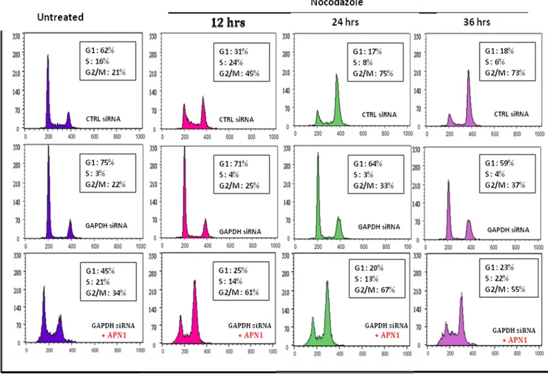
1. GAPDH knockdown in HCT116 causes G1 arrest………. 57
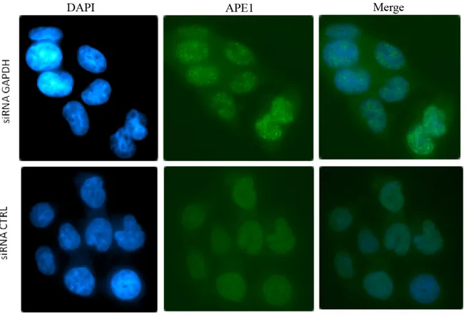
2. SiRNA mediated GAPDH silencing triggers
the formation of APE1 aggregates……… 61
3. Identification of the cysteine residue(s) in APE1
that are reduced by GAPDH………. 62
4. In vitro reduction of APE1 by GAPDH... 67
5. Cloning of APE1 variants in pEYFP-N1 vector
for expression in mammalian cells………. 71
6. Establishing Stable clones expressing APE1
cysteine variants………. 72
Chapter 4: Discussion……….. 73
1. GAPDH deficiency causes G1 arrest………. 74
2. Use of Apn1 for complementation assays………. 75
3. Apn1 expression in HCT116 cells is lethal……… 75
4. Apn1 Stable clones………. 76
5. APE1 foci-like structure could be aggregates
of oxidized species………... 78
6. Purified Cysteine mutants aggregate………. 79
7. Expression of APE1 mutants in AP endonuclease
deficient bacteria……….. 80
8. Conclusion and future work………. 81
8.1. Study the AP endonuclease activity
of APE1 variants……… 82
8.2. Expression of APE1 cysteine mutants in
GAPDH knockdown cells……… 83
8.3. Expression of APE1 cysteine mutants in
AP endonuclease deficient yeast strain……….. 83
8.4. GAPDH-induced APE1 conformational change…… 84
8.5. Study the effect of GAPDH on APE1
redox function……….. 84
9. Perspectives………... 85
Figure 1. Abasic site………... 5
Figure 2. Examples of oxidized bases……….
6
Figure 3. Examples of alkylated bases………. 7
Figure 4. Main DNA lesions induced by UV-light... 8
Figure 5. Cell cycle checkpoint pathways... 11
Figure 6. The main damage-specific DNA repair pathways in human cells… 13
Figure 7. Overview of the base excision repair pathway………. 14
Figure 8. APE1 active site interactions with the AP site……….. 31
Figure 9. AP site cleavage mechanisms by APE1 catalytic
site
residues……… 31
Figure 10. GAPDH structure………..
36
Figure 11. Verification of GAPDH knockdown in HCT116………..
57
Figure 12. GAPDH knockdown causes a G1 arrest………
60
Figure 13. APE1 nuclear aggregates in GAPDH knockdown cells…….. …..
62
Figure 14. His-APE1 cloning sites in pET-14b……….... …..
63
Figure 15. APE1 linear map……… 64
Figure 16. Positions of Cysteine residues within APE1 positioned
relative to the active site H309……….. ….
64
Figure 17. Purification of His-APE1, native and cysteine mutants………..
66
Figure 18. GST-GAPDH purification………..
67
Figure 19. In vitro reduction of His-APE1 by GST-GAPDH………….
69
Figure 20. GAPDH reduction of APE1 is a fast reaction………
70
List of tables
Table 1. Site-directed mutagenic primers……….47
Table 2. Effects of the created mutations on APE1 redox and DNA repair
3MeA 3-methyladenine
4NQO 4-Nitroquinoline 1-oxide 7MeG 7-methylguanine
8oxoG 8-oxo-7,8-dihydrodeoxyguanosine A Adenine
AAG Alkyladenine-DNA glycosylase AD Alzheimer’s disease
AID Activation-Induced cytidine Deaminase AP site Apurinic-apyrimidinic site
AP-1 activator protein-1 APE1 AP endonuclease 1
APOBEC1 Apolipopro tein B mRNA Editing Catalytic subunit1 Asp aspartic acid
AT ataxia telangiectasia
ATM Ataxia telangiectasia mutated
ATR ataxia telangiectasia and Rad3-related BER Base excision repair
C.elegans Caenorhabditis elegans
CPDs cyclobutane pyrimidine dimmers CS Cockayne syndrome
Cys Cysteine
DDR DNA damage response DHT Dihydrothymidine DNA deoxyribonucleic acid DNase I Deoxyribonuclease I dRP Deoxyribosephosphate DSB double strand breaks dsDNA double-stranded DNA DTT Dithiothreitol
E.Coli Escherichia coli
EDTA ethylenediaminetetraacetic acid Endo IV Endonuclease IV
ER endoplasmic reticulum EXO III Exonuclease III
EYFP enhanced yellow fluorescent protein G Guanine
GAPDH glyceraldehyde-3-phosphate dehydrogenase GST Glutathione S-transferase
H2O2 Hydrogen Peroxide
HD Huntington’s disease HDACs histone deacetylases
HIF-1α hypoxia inducible factor-1α His Histidine
HR Homologous recombination
IPTG Isopropyl β-D-1-thiogalactopyranoside IR ionizing radiation
kb Kilo bases kDa Kilo Dalton LB Luria Broth LP Long patch
MDM2 mouse double minute 2 MMR Mismatch repair
MMS methyl methanesulfonate
MTS mitochondrial targeting sequence MW molecular weight
NAC N-acetyl cysteine
NCA Lucanthone and 7-Nitroindole-2-carboxylic acid nCARE negative calcium regulatory element
NER Nucleotide Excision Repair NF-κB nuclear factor-κB
NHEJ Non-homologous end joining NIR Nucleotide Incision Repair NLS Nuclear localization signal OGG1 8-oxoguanine glycosylase
PAGE Polyacrylamide gel electrophoresis PARP1 poly(ADP-ribose) polymerase 1 PCNA proliferating cell nuclear antigen PCR Polymerase Chain Reaction PD Parkinson’s disease
PTEN Phosphatase and tensin homolog RNA Ribonucleic Acid
RNS reactive nitrogen species ROS reactive oxygen species rpm Revolutions Per Minute SAM S-adenosylmethionine SDS Sodium Dodecyl Sulfate siRNA small interference RNA
TRX Thioredoxin
UDG Uracil-DNA glycosylase UV UltraViolet
V(D)J Variable Diversity Joining XP Xeroderma Pigmentosum
Acknowledgment
The writing of this thesis has been one of the most challenging academic realizations I’ve ever had to do. Without the support, patience and guidance of the following people, this work would have not been possible, I am gratefulto:
Dr Dindial Ramotar, my research supervisor who gave me the opportunity to be part of his lab, who guided me throughout the one year and half that I spent working on the research project, and who made the stressful times easier by his enthusiasm and energy. Now I’ve come to believe in the equation written on his office door: work=fun!
Dr El Bachir Affar, my academic “Godfather”, mostly for his guidance and suggestions that contributed a lot to my project. He was always there when I needed advice and encouragement. I am also grateful that he accepted to be part of the jury even though he is so occupied during the period of the submission and review of my thesis.
My labmates: Siham Berra, Rim Marrakchi, Karima El Fadili, Nathalie Jouvet, Jim Daley for providing a warm and cheerful environment. BIG thanks to Nath and Karima for their help in some procedures. A special thanks to Jim, our bright post-doc, for giving me his helpful comments on this thesis, I’m sure he’s going to be a great scientist.
Our lab technicians, Rad and xiaoming. Rad for her amazing spirit, her humour, patience, generosity and her sound mother advice. Xiaoming, for his constant happy mood, for cheering me up when an experiment failed me, for praising my work even when I screwed up.
Dr Martine Raymond, Dr Pascal Chartrand, Dr El Bachir Affar, members of my thesis jury for their efforts, time and comments. A special thanks For Dr Chartrand for being my supervisor at the Biochemistry department.
My big family: cousins, uncles and aunts for supporting me in all aspects. I am so grateful to my grandma, Emily, who always thought of me as a successful intelligent person, someone that I am trying to be, for her love and prayers and her patience in raising me as a second mother.
The Canadian International development agency and the “Programme canadien de bourses de la francophonie” for the merit scholarship, their financial and moral support over the past two years especially Mme Jeanne Gallagher for her loving and caring spirit. Chadi, my labmate, my friend, and my other half, for being the man he is, for always being there for me for his patience during my thesis writing, for believing in me and making me believe in myself, for lifting my spirit when I felt so down, for his constant love and care, for encouraging me when I was about to give up, for him I owe the most... My dear parents and sisters to whom this thesis is dedicated, for standing beside me in my success and failures, for giving me more than everything to survive a tough life in Lebanon, for making me dream, for giving me wings and make me reach the
unreachable, for their endless sacrifices, for their trust, for everything I am... for just being the ideal warm family anyone dream to have...
To Mom, Dad, Tina and Perla... For the good times and the time yet to come ...
1. General Overview
140 years after its discovery, deoxyribonucleic acid (DNA), the genetic material and the blueprint of the cell, is known to be responsible for holding the information needed to constitute all the components of a living cell, hence of a whole organism. Today, we are still studying its structure, its transmission from one generation to the other, its
interaction with its environment and its regulation by different known and unknown factors in a cell’s micro- and macro-environment.
The cell faces many challanges throughout its life, leaving it with different kinds of damaging agents that cause damage to proteins (enzymes, structural proteins, etc...), to DNA and to other important components. The cellular response to damage depends on the cell type and the nature of damage. It either resolves the problem or bypasses it, leaving in the latter case a potential permanent damage [1]. As one would expect, damage to DNA is the most harmful damage that a cell might undergo due to many facts. First, DNA replication will allow this damage, or “mutation” in other terms, to be transmitted to daughter cells; second, DNA transcription will give a modified protein product that might be non-functional. In fact, damage to DNA is considered to be the early step to carcinogenesis, mutagenesis, and aging [2, 3]. Consequently, the cell has evolved DNA repair pathways to overcome these challenges, as one of its surviving tools.
2. DNA Damage
Genome integrity is continuously challenged by endogenous and exogenous agents, which might generate different unwanted insults. Identifying the sources and nature of these insults is of crucial importance for the cell to determine the damage-specific strategy in order to remove a given lesion.
2.1 Sources of DNA damage
2.1.1 Endogenous origin
While the cell struggles to protect its genome from different damaging agents, it produces, ironically, its own “homemade” DNA damaging products, mainly reactive oxygen species (ROS) and reactive nitrogen species (RNS).
ROS are oxygen-derived free radicals that have one or more unpaired electrons. Superoxide (O2−), Hydrogen peroxide (H2O2) (non radical), hydroxyl radical (OH·)
among others belong to ROS family.On the other hand, RNS are nitrogenous products of nitric oxide synthases, ranging from Nitric Oxide to Nitrates [4].ROS and RNS are produced by cellular metabolisms as by-products of the mitochondrial electron transport reactions and some metal-catalyzed reactions. They are also produced by immune system cells like neutrophils and macrophages during inflammation. Moreover, their formation can be induced by external factors like ultra-violet (UV) light, gamma radiation and X-rays. ROS can exist as pollutants in the atmosphere as well [5]. Once in the cell, these species may damage proteins, lipids and nucleic acids [6]. The harmful effects of ROS
dismutase SOD) and non-enzymatic antioxidants (e.g. vitamin E). Oxidative stress occurs when the balance between antioxidant and ROS production favours the latter. In DNA, ROS primarily induces oxidized bases and single-strand breaks (See section 2.2 for details). Paradoxically, at low concentrations, ROS and RNS can benefit the cell by increasing defence against infectious agents [6].
In addition to ROS, some naturally occurring phenomena, such as meiosis, V(D)J and class switch recombination in immunoglobulin rearrangements may also cause damage to DNA [7].
2.1.2 Exogenous Sources
Besides endogenous sources of DNA damage, the cell is subject to many exogenous factors that are either naturally occurring in a given cellular environment, like viral infection, UV-light from the sun and ionizing radiations (e.g. gamma radiation, X-rays...), or that are human-made like many mutagenic/carcinogenic substances (e.g. tobacco, DNA intercalating agents like ethidium bromide, etc...). These factors might act directly on DNA or indirectly by inducing an increase in ROS levels which, subsequently will damage the DNA. Chemotherapeutic drugs targeting the DNA are another example of man-made damaging agents [6]. They are divided into many categories: alkylating agents, antimetabolites, anthracyclines, plant alkaloids and topoisomerase inhibitors. All of these drugs lead to accumulated damage on the DNA which might interfere with cancer cell division and survival. Many secondary effects were associated with this type
of cancer treatment and the main reason is the untargeted mode of action of many of these drugs and their impact on healthy cells [8].
2.2 Types of DNA damage
DNA damage refers to any structural alteration of the DNA that will interfere with its normal conformation and composition leading to errors in transcription and replication. The types of DNA lesions vary depending on the origin of the damage and the targeted part of the DNA. DNA damaging agents can affect the nitrogenous bases as well as the sugar backbone. Base damage includes: oxidation, alkylation and deamination. Bases can also be spontaneously lost by hydrolysis. The DNA backbone can be broken on one or both strands leading to single strand breaks (SSBs) and double strand breaks (DSBs) [9].
2.2.1 Spontaneous base loss and AP sites
The N-glycosidic bonds that hold the nitrogenous bases to the sugar-phosphate backbone are weak points in the structure of DNA. Hydrolysis of this bond by a water molecule leads to the loss of the base and creating an apurinic-apyrimidinic (AP) site, also known as abasic site (figure1) [10]. The double ring structure of purines, adenine (A) and guanine (G), makes them more susceptible for hydrolysis than pyrimidines [10, 11]. AP sites are highly mutagenic lesions that interfere with DNA transcription and replication by blocking DNA and RNA polymerases. Some DNA polymerases bypass AP sites, incorporating an untemplated nucleotide into the newly synthesized strand, introducing
lesions by the base excision repair (BER) pathway, the main DNA repair pathways that repairs AP sites. (See section 4.1.1).
Figure 1. Abasic site. Adapted from [14].
2.2.2 Base modification
2.2.2.1
Base oxidation
DNA bases are subject to oxidation by different radicals, the most prominent of which is hydroxyl radical (OH·). Oxidation usually occurs on carbons 5 and 6 of pyrimidines and carbons 4 and 8 of purines [15]. Bases can be oxidized either within the DNA strand or in
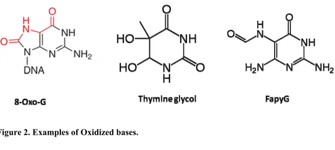
the dNTP pool before incorporation [16].Some common oxidized bases include 8-oxoguanine (8-oxo-G), thymine glycol, uracil glycol, FapyG, and 5-formyluracil
(figure 2). High levels of 8-oxo-G and thymine glycol have been reported under normal conditions as well as under oxidative stress in both nuclear and mitochondrial DNA [17]. They are highly mutagenic, if left unrepaired, they will lead to mutations that are lethal in some cases. The DNA polymerase might bypass this type of lesions, inserting the wrong nucleotide, thus creating transversion mutations as a consequence. Moreover, they can
lead to replication fork arrest after blocking the DNA polymerase, which might induce checkpoint responses and apoptosis. Modified bases can be removed by DNA
glycosylases which cleave the N-glycosylic bond between the sugar and the base, leaving behind an AP site [16]. It is noteworthy that the sugar moiety of the DNA might undergo oxidation as well. In this latter case, oxidized AP sites and SSBs with 3’ blocking groups are the possible outcomes [18] (see section 2.2.4).
Figure 2. Examples of Oxidized bases.
2.2.2.2
Base alkylation
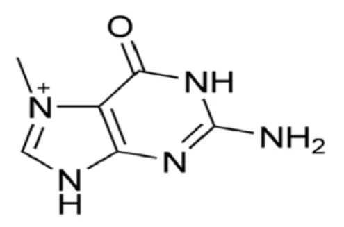
The transfer of an alkyl group to a nitrogenous base is another common base modification [19]. S-adenosylmethionine (SAM) is a well-characterized endogenous source of
methylated bases, as it has a reactive methyl group that can be transferred to a DNA base, giving rise to 7-methylguanine (7-MeG) or 3-methyladenine (3-MeA) (figure 3) [20]. The drug methyl methanesulfonate (MMS) also induces these lesions [21]. 3-MeA blocks RNA and DNA polymerases and is highly toxic intermediates, whereas 7-MeG seems harmless. Alkyladenine-DNA glycosylase (AAG) is the only human enzyme that excises
Figure 3. Examples of alkylated bases
2.2.2.3
Base deamination
Base deamination is the removal of the amine group, usually by hydrolysis, from DNA nucleobases. Hydrolytic deamination of cytosine generates uracil, which leads to GC to AT transversion mutations after DNA replication. Uracil-DNA glycosylase (UDG)
hydrolyses the glycosylic bond between the uracil and the sugar, creating an AP site. 5-Methylcytosine can undergo a spontaneous deamination, generating thymine.
Consequently, C to T transversions are the most common single point mutations. Such mutations are particularly dangerous because the thymine is not recognized as a damaged base [23].Mismatch repair pathway allows the recognition and repair of mismatched bases as well as some insertions and deletions that occur during DNA replication or recombination [24]. Another way to deaminate DNA bases is by the action of reactive nitrogen species (RNS) in addition to several proteins such as activation-induced cytidine deaminase (AID) and Apolipoprotein B mRNA editing catalytic subunit 1
2.2.3 Bulky lesions and cross links
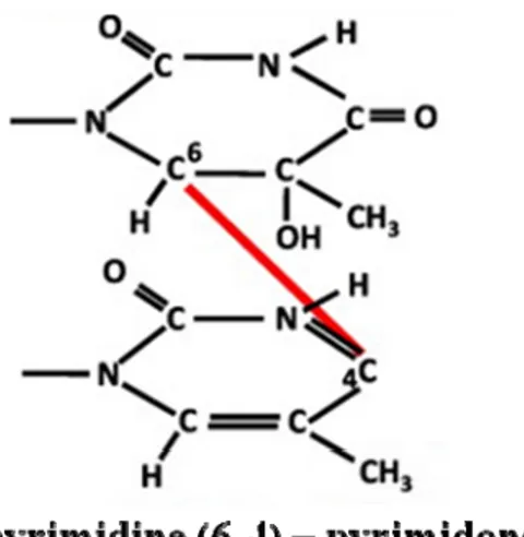
Bulky lesions and inter- or intra-strand cross links are caused by the UV-light. The main lesions created by UVB are cis-syn cyclobutane pyrimidine dimers (CPDs) and
pyrimidine (6-4) – pyrimidone photoproducts (figure 4), which connect adjacent pyrimidine molecules present in the same or different strands of the DNA to create a distortion in the DNA structure . 4-Nitroquinoline 1-oxide (4NQO) is a UV-mimetic drug that causes the above mentioned DNA lesions that are repaired by the nucleotide excision repair (NER) pathway. Deficiency in repairing such lesions has been associated with important human DNA repair syndromes such as xeroderma pigmentosum (XP) and Cockayne syndrome (CS) [27].
Figure 4. Main DNA lesions induced by UV-light.
2.2.4 Sugar-phosphate backbone breakage
2.2.4.1
Single strand breaks
spontaneous hydrolysis of the sugar-phosphate backbone, as a consequence of ionizing radiation (IR), or elevated ROS levels. SSBs can form directly by the removal of the oxidized sugar or indirectly during the base-excision repair (BER) of damaged bases. In the latter case, a DNA glycosylase cleaves off the damaged base to create an AP sites which is then cleaved by an AP endonuclease or lyase to create a SSB [28-30]. In addition to the sources mentioned above, some chemotherapeutic drugs directly induce SSBs. For example, bleomycin creates SSBs with 5’-phosphate and 3’-phosphoglycolate or 3’-phosphate termini [31].
If not repaired rapidly or appropriately, SSBs create a serious threat to chromosomal stability and hence cell survival. Consequently, cells have evolved rapid and efficient mechanisms for their repair during later stages of the BER pathway for instance [32]. (See section 4.1.1 for more details).
2.2.4.2
Double strand breaks
DSBs are the most dangerous DNA lesions because they can lead to large-scale genomic rearrangement and chromosomal fusions (translocations, deletions). A lot of naturally occurring phenomena prompt the formation of DSBs like: meiosis, mating type switching (in yeast), V(D)J and immunoglobulin class switch recombination. External factors like IR, drugs like bleomycin and chemicals also contribute to the formation of DNA DSBs. They are repaired mainly by two pathways: Homologous recombination (HR) and Non-homologous end joining (NHEJ) [7, 33-35].
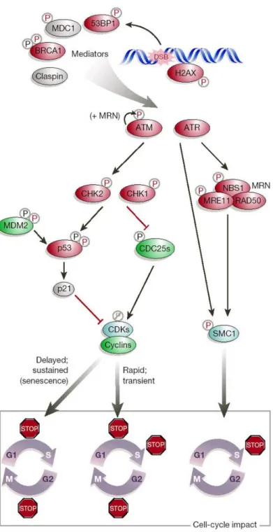
3. DNA Damage response
Cells have developed elaborate mechanisms to sense and respond to DNA damage. These mechanisms are collectively called DNA damage response (DDR). DDR is today known to be a signal transduction pathway as it appeared to be a kinase cascade activated by DNA damage and replication stress. The main kinases of this pathways are ataxia telangiectasia and Rad3-related (ATR) and Ataxia telangiectasia mutated (ATM), a defect in which was associated with ataxia telangiectasia (AT) disease[36]. Although it was thought for years that the purpose of this cascade is to regulate the cell cycle transition, in the last decade it was shown that the DDR is versatile and that cell cycle control, at checkpoints level, is only one of many outcomes controlled by this sensory network whose central goal is the repair of DNA [37].
In human cells, DNA damage activates p53 which then induces the transcription of p21, a major inhibitor of the Cyclin-dependent kinases (CdKs). The inhibition of CdKs induces cell cycle arrest [38]. Moreover, Chk1 and Chk2, the two serine/threonine kinases that act downstream of ATR/ATM, are activated by phosphorylation in response to DNA damage. The activation of Chk1 and Chk2 leads to the inhibition of Cdc25, a dual specificity phosphatase that ensures cell cycle progression by promoting phase transition [39, 40]. Chk1 is the primary effector of the intra-S and G2/M phase
checkpoints, whereas Chk2 plays a minor role on the intra-S and G1/S checkpoints [41].
Figure 5 represents a simplified scheme of the cell cycle checkpoint pathways and the
Figure 5. Cell cycle checkpoint pathways induced in response to DNA damage. In red: Tumor suppressors ; in green: proto-oncogenes. Adapted from
4. DNA repair: a crucial line of defence
4.1 DNA repair pathways
The diversity and specificity of DNA repair pathways come from the need to repair different types of lesions that arise in the DNA. DNA repair pathways are damage- specific as shown in figure 6 that summarizes different types of damages, types of lesions created and the major DNA repair pathway involved.
4.1.1 Base excision repair
This section will summarize BER in more details, focusing mainly on the pathway as it occurs in humans.
Base excision repair (BER) is a vital DNA repair pathway for living cells since it is responsible for correcting most common types of damages occurring in the genome: damaged bases (e.g. deaminated, oxidized and alkylated bases), AP sites, as well as single strand breaks with 3’-blocked termini [43, 44]. Given this role, BER pathway is considered one of the major DNA repair pathways that deals with most DNA damage [45, 46]. Moreover, this pathway is well conserved from bacteria to human in terms of the core components of its DNA repair machinery [47, 48].
The BER pathway was discovered nearly 35 years ago by Thomas Lindahl who searched for an enzymatic activity that catalyzed the removal of uracil from DNA. In E.coli, he identified uracil DNA glycosylase (UDG) [49, 50], and other groups subsequently demonstrated that this enzyme is conserved in most organisms [51, 52].
Figure 6. The main damage-specific DNA repair pathways in human cells. DNA-damaging agents
(top), examples of DNA lesions (middle), and the Corresponding DNA repair pathways (bottom). The essential genes involved in each DNA repair pathway are shown below the corresponding titles. HR, homologous recombination; NHEJ non-homologous end-joining. Adapted from [53], [54] and [55].
Subsequent work over the next two decades identified more glycosylases specific for
BER occurs in five major steps: (1) Recognition and removal of the damaged base, (2) enzymatic incision of the AP site, (3) processing of the ends of the SSB by AP site incision, (4) repair DNA synthesis, and finally (5) ligation of nick to restore the continuity of the DNA strand [58]. These will be discussed in detail below, with an emphasis on step 2. Figure 7 represents an overview of the pathway.
4.1.1.1
Damage Recognition
The first step in BER is the recognition of the modified base by a DNA glycosylase. Glycosylases cleave the N-glycosylic bond linking the damaged base to its corresponding deoxyribose sugar. DNA glycosylases are classified as either mono or bi-functional. While both classes of glycosylases hydrolyze the N-glycosylic bond, bifunctional glycosylases, such as 8-oxoguanine glycosylase (OGG1) and NEIL1/2/3, are endowed with an additional AP lyase activity. These enzymes can further process the AP site through β or β-δ-elimination to generate SSB with polyunsaturated aldehyde or 3’-phosphate termini, respectively [59-61]. Although DNA glycosylases are substrate specific, the recognition of different modified bases by the same glycosylase, or the same damaged base by multiple glycosylases still occur, thus providing redundancy [62].
4.1.1.2
AP sites and blocking groups processing
The removal of the modified base by the DNA glycosylase generates the highly
mutagenic AP site intermediate. These lesions are substrates of the catalytic activity of AP endonuclease family, AP lyases and delta lyases. While delta lyases create an incision at AP sites and process 3’-aldehyde to create 3’-phosphate termini, AP endonuclease enzymes incise the phosphodiester backbone at the 5’side of AP sites to generate 3’-hydroxyl and 5’-deoxyribosephosphate (dRP) termini [61].These unconventional termini have to be restored to 3’-OH and 5’-P through deoxyribose-phosphatase diesterase
(dRPase) activity of Pol β (5’-dRP), 3’ diesterase activity of the Apurinic/apyrimidinic endonuclease 1 APE1 (3’-PUA) and the phosphatase activity of PNKP in order to allow further repair [14, 58].
However, between the AP endonucleases and the AP lyases cleavage of AP sites, the AP endonuclease incision is the predominant pathway [61]. The importance of AP
endonucleases functions over the AP lyases has also been supported by findings
showing that endonuclease deficient yeast strains are more sensitive to MMS, but not AP lyase deficient yeast strains [63, 64]. AP endonuclease families will be discussed in further details in sections 5.
4.1.1.3
BER sub-pathways: the short and long patch
4.1.1.3.1 Short Patch BER
In SP-BER, DNA polymerase β (POL β) performs a one-nucleotide gap-filling reaction and removes the 5’dRP via its lyase activity. This is then followed by sealing of the remaining nick by the XRCC1–ligase3 complex. XRCC1 is a scaffold protein that interacts with several of the BER core components and may therefore be important in protein exchange [65].
In long-patch BER (LP-BER), APE1 catalyzes the formation of a nick 5’ to the AP site. This action recruits POL β or DNA polymerase δ (POL δ), PCNA, flap structure-specific endonuclease 1 (FEN1), and DNA ligase 1 (LIG1). In a PCNA-dependent manner POL β performs displacing synthesis and polymerizes a tract of DNA from ~2-10 bases, which produces a flapped substrate that is refractory to ligation. FEN1 cleaves the flap at the ssDNA-dsDNA junction, creating a ligatable nick, which is then sealed by DNA ligase I [66].
It was concluded that the short-patch BER (SP-BER) repair is probably the dominant subpathway of BER. [53]. However, it was shown that many factors such as cell state (cell cycle phase, differentiation stage), BER protein–protein interactions and the types of lesions are all involved in the selection between SP- and LP-BER [67].
4.1.1.4
BER and cancer
Cancer is, without any doubt, the disease of the past century. Since AP sites are considered the most occurring type of lesions arising in the DNA, they are major contributors to DNA mutations. Accumulated mutations often lead to subsequent
activation of proto-oncogenes and inactivation of tumour-suppressor genes, the hallmarks of cancer. Therefore, BER implication as a cancer-prevention pathway is essential. Indeed, numerous links have been identified between oncogenesis and acquired or inherited alterations in the genome “guardians”, highlighting the key role of DNA repair
systems in tumour prevention. In the last decade, evidence of association of BER deficiency with cancer has started to emerge. For instance, studies have demonstrated a link between defects in the adenine–DNA glycosylase MYH and adenomatous colorectal polyposis and colorectal cancer risk [68]. Reduced activity of OGG1 was associated with elevated lung cancer [69]. Moreover, BER enzymes are attractive targets for inhibitor-based cancer therapy. Many chemotherapeutic drugs act by damaging the DNA, leading to cell death. Therefore, selective inhibition of repair in cancer cells could sensitize them to the drug [70].
5. AP Endonuclease/3’ -Diesterase
AP sites are very common because, besides being formed spontaneously, they are intermediates in repair of most types of base damage. AP sites are repaired primarily by AP lyases and AP endonucleases. AP endonuclease family members cleave the sugar-phosphate bond 5′ of an AP site to produce a 3′ hydroxyl group and a 5′-deoxyribose phosphate [71, 72]. The first evidence of the presence of an enzyme that is capable of processing AP sites emerged from studies in E.coli. These studies showed that an enzyme termed exonuclease III, first described as a 3’ to 5’ exonuclease with a 3’-phosphatase activity, also possesses an AP endonuclease activity [73, 74]. To date, two families of AP endonucleases / 3’-diesterases, named Endonuclease IV (Endo IV) and Exonuclease III (Exo III) after the ancestral bacterial enzymes, have been well characterized [61]. In addition to AP endonucleases, AP lyases are also capable of processing AP sites. In fact, the E. coli endonuclease III is an AP lyase that cleaves on the 3′ side of the AP site
processed by AP endonucleases [75, 76].
5.1 Two Distinct families of AP endonucleases/3’
Diesterase
Despite a certain degree of redundancy between the Exo III and Endo IV AP
endonucleases families, some differences in expression and substrate specificity do exist. However, the key feature used to differentiate between the two families is their
magnesium dependency. While members of the Endo IV family are Mg2+ independent, Exo III family members require magnesium for their DNA repair activity [61].
Interestingly, these two proteins use completely different structural folds to accomplish
basically the same set of activities [77].
In E.coli, Exo III is encoded by the xth gene and is the major AP endonuclease accounting for around 90% of the total AP endonuclease activity in extracts. Exo III possesses four different catalytic activities. First, Exo III is an apurinic/apyrimidinic endonuclease activity. Second, it has a 3’ to 5’ exonuclease with an activity specific for dsDNA allowing it to degrade blunt ends and 5’ overhangs. Third, it can also act as a 3’ -phosphodiesterase that removes groups including, phosphates and phosphoglycolate in addition to the aldehydes left by AP lyases [78-80] to allow the DNA polymerase to prime DNA synthesis from a 3’ -OH [71]. Fourth, it has an RNase H activity to degrade RNA in DNA-RNA hybrids [57, 81, 82].
While Exo III family members are present in all kingdoms of life, Endo IV homolog exist in prokaryotes and lower eukaryotes, but not in plants, mammals and many other
vertebrates. Like Exo III family members, the Endo IV enzymes possess 3’-diesterase,
3’ -exonuclease and RNase H activities [61].
In E.coli Endo IV is encoded by the nfo gene. It accounts for only 10% of the basal AP
endonuclease activity but is induced by oxidative stress [57, 82]. In contrast, Apn1, the
S. cerevisiae Endo IV homolog, is the major AP endonuclease in yeast and shares 41%
sequence identity with the bacterial enzyme [83]. Yeast apn1Δ cells show less than 1% of AP endonuclease/3’ diesterase activities of wild-type, a 60 fold increase in ATGC transversion mutations, and hypersensitivity to alkylating agents and chemical oxidants[84, 85]. Apn2, the E.coli Exo III homolog in yeast was discovered as the second and the minor AP endonuclease in yeast after Apn1 [86]. As in E.coli, yeast Apn2 was shown to possess AP endonuclease, 3’ diesterase and 3’ to 5’ exonuclease activities that require magnesium [63]. The transcription of Apn2 gene was found to be induced up to six-fold by treatment with MMS [87] and its 3’-diesterase and 3’→5’ exonuclease activities stimulated by proliferating cell nuclear antigen (PCNA) [88].
In human cells, AP endonuclease 1 (APE1) and AP endonuclease 2 (APE2) are the human homologues of the Exo III family. APE1 is the major human AP endonuclease/3’ diesterase and since the research project concentrates on the DNA repair capacity of APE1, this enzyme will be discussed in more details in section 6 below. This enzyme has previously been known as APEX in mice [89], “HAP1” for Human AP endonuclease 1 [90], and redox factor-1 (Ref-1) for its role as a redox regulator [91], but will be referred
and mitochondrial AP endonuclease in human cells[92]. APE2 was found to possess a very weak AP endonuclease activity and an even weaker (7-fold lower) 3’ diesterase activity [93]. Moreover, APE2, unlike APE1, could not complement yeast AP endonuclease mutants [94]. While APE1 has been reported to have no functional
mitochondrial targeting sequence (MTS), it was shown that APE2 possesses a MTS and has a potential role in the repair of mitochondrial DNA lesions [92]. Moreover, it was reported that APE2 has a long C-terminal domain that contains a PCNA binding motif, similar to the one present in S. cerevisiae Apn2, suggesting a possible role in LB-BER [92]. However, the presence of APE2 did not rescue the lethal phenotypes of the knockout and the knockdown of APE1, which rule out possible significant roles for APE2 in DNA repair. The in vivo role of APE2 remains unclear; it might be a pseudogene or an evolutionary relic.
Many attempts failed to identify the Endo IV homolog in human. The highest organism where both AP endonucleases were identified and characterized is Caenorhabditis
elegans (C.elegans) [95, 96]. In addition, Exo III and Endo IV were both identified but
not yet characterized in the frog Xenopus tropicalis and the fish Danio rerio [97].
6. APE1
APE1 was first characterized and purified from HeLa cells in 1981 by the group of Stuart Linn [98]. Later, cDNAs encoding several mammalian AP endonucleases (human,
murine and bovine) were published [99-102]. The APE1 protein is closely related to other mammalian AP endonucleases (91-93% identity) and more distantly to E.coli Exo III (28% identity). Like Exo III, APE1 has a Mg2+- dependent AP endonuclease activity in
vitro [98, 103].
6.1 APE1: a DNA repair enzyme
As described earlier, APE1 is considered of major importance in protecting the cell from
the effects of cytotoxic and mutagenic unrepaired AP sites. The expression of the APE1 protein in human cells is activated by DNA damaging agents
and ROS, in order to process the resulting AP sites during the BER [104]. The second major role of APE1 in the BER is its 3’ diesterase activity. Although the diesterase activity of APE1 is about 200-fold lower than its AP endonuclease activity, this function allows it to remove a variety of 3’ blocking groups including 3’phosphate and 3’
phosphoglycolate occurring at single-strand breaks created by oxidative agents, ionizing radiation or by a chemotherapeutic agent like Bleomycin which creates 3’
phosphoglycolate [105, 106].
APE1 is one of the major enzymes orchestrating the mammalian BER. In addition to its AP endonuclease/3’ –diesterase activity, APE1 may have also an implication in the selection between the SP- and LP-BER subpathways. It was proposed that APE1 is active in both LP- and SP-BER and that it interacts with several proteins in these two
subpathways including OGG1, XRCC1, PCNA, FEN1, and DNA polymerase β. Hence, APE1, owing to its interactions with other BER proteins and to its structural mechanism
subsequent proteins after its initial cleavage of the DNA [107, 108].
In the first step of BER, after the DNA glycosylase removes the damaged base, it remains bound and presents the AP site to APE1. Due to the extensive surface of APE1-DNA interaction that covers both DNA strands and the DNA kinking, APE1 displaces the glycosylase allowing it to release the damaged base. After cleaving the AP site, the nicked DNA is exposed to DNA polymerase and/or XRCC1. This product-substrate exchange might be partially mediated by APE1 N-terminal and the DNA polymerase dRP-diesterase domain. Following repair, the DNA can no longer accommodate the bending and the component enzymes likely dissociate [109]. It appears that APE1 interactions with other BER proteins show a high level of DNA repair organization for maximal efficiency in a relatively large mammalian genome.
Although they belong to different AP endonucleases families and to two different species, it is noteworthy that the human APE1 shares many common features with the yeast Apn1. First, each one of them is the major AP endonuclease/3’ diesterase enzyme in its corresponding cellular context. Second, they are both capable of incising the phosphodiester bond at the 5’ of an AP site and of removing similar 3’ blocking groups,
e.g. 3’ phosphate, 3’phosphoglycolate that are associated with Ionizing radiation (IR) and
ROS [71, 110, 111]. However, in contrast to what might be predicted based of the biochemical repair activities of APE1 and Apn1, APE1 knockout mouse blastocysts and antisense-expressing human cells are hypersensitive to γ rays and oxidizing agents, whereas Apn1 yeast mutants are at best mildly sensitive to γ rays and oxidizing agents like (e.g. H2O2) [105]. These results suggest that there might be other species-specific
factors implicated in alternative repair mechanisms that need to be taken into
consideration when assessing the right model (yeast or mammalian cells) to study DNA damage response to certain genotoxic agents.
Besides its activity in the BER pathway, APE1 was shown to be the damage-specific endonuclease in the nucleotide incision repair (NIR) pathway, a known feature in Endo-IV family of endonucleases [112]. NIR pathway was proposed as an alternative pathway to classic BER. In this pathway, APE1 nicks oxidatively damaged DNA (e.g.
5,6-dihydro-2’-deoxyuridine, 5,6-dihydrothymidine (DHT), 5-hydroxy-2’-deoxyuridine, alpha-2’-deoxyadenosine and alpha thymidine adducts) in a DNA glycosylase-independent manner, generating 3’-OH and 5’ phosphate. This mechanistic feature is distinct from DNA glycosylase-mediated BER and has the advantage of avoiding the
genotoxic intermediates (i.e. AP sites) generated in the BER pathway [113]. A recent study showed that APE1 appears to suppress the activation of poly(ADP-ribose) polymerase 1 (PARP1) during oxidative damage repair which promotes cell survival [114].
Furthermore, some groups showed that APE1 also possesses 3’ to 5’ exonuclease activity which plays a role in the excision of deoxyribonucleoside analogs from the DNA [115, 116]. If this is the case, the inhibition of this activity could have implications in treating some cancers where nucleoside analogs such as gemcitabine are used. Conflicting data are present in the literature regarding this latter activity of APE1. Some groups showed that this activity is structure specific and presented a preference for 3’-mismatched
nucleotides [115, 117, 118], while others showed that it presented no exonuclease activity against blunt ended dsDNA [93, 119]. Interestingly, the yeast Apn1 is endowed with a
8-oxo-G from the DNA, a function that possibly APE1 might have [120].
Given the many DNA repair functions of APE1, it is clear that this enzyme is fundamental to the maintenance of the genome and survival of cells. Hence the
observation that even in the absence of exogenous DNA damage, siRNA directed against APE1 results in a decrease in proliferation, an increase in AP sites and increased levels of apoptosis [121].
6.2 APE1: a Redox enzyme
In addition to its role in DNA repair, APE1 has also been implicated in transcription regulation by reducing and thus activating many transcription factors. APE1 was first shown to reduce the conserved cysteine residues in Fos and Jun. Fos and Jun form a heterodimeric complex that regulates gene transcription by binding to the activator protein-1 (AP-1) DNA sequence motif. APE1 activation of Fos and Jun stimulates AP-1 DNA-binding activity [91].
Later reports have revealed a similar role of APE1 in activating p53, Fos, Jun, nuclear factor-κB (NF-κB), PAX (paired box-containing family of genes), hypoxia inducible factor-1α (HIF-1α), HIF-1-like factor and others [105]. The biological implication of these transcription factors activation is no fully clear yet, but what is known so far that APE1 redox function facilitates the DNA binding of some of these transcription factors or change the transcriptional activity of others [105]. Interestingly, it was demonstrated
that p53 binding to DNA and the subsequent transcriptional activation of p21, BAX or cyclin G is dependent on APE1. Moreover, APE1 protein was reported to stimulate p53 activity by both redox-dependent and -independent means [122].
APE1 has also been implicated in a number of other redox activations, one of which is the activation of bioreductive drugs requiring reduction for their activity [123].Hence APE1 acts as a redox factor that senses the redox state of the cell.
APE1 redox activity requires the N-terminal region of the protein as the truncation of the 62 N-terminal residues renders the enzyme redox deficient (Figure 15 and 16) [124, 125]. It is noteworthy that human APE1 has 61 N-terminal additional amino acids that are not present in the E.coli Exo III. It was proposed that APE1 redox activity involves a cysteine residue(s) [124] that according to LJ Walker et al. was Cys 65 that forms a disulfide bridges with Cys 93 [125]. However, several reported crystal structures of APE1 showed three different observations contradicting the latter finding. First, no disulfide bridges were found in the crystal structures. Second, Cys 65 is buried inside the protein and is not accessible by the target transcription factors reduced by APE1 [77, 126, 127]. Third, all the reported APE1 crystallographic lattices are similar, suggesting the absence of conformational changes that could reposition Cys 65 [77]. Moreover, mice in which Cys 64 (equivalent to human Cys 65) is mutated to Ala show no developmental defects [128]. Thus, the mechanism by which Cys 65 acts in APE1 reduction of transcription factor needs to be further investigated.
Given the mild phenotype seen in other AP endonuclease-deficient organisms, lethality of APE1 -/- mice and knockout cells was a big surprise.
Unlike bacteria and yeast, which tolerate the complete loss of AP endonuclease activity with little consequence unless challenged with DNA damaging agents, APE1 knockout in mice is lethal at early embryonic stages, E3.5–E9.5 [129, 130].
However, heterozygous mutant mice developed into adulthood without any apparent abnormalities [129]. Although it was not clear at the time which function(s) of APE1 is contributing to the lethality phenotype seen in mice, a recent study using a specific inhibitor of APE1 redox function demonstrated that APE1 may play a role in normal embryonic hematopoiesis and that its redox function, but not the DNA repair
endonuclease activity, is critical in normal embryonic hematopoietic development [131]. On the contrary, evidence for importance of DNA repair function was presented by another group who showed that epiblast cells have fragmented nuclei and undergo apoptosis and that explanted homozygous APE1-null blastocysts displayed increased sensitivity to γ-irradiation [130].
Further research showed that loss of APE1 is lethal at the level of the cell. Human cells in which APE1 is deleted by a microinjection of a Cre expression plasmid, undergo
apoptosis within 48 hours [132].
Since APE1 deletion is lethal, many groups manipulated APE1 expression levels by either downregulation or overexpression. RNA interference (RNAi) knockdown resulted
in decreased cell proliferation and apoptosis which was correlated with an accumulation of AP sites [121]. Moreover, lower levels of APE1 render mammalian cells
hypersensitive to MMS and H2O2 [133]. Importantly, the defects seen in knockdown cells
were rescued by the expression of the yeast Apn1. Thus, the AP endonuclease activity of APE1 was shown to be essential for cell viability.
To date, many types of inhibitors of APE1 activities have been discovered. Lucanthone and 7-Nitroindole-2-carboxylic acid (NCA) were shown to be direct inhibitors of APE1 DNA repair activity [70, 134]. Recently, a novel quinone derivative that inhibits APE1 redox activity was identified, namely E3330 ([(2E)-3-[5-(2,3dimethoxy-6-methyl-1,4-benzoquinolyl)]-2-nonyl-2-propenoic acid]) [135].
In this context, a beneficial effect of the reduction of APE1 levels is to sensitize cells to chemotherapeutic agents such as bleomycin, carmustine, gemcitabine [136-138].
On the other hand, APE1 overexpression in human cells enhances tumour resistance to DNA damaging drugs and radiotherapy, as expected, a phenotype seen in many cancer cells [139].
6.4 Regulation of APE1 Activity
APE1 is regulated at two different levels: transcriptional and post-translational.
Regulation of the activity of an essential enzyme like APE1 is of great importance for the cell to respond properly to DNA damage and to other diverse stimuli where it is involved.
APE1 transcription is known to be induced by ROS. This regulation was proposed to happen in two steps: first APE1 translocates from the cytoplasm to the nucleus. Second,
de novo protein synthesis takes place via transcriptional activation at the promoter level
[104, 140-142]. Interestingly, APE1 was shown to autoregulate its own transcription by binding to its own promoter followed by inhibition of transcription at the level of its negative calcium regulatory element (nCARE) [143].
6.4.2 Post-translational modifications
APE1 is an abundant protein (~104–105 copies/cell) within eukaryotic cells and with a relatively long half-life of about 8 hours [144]. Therefore, the needed regulation of this multifunctional enzyme must lie in the many post-translational modifications that this enzyme undergoes.
6.4.2.1
APE1 structure
The N-terminal region contains the nuclear localization signal (NLS) and also appears to regulate the DNA binding activity of many transcription factors in vitro. Particularly, the residues 43-62 are necessary for APE1 redox activity [91, 122, 124, 125].
The active site residues of APE1 have been determined by site-directed mutagenesis and by analysis of sequence conservation between APE1 and Exo III [72, 145, 146]. There is
an absolute requirement for divalent metal ions for APE1 catalytic activity, with a distinct preference for magnesium [145]. Figure 8 highlights APE1 active site, illustrating the main residues implicated in its catalytic activity along with the ones critical for binding to AP sites in DNA.
Comparison of the crystal structures of APE1 and Exo III reveals three loop regions that act in AP site recognition and cleavage. While many structural similarities between APE1, Exo III and the bovine endonuclease Deoxyribonuclease I (DNase I) have been reported, DNase I does not have an AP endonuclease activity since it has different loops and α-helices from APE1 and Exo III. These loops contribute to the positively charged grooves that form the DNA-binding faces of APE1 and Exo III thus dictating their specificity as AP endonucleases [77, 81, 109].
Although they are structurally unrelated enzymes, analysis of the crystal structures of APE1 and the bacterial Endo IV binding to an AP site shows that both enzymes orient the AP site via positively charged surfaces and insert loops into the DNA base stack, bending and kinking the DNA to promote flipping of the AP site into a sequestered enzyme pocket that contains only the damaged nucleotide [109].
His309 is considered the catalytic residue of APE1. It acts as the general base to abstract a proton from a water molecule, while Asp283 orients the imidazole ring of His309 and stabilizes its transiently- positive charged state along with Mg2+. The resulting hydroxide ion then attack the 5’ phosphate of an AP site via an inversion of configuration. The mutations of His309 to Asn and of Asp283 to Ala result in the elimination of enzymatic activity, showing the importance of these two residues for APE1 activity [77]. The
Figure 9.
Figure 8. APE1 active site interactions with the AP site. Adapted from [77].
Figure 9. AP site cleavage reaction mechanisms by APE1catalytic site residues.
6.4.2.2
Phosphorylation
It was demonstrated that APE1 is a substrate for phosphorylation by different kinases such as serine/threonine casein Kinases (CK) I and II and protein kinase C. However, phosphorylation uniquely by CK II was shown to inhibit the AP endonuclease activity of APE1 on AP sites [147]. Conversely, two years after this discovery another group showed that this phosphorylation by CK II does not have any impact on the DNA repair capacity of APE1, instead it enhance the redox function of APE1 shown by the increase of AP-1 binding to DNA [148].
6.4.2.3
Acetylation
p300 is a transcriptional co-activator that was shown to acetylate APE1 at Lys6 and Lys7
in vitro and in vivo in a Ca2+-dependent manner. This modification enhances APE1 binding to nCARE which was shown to downregulate the parathyroid hormone (PTH) expression [149]. This acetylation is reversed by class I histone deacetylases (HDACs). Moreover, it was demonstrated that APE1 acetylation is needed for the activation of the Phosphatase and tensin homolog (PTEN) gene [150].
A recent discovery identified APE1 as a novel target of ubiquitination. This modification occurs on Lys residues of the N-terminus of the protein and involves the E3 ubiquitin ligase mouse double minute 2 (MDM2), in a p53-dependent manner. The ubiquitinated APE1 was shown to be predominantly present in the cytoplasm compared to nuclear localization of the unmodified protein. Although it was proposed that cytoplasmic localization of the ubiquitinated APE1 might be an indication of either its degradation or an alteration of its function, the effect of ubiquitination on APE1 is still not clear [151].
6.4.2.5
Nitrosylation
Oxidative stress was shown to have an effect on APE1 activity. Interestingly, nitrosative stress was also shown to modify this enzyme. The nitrosation of APE1 on Cys93 and Cys310 induces the translocation of APE1 to the cytoplasm, a phenomenon reversed by treatment with reducing agents [152]. The biological implication of this modification needs more investigation.
6.4.2.6
Redox regulation
In addition to the post-translational modifications listed above, APE1 diverse activities were found to be regulated by the reduction and oxidation. Thioredoxin (TRX) is a pleiotropic cellular factor that has thiol-mediated redox activity implicated in many cellular processes, including the reduction and activation of APE1 redox function [153].
The reduction of oxidized APE1 by TRX leads to intensified APE1-mediated p53 activation and enhanced AP-1 DNA binding [154]. Although the target residues of the reduction on APE1 are not known, they may include Cys65 and Cys93 since these sites are thought to be redox sensitive [125]. Moreover, it was shown that the redox state of APE1 affects its AP endonuclease activity in vitro and that this modification involves a specific cysteine residue of this enzyme [155].
Recently in our laboratory, we showed that APE1 interacts directly with the glycolytic enzyme glyceraldehyde-3-phosphate dehydrogenase (GAPDH), which converts the oxidized form of APE1 to the reduced active form, thus maintaining its AP endonuclease activity to repair AP sites [156]. Investigating the implications and aspects of this novel finding will be the object of my research project.
7. GAPDH: a multifunctional protein
GAPDH is a tetrameric enzyme (Figure 10) thatuses the oxidized form of nicotinamide adenine dinucleotide (NAD+) to converts glyceraldehyde-3-phosphate to
1,3-bisphosphoglycerate, releasing NADH [157]. GAPDH is regarded as a housekeeping gene whose transcript level remains constant under most experimental conditions, and it has been frequently used as an internal control in studying the regulation of gene
expression. Mounting evidence, however, has started to support the multi-functionality of this enzyme, identifying roles in a variety of cellular processes, independent from its glycolytic role.
phosphorylate other proteins [158-162]. Interestingly, GAPDH was shown to have autophosphorylation activity as well [163]. Other new functions includes regulation of the cytoskeleton [164], membrane fusion and transport [165, 166], transport of nuclear RNA [167], glutamate accumulation into presynaptic vesicles [168], activation of transcription in neuronal cells [169], and protection of telomeresagainst chemotherapy-induced degradation [170]. Moreover, GAPDH exhibited a DNA repair activity by acting as a Uracil DNA glycocylase (UDG) which removes mis-incorporated uracil from the DNA [171, 172].
Notably, GAPDH was demonstrated to play a crucial role in neuronal apoptotic cell death that has pathophysiological implications in many neurodegenerative diseases such as Alzheimer’s disease (AD) [173], Huntington’s disease (HD) [174] and Parkinson’s disease (PD) [175].
Under oxidative stress, GAPDH undergoes oxidation and manifests a reduction in its enzymatic activity [176]. This oxidation also enhances the binding of GAPDH to RNA and DNA [177]. Treatment with H2O2 induces the translocation of GAPDH from the
cytoplasm to the nucleus in mammalian cells [178]. Moreover, a recent study showed that nitric oxide (NO) induces GAPDH nuclear localization followed by its acetylation by p300/CBP that will result in the transcriptional activation of many genes such as p53 [179]. Considering all of these alterations of function upon oxidative conditions, GAPDH seems to be playing an important role in cellular response to oxidative stress.
Figure 10. Overall structure of GAPDH homotetramer. Adapted from[180].
8. Research project
8.1 Background
As described earlier, APE1, a member of the Exo III family, was found to be the major AP endonuclease present in mammalian cells, unlike the Endo IV family members that are only present in lower organisms. Many previous attempts to identify an Endo IV enzymatic activity in mammalian cells that acts independently of Mg2+ failed to detect it. It was recently documented in our laboratory [156] a trial to identify such activityusing total cell extracts derived from human lung fibroblast that resulted in the detection of a weak Mg2+- independent that was reflected by the cleavage of a 42-mer [γ-32P] labeled
steps followed by mass spectrometry analysis of the protein fraction having this activity revealed the presence of GAPDH protein. Purified recombinant GAPDH lacks an AP endonuclease activity, the only explanation to the observed activity is that GAPDH was co-purified with another protein possessing this activity, namely APE1. Since APE1 has an Mg2+-dependent activity, a possible explanation is the EDTA concentration in the buffer used was not enough to chelate all the Mg2+ from the extract in the reaction, leaving some APE1 enzymes active, contributing to the detected AP endonuclease activity.
Further experiments showed that GAPDH interacts with APE1 in vivo and in vitro. This interaction causes the conversion of the oxidized and AP endonuclease deficient APE1 to a reduced and active protein capable of cleaving AP sites. This reducing potential of GAPDH was shown to be dependent on Cys 152 residue of its active site. Other reports showed that APE1 exists as heterogeneous molecules that are either reduced or oxidized [153]. Moreover, it was reported that APE1 can be oxidized by H2O2 to the inactive form,
a phenomenon partially reversed by the treatment with dithiothreitol (DTT) [155].
Interestingly, siRNA knockdown of GAPDH in HCT116 cells caused sensitivity to MMS and bleomycin, two drugs known to create DNA lesions that are repairable by APE1, but not to UV irradiations which produces bulky lesions. A similar phenotype was observed when using siRNA directed against APE1 [138]. Furthermore, total extracts of these cells showed a reduced AP endonuclease activity. They also exhibited an increase in
8.2 Hypothesis
Since GAPDH interaction with APE1 activates the AP endonuclease activity of this enzyme in vitro, and since the deficiency in GAPDH sensitized cells to DNA damaging drugs, induced an increase in AP sites formation and reduced the AP endonuclease capacity, we hypothesize that deficiency in GAPDH affects cell cycle progression and APE1 cellular distribution.
Moreover, since GAPDH activates APE1 by reduction, we postulate that this reduction occurs at the level of disulfide bridge(s) formed between APE1 cysteine residues.
8.3 Objectives
Objective 1: To determine whether activation of APE1 by
GAPDH is required for efficient cell cycle progression
It is critical for dividing cells to ensure their DNA integrity in order to avoid any inherited mutations in daughter cells. DNA lesions activate checkpoint pathways that regulate specific DNA repair mechanisms during different phases of the cell cycle. Checkpoint-arrested cells resume their cell cycle progression once the damage has been repaired, whereas cells with unrepaired DNA lesions undergo permanent cell cycle arrest or apoptosis.
Since GAPDH Knockdown cells showed a defect in proliferation and since this

![Figure 7. Overview of the BER pathway and its subpathways. Adapted from [47]](https://thumb-eu.123doks.com/thumbv2/123doknet/2166080.9890/31.918.45.872.364.954/figure-overview-ber-pathway-subpathways-adapted.webp)
![Figure 8. APE1 active site interactions with the AP site. Adapted from [77].](https://thumb-eu.123doks.com/thumbv2/123doknet/2166080.9890/48.918.150.851.662.997/figure-ape-active-site-interactions-ap-site-adapted.webp)
![Figure 10. Overall structure of GAPDH homotetramer. Adapted from[180].](https://thumb-eu.123doks.com/thumbv2/123doknet/2166080.9890/53.918.152.673.106.571/figure-overall-structure-gapdh-homotetramer-adapted.webp)