Université de Montréal
Immunopathogenèse de la cryptococcose chez la souris transgénique exprimant le génome du VIH-1
par
Vincent Cousineau-Côté
Département de microbiologie et immunologie Faculté de médecine
Mémoire présenté à la Faculté des études supérieures en vue de l’obtention du grade de
Maître ès sciences (M.Sc.) en microbiologie et immunologie
Mai 2013
Université de Montréal Faculté des études supérieures
Ce mémoire intitulé :
Immunopathogenèse de la cryptococcose chez la souris transgénique exprimant le génome du VIH-1
Présenté par : Vincent Cousineau-Côté
A été évalué par un jury composé des personnes suivantes :
Dr. Carolina Alfieri
Président-rapporteurDr. Louis de Repentigny
Directeur de rechercheDr. France Daigle
Membre du juryRESUMÉ
La cryptococcose chez les patients atteints du VIH-1 est principalement causée par Cryptococcus neoformans var. grubii tandis que Cryptococcus gattii infecte surtout les personnes immunocompétentes. Afin d’élucider les mécanismes causant la susceptibilité différentielle à l’égard de ces deux espèces de Cryptococcus dans le contexte de l’infection au VIH-1, nous avons utilisé un modèle novateur de la cryptococcose chez la souris transgénique CD4C/HIVMutA, qui exprime les gènes nef, env et rev du VIH-1. L’expression du transgène VIH-1 a augmenté le recrutement pulmonaire des macrophages alvéolaires mais a diminué celui des lymphocytes T CD4+ et CD8+ en réponse à l’infection par le C. neoformans ou le C. gattii. La production pulmonaire des chimiokines MCP-1 (CCL2) et RANTES (CCL5) était également réduite chez les souris transgéniques infectées par l’une ou l’autre de ces espèces de Cryptococcus. La production pulmonaire de MIP-1α, MIP-1β, TNF-α, TGF-β, IL-2, IL-4 et IL-13 était augmentée chez la souris infectée au C. neoformans comparativement à C. gattii. In vitro, les macrophages alvéolaires prélevés chez la souris Tg et stimulés par des agonistes ont produit davantage de MIP-1β, alors que les chimiokines MCP-1 et RANTES n’ont pas été détectées. MOTS-CLÉS Cryptococcose Cryptococcus neoformans Cryptococcus gattii VIH-1 Souris transgénique Cytokines
SUMMARY
The most common cause of cryptococcosis in HIV-1-infected patients is Cryptococcus neoformans var. grubii, while Cryptococcus gattii usually infects immunocompetent people. We used a novel inhalation model of cryptococcosis in CD4C/HIVMutA transgenic mice expressing nef, env, and rev of HIV-1 to examine the mechanisms that cause differential susceptibility to these species of Cryptococcus in the context of HIV-1 infection. HIV-1 transgene expression increased alveolar macrophage but decreased pulmonary CD4+ and CD8+ T lymphocyte recruitment. Pulmonary production of the CC chemokines MCP-1 (CCL2) and RANTES (CCL5) was reduced in transgenic mice infected with C. neoformans or C. gattii, and concentrations were lower after infection with C. gattii compared to C. neoformans. Production of MIP-1α, MIP-1β, TNF-α, TGF-β, IL-2, IL-4 and IL-13 was increased in mice infected with C. neoformans compared to C. gattii. Production of MIP-1β by alveolar macrophages harvested from Tg mice was enhanced after agonist exposure in vitro, but production of the chemokines MCP-1 and RANTES was undetectable.
KEYWORDS Cryptococcosis Cryptococcus neoformans Cryptococcus gattii HIV-1 Transgenic mice Cytokines
TABLE OF CONTENTS
Table list ... v
Figure list ... vi
Abbreviation list ... vii
Acknowledgements ... xi
Chapter 1: Cryptococcus and cryptococcosis ... 1
1.1 Taxonomy and reproduction ... 1
1.2 Ecology and epidemiology ... 4
1.3 Pathogenesis ... 9
1.4 Virulence factors ... 13
1.5 Clinical manifestations ... 19
1.6 Treatment ... 21
Chapter 2: Host immune response to Cryptococcus ... 24
2.1 Immune cell response to Cryptococcus ... 24
2.1.1 Alveolar macrophages ... 24 2.1.2 Dendritic cells ... 26 2.1.3 Neutrophils ... 27 2.1.4 CD4+ T lymphocytes ... 28 2.1.5 Th1, Th2, Th17 response ... 29 2.1.6 CD8+ T lymphocytes ... 31
2.1.7 Natural Killer cells ... 31
ii
2.2 Protective and non-protective cytokine response to Cryptococcus ... 33
2.2.1 IL-1β ... 33 2.2.2 IL-2 ... 33 2.2.3 IL-4 ... 34 2.2.4 IL-6 ... 34 2.2.5 IL-10 ... 34 2.2.6 IL-12 ... 35 2.2.7 IL-13 ... 35 2.2.8 IL-17A ... 35 2.2.9 IL-21 ... 36 2.2.10 TNF-α ... 36 2.2.11 TGF-β... 37 2.2.12 IFN-γ ... 37 2.2.13 MIP-1α ... 38 2.2.14 MIP-1β ... 38 2.2.15 MCP-1 ... 39 2.2.16 RANTES ... 39
2.3 Toll-like receptors 2 and 4 ... 40
Chapter 3: HIV and HIV infection... 41
3.1 Human immunodeficiency virus type 1 (HIV-1) ... 41
3.2 HIV-1 replication ... 43
3.3 Natural course of HIV infection ... 44
iii
3.5 Immunological effects of HIV-1 ... 48
3.6 Altered innate and adaptive immune response to Cryptococcus ... 49
Chapter 4: Models of cryptococcosis and HIV-1 infection ... 52
4.1 Invertebrate models of crytococcosis ... 52
4.2 Mammalian models of crptococcosis ... 55
4.3 Murine models of HIV infection and AIDS ... 58
4.4 CD4C/HIVMut mice ... 60
Hypothesis ... 63
Chapter 5: Materials and methods ... 64
5.1 CD4C/HIVMutA transgenic mice... 64
5.2 Preparation of C. neoformans and C. gattii inocula ... 65
5.3 Intranasal inoculation of mice ... 65
5.4 Flow cytometry analysis of lung immune cell populations ... 66
5.5 Production of cytokines in the lungs 7 and 14 days postinoculation ... 69
5.6 Cytokine analysis of alveolar macrophage supernatants ... 70
5.7 Statistical analysis ... 71
Chapter 6: Results ... 72
6.1 Quantification of pulmonary immune cell populations 14 days postinfection in Tg and non-Tg mice ... 72
6.2 Altered immune response to Cryptococcus in Tg mice and non-Tg mice ... 75
6.3 Cytokine production by alveolar macrophages of Tg and non-Tg mice 24 and 48 h after agonist exposure ... 76
iv Chapter 7: Discussion ... 79 Perspectives ... 87 Bibliography ... 89 Annex 1 ... xii Annex 2 ... xiii
v
TABLE LIST
Table I: Antibodies used to analyze immune cell populations by flow cytometery ... 67 Table II: Combinations of antibodies used in flow cytometery analysis of lung immune cell populations ... 68 Table III: Cytometric bead array used to analyze pulmonary cytokines by
vi
FIGURE LIST
Figure 1: Mating and monokaryotic fruiting of Cryptococcus ... 3
Figure 2: Distribution of reported human and veterinary cases of cryptococcosis in British Columbia between 1999 and 2011 ... 7
Figure 3: Cryptococcal mechanisms of crossing the blood-brain barrier ... 13
Figure 4: Human Immunodeficiency Virus (HIV) structure ... 43
Figure 5: Progression of HIV-1 infection ... 46
Figure 6: Genetic construction of CD4C/HIVMut transgenic mice ... 62
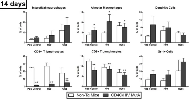
Figure 7: Percentages of lung immune cell populations, quantified by flow cytometry, in CD4C/HIVMutA Tg and non-Tg mice 14 days after inoculation with PBS control, C. neoformans H99, or C. gattii R265 ... 72
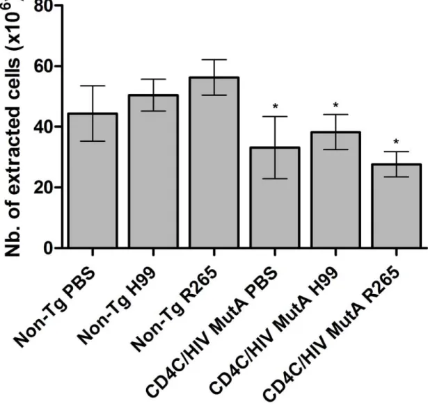
Figure 8: Total number of extracted pulmonary cells 14 days after inoculation of CD4C/HIVMutA Tg and non-Tg mice with PBS control, C. neoformans H99, or C. gattii R265 ... 74
Figure 9: Cytokine production in the lungs of CD4C/HIVMutA Tg mice and non-Tg mice 7 and 14 days after inoculation with PBS control, C. neoformans H99, or C. gattii R265 ... 75
Figure 10: Cytokine production by alveolar macrophages of CD4C/HIVMutA Tg and non-Tg mice 24 or 48 hours after exposure to KRPG control, heat-killed C. neoformans H99 and C. gattii R265, live H99 and R265, lipoteichoic acid, or lipopolysaccharide ... 77
vii
ABBREVIATION LIST
AIDS: acquired immunodeficiency syndrome APC: antigen-presenting cell
APP1: anti-phagocytic protein 1 AZT: azidothymidine
BBB: blood-brain barrier
BCCDC: British Columbia Centre for Disease Control BMEC: brain microvascular endothelial cells
CAP: cyclase-associated protein CCR2: chemokine (C-C motif) receptor CD: cluster of differentiation
CDEA: Comité de déontologie de l’expérimentation sur les animaux CFU: colony forming unit
CNA1: calcineurin A 1 CNS: central nervous system CR: complement receptor CSF: cerebrospinal fluid CTL: cytotoxic T lymphocyte DC: dendritic cell
DTH: delayed-type hypersensitivity
FACS: fluorescence-activated cell sorting FIV: feline immunodeficiency virus
viii GalXM: galactoxylomannan
GXM: glucuronoxylomanan
HAART: highly active anti-retroviral therapy HIV: human immunodeficiency virus
HOCl: hypochlorous acid
ICAM: inter-cellular adhesion molecule IFN: interferon
Ig: immunoglobulin IL: interleukin IV: intravenous
JAK/STAT: janus kinase/ signal transducer and activator of transcription KRPG: Krebs ringer phosphate glucose
LAC1: laccase1
L or D DOPA: L- or D- 3, 4-dihydroxyphenylalanine LPS: lipopolysaccharide
LTA: lipoteichoic acid
MAIDS: murine acquired immunodeficiency syndrome MALT: mucosal-associated lymphoid tissue
MCP: monocyte chemotactant protein MHC: major histocompatibility complex MIP: macrophage inflammatory protein MPO: myeloperoxidase
ix MyD88: myeloid differentiation factor 88 NK: natural killer
NO: nitric oxide
NOS: nitric oxide synthase OH: hydroxyl radicals
PBL: peripheral blood lymphocytes
PBMC: human peripheral blood mononuclear cells pDC: plasmacytoid dendritic cells
PKA: protein kinase A PKC: protein kinase C PLB: phospholipase B
PMN: polymorphonuclear neutrophil
Rac1: ras-related C3 botulinum toxin substrate 1
RANTES: regulated upon activation normal T-cell expressed and secreted Ras: rat sarcoma
ROM2: RhO1 multicopy 2 ROS: reactive oxygen species
SCID: severe combined immunodeficiency SIV: Simian immunodeficiency virus SOD: superoxide dismutase
TCR: T-cell receptor Tg: transgenic
x TLR: toll like receptor
TNF: tumor necrosis factor
TNFR: tumor necrosis factor receptor UV: ultraviolet
xi
ACKNOWLEDGEMENTS
Thank you Dr. Louis de Repentigny, for welcoming me to your laboratory, and for your guidance, patience, and availability throughout my research.
Thank you Mathieu Goupil, for your friendship and for helping me through every step of my graduate career.
Thank you Kassandre Leongson, for always being available to consult with, even though you graduated.
Thank you Francine Aumont, for helping me adjust to life in a research laboratory.
Thank you Serge Sénéchal, for your just consult on anything concerning flow cytometry.
Finally, thank you to my family and friends for your support, from as close as Montréal to as far away as Arizona.
CHAPTER 1- CRYPTOCOCCUS AND CRYPTOCOCCOSIS
Taxonomy and Reproduction
In 1894, an Italian scientist named Francesco Sanfelice isolated a yeast from fermenting peach juice which he called Saccharomyces neoformans (Bovers et al. 2008; Dixit et al. 2009; Barnett 2010). In the same year, a German professor, named Otto Busse, observed a pathogen isolated from a woman’s tibia and concluded that it resembled organisms from the genus Saccharomyces (Barnett 2010). In 1901, Jean-Paul Vuillemin examined these cultures and placed them in the genus Cryptococcus because they were unable to produce ascospores, a characteristic of the Saccharomyces genus. Both of these cultures later became known as C. neoformans (Bovers et al. 2008; Barnett 2010). C. gattii was first isolated from a leukemia patient in 1970 (Bovers et al. 2008; Dixit et al. 2009). The genus Cryptococcus consists of basidiomycetous yeasts that are part of the order Tremellales (Loftus et al. 2005; Byrnes et al. 2009). There are currently 37 recognized species in the genus Cryptococcus, but only C. neoformans, C. gattii, C. laurentii, and C. albidus are pathogenic in humans and animals (Li and Mody 2010). Both C. neoformans and C. gattii are major human pathogens, while C. laurentii and C. albidus rarely cause disease in immunocompromised patients. C. neoformans was originally classified into four serotypes (A-D) based on capsular agglutination reactions, but due to molecular analysis serotypes B and C have been reclassified as a separate species, C. gattii (Li and Mody 2010). C. neoformans has been divided into two varieties, C. neoformans var. grubii (serotype A) and C. neoformans var. neoformans (serotype D) (Dixit et al. 2009; Li and Mody 2010). The third serotype of C. neoformans is the hybrid serotype AD (Dixit et al. 2009; Li and Mody 2010; Li et al. 2012). Other hybrids, such as BD and AB,
2
have been observed, but are extremely rare (Dixit et al. 2009). Both species have been further divided into four major molecular types, VNI-VNIV for C. neoformans and VGI-VGIV for C. gattii (Li and Mody 2010). The two most prevalent molecular types are VNI and VGI (Dixit et al. 2009). Both in nature and patients, Cryptococcus is most commonly found as unicellular budding yeast (Lin and Heitman 2006; Kozubowski and Heitman 2012). It has also been occasionally observed as pseudohyphae, which could serve as a strategy to avoid predators in the environment (Kozubowski and Heitman 2012). Cryptococcus can also be found in hyphal form.
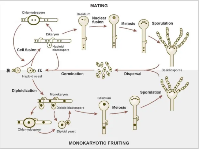
Sexual reproduction of Cryptococcus has never been observed in nature or within a host, and only specific conditions in the laboratory have been able to trigger mating between compatible yeast cells (Kozubowski and Heitman 2012) (Figure 1). The perfect states of C. neoformans and C. gattii, discovered by Dr. Kwon-Chung, are named Filobasidiella neoformans and F. bacilispora respectively (Kwon-Chung 1976; Kwon-Chung 1976). Different serotypes differ in their ability to mate; most of the serotype D strains mate, while the ability of serotype A and C. gattii to mate is strain specific. Cryptococcus has a bipolar mating system, where there is only a single mating locus called the MAT locus (Kozubowski and Heitman 2012). The MAT locus of C. neoformans, which is greater than 100kb and codes for over 20 genes, is longer than the MAT loci of other fungi (Kozubowski and Heitman 2012). The mating type of Cryptococcus is called either a or α, depending on the MAT locus (Lin and Heitman 2006; Kozubowski and Heitman 2012). There is an overwhelming predominance of mating type α in the environment (98-99.9%), which could explain why sexual reproduction in nature is rare (Lin and Heitman 2006). Sexual reproduction of Cryptococcus begins when fusion occurs between haploid yeast MATa and MATα, to create a dikaryon (Lin and Heitman 2006)
3
(Figure 1). The dikaryon undergoes a dimorphic transition and becomes dikaryotic hyphae (Lin and Heitman 2006). The hyphal tips swell to create basidia, where nuclear fusion occurs (Lin and Heitman 2006). Meiosis then follows to create four haploid meiotic daughter nuclei (Lin and Heitman 2006). This leads to the production of four chains of basidiospores, which are readily aerosolized (Lin and Heitman 2006).
Figure 1. Mating and monokaryotic fruiting of Cryptococcus (Lin and Heitman 2006)
Used with permission from Annual Review of Microbiology (License number 3172580616034)
C. neoformans can also undergo same-sex mating when exposed to the right conditions, also known as monokaryotic fruiting (Lin and Heitman 2006; Kozubowski and Heitman 2012)
4
(Figure 1). Although it was originally thought to be asexual haploid fruiting, it has later been shown to be a modified version of sexual reproduction occurring between strains of the same mating type (Lin and Heitman 2006; Kozubowski and Heitman 2012). This can occur with both a and α mating types, and similar to sexual reproduction, monokaryotic fruiting has not been observed in the environment (Kozubowski and Heitman 2012). Monokaryotic fruiting is very similar to sexual reproduction, except the hyphal cells only contain one nucleus (Kozubowski and Heitman 2012). The spores generated from monokaryotic fruiting are also smaller and rounder than those produced from sexual reproduction (Kozubowski and Heitman 2012).
Ecology and Epidemiology
In the environment, C. neoformans can be isolated worldwide from avian excreta and the soil surrounding it (Harrison 2000; Litvintseva and Mitchell 2009). Aged avian excreta and the soil surrounding it are more likely to contain C. neoformans compared to fresh avian guano (Lin and Heitman 2006; Lin 2009). C. neoformans thrives on the nitrogenous components associated with avian excreta (Mitchell and Perfect 1995). Although it is more commonly isolated from avian guano, C. neoformans has also been isolated from decaying wood and tree hollows (Harrison 2000; Lin and Heitman 2006; Litvintseva and Mitchell 2009). Cryptococcus can infect a wide variety of domestic and wild animals, though no transmission between animals and humans has been reported (Lin and Heitman 2006). However, substantial evidence has shown that birds, specifically pigeons, are directly linked to the worldwide distribution of C. neoformans (Mitchell and Perfect 1995; Lin and Heitman 2006; Lin 2009). Most evidence shows that pigeons themselves are not infected and act as carriers
5
(Lin and Heitman 2006; Lin 2009). Human infection usually occurs without direct contact with birds (Harrison 2000; Warkentien and Crum-Cianflone 2010). Prior to 1955, there had only been less than 300 reported cases of cryptococcosis (Perfect 2005). Currently, C. neoformans is responsible for an estimated one million cases a year resulting in approximately 625,000 deaths (Kronstad et al. 2011). The majority of these reported cases occur in Sub-Saharan Africa, where the number of fatal cases can surpass the number of deaths due to tuberculosis in some areas (Kronstad et al. 2011). The increase in incidence of C. neoformans infections can be attributed to the increased number of immunocompromised individuals, including HIV/AIDS and transplant patients (Perfect 2005; Li and Mody 2010). Approximately 90% of all cryptococcal infections and 99% of cryptococcosis cases in AIDS patients are attributed to C. neoformans var. grubii (serotype A) (Bovers et al. 2008; Litvintseva and Mitchell 2009). Of the patients that are HIV-uninfected, over 90% displayed some form of immunodeficiency (Li and Mody 2010). C. neoformans var. neoformans (serotype D) also infects mainly immunocompromised patients, but is less common worldwide and considered less virulent (Litvintseva and Mitchell 2009). In Europe, serotype D is more common and is responsible for 30% of reported cases (Bovers et al. 2008). Hybrid serotype AD has been isolated from patients in North America and Europe, but is uncommon (Litvintseva and Mitchell 2009).
Unlike C. neoformans, the most common environmental niche for C. gattii is the red gum group of eucalyptus trees, primarily Eucalyptus camaldulensis (Mitchell and Perfect 1995; Sorrell 2001; Lin and Heitman 2006; Dixit et al. 2009). It has also been isolated from other tree species including almond, golden shower and Douglas fir (Lin and Heitman 2006; Dixit et al. 2009). Like C. neoformans, C. gattii has been isolated from decaying trees and tree
6
hollows (Lin and Heitman 2006). C. gattii can also infect both domestic and wild animals including cats, dogs, sheep, rabbits, foxes, and koalas (Lin and Heitman 2006). There are 1.5 times more veterinary cases of C. gattii infections compared to human cases (Mak et al. 2010). Although C. gattii infections have been occasionally reported in immunocompromised patients, 70-80% of cases are associated with apparently healthy individuals (Sorrell 2001; Lin and Heitman 2006). The AIDS pandemic has not had an effect on the incidence of C. gattii infections (Morgan et al. 2006). C. gattii is endemic to tropical and subtropical regions, and a majority of reported cases C. gattii infections occur in Australia and Papua New Guinea (Dixit et al. 2009; Litvintseva and Mitchell 2009; Mak et al. 2010). Serotype B is more commonly isolated in clinical and environmental samples compared to serotype C (Springer and Chaturvedi 2010). Serotype C has been isolated from clinical samples from India, Africa, and Southern California, but is rarely isolated from the environment (Sorrell 2001; Springer and Chaturvedi 2010).
Beginning in 1999, there was a dramatic increase in the incidence of C. gattii infections, in both humans and animals, on the east coast of Vancouver Island in British Colombia, Canada (Mak et al. 2010; Kronstad et al. 2011). British Columbia Centre for Disease Control (BCCDC) recognized the increased incidence of C. gattii infections as an outbreak in 2002 (Hoang et al. 2004). The current incidence of C. gattii infections in British Columbia is 5 cases per million inhabitants, which is superior to the Australian average (0.94 cases/ million), where C. gattii is more common (Hoang et al. 2004; Dixit et al. 2009). The central eastern coast of Vancouver Island has the highest annual incidence rate for both animal and human cases of C. gattii infections (Figure 2) (Duncan et al. 2006). In the decade following the initial
7
outbreak, there have been 236 reported cases of C. gattii infections in humans, resulting in 19 deaths, as well as numerous reports of veterinary cases (Kronstad et al. 2011).
Figure 2. Distribution of reported human and veterinary cases of cryptococcosis in British Columbia between 1999 and 2011. (BC CDC)
The molecular types of C. gattii responsible for this outbreak are two sub-genotypes of VGII (Ngamskulrungroj et al. 2011). VGIIa is the predominant sub-genotype in both the environment and in patients, and shows a higher virulence than other C. gattii genotypes (Kidd, Bach, et al. 2007; Kronstad et al. 2011; Ngamskulrungroj et al. 2011). VGIIb is the minor sub-genotype and was responsible for a few cases on Vancouver Island (Kidd, Bach, et al. 2007). The original outbreak strain of C. gattii is R265 of the VGIIa molecular type
8
(Ngamskulrungroj et al. 2011). The endemic area for C. gattii in the Pacific Northwest has expanded past Vancouver Island; in 2004, cases of C. gattii infections were reported from mainland Vancouver, Oregon, and Washington (Kronstad et al. 2011). There have been approximately 60 reported cases in Washington, Oregon, Idaho, and California since 2004 (Kronstad et al. 2011). The molecular type of C. gattii most commonly isolated from this region is VGIIa, the same as the major type in British Columbia (Ngamskulrungroj et al. 2011). VGIIc, a new molecular type, has been isolated exclusively from Oregon (Byrnes and Heitman 2009; Datta et al. 2009). The emergence of C. gattii in the Pacific Northwest is thought to have partly originated from Australia because the VGIIb strain from Vancouver Island is identical to the Australian VGIIb clinical isolate NT-13 (Dixit et al. 2009). The VGIIa strain has been hypothesized to originate from South America, because VGIIa isolates from both Brazil and Vancouver Island are of mating type α (Dixit et al. 2009). Some possible methods of dispersal of C. gattii to the Pacific Northwest include human-mediated spread (C. gattii transferred by contact surfaces like shoes), passive transport by wild and domestic animals, and airborne dispersal through deforestation (Kidd, Bach, et al. 2007). C. gattii has been observed to be able to survive on shoes for over 144 days, but the active usage of the shoes reduced the levels of viable C. gattii (Kidd, Bach, et al. 2007). Samples taken from cutting down and chipping of a Red alder (Alnus rubra) and a Douglas fir (Pseudotsuga menziesii) resulted in high levels (up to 53,125 CFU/m3) of C. gattii 0-15 meters above the ground (Kidd, Bach, et al. 2007).
9 Pathogenesis
Natural Cryptococcus infections begin with the inhalation of infectious propagules (Botts and Hull 2010; Kronstad et al. 2011). Desiccated yeast cells or spores are considered to be the infectious propagule for Cryptococcus, because their small size, 1-3 μm, allows them to be deposited more easily deep in the alveoli of the lung (Botts et al. 2009; Giles et al. 2009; Kronstad et al. 2011). Spores have been more generally accepted as the infectious propagules of Cryptococcus because they are more resistant to environmental stress compared to desiccated yeast cells, making them better suited for air dispersal and survival (Giles et al. 2009; Botts and Hull 2010; Kronstad et al. 2011). Purified spores have thick cell walls, which allow them to have a greater resistance to desiccation, oxidative stress, and temperature compared to desiccated yeast (Botts et al. 2009; Kronstad et al. 2011). Cryptococcus spores have been shown to be very infectious, having a lethal dose of as few as 500 cells in a murine model (Velagapudi et al. 2009; Kronstad et al. 2011). In the lungs, Cryptococcus is phagocytized by alveolar macrophages through interactions between fungal β- (1,3)- glucan and host receptors Dectin-1 and CD11b (Giles et al. 2009). Cryptococcus is well adapted to survive and reproduce in an acidic environment, such as the microenvironment of macrophage phagolysosomes (Levitz et al. 1999). Cryptococcus is able to produce extracellular vesicles, named “virulence factor delivery bags”, that allow it to export protein components important to virulence outside of the cell wall (Rodrigues et al. 2007; Rodrigues et al. 2008; Oliveira et al. 2010; Kronstad et al. 2011). The major capsular polysaccharide, glucuronoxylomanan (GXM) is exported in these vesicles which are necessary for the formation of the capsule (Rodrigues et al. 2008). The enzymes laccase, which synthesizes melanin, phospholipase B, and urease are also transported in the “delivery bags” (Rodrigues et al. 2008; Kronstad et al.
10
2011). In mice, these “delivery bags” induce production of TNF-α, IL-10, TGF-β, and nitric oxide (NO), and alternatively activate macrophage antimicrobial activity (Oliveira et al. 2010). Cryptococcus is a facultative intracellular pathogen and has developed a variety of strategies to avoid being killed by macrophages (Garcia-Rodas and Zaragoza 2012). These mechanisms are separated into two groups, capsule-dependent and capsule-independent (Garcia-Rodas and Zaragoza 2012). The capsule itself acts as a physical barrier that successfully inhibits mannose-binding lectin, binding and conceals surfactant protein A binding sites, which can opsonize fungal cells (Seider et al. 2010; Garcia-Rodas and Zaragoza 2012). It is also able to bind to CD14 and both TLR2 and TLR4, which translocate NF-κB to the nucleus, inhibiting the secretion of TNF-α; this inhibition causes a deficient activation of macrophages (Garcia-Rodas and Zaragoza 2012). Glucuronoxylomannan (GXM) affects neutrophils by reducing expression of L-selectin (CD62L), thus impairing neutrophil migration; restraining neutrophil rolling on the endothelium; and inducing the loss of tumor necrosis factor receptor (TNFR), which inhibits neutrophil activation by TNF- α (Urban et al. 2006). C. neoformans is also able to produce enlarged “titan” cells (Garcia-Rodas and Zaragoza 2012; Okagaki and Nielsen 2012). Titan cells are 5 to 10 times larger than normal yeast cells and are characterized by an altered capsule structure, thickened cell wall, and increased ploidy (Okagaki and Nielsen 2012). Titan cells represent approximately 20% of the cryptococcal cell population during pulmonary infections (Okagaki and Nielsen 2012). These titan cells are more resistant to phagocytosis as well as oxidative stress and nitrosative antimicrobial mechanisms (Okagaki and Nielsen 2012). Cryptococcus is able to secrete antiphagocytic protein 1 (APP1) which binds to the complement receptors CR2 and CR3, thereby inhibiting phagocytosis mediated by these receptors (Stano et al. 2009; Garcia-Rodas and Zaragoza 2012). Recently, the
11
pleiotropic virulence determinant Gat201 has been shown to be important in the antiphagocytic activity of Cryptococcus (Garcia-Rodas and Zaragoza 2012). Mutants lacking Gat201 had a basal capsule and were more readily phagocytized than acapsular mutants (Garcia-Rodas and Zaragoza 2012).
In vitro and in vivo, Cryptococcus has been observed to be able to exit macrophages through phagosomal extrusion, avoiding pathogen and host cell death, as quickly as 2 hours after phagocytosis (Alvarez and Casadevall 2006). This process occurs when a mature phagosome containing Cryptococcus fuses with the plasma membrane and the yeast cells are expulsed into the extracellular space (Ma et al. 2006; Casadevall 2010). This mechanism is used by both C. neoformans and C. gattii, but they differ slightly from one another (Alvarez and Casadevall 2006). C. neoformans var. grubii has been observed being ejected as individual cells, while C. neoformans var. neoformans and C. gattii are expulsed as yeast cells accumulated in a polysaccharide matrix (Alvarez and Casadevall 2006).
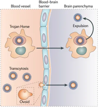
Systemic dissemination of Cryptococcus from the lungs can occur though different mechanisms. In vitro, Cryptococcus has been shown to adhere and be internalized by pulmonary epithelial cells (Filler and Sheppard 2006). Also, human type II pneumocytes, which cover approximately 5% of the surface area of alveoli, have a receptor for GXM, which allows them to internalize Cryptococcus (Filler and Sheppard 2006; Zhao et al. 2010). Another mechanism Cryptococcus can use to disseminate from the lungs is a “Trojan horse” approach (Figure 3), in which Cryptococcus uses an infected phagocyte as transportation (Casadevall 2010). Infected phagocytes can then migrate through blood vessels and cross the blood-brain barrier (BBB) carrying Cryptococcus (Casadevall 2010). The second strategy that Cryptococcus can use to cross the BBB is by direct transcytosis of free yeast cells (Casadevall
12
2010). This is achieved when cryptococcal cells stop suddenly, without rolling or tethering, in the capillaries adjacent to the meninges, most likely due to their inability to pass through the narrow capillary (Casadevall 2010; Shi et al. 2010). Following this microembolic event, cryptococcal cells are observed crossing the capillary wall in a manner that requires the deformation of cell morphology, and is urease dependent (Casadevall 2010; Shi et al. 2010). Cryptococcal hyaluronic acid interacts with CD44 of brain microvascular endothelial cells (BMEC), which activates protein kinase C α (PKCα) (Jong, Wu, Prasadarao, et al. 2008; Jong, Wu, Shackleford, et al. 2008). PKCα in turn regulates actin rearrangement in BMEC, facilitating yeast internalization (Jong, Wu, Prasadarao, et al. 2008). It has also been shown that dual specificity tyrosine phosphorylation-regulation kinase 3 is required for internalization of Cryptococcus, suggesting that Cryptococcus may use the endocytic signaling pathway to facilitate transcytosis of BMEC (Huang et al. 2011).
13
Figure 3. Cryptococcal mechanisms of crossing the blood-brain barrier (Kronstad et al. 2011). Used with permission from Nature Publishing Group (License number
3172560360717)
Virulence Factors
The ability to grow at 37°C, melanin synthesis, and the capsule are the three major virulence factors of Cryptococcus.
The only Tremellales that are capable of growing optimally at temperatures superior to 30°C are C. neoformans and C. gattii (Bovers et al. 2008). This allows Cryptococcus to grow in the environment of the human body which is at 37°C (Perfect 2005). Mutants of Cryptococcus that are unable to grow at temperatures above 30°C have been shown to be avirulent in mammalian models (Perfect 2005). Over 15 genes have been shown to be associated with high temperature growth of Cryptococcus, but these most likely represent a fraction of the genes necessary for growth at 37°C (Perfect 2005). Calcineurin A (CNA1) gene has been shown to
14
be necessary for survival at 37°C, and regulates pathogenicity (Buchanan and Murphy 1998; Alspaugh et al. 2000; Perfect 2005). Ras proteins serve as molecular switches and are implicated in the activation of many signaling pathways; Cryptococcus with a mutated RAS1 gene is not viable at 37°C, has a severe defect in mating and poorly adheres to the surface of the agar (Alspaugh et al. 2000). The guanine nucleotide exchange factor Cdc24, a RAS1 effector, functions in a RAS1 signaling cascade and is important for Cryptococcus growth at 37°C (Nichols et al. 2007). Rac1, a small G protein, interacts with Ste20, a PAK kinase, and acts downstream of Ras proteins to control both growth at elevated temperatures and cellular differentiation (Vallim et al. 2005). ROM2 is necessary for growth at 37°C because it is involved in actin and microtubule organization specifically at high temperatures (Fuchs et al. 2007).
The production of melanin, a brown or black pigment, protects Cryptococcus from oxidative host defenses, phagocytosis, ionizing radiation, heavy metals and UV light (Mitchell and Perfect 1995; Buchanan and Murphy 1998; Langfelder et al. 2003; Eisenman et al. 2009). Melanized Cryptococcus cells are also less susceptible to the antifungal drugs amphotericin B and caspofungin (van Duin et al. 2002; Walton et al. 2005). Melanin is produced in intracellular vesicles and transported to the cell wall where melanin granules are incorporated into the cell wall (Eisenman et al. 2007; Eisenman et al. 2009). Melanogenesis is accomplished when cryptococcal laccase catalyzes phenol components, such as both L- and D- 3, 4-dihydroxyphenylalanine (L- or D- DOPA) but not tyrosine, to dopaquinone (Buchanan and Murphy 1998; Eisenman et al. 2007; Frases et al. 2007). Dopaquinone is then rearranged to dopachrome and polymerized to melanin, both of which are spontaneous events (Buchanan and Murphy 1998). The brain is a tissue that is rich in phenol components, which could partly
15
explain why the brain is a target of Cryptococcus (Buchanan and Murphy 1998; Nosanchuk et al. 2000). Melanization is also dependent on other factors such as the copper transporter CC2, the copper chaperone Atx1, the chitin synthase Chs3, the transcriptional coactivator Mbf1, and the chromatin-remodeling enzyme Snf5 (Walton et al. 2005). Melanin reduces the production of TNF-α from alveolar macrophages and shields the yeast from microbicidal proteins (Jacobson 2000). Since melanin is negatively charged it effectively neutralizes neutrophil defensins as well as other cationic antimicrobial peptides (Liu and Nizet 2009). It can also reduce the amount of ferric iron, by converting Fe3+ to Fe2+, thus improving survival of Cryptococcus in vivo (Liu and Nizet 2009).
The polysaccharide capsule is the most important virulence factor of Cryptococcus. It is responsible for inhibition of phagocytosis, alterations in cytokine secretion by leukocytes, impairment of complement recognition, resistance to NO and reactive oxygen species (ROS), reduction of antibody production and leukocyte migration, down-regulation of MHC I, II, and CD83, inducing the shedding of L-selectin from neutrophils and a non-protective Th2 response to Cryptococcus (Kozel et al. 1991; Buchanan and Murphy 1998; Lupo et al. 2008; Zaragoza et al. 2008; De Jesus et al. 2009). Cryptococcal polysaccharides interact with CD18 on neutrophils, inhibiting them from adhering to endothelial cells, thus inhibiting their migration to the site of infection (Dong and Murphy 1997). The capsule can also increase the expression of CTLA-4 on CD4+ T cells, which reduces the production of IFN-γ and IL-2, and inhibits T cell proliferation (Pietrella, Perito, et al. 2001). It has the ability to mask C3b and C3bi deposits, which facilitate binding of Cryptococcus to CR3 on leukocytes, and block the Fc fragment on antibodies, which binds to the Fc receptor on phagocytes (Buchanan and Murphy 1998).
16
The capsule is composed of approximately 90% glucuronoxylomannan (GXM), 7% galactoxylomannan (GalXM), and the remaining 3% is composed of mannoprotein (Bose et al. 2003). CAP genes (CAP59, CAP64, CAP60, CAP10) are all individually necessary for capsule biosynthesis (Janbon 2004; Zaragoza et al. 2009). GXM is responsible for many different pathological properties that are attributed to the capsule (Perfect 2005). GXM can interfere with E-selectin binding, as well as down-regulate C5aR on neutrophils, which inhibit migration (Ellerbroek et al. 2004; Monari, Kozel, et al. 2006). GXM is handled differently by PMNs and macrophages; in macrophages there is continuous intracellular accumulation of GXM while in PMNs it is expulsed from the cell (Zaragoza et al. 2009). This differential handling of GXM is reflected on cytokine production, where in macrophages GXM induces IL-10, IL-8, TNF-β, and IL-6; while TNF-α, IL-1β, IL-6 and IL-8 production is increased in neutrophils (Zaragoza et al. 2009). The production of TNF-β inhibits T cell proliferation and down-regulates MHC-II and B7 expression (Monari, Bistoni, et al. 2006). GXM can also induce macrophage apoptosis by dissociating the tetramers of 6-phosphofructo-1-kinase (PFK), therefore inhibiting the glycolytic pathway (Grechi et al. 2011). GXM also interacts with CD18 and FcγII, which down-regulates caspase-3 activity, promoting NO-dependent apoptosis of macrophages (Chiapello et al. 2008). GalXM is located in discrete pockets on the outer edge of the capsule, and is a transient component of the capsule (De Jesus et al. 2009). It strongly induces the production of TNF-α and NO, as well as the production of IL-6, IL-10, and IFN-γ (Pericolini et al. 2006; Villena et al. 2008; Zaragoza et al. 2009). GalXM is also able to induce Fas/ FasLigand expression, which leads to macrophage apoptosis (Villena et al. 2008). Increased Fas/FasL expression also induces apoptosis of T lymphocytes by activating caspase-8 (Pericolini et al. 2006). Mannoprotein induces an increase in TNF-α production,
17
and regulates the expression of other cytokines in monocytes such as IL-12, IL-6, IL-8, IFN-γ, and IL-10 (Zaragoza et al. 2009). Mannoprotein 4 reduces the expression of L-selectin and TNF receptor on neutrophils (Coenjaerts et al. 2001).
The ability of Cryptococcus to secrete extracellular enzymes, laccase, phospholipase, and urease, is an important contribution to its virulence (Kronstad et al. 2011). Only C. neoformans and C. gattii produce laccase (Chan and Tay 2010). Laccase is important to virulence because it facilitates the production of melanin (Buchanan and Murphy 1998; Frases et al. 2007). The two conditions that induce LAC1 are a low concentration of glucose or a high concentration of copper (Zhu and Williamson 2004). IPC1, GPA1, MET3, and STE12 are all involved in the regulation of laccase (Noverr et al. 2004). Laccase is able to oxidize phagosomal iron in macrophages, limiting the formation of hydroxyl radicals (Liu et al. 1999). Cryptococcus also secrets phospholipases, including lysophospholipase, lysophospholipase transacylase, and phospholipase B (PLB), which are most active between 25°C and 40°C (Santangelo et al. 1999). The activation of SEC14 is required for the secretion of PLB (Chayakulkeeree et al. 2011). PLB can hydrolyze the phospholipids PG and DPPC, the most common components of lung surfactant, which facilitates adherence of Cryptococcus to lung epithelial cells and dissemination (Santangelo et al. 1999; Djordjevic 2010). In macrophages, the disruption of the phagolysosome membrane by PLB1 is required for non-lytic extrusion of Cryptococcus (Djordjevic 2010; Chayakulkeeree et al. 2011). The enzyme urease is a key virulence factor, because it has been shown to increase the accumulation of immature dendritic cells and induce a non-protective Th2 response (Osterholzer, Surana, et al. 2009; Li and Mody 2010). Urease also promotes microvascular sequestration, enhancing the ability of Cryptococcus to invade the central nervous system (CNS) (Olszewski et al. 2004).
18
Cryptococcus also has other elements that play a minor role in virulence. Cryptococcus encounters a hostile nutrient environment in the phagosomes of macrophages, and in order to survive Cryptococcus undergoes autophagy (Hu, Hacham, et al. 2008). Autophagy is the recycling of the cells` cytoplasm and defective organelles in order to survive in high stress conditions (Hu, Hacham, et al. 2008; Palmer et al. 2008). PI3K signaling is required in Cryptococcus to survive during nutrient-deprived conditions; it also plays a role in vesicular transportation of vacuolar hydrolases (Hu, Hacham, et al. 2008; Palmer et al. 2008). The interconversion of CO2 and HCO3 is catalyzed by carbon anhydrases, which allow
Cryptococcus to regulate CO2 levels (Elleuche and Poggeler 2010). Adenylyl cyclase helps
regulate CO2 concentrations by acting as a CO2 chemosensor (Klengel et al. 2005).
Glucosylceramide has been shown to be associated with the cell wall, and allows Cryptococcus to survive in alkaline conditions (Rhome et al. 2007). Gcn5, a histone acetyltranferase, facilitates survival at high temperatures, decreases sensitivity to oxidative stress, and is important in capsule attachment to the cell surface (O'Meara et al. 2010). The sexual mating type of Cryptococcus plays a role in virulence, and the majority of clinical samples are MATα (Nielsen et al. 2005; Li and Mody 2010). The STE12α gene exists only in MATα cells, and is involved in capsule and melanin production (Chang et al. 2000). Ctr2 regulates copper homeostasis and is important in the production of the polysaccharide capsule and inhibition of phagocytosis (Chun and Madhani 2010). The PKC1 protein and its downstream components are essential for cryptococcal defense against nitrosative and oxidative stresses, as well as playing a role in temperature sensitivity, capsule production, and the synthesis of melanin (Gerik et al. 2008). C. neoformans produces D-mannitol, which protects the yeast cells from free radicals (Niehaus and Flynn 1994; Guimaraes et al. 2010).
19
Superoxide dismutase (SOD) is an enzyme that has the ability to neutralize toxic levels of superoxide radicals by converting them into hydrogen peroxide and oxygen (Cox et al. 2003). Copper and zinc SODs are vital for the survival of C. gattii in neutrophils, and in the expression of laccase, urease, and phospholipase (Narasipura et al. 2003). The trehalose synthesis pathway, controlled by synthesis genes TPS1 and TPS2, regulates protein secretion, mating, and cell wall integrity in C. gattii (Ngamskulrungroj et al. 2009).
Clinical Manifestations
The majority of humans have already been exposed to Cryptococcus before the age of 5 years (Bovers et al. 2008). Humans frequently come in contact with Cryptococcus, but immunocompetent individuals are able to either clear it or it remains latent (Bovers et al. 2008). The incubation period for C. neoformans infection is on average 110 months, while the incubation of C. gattii from Vancouver Island was determined to have a shorter incubation period of between 2 and 11 months, with the average being 6 or 7 months (MacDougall and Fyfe 2006; Kidd, Chow, et al. 2007). A majority of cryptococcosis patients are immunodeficient, including patients with AIDS, organ transplant recipients (primarily kidney and liver), patients receiving immunosuppressive medications, and patients with diabetes, or an autoimmune disease (Shirley and Baddley 2009; Li and Mody 2010; Pfaller and Diekema 2010; Warkentien and Crum-Cianflone 2010). Interestingly, it is extremely rare for a patient with either cancer or bone marrow transplant to be infected with Cryptococcus (Pukkila-Worley and Mylonakis 2008). The age of the patient also seems to be a factor in Cryptococcus infections. Children are rarely affected by Cryptococcus, and the incidence of cryptococcosis in children with AIDS is extremely low at around 1% (Subramanian and
20
Mathai 2005; Severo et al. 2009; Pfaller and Diekema 2010). The mean age of Cryptococcus-infected HIV-negative individuals is ≥45 years (Pfaller and Diekema 2010). The sex of the individual also seems to be an important risk factor as males are 3 times more likely to be infected by Cryptococcus than females (Chen et al. 2000; Subramanian and Mathai 2005; Li and Mody 2010). A retrospective study done by Subramanian et al. showed that, out of the 105 cryptococcosis cases in their center, 90% of the patients were male (Subramanian and Mathai 2005).
Cryptococcosis is caused by C. neoformans and C. gattii and most commonly affects the lungs and the brain (Sorrell 2001). It is considered to be the most common cause of fungal meningitis (Sorrell 2001). The site, degree of severity of cryptococcal infection, and the health status of the patient can affect the clinical manifestations of cryptococcosis, ranging from being asymptomatic or a cough to meningoencephalopathy or even death (Li and Mody 2010). There are numerous signs and symptoms of cryptococcal infection, and they do not all necessarily manifest in every case (Li and Mody 2010). These include: cough, headache, fever, nausea, chest pain, loss of weight, profound hearing or visual loss, altered mental state, and coma (Pappalardo and Melhem 2003; Black and Baden 2007; Baddley et al. 2008; Costa et al. 2009; Li and Mody 2010). Chest X-rays are performed when the patient displays pulmonary disease. Chest X-ray findings in cryptococcosis include solitary or multiple small nodules (60-80% of cases), which resemble those of tuberculosis (Subramanian and Mathai 2005; Shirley and Baddley 2009). The diagnosis is then confirmed through biopsy, cultures of bronchoalveolar lavage and cerebrospinal fluid (CSF), and detection of Cryptococcus polysaccharide in serum or CSF (Goldman et al. 1995; Subramanian and Mathai 2005). The lungs are primarily infected by Cryptococcus since they are the portal of entry (Dixit et al.
21
2009; Li and Mody 2010; Kronstad et al. 2011). Immunocompetent patients are normally able to contain Cryptococcus infection in the lungs (Shankar et al. 2007). Dissemination through the bloodstream most commonly leads to infection of the CNS, but Cryptococcus can also infect other organs including the skin, eyes, prostate, liver, urinary tract, bones, mucus membranes (mouth, larynx and anal region), and joints (Subramanian and Mathai 2005; Dixit et al. 2009; Li and Mody 2010). Dissemination from the lungs to other organs occurs in 50% of immunocompromised patients (Shankar et al. 2007). A meningoencephalitis occurs when Cryptococcus disseminates to the brain, and predominantly occurs in AIDS patients (Li and Mody 2010; Pfaller and Diekema 2010). Dissemination to the brain is more common with C. gattii than with C. neoformans (Galanis et al. 2009). On chest X-rays, C. gattii appears as large inflammatory masses while C. neoformans presents as small pulmonary lesions (Severo et al. 2009). C. gattii can also be differentiated from C. neoformans because of its slightly greenish coloration when growing on creatinine dextrose bromothymol blue thymine medium, while C. neoformans develops as either bright red colonies (serotype D) or pale colonies (serotype A) (Irokanulo et al. 1994).
Treatment
Untreated cases of cryptococcal meningitis have a mortality rate of 100% (Pfaller and Diekema 2010). In the 1950’s, the introduction of amphotericin B monotherapy (0.4 mg/kg/day), given intravenously (IV) for six weeks, improved the cure rate of cryptococcal meningitis to over 50%, but dose-related nephrotoxicity was a frequent adverse event (Subramanian and Mathai 2005; Pfaller and Diekema 2010). To decrease toxicity, liposomal amphotericin B was developed, and has been shown to be safe and effective, although more
22
costly than regular amphotericin B (Subramanian and Mathai 2005). Flucytosine monotherapy has been previously used, but usually results in Cryptococcus developing resistance (Subramanian and Mathai 2005). The combination of amphotericin B (0.5-1 mg/kg/day) or liposomal amphotericin B (3-4 mg/kg/day) with oral flucytosine (150 mg/kg/day) for 2 weeks (induction phase) followed by oral fluconazole (400 mg/day) maintenance therapy for 10 weeks is now the standard treatment regimen for cryptococcal meningitis (Subramanian and Mathai 2005; Perfect et al. 2010; Pfaller and Diekema 2010). This combination therapy has shown sustained clearance of Cryptococcus from the CNS (60%) compared to amphotericin B monotherapy (51%) (Subramanian and Mathai 2005). In the case of mild to moderate pulmonary infections the recommended treatment is fluconazole (200-400 mg/day) for up to 36 months (Subramanian and Mathai 2005; Ritter and Goldman 2009; Perfect et al. 2010). Certain patients cannot tolerate fluconazole; these patients are then treated with itraconazole (200-400 mg/day) for 4-12 months (Subramanian and Mathai 2005; Ritter and Goldman 2009). Itraconazole is hydrophobic, thus the drug is accumulated in the host cells and reduces its penetration of the CNS (Subramanian and Mathai 2005; Gomez-Lopez et al. 2008). AFR1 efflux pumps of C. neoformans have been shown to be important to its ability to become resistant to azoles (Morschhauser 2010). Cryptococcosis patients with AIDS are given chronic suppressive therapy with fluconazole (200 mg/day) to prevent relapse (Subramanian and Mathai 2005). In Africa, amphotericin B is not readily available, thus patients are treated with fluconazole monotherapy, with a clinical cure rate of 63% in AIDS patients (Subramanian and Mathai 2005). Dosage of fluconazole can be increased to 800 mg/day for patients who show no improvement (Subramanian and Mathai 2005). If the infection is persistent, then the induction phase can be reinstituted for a longer period, as well
23
as increasing the dosage for amphotericin B and flucytosine (Perfect et al. 2010). Also, recombinant IFN-γ treatment (100 μl/m2 for adults who weigh over 50 kg) 3 times a week for 10 weeks can be added to the patients’ treatment (Perfect et al. 2010). Dexamethasone, a corticosteroid, has been used to treat persistent C. gattii infections (75%), where patients present with worsening mental status and/or inflammatory lesions on brain images (Phillips et al. 2009). Interestingly, radioimmunotherapy has been shown to be more effective than amphotericin B by almost completely eliminating Cryptococcus from the lungs and brain in mice (Bryan et al. 2010).
24
CHAPTER 2- HOST IMMUNE RESPONSE TO CRYPTOCOCCUS
Immune cell response to CryptococcusAlveolar Macrophages
The first immune cells exposed to Cryptococcus following its inhalation into the lungs are alveolar macrophages (Osterholzer, Milam, et al. 2009; Garcia-Rodas and Zaragoza 2012). Macrophages are derived from granulocyte/macrophage progenitor cells in the bone marrow (Goldsby and Goldsby 2003). Progenitor cells differentiate into pro-monocytes and enter the circulation, where they mature into monocytes (Goldsby and Goldsby 2003). Monocytes leave the circulation and then differentiate into different phenotypes according to their location: osteoclasts in bones, Kupffer cells in the liver, microglia in neural tissue, histiocytes in connective tissue, and alveolar macrophages in the lungs (Goldsby and Goldsby 2003). Phagocytosis by macrophages can be initiated through adherence to a microorganism, viral particles, or through the use of antibodies or complement particles that act as opsonins (Goldsby and Goldsby 2003).
Macrophage recognition normally occurs through antibodies that are bound to Cryptococcus and bind to the Fcγ receptors, or by complement component C3b which binds to CR3 on macrophages; however, it has been shown that alveolar macrophages do not need opsonins to phagocytize Cryptococcus (Casadevall and Pirofski 2005). Macrophages have been shown to recognize GXM of the cryptococcal capsule (Chang et al. 2006). Chitosan, which is present in the cell wall of Cryptococcus, can also be recognized by macrophages and this interaction induces an inflammatory response (Gorzelanny et al. 2010). Other than direct antifungal activity, macrophages have an assortment of roles, which include antigen presentation, polysaccharide sequestration, and cytokine and chemokine production (He et al. 2003). There
25
is an increased production of MCP-1, which stimulates recruitment of monocytes and T cells to the site of inflammation, and TNF-α, which promotes DC cell migration from tissues to lymph nodes, induces chemokines, and up-regulates antigen presentation by macrophages in response to Cryptococcus (Herring et al. 2002; He et al. 2003). Macrophages can be classically activated by IFN-γ, which induces an anti-cryptococcal Th1 response, or they can be alternatively activated by IL-4, prompting a non-protective Th2 response (Arora et al. 2011). If the concentrations of both IL-4 and IFN-γ are relatively equal, macrophages are intermediately activated and express both nitric oxide synthase (Th1 response) and arginase (Th2 response) (Arora et al. 2011). The activation phenotype of macrophages is important, because it predicts fungal clearance or persistence (Hardison, Ravi, et al. 2010). Classically activated macrophages also produce IL-6 and IL-23, which are important for differentiating naïve CD4+ T lymphocytes into the Th17 subset (Hardison, Wozniak, et al. 2010). Activation markers, MHC-II, adhesion molecule ICAM-1 and FcγR, are all up-regulated when macrophages are exposed to Cryptococcus (Kawakami et al. 1994). In both rat and rabbit models, alveolar macrophages showed increased levels of oxidative metabolism, phagocytosis, as well as lower pH in phagolysosomes as soon as 24 h after exposure to Cryptococcus (Gross et al. 1997; Nessa et al. 1997). Macrophages are also able to produce reactive oxygen intermediates and nonoxidative mediators, which can kill Cryptococcus (Schop 2007). After phagocytosis of Cryptococcus, macrophages fail to respond as well to chemokines, which allows the host to contain the infected macrophages and reduce dissemination (Luo et al. 2009).
26 Dendritic Cells
Dendritic cells (DC) are, along with alveolar macrophages, one of the first immune cells to interact with Cryptococcus (Osterholzer, Milam, et al. 2009). There are two major types of DCs: myeloid DCs, which are important for antigen presentation and the induction of the adaptive immune response, and plasmacytoid DCs, which play a role in antiviral immunity and produce type I IFN (Ueno et al. 2010). Immature DCs capture foreign antigens through phagocytosis, receptor-mediated endocytosis, or pinocytosis; they then mature and migrate to secondary lymphoid organs in order to present the antigen to T lymphocytes (Goldsby and Goldsby 2003; Andersson et al. 2008). All dendritic cell populations constitutively express MHC class I and II as well as the co-stimulatory molecules, CD80 and CD86 (Goldsby and Goldsby 2003). They also express CD40, which activates T and B lymphocytes by interaction with CD40L (Goldsby and Goldsby 2003).
DCs have the capacity to perform phagocytosis and kill Cryptococcus, but require the use of opsonins, either complement or antibodies (Wozniak and Levitz 2008). Following phagocytosis, Cryptococcus is processed via the endocytic pathway for presentation with MHC-II (Wozniak and Levitz 2008). In a murine model, DCs have been shown to be necessary for survival to a cryptococcal infection (Osterholzer, Milam, et al. 2009). In DC-depleted mice, death was caused by a massive accumulation of neutrophils and B lymphocytes that causes neutrophil bronchopneumonia, cyst formation, and alveolar damage (Osterholzer, Milam, et al. 2009). The co-stimulation of CD40 and CD40L is necessary for an efficient immune response to Cryptococcus, because it regulates cytokine production, T cell activation, CD28 co-stimulatory molecule expression, and NO2- production (Chen et al. 2010).
27
and MCP-3 (Osterholzer et al. 2008). Both macrophages and DCs have mannose receptors and FcγR II, required for the uptake of Cryptococcus, but DCs are more efficient at presenting antigens to T lymphocytes (Syme et al. 2002). The interaction of cryptococcal mannoproteins with mannose receptor CD206, CD209 (DC-SIGN), and FcγRII of dendritic cells induces differentiation of naïve T lymphocytes towards a Th1 response, through the increased expression of maturation markers CD80, CD86, MHC II, and co-stimulatory molecule CD40, which increases IL-12 production (Mansour et al. 2002; Syme et al. 2002; Dan et al. 2008). In a murine model, DCs originating from the bone marrow can recognize C. neoformans with TLR9 and TLR2, but not TLR4, and activate the MyD88 pathway, leading to the production of IL-12 and IL-23 (Netea et al. 2004; Yauch et al. 2004; Biondo et al. 2005). GXM can associate with TLR4 and CD14, but DC activation is incomplete and does not stimulate TNF-α production (Shoham et al. 2001).
Neutrophils
Neutrophils are formed in the bone marrow through hematopoiesis and circulate in the peripheral blood (Goldsby and Goldsby 2003). Upon inflammatory stimulation, neutrophils rapidly migrate to infected tissue sites (Zhang et al. 2005). The lung vasculature contains approximately 40% of the total body’s polymorphonuclear neutrophil (PMN) population, although only a few PMNs can be observed in the alveolar space in normal states (Zhang et al. 2005).
PMN recruitment is vital to the protective immune response to Cryptococcus infections (Ye et al. 2001). Cryptococcus stimulates the production of IL-17 by T lymphocytes, which recruits PMNs to the site of infection and enhances neutrophil phagocytosis and antimicrobial
28
respiratory burst (Ye et al. 2001; Hardison, Wozniak, et al. 2010). PMNs have been shown to be able to phagocytize Cryptococcus (Kozel et al. 1987). Mannose binding lectin, a carbohydrate-binding protein, activates the lectin pathway of complement, as well as increases PMN phagocytosis of acapsular strains of Cryptococcus (van Asbeck et al. 2008). PMNs can also directly kill Cryptococcus through the use of oxidative and non-oxidative mechanisms (Mambula et al. 2000). Myeloperoxidase (MPO) is found in PMNs and, in the presence of hydrogen peroxide, produces hypochlorous acid (HOCl) and hydroxyl radicals (OH), which can kill Cryptococcus but requires a higher concentration than bacteria (Chaturvedi et al. 1996; Mambula et al. 2000; Aratani et al. 2006). PMNs have three major elements that have antimicrobial activity: primary and secondary granules and cytoplasm (Mambula et al. 2000). Granules fuse with phagosomes, after phagocytosis, and release their contents on the pathogen (Mambula et al. 2000). Primary granules contain the antimicrobial substances: defensins, elastase, cathespsin G, collagenase, proteinase 3, bacterial permeability factor, and azurocidin (Mambula et al. 2000). Lysozyme and lactoferrin are the antimicrobial proteins in secondary granules. The cytoplasm contains the zinc-binding protein, calprotectin (Mambula et al. 2000). Of these substances, calprotectin, lysozyme, lactoferrin, and defensins inhibit or kill Cryptococcus (Mambula et al. 2000).
CD4+ T lymphocytes
CD4+ T lymphocytes circulate in the blood and lymph nodes until they are activated through contact with an antigen by antigen-presenting cells (Goldsby and Goldsby 2003). CD4+ T lymphocytes can then differentiate into either Th1, Th2, Th17, or Treg sub-populations (Goldsby and Goldsby 2003). The Th1 sub-population secretes IL-12, IFN-γ, and TGF-β; it is
29
also responsible for activating CD8+ cytotoxic lymphocytes and delayed type hypersensitivity (DTH) (Goldsby and Goldsby 2003). Th2 lymphocytes secrete IL-4, IL-5, Il-6, and IL-10 and also activate B lymphocytes (Goldsby and Goldsby 2003). The Th17 sub-population is involved in inflammatory responses, auto-immune diseases, and resistance to pathogens (Goldsby and Goldsby 2003; Harrington et al. 2005). Tregs have a strong immunosuppressive activity (Honda et al. 2011). CD4+ T cells located in secondary lymphoid tissue are specialized for proliferation, but are poor effectors, while CD4+ T lymphocytes from the site of infection readily produce effector cytokines, but lack proliferative capacities (Lindell et al. 2006). In pulmonary C. neoformans infections, CD4+ T lymphocytes are recruited to the lungs through MCP-1 (Huffnagle et al. 1995). CD4+ T lymphocytes are able to kill C. neoformans through granulysin, which is dependent on IL-2, STAT5, and PI3K (Zheng et al. 2007; Xing et al. 2010). Activated CD4+ T lymphocytes that are present in the CNS play an important role in leukocyte accumulation at this site (Buchanan and Murphy 1998). CD4+ T lymphocytes are involved in the formation of granulomas, which traps Cryptococcus in multinucleated giant cells, and helps prevent dissemination (Hill 1992).
Th1, Th2, Th17 response
Th1 cell-mediated immunity is driven by the production of IFN-γ, TNF-α, and IL-12 (Herring et al. 2002; Arora et al. 2011). The Th1 response results in leukocyte recruitment and the production of granulomas, containing classically activated macrophages, promoting cryptococcal clearance (Huffnagle et al. 1995; Jain et al. 2009). CCR2 is required for the promotion of Th1 differentiation in the lymph nodes in response to C. neoformans (Traynor et al. 2002). TNF-α, one of the first cytokines produced by activated macrophages, is necessary
30
for driving the production of IFN-γ by APCs and IL-12 by T and NK cells, which induces a protective Th1 response (Herring et al. 2002). Higher concentrations of IFN-γ and TNF-α, as well as G-CSF and IL-6, in the cerebrospinal fluid (CSF) resulted in a faster decline in cryptococcal CFUs in the CSF (Siddiqui et al. 2005).
Contrary to Th1 responses, Th2 immune responses are non-protective (Jain et al. 2009). Th2 responses are driven by the production of IL-4, IL-5, and IL-13 and are characterized by pulmonary eosinophilia, alternatively activated macrophages, increased mucus production, and elevated airway hyperactivity (Muller et al. 2007; Jain et al. 2009). Th2-activated macrophages have significantly lowered anticryptococcal activities, and they also have a lower rate of cryptococcal expulsion, compared to Th1- or Th17-activated macrophages (Voelz et al. 2009). GM-CSF, which activates macrophages and increases their antifungal and antibacterial activities, plays a dual role in the immune response to C. neoformans (Chen et al. 2007). It induces the production of Th2 cytokines, producing a nonprotective Th2 response, but also stimulates the production of Th1 cytokines, producing a protective Th1 response (Chen et al. 2007). Th2 responses are responsible for inhibiting anticryptococcal functions, thus promoting a chronic Cryptococcus infection (Voelz et al. 2009; Voelz and May 2010).
The Th17 immune responses are characterized by the secretion of IL-17, IL-6, and TNF-α, which leads to an increased inflammatory response and clearance of C. neoformans in the lungs (Kleinschek et al. 2006; Kleinschek et al. 2010). IL-23, secreted by macrophages and DCs, stabilizes the differentiation of Th17 CD4+ T lymphocytes during the immune response (Kleinschek et al. 2010). IL-13 expression down-regulates the Th17 response and induces a non-protective Th2 immune response (Muller et al. 2007). Both Th1 and Th17 cells are essential to the anticryptococcal immune response by decreasing cryptococcal intracellular
31
proliferation and expulsion, but have no effect on dissemination (Voelz et al. 2009; Zhang et al. 2009).
CD8+ T lymphocytes
CD8+ T lymphocytes are antigen specific cytotoxic cells that must first be activated by an antigen-presenting cell (Goldsby and Goldsby 2003). The two mechanisms that CD8+ T lymphocytes use to kill a pathogen are by either mediating cell death through the Fas/FasL pathway, or through the production of cytotoxic proteins, such as perforin and granulysin (Waring and Mullbacher 1999; Goldsby and Goldsby 2003). Anticryptococcal activity due to CD8+ T lymphocytes, in humans, is attributed to the secretion of granulysin (Ma et al. 2002). Granulysin secretion is dependent on presentation of cryptococcal mitogen to CD4+ T lymphocytes, which activates accessory cells such as monocytes or macrophages (Ma et al. 2002). The accessory cells produce IL-15, activating CD8+ T lymphocytes to produce granulysin (Ma et al. 2002). The JAK/STAT pathway was also found to be necessary for granulysin production in response to IL-15 and IL-21 (Oykhman and Mody 2010). Granulysin causes cell death by interacting with lipids in the cell membrane and also by activating lipid-degrading enzymes (Ma et al. 2002; Oykhman and Mody 2010).
Natural killer cells
Natural killer (NK) cells are important during the early immune response to Cryptococcus due to their ability to directly kill Cryptococcus without the help of accessory cells (Marr et al. 2006). After the direct contact of NK cells and Cryptococcus, NK cells are activated and undergo degranulation and release granules containing perforin and granzyme A, B, and H
32
(Marr et al. 2009). Perforin is necessary for NK cell anticryptococcal activity (Marr et al. 2009). Following direct contact with Cryptococcus, NK cells produce IFN-γ inducing the transcription of perforin (Marr et al. 2006; Marr et al. 2009). Perforin secretion is dependent on activation of ERK1 and ERK2 through the PI3K pathway (Wiseman et al. 2007). The interaction between NK cells and Cryptococcus also inhibits the production of GM-CSF and TNFα (Murphy et al. 1997). NKT cells, which express both T cell and NK cell receptors, interact with C. neoformans and produce IFN- γ, increasing the protective Th1 response (Kawakami 2004).
B lymphocytes
B lymphocytes have been shown to play an important role against Cryptococcus in the absence of T lymphocytes, or T lymphocyte impairment, in the brain (Aguirre and Johnson 1997). The Cryptococcus capsule has been shown to stimulate B lymphocytes, but this interaction is not accompanied by increased antibody production (Rodrigues et al. 2005). IgM+ memory B lymphocytes are essential to encapsulated pathogens, due to their production of TNF-α, IFN-γ, and IL-12 (Subramaniam et al. 2009). Antibody production by B lymphocytes plays a role in the cryptococcal immune response, but its protective effect is dependent on the quantity, specificity, and isotype composition of the antibody, and also the susceptibility of Cryptococcus (Casadevall and Pirofski 2005). Antibodies can be specific to the capsule, proteins, and mannoproteins of Cryptococcus (Casadevall and Pirofski 2005; Robertson and Casadevall 2009). Antibodies specific to GXM play a role in the inhibition of biofilm formation (Robertson and Casadevall 2009). They can also bind to extracellular GXM, preventing GXM from inhibiting leukocyte recruitment (Casadevall and Pirofski 2005).
33
IgG1 bound to the cell wall of C. neoformans caused changes in lipid metabolism and produced specific differences in the pattern of phosphorylated proteins, which resulted in increased susceptibility to amphotericin B (McClelland et al. 2010). Antibodies also act as opsonins, with complement components, due to the fact that macrophages phagocytize Cryptococcus only in the presence of opsonins (Macura et al. 2007).
Protective and Non-protective Cytokine Response to Cryptococcus IL-1β
The expression of IL-1β is induced by the activation of NF-κB, and is synthesized by monocytes, macrophages, and dendritic cells as an inactive precursor molecule (proIL-1β) (Fettelschoss et al. 2011; Contassot et al. 2012). ProIL-1β is processed by a protease, usually caspase-1, to the active form of IL-1β (Fettelschoss et al. 2011). IL-1β plays a critical role in the inflammatory response (Fettelschoss et al. 2011; Contassot et al. 2012). GXM induces the production of IL-1β in both monocytes and neutrophils (Zaragoza et al. 2009).
IL-2
IL-2 is produced by recently activated T-cells (Goldsby and Goldsby 2003; Malek 2003). The main function of IL-2 is to induce the production of Tregs and the differentiation of CD4+ T lymphocytes to effector T subsets following antigen-mediated activation (Malek 2003; Boyman and Sprent 2012). IL-2 also plays a role in NK cell activation and proliferation, and B cell proliferation (Goldsby and Goldsby 2003).
34 IL-4
IL-4 is produced by both Th2 lymphocytes and mast cells (Goldsby and Goldsby 2003). It induces the differentiation of naïve CD4+ T lymphocyte to the Th2 subset (Goldsby and Goldsby 2003). IL-4 is also the first signal in the induction of IgE synthesis by B cells (Jabara et al. 1991). The secretion of IL-4 also induces the development of alternatively activated macrophages (Byers and Holtzman 2011).
IL-6
IL-6 is a pleiotropic cytokine that has a wide range of activities in inflammation, hematopoiesis, immune regulation, and ontogenesis (Furuya et al. 2010). It was originally known as B cell differentiation factor, because it induces the final maturation of B cells into antibody-producing B cells (Kishimoto et al. 1995). It is produced by both macrophages and endothelial cells (Goldsby and Goldsby 2003). The production of IL-6 can be induced by all three capsule components of Cryptococcus (Zaragoza et al. 2009).
IL-10
IL-10 is an anti-inflammatory cytokine produced by both myeloid cells and lymphocytes, that can have effects on both the innate and adaptive immune responses (Trinchieri 2007; Bolpetti et al. 2010). IL-10 has the ability to inhibit the pro-inflammatory cytokine production of APCs, which can suppress the function of NK cells and T lymphocytes (Trinchieri 2007). All three components of the Cryptococcus capsule have the ability to induce IL-10 production (Zaragoza et al. 2009).