Delayed neuroendocrine sexual maturation in female
rats after a very low dose of Bisphenol A through
altered GABAergic neurotransmission and opposing
effects of a high dose
Delphine Franssena, Arlette Gérarda,b, Benoit Hennuyc, Anne-Françoise Donneaud, Jean-Pierre Bourguignona,b*, Anne-Simone Parenta,b*
aNeuroendocrinology Unit, GIGA Neurosciences, University of Liège, Sart-Tilman, B-4000, Liège,
Belgium;bDepartment of Pediatrics, CHU de Liège, Rue de Gaillarmont 600, B-4032 Chênée, Belgium;
cGIGA Transcriptomic platform, University of Liège, Sart-Tilman, B-4000, Liège, Belgium;dDepartment of
Public Health, Biostatistics Unit, University of Liège, Sart-Tilman, B-4000, Liège, Belgium
Rat sexual maturation is preceded by a reduction of the interpulse interval (IPI) of gonadotropin-releasing hormone (GnRH) neurosecretion. This work aims at studying disruption of that neu-roendocrine event in females after early exposure to a very low dose of Bisphenol A (BPA), a ubiquitous endocrine disrupting chemical.
Female rats were exposed to vehicle or BPA 25 ng/kg.day, 25g/kg.day, or 5 mg/kg.day from
postnatal day (PND) 1 to 5 or 15. Exposure to 25 ng/kg.day of BPA for 5 or 15 days was followed by a delay in developmental reduction of GnRH IPI studied ex vivo on PND 20. After 15 days of exposure to that low dose of BPA, vaginal opening tended to be delayed. In contrast, exposure to BPA 5 mg/kg.day for 15 days resulted in a premature reduction in GnRH IPI and a trend toward early vaginal opening. RNAseq analysis on PND20 indicated that exposure to BPA resulted in opposing
dose effects on the mRNA expression of hypothalamic genes involved in GABAAneurotransmission.
The study of GnRH secretion in vitro in the presence of GABAAreceptor agonist/antagonist
con-firmed an increased or a reduced GABAergic tone after in vivo exposure to the very low or the high dose of BPA, respectively.
Overall, we show for the first time that neonatal exposure to BPA leads to opposing dose-depen-dent effects on the neuroendocrine control of puberty in the female rat. A very low and environ-mentally relevant dose of BPA delays neuroendocrine maturation related to puberty through increased inhibitory GABAergic neurotransmission.
B
isphenol A (BPA), a most ubiquitous Endocrine Dis-rupting Chemical (EDC), is used in the production of polycarbonate plastics and epoxy resins (1). In a recent review of thirteen studies addressing the issue of BPA ef-fects on female rodent sexual maturation (2), exposure during the first two weeks after birth was associated with either normal (3– 6) or early vaginal opening (4, 6 – 8) de-pending on the dose of BPA but without consistent dose-response effect. None among those studies found puberty to be delayed. A neuroendocrine mechanism wassug-gested in some studies (7, 9) and invalidated in others (4). In the 13 studies summarized in the above-mentioned re-view, BPA was used in theg/kg.day or mg/kg.day body weight ranges of daily exposure (2). So far, pubertal timing had never been studied after BPA administration in the ng/kg.day range which is consistent with current human exposure (0.01 to 1g/kg.day) (10). In humans, equivocal data have been obtained. BPA excretion was not associ-ated with changes in pubertal timing in several studies (11–13). Increased urinary BPA levels were observed
ei-ISSN Print 0013-7227 ISSN Online 1945-7170 Printed in USA
Copyright © 2016 by the Endocrine Society Received November 5, 2015. Accepted March 1, 2016.
Abbreviations:
ther in precocious puberty (14) or in relation with late menarche (15). The purpose of the present study was to further elucidate the impact of the dose of BPA on female puberty and the underlying neuroendocrine mechanism with emphasis on rat exposure to low environmentally relevant doses during the early postnatal window. The mechanistic issue was addressed using an ex vivo para-digm of pulsatile GnRH secretion from hypothalamic ex-plants (16) that was shown to be affected by other EDCs in previous studies (17, 18).
The effects of EDCs on female puberty can result either from peripheral effects at the level of estrogen sensitive tissues eg, breast, uterus and vagina or from an activation of the hypothalamic-pituitary-ovarian axis (19, 20). The question arises as to whether early life exposure to BPA can affect the neuroendocrine control of puberty. During perinatal life, a critical window for organization of the central control of the reproductive axis, the female neu-roendocrine system is particularly sensitive to sex steroids. In previous studies, we have shown that a single estradiol administration caused central sexual precocity and dis-rupted the central mechanism of ovulation (21). Also, early postnatal exposure of female rats to the estrogenic insecticide dichlorodiphenyltrichloroethane (DDT) re-sulted in early neuroendocrine maturation with preco-cious vaginal opening (VO) (17). Neonatal exposure to DES for 5 days was also associated with early puberty following a dose of 10g/kg.day and, unexpectedly, with a delayed puberty preceded by a delayed maturation of GnRH secretion after 1g/kg.day of DES (18).
Since we used exposure to DES for 5 days during the neonatal programming window in our previous study, a similar window of exposure was used initially for BPA. In such conditions, effects on GnRH secretion were observed after exposure to 25 ng/kg.day and 5 mg/kg.day whereas no effect was seen using 25g/kg.day. None of the three doses was associated with differences in pubertal timing. Therefore, we studied the effects of a longer exposure (15 days) to the lowest and the highest doses in further exper-iments. For the first time in investigating puberty, we used a very low dose of BPA of (25 ng/kg.day), which is an equivalent low dose of environmental exposure in humans (10). The delaying effects of this low dose on developmen-tal changes in GnRH secretion and puberty were opposed to the acceleration seen after exposure to 5 mg/kg.day. This contrast provided the rationale for RNAseq study to delineate possible opposing changes in mRNA expression of some hypothalamic genes involved in the maturational control of GnRH secretion. It appeared that the most sig-nificantly affected genes were related to GABA neu-rotransmission, a finding confirmed using quantitative PCR (qPCR). The involvement was interesting given the
developmental reduction in inhibitory GABA control of GnRH secretion that was substantiated in the primate in the period preceding the onset of puberty (22, 23) and also shown to occur in the rat using our hypothalamic explant paradigm (24, 25).
Materials and Methods
Animal care and exposure
Female Wistar rats were purchased from the University of Liège. They were housed in standardized conditions (22.8°C, lights on from 6.30 am to 6.30 pm) and mated. Litters were standardized for size and sex ratio on the first postnatal day of life in order to have 10 –12 pups per litter and an identical num-ber of males and females. The day of birth was considered as postnatal day 1. Weaning occurred at 21 days of age. In order to avoid a litter effect, a maximum of two pups coming from the same dam were studied in a same group for a given experiment. In each litter, female pups were exposed either to corn oil or to one of the three BPA doses. The animals were raised in BPA-free cages (Polypropylene cages, Ref 1291H006, Tecnilab, Nether-lands) and fed EDC- and phytoestrogen-free chow (V135 R/Z low phytoestrogen pellets, SSNIFF Diet, Netherlands). Water was supplied in glass bottles. Beginning on PND 1, female pups were subcutaneously injected with vehicle (corn oil) or BPA 25 ng/kg.day (consistent with low environmental exposure) or 5 mg/kg.day (10 times below LOAEL – Low observed adverse effect level) or an intermediate dose of 25g/kg.day. BPA (Ref: 23,9658; Sigma–Aldrich, Saint Louis, USA) was dissolved in eth-anol at an initial concentration of 100 mg/ml and then diluted in corn oil. Injections (0.05 ml) were given every 24 hours, between 11.00 am and 1.00 pm, from PND 1 to 5 or from PND 1 to 15. All experiments were carried out with the approval of the Belgian Ministry of Agriculture and the Ethical Committee at the Uni-versity of Liège.
Experimental design
Effects of exposure to BPA from PND 1 to 5 on pubertal timing and GnRH secretion. Ninety female pups from 16 dams were exposed from PND 1 to 5 to corn oil, 25 ng, 25g or 5 mg/kg.day of BPA. Females were sacrificed on PND 15 (n⫽ 16), 20 (n⫽ 12), or 25 (n ⫽ 8) to study GnRH secretion ex vivo (4 animals/ BPA dose group) while 54 animals were followed for
VO and estrous cyclicity (n⫽ 6–14/ BPA dose group and 21
controls).
Effects of exposure to BPA from PND 1 to 15 on pubertal timing, GnRH secretion and hypothalamic gene expression.One hundred and four female pups born from 18 dams were exposed to corn oil, 25 ng or 5 mg/kg.day of BPA from PND 1 to 15. Thirty-four females were followed for VO/estrus cyclicity (n⫽ 8–14/group) while 46 were sacrificed on PND 20 to study GnRH secretion ex vivo. Explants were incubated in control medium or in the presence of 10-4
M of bicuculline
(B7561, Sigma) or muscimol (M1523, Sigma) (25) (n⫽ 4 to
10/group). In 24 females also sacrificed on PND 20, the retro-chiasmatic hypothalamus was dissected and RNA was extracted
for RNA sequencing or real-time PCR (n⫽ 8/control and each BPA dose group). In Supplemental Table 1, the number of ani-mals per experiment is specified.
Vaginal opening and estrous cyclicity
From PND 25, female rats were examined daily for imper-foration of the vaginal membrane to determine age at VO. There-after, vaginal smears were taken every day in the afternoon until PND 80 as described previously (18). A regular cycle was defined by a regular sequence of diestrus 1, diestrus 2, proestrus and estrus (26).
Hypothalamic explant incubation and GnRH assay
GnRH pulse frequency was studied through calculation of the interpulse interval (IPI) using GnRH release from female rat hy-pothalamic explants. GnRH secretion was studied after expo-sure to different doses of BPA from PND 1 to 5 or 15 (Supple-mental Table 1). After exposure to corn oil (control) or different doses of BPA from PND 1 to 5, 16 explants were studied on PND
15 (corn oil and BPA 25 ng, 25 g or 5 mg/kg.day) and 12
explants on PND 20 after exposure to the low (25 ng/kg.day) or the high (5 mg/kg.day) dose of BPA. Eight explants were also studied on PND 25 after exposure to the low dose of BPA (25 ng/kg.day) from PND 1 to 5. After a longer exposure (PND 1 to 15) to the low (25 ng/kg.day) or the high (5 mg/kg.day) dose of BPA, explants were studied on PND 20 (8 controls, 16 low dose of BPA and 22 high dose). The GnRH IPI was studied in the presence of a GABAAreceptor agonist, muscimol (10
-4
M) and an antagonist, bicuculline (10-4M). In previous in vitro studies
using hypothalamic explants, a high GABAergic inhibitory tone on GnRH release was revealed by a reduction of the IPI in the presence of the antagonist bicuculline on PND 15. On PND 25, a lowered GABAergic tone was suggested by the disappearance of bicuculline effects and the occurrence of an increase in GnRH IPI caused by the agonist muscimol (24, 25). We used the same conditions here including bicuculline and muscimol concentra-tions (10-4
M), to investigate the effects of BPA on the regulation of GnRH secretion by GABAergic neurotransmission.
After decapitation, a tissue fragment including the preoptic region and the medial basal hypothalamus was rapidly dissected, as described in supplementary data. Each explant was trans-ferred into an individual chamber in a static incubator (21). The incubation medium was collected and renewed every 7.5 minutes for a period of 4 hours. The GnRH release in the incubation medium was measured in duplicate using a radioimmunoassay (RIA) method with intra-assay and interassay coefficients of variation of 14% and 18%, respectively (27, 28). The highly specific CR11-B81 anti-GnRH antiserum (final dilution 1:80,000) was kindly provided by Dr. V.D. Ramirez (Urbana, IL) (29). The properties of this antibody are given in Table 1. Data below the limit of detection (5 pg/7.5-minute fraction) were as-signed that value.
RNA sequencing
RNAseq analysis was carried out on total RNA extracted from retrochiasmatic hypothalamus of female rats on PND 20 (control: n⫽ 3; BPA 25 ng: n ⫽ 3; BPA 5 mg: n ⫽ 3). Previous studies have shown that the retrochiasmatic hypothalamus con-tains neurono-glial networks involved in the regulation of pul-satile GnRH secretion (24, 25, 30). The extraction was done with the RNeasy Mini kit (Qiagen, Netherlands). Library preparation and sequencing were performed at the GIGA Genomics facility (University of Liège, Belgium). RNA integrity was verified on the Bioanalyser 2100 with RNA 6000 Nano chips, RIN scores were⬎ 8 for all samples. Illumina Truseq stranded mRNA Sam-ple Preparation kit was used to prepare libraries from 1g of total RNA. Poly-A RNAs were purified with polyT-coated mag-netic beads and chemically fragmented. They were used as tem-plate for first strand synthesis in the presence of random hex-amers followed by second strand synthesis. cDNA ends were adenylated at 3⬘OH extremities before the ligation to adaptors containing the indexes. Finally, the adapters-ligated library frag-ments were enriched by PCR following Illumina’s protocol and purified with Ampure XP magnetic beads. Libraries were vali-dated on Bioanalyser DNA 1000 chip and quantified by qPCR with the KAPA library quantification kit. Sequencing was per-formed on Illumina NextSeq500. For the data analysis, Fastq files were trimmed for adaptor sequences. The reads were aligned with Tophat 2.0.9 (http://ccb.jhu.edu/software/tophat/index-.shtml) to the rat genome. The number of reads per sample was between 11.106
and 22.106
. The overall read aligment rate was between 77.7 to 83%. Cufflinks 2.2.0 suite (http://cole-trapnell-lab.github.io/cufflinks/manual/) was used to generate Fragments Per Kilobase of transcript per million fragments mapped (FPKM) values and CuffDiff was used to identify significantly differen-tially expressed genes between the 3 conditions of treatment
(q-value, ie, corrected p valueⱕ 0.05). The data are presented in log2 (fold change) to allow a representation of an increase as a positive value and a decrease as a negative value on a single plot. More details concerning these softwares and their analytical and statistical methods are given in supplemental materials and methods (31).
Data Mining Analysis
In order to identify the pathways most affected by BPA, sig-nificant RNAseq data were uploaded on QIAGEN’s Ingenuity® Pathway Analysis (IPA®, QIAGEN Redwood City, www.q-iagen.com/ingenuity). Canonical pathway analysis was per-formed on genes with a p-value⬍ 0.05 in order to identify the pathways most affected by BPA. More information concerning this software and the analysis are given in supplemental materials and methods.
Table 1.
Peptide/protein
target Antigen sequence(if known) AntibodyName of name of individual providing the antibodyManufacturer, catalog #, and/or monoclonal or polyclonalSpecies raised in; Dilutionused
GnRH
Real-time PCR
Quantitative PCR (qPCR) analysis was used to assess mRNA levels of GAD2 and GAT2 on PND 20 (n⫽ 8/group). Total RNA was extracted from retrochiasmatic hypothalamus of female rats using the RNeasy Mini kit (Qiagen, Netherlands). Five hundred nanograms of RNA for each sample were reverse transcripted using the Transcriptor first strand cDNA synthesis kit (Roche, Germany). Real-time quantitative PCR reactions were per-formed using FastStart Universal SYBR Green Master (Rox) (Roche, Germany) and a LightCycler 480 system (Roche, Ger-many). Samples were run in triplicate. Four microliters of cDNA (previously 5-fold diluted) were added to a mix of 5l of Sybr green, 0.3l of both forward and reverse primers and 0.4 l of nuclease-free water. Ct values were obtained from each individ-ual amplification curve and the average Ct was calculated for each target gene in each sample. Quantification of relative gene expression was performed according to the 2-⌬⌬Ctmethod using
-actin as housekeeping gene (32). The primers sequences and information are provided in Supplemental Table 2.
Statistics
Numerical values were expressed as mean⫾ SEM. For the
analysis of GnRH IPI and mRNA expression (after RTqPCR), we performed a Student’s t test to compare two groups of the same age. For data sets containing more than two groups of the same age, a one-way ANOVA followed by a Newman–Keuls multiple test was used. To compare more than two groups at different ages, a two-way ANOVA followed by a Newman– Keuls multiple test was used. To analyze the age at VO, a Cox regression model was applied. This model is an adaptation of probit analyses commonly used to study pubertal onset in hu-mans (33). Probit analysis is a type of regression used with bi-nomial response variable (in this study: vaginal opening, yes/no). Differences with a p value lower than 0.05 were considered to be statistically significant. Statistical analyses were carried out us-ing Statistica® software.
Results
Pubertal timing after exposure to BPA from PND 1 to 5
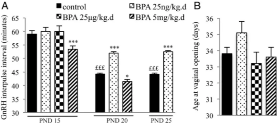
In agreement with our previous findings (16), matura-tion of GnRH secrematura-tion preceding puberty was character-ized by a reduction of the GnRH IPI between PND 15 and PND 25 (Figure 1) using hypothalamic explants of control female rats ex vivo. Exposure to 25 ng/kg.day of BPA from PND 1 to 5 resulted in a significant increase of GnRH IPI on PND 20 and 25, consistent with delayed maturation. In contrast, exposure to 5 mg/kg.day of BPA resulted in a significant reduction of GnRH IPI on PND 15 and 20, consistent with advanced maturation. The intermediate dose of 25g/kg.day did not affect GnRH IPI. Though the low dose of BPA caused a slight delay of age at VO (Figure 1), there was no significant difference between age at VO among the 4 groups (control, BPA 25 ng/kg.day, BPA 25 g/kg.day, BPA 5 mg/kg.day). The BPA-exposed animals showed normal estrous cyclicity from vaginal opening un-til PND 80. No significant difference in average weight was observed between the control and BPA groups from PND 1 to 90.
Pubertal timing after exposure to BPA from PND 1 to 15
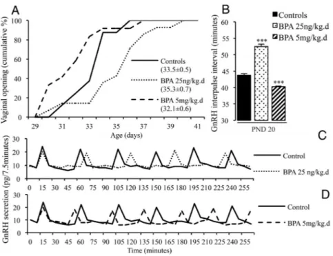
After a longer exposure (from PND 1 to 15), the GnRH IPI on PND 20 was significantly increased by the low dose of BPA and reduced by the high dose (Figure 2), similarly to the effects observed after a shorter exposure. As illus-trated by the cumulative percentage curves in Figure 2 (panel A), exposure to the low dose of BPA tended to delay VO while the high dose of BPA tended to advance VO. The Cox regression regarding age at VO revealed a significant difference (P⫽ .016) between the three groups (control, BPA 25ng/kg.day, BPA 5 mg/kg-.day). Pair analysis indicated that the difference was only significant be-tween the groups exposed to the low and high doses of BPA (P⫽ .0043) and not when these exposed groups were compared to controls (Figure 2). The BPA-exposed animals exhib-ited normal estrous cyclicity from vaginal opening until PND 80. No significant difference in average weight was observed between the control and BPA groups from PND 1 to 90, suggesting that differences in maturation of GnRH secretion and pubertal timing did not result from differences in nutritional status.
Figure 1. Effects of postnatal BPA exposure for 5 days on pulsatile GnRH secretion and female
pubertal timing. A, GnRH IPI in vitro using hypothalamic explants obtained on PND 15, 20 or 25
from female rats after exposure from PND 1 to 5 to vehicle, 25 ng/kg.day, 25g/kg.day or 5 mg/
kg.day of BPA. B, Age at vaginal opening in female rats exposed from PND 1 to 5 to vehicle, 25
ng/kg.day, 25g/kg.day or 5 mg/kg.day of BPA. Data are mean ⫹ SEM; *P ⬍ .05 and ***P ⬍
BPA exposure and changes in hypothalamic mRNA expression
In order to identify the hypotha-lamic genes possibly involved in the dose-related opposing effects of BPA on GnRH secretion and pubertal timing, a RNA sequencing study was performed using retrochiasmatic hy-pothalamic extracts obtained on PND 20 in female rats. After expo-sure to BPA 25 ng/kg.day and 5 mg/ kg.day (PND 1–15), the mRNA ex-pression of 34 and 472 genes was significantly affected, respectively (q-value⬍ 0.05 vs controls). When comparing the two BPA doses, 1407 genes were differentially expressed. A bioinformatic analysis (Ingenuity Pathway Analysis) revealed that GABAAreceptor neurotransmission was among the systems most af-fected by both doses of BPA (Figure 3), based on both the number of af-fected genes and the statistical signif-icance of changes in expression. The 10 canonical pathways most affected
Figure 2. Effects of postnatal BPA exposure for 15 days on pulsatile GnRH secretion and female
pubertal timing. A, Cumulative percentage of female rats displaying vaginal opening in relation to age in animals exposed from PND 1 to 15 to vehicle or 25 ng/kg.day or 5 mg/kg.day of BPA.
Mean age at VO⫾ SEM for each group is indicated in brackets. B, GnRH IPI in vitro using
hypothalamic explants obtained on PND 20 from female rats after exposure from PND 1 to 15 to
vehicle or 25 ng/kg.day or 5 mg/kg.day of BPA. Data are mean⫹ SEM. *** P ⬍ .001, vs control
group. C, and (D) Individual representative profiles of GnRH secretion after neonatal exposure to vehicle or BPA 25 ng/kg.day or 5 mg/kg.day.
Figure 3. Hypothalamic pathways affected by 25 ng or 5 mg/kg.day of BPA. A. Top 10 canonical pathways derived from Ingenuity Pathway
Analysis based on genes that are significantly down- or upregulated in the retrochiasmatic hypothalamus on PND 20 after 15 days of exposure to BPA 25 ng/kg.day. B. Top 10 canonical pathways derived from Ingenuity Pathway Analysis based on genes that are significantly down- or upregulated in retrochiasmatic hypothalamus on PND 20 after 15 days of exposure to BPA 5 mg/kg.day. The pathways are ranked based on –log (p-value) in order of decreasing significance that is illustrated by the bold line. The black and the white bars refer to the percentage of upregulated and downregulated genes that is calculated relative to the total number of genes involved in each pathway (given in the brackets).
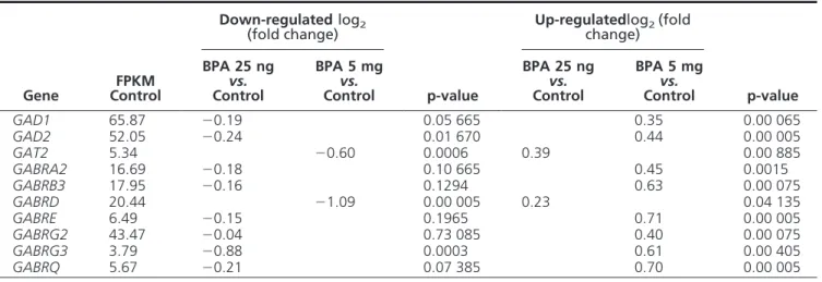
by the two doses of BPA are listed in Supplemental Tables 3 and 4. The two doses resulted in opposing changes of mRNA expression of genes involved in GABA synthesis (glutamic acid decarboxylase, GAD), reuptake (GABA transporter, GAT) and signaling (GABAAReceptor sub-units) (Table 2). Using qPCR on a larger number of sam-ples (n⫽ 8), the increase in mRNA expression of GAD2 was confirmed after 5 mg/kg.day of BPA (1.3⫾ 0.07 vs 1.04⫾ 0.09 in control). Likewise, the increased mRNA expression of GAT2 was confirmed after 25 ng/kg.day of BPA (1.5⫾ 0.27 vs 1.09 ⫾ 0.13 in control).
BPA exposure and GABAergic regulation of GnRH release
Based on the RNA sequencing results, we studied whether GABAergic regulation of pulsatile GnRH secre-tion was affected by BPA. Exposure to the low BPA dose from PND 1 to 15 resulted in a significant increase of GnRH IPI on PND 20 (51.7⫾ 0.9 minutes vs 43.5 ⫾ 0.0 minutes) while the high dose caused a significant reduction of GnRH IPI (39.4⫾ 0.5 minutes), confirming the data of earlier experiments. The increase in GnRH IPI that oc-curred after 25 ng/kg.day of BPA likely involved increased GABAergic tone because it was no longer observed (Figure 4) when hypothalamic explants were incubated in the presence of bicuculline, a GABAA receptor antagonist (45.8⫾ 0.9 minutes vs 51.65 ⫾ 0.9 minutes, bicuculline vs control). Moreover, muscimol did not affect the in-crease in GnRH IPI seen after exposure to 25 ng/kg.day of BPA. The reduction in GnRH IPI that occurred after 5 mg/kg.day of BPA (43.8⫾ 1.3 minutes vs 40 ⫾ 1.25 min-utes, muscimol vs control) was no longer observed when hypothalamic explants were incubated in the presence of
muscimol, a GABAAreceptor agonist. The GABAA recep-tor antagonist did not affect the reduction in GnRH IPI seen after exposure to 5 mg/kg.day of BPA (Figure 4). Taken together, these data confirm that postnatal expo-sure to a very low dose of BPA accounts for delayed mat-uration of GnRH secretion through increased GABAergic tone whereas a high dose of BPA accounts for early mat-uration through reduced GABAergic tone.
Discussion
We provide here the first evidence that BPA can have op-posing dose-dependent effects on the neuroendocrine con-trol of puberty in the female rat. Postnatal exposure to a high dose of BPA led to accelerated maturation of GnRH secretion and a trend toward early onset of puberty. Con-versely, neonatal exposure to a very low dose of BPA led to a delay in developmental acceleration of GnRH secre-tion and a trend toward delayed vaginal opening. Based on these opposing effects on neuroendocrine maturation, we hypothesized that some hypothalamic genes could be dif-ferentially affected by a low vs high dose of BPA. An RNAseq analysis showed opposing variations in mRNA expression of genes related to GABA neurotransmission with confirmation using qPCR. Using GABA agonist and antagonist, a decrease or an increase of GABA inhibitory tone on pulsatile GnRH secretion was shown after expo-sure to a high or low dose of BPA, respectively.
As detailed in the Supplemental Table 5, a number of studies have reported that GABA neurotransmission could be affected in different brain areas after exposure to BPA during early life (34 – 41). GABAergic tone appeared
Table 2. Genes related to GABA neurotransmission showing significant changes in hypothalamic mRNA expression after exposure to low or high BPA doses from PND 1 to 15
Gene FPKM Control Down-regulated log2 (fold change) p-value Up-regulatedlog2(fold change) p-value BPA 25 ng vs. Control BPA 5 mg vs. Control BPA 25 ng vs. Control BPA 5 mg vs. Control GAD1 65.87 ⫺0.19 0.05 665 0.35 0.00 065 GAD2 52.05 ⫺0.24 0.01 670 0.44 0.00 005 GAT2 5.34 ⫺0.60 0.0006 0.39 0.00 885 GABRA2 16.69 ⫺0.18 0.10 665 0.45 0.0015 GABRB3 17.95 ⫺0.16 0.1294 0.63 0.00 075 GABRD 20.44 ⫺1.09 0.00 005 0.23 0.04 135 GABRE 6.49 ⫺0.15 0.1965 0.71 0.00 005 GABRG2 43.47 ⫺0.04 0.73 085 0.40 0.00 075 GABRG3 3.79 ⫺0.88 0.0003 0.61 0.00 405 GABRQ 5.67 ⫺0.21 0.07 385 0.70 0.00 005
GAD: Glutamic Acid Decarboxylase; GAT: GABA Transporter; GABR: GABA Receptor
to be increased after BPA exposure in some studies (34, 37) or reduced in others (36, 38, 39). In a recent study close to our conditions using whole brain extracts from animals exposed to different doses of BPA from gestational day 8 to PND 16, Cabaton et al, investigated brain composition by nuclear magnetic resonance spectroscopy on PND 21. In this non targeted metabolomics study, these authors reported that 25 g/kg.day of BPA could reduce GABA concentrations in the brain (36). Interestingly enough they also used a very low BPA dose of 25 ng/kg.day and it appears that opposing effects were obtained with the low dose ie, increase in GABA concentrations (N. Cabaton and D. Zalko, personal communication). These opposing dose effects are consistent with the present study. Also, such an observation in the whole CNS could suggest that the ef-fects reported here about a specific neuroendocrine func-tion could be part of a much larger effect of BPA in the brain. Opposing dose effects of BPA are supported by in vitro studies of GABA-induced currents in hippocampal neurons that are inhibited or potentiated by high or low BPA concentrations, respectively (41). It has been shown that GnRH secretion can be regulated by GABA in two manners (42). Synaptic receptors generate fast (phasic) postsynaptic currents and extrasynaptic receptors gener-ate a persistent (tonic) current controlling neuronal excit-ability. The latter GABAAreceptors accounting for tonic
current involve the delta subunit. Among the genes en-coding the GABAAreceptor subunits, mRNA expression of Delta subunit is the only one repressed after exposure to 5 mg/kg.day of BPA and one among two subunit genes showing increased mRNA expression after 25 ng/kg.day of BPA (Table 2). This is consistent with dose-dependent dual effects of BPA on tonic GABA neurotransmission. Only a few studies addressed the neuroendocrine events that can precede or follow the changes in GABAergic tone (Supplemental Table 5). Either two separate mechanisms involving different factors or opposing variations of a common factor could explain the opposing effects of BPA on GABA neurotransmission. An insight into a possible upstream mechanism was recently provided by a study reporting that delayed neonatal maturation of a chloride cotransporter after in utero exposure to BPA contributes to activate GABA neurotransmission through reduction of intraneuronal chloride concentrations (40). The gene en-coding this cotransporter (Kcc2) was not affected in our RNAseq study performed on PND20. Studies performed in younger animals with emphasis on epigenetic mecha-nisms are warranted because such mechamecha-nisms were found to account for BPA alteration of Kcc2 expression (40) and DNA methyltransferase 1 mRNA expression was altered following BPA exposure in another study (39). In the pres-ent study, DNMT1 mRNA expression in the female hy-pothalamus was found to increase significantly at PND 20 after exposure to 5 mg/kg.day of BPA (data not shown). Such effects consistent with involvement of epigenetic mechanisms could suggest that BPA has organizational together with activational effects. The issue is complex in the reproductive axis since some effects are likely occur-ring at other levels including the gonads (43, 44).
Changes in mRNA expression of the components of GABAergic neurotransmission (GAD, GAT, GABAR) can be viewed either as part of the altered GABAergic tone or as a downstream reaction to the altered GABAergic tone. In the first case, increased GAD and reduced GAT expres-sion will be associated with increased GABAergic tone (and conversely) such as found for GAD mRNA in the basolateral amygdala (39). In the present study, the verse observation was made since reduced GAD and in-creased GAT expression were associated with inin-creased GABAergic tone (and conversely). Such findings can be consistent with effects downstream from the increased GABA tone because it was shown in neuronal culture that GABA could down-regulate GAD expression and activity (45) and up-regulate GAT activity (46). Confirmation of such an explanation would require a time-lapse study of GAD and GAT expression during and after exposure to different doses of BPA. A prerequisite to the interpretation of changes in GAD and GAT mRNA expression was to
Figure 4. Effects of in vivo exposure to BPA on GABAergic regulation
of GnRH secretion in vitro. GnRH IPI was calculated using secretion from hypothalamic explants obtained on PND 20 from female rats exposed in vivo to vehicle (corn oil) or 25 ng/kg.day or 5 mg/kg.day of BPA from PND 1 to 15. The in vitro conditions involve control or
presence of a GABAAreceptor antagonist (bicuculline) or a GABAA
receptor agonist (muscimol) both used at 10-4M concentration shown
previously to be effective in similar conditions. A reduction in GnRH IPI by bicuculline and absent effects of muscimol indicated a high tone of GABAergic inhibition of GnRH release. Absence of bicuculline effects and increase in GnRH IPI by muscimol indicated a low tone of
GABAergic inhibition of GnRH release. *** P⬍ .001. Data are mean
clarify whether the GABAergic control of GnRH secretion was increased or reduced. An answer was obtained indi-rectly using the GABAAR agonist muscimol and antago-nist bicuculline. Interestingly, Zhou et al (38) also found that the GABAAR agonist was effective in conditions of BPA-induced reduction of GABAergic tone using another paradigm, whereas the antagonist was effective in control conditions. A body of literature shows that GABA is a major contributor to the prepubertal inhibition of GnRH secretion in several species (23). However, there is evi-dence of stimulatory GABA in adult rats and mice (47, 48) and in the fetal hypothalamus (49). Since GABA has dual effects on GnRH neurosecretion, one could hypothesize that BPA resulted in increased or reduced GnRH secretion through antagonism of inhibitory or stimulating GABA inputs. In such event however, the effects of muscimol and bicuculline would have been opposed. In addition, dual GABA effects could be determined by the developmental stage and the neuronal apparatus (GnRH neurons vs im-pinging regulatory neurons). Here, opposing effects were obtained in similar conditions of age and hypothalamic structure.
Based on RNAseq data, G-Protein-coupled receptors represent another mechanistic component that bioinfor-matics analysis identified as a candidate system possibly involved. This was particularly interesting given the crit-ical stimulating role of GPR54 and its ligand kisspeptin in the neuroendocrine control of puberty and reproduction (50, 51). However, neither Kiss1 nor GPR54 mRNA ex-pression appeared to be affected in the present RNAseq analysis. We reported recently that DES exposure (1 and 10g/kg.day) reduced KISS1 expression in the hypothal-amus (18). In the present study however, exposure to BPA did not affect KISS1 mRNA level in the mediobasal hy-pothalamus (data not shown). After neonatal exposure of female rats to 0.1 or 0.5 mg/kg.day of BPA for 5 days, Navarro et al (9) reported reduced KISS1 mRNA expres-sion on PND 30 and Patisaul et al (52) found reduced kisspeptin immunoreactivity in the arcuate nucleus after neonatal exposure to 50 mg/kg.day of BPA for 4 days. Because kisspeptin is a gatekeeper of GnRH neuron stim-ulation, these findings are in contrast to the early vaginal opening that was reported after neonatal or early postna-tal exposure to BPA doses ranging 0.05–100 mg/kg.day (4, 7, 8). These discordant findings could suggest that BPA effects on kisspeptin are not causal in the mechanism of early sexual maturation and might be reactive to that pro-cess, such as discussed about GAT and GAD mRNA expression.
Several studies have reported an effect of BPA on pu-bertal onset in female rodents. Taken as a whole, those studies show that BPA effects depend markedly on the age
window and dose of exposure, with possible nonlinear dose-response relationship (2, 53). To our knowledge, our study is the first one demonstrating the impact of such a low and environmentally relevant dose of BPA on the neu-roendocrine mechanism of sexual maturation. Twenty-five ng/kg.day of BPA affect the frequency of GnRH se-cretion and mRNAs expression of several hypothalamic genes. The effect of the very low dose of BPA on frequency of GnRH secretory pulses is robust and reproducible in our model. Moreover, it is consistent with the trend to-ward delayed VO that is seen subsequently. The amplitude of GnRH secretory pulses is not affected (data not shown). Using the ex vivo paradigm of hypothalamic explant, changes in amplitude of GnRH release were only apparent after in vitro stimulation with secretagogues such as glu-tamate or gluglu-tamate receptor agonists (21, 54). Since we aimed at changes in endogenously driven pulsatile secre-tion of GnRH in the present study, GnRH secretagogues have not been used.
The neuroendocrine system appears to be exquisitely sensitive to BPA endocrine disruption that may not be manifested phenotypically. When the female rats are post-natally exposed to 25 ng/kg.day for 5 or 15 days, age at puberty is not significantly different from controls but the frequency of GnRH secretion is significantly decreased. Presumably, the effects on maturation of GnRH secretion evidenced at PND 20 and 25 are not lasting enough or sufficient to affect significantly pubertal timing about ten days later. We made a similar observation in male rats exposed for 5 days after birth (2). Thus, absence of phe-notypic changes in some of the previous studies does not necessarily mean absence of effect. Such high neuroendo-crine sensitivity to BPA, functionally evidenced through changes in pulsatile GnRH secretion, is also consistent with a significant change in hypothalamic mRNA expres-sion of 34 genes following exposure to that very low dose of BPA.
The study of mRNAs expression showed some limita-tions. Our RNAseq analysis was limited to 3 samples for each group and confirmed on 8 samples only for some of the genes involved in GABA synthesis, reuptake and sig-naling. However, the incubation of control or BPA-ex-posed hypothalamic explants with a GABA receptor ag-onist or antagag-onist robustly confirmed the effects of BPA on GABAergic neurotransmission. A short exposure of 5 or 15 days differs from the constant human exposure throughout life. However, such short exposures allow the delineation of a window of sensitivity to BPA. Also, the effects of BPA on puberty are unlikely to result from direct effects on target peripheral tissues such as the female gen-ital tract because the exposure ended several weeks before puberty and BPA is a nonpersisting chemical. Accordingly,
the data are likely to reflect alterations of functions orga-nized during early postnatal life.
Human exposure to BPA varies between 0.01 and 1 g/kg.day (10). Because we have studied a very low dose of 25 ng/kg.day, we chose subcutaneous injections in or-der to guarantee accuracy of the exposure. This route of administration was used as well in all the other studies with BPA exposure in early postnatal life (reviewed in (2)). While not relevant to the human route of exposure, sub-cutaneous injections have been shown to lead to similar serum concentration as oral administration (55). The question remains as to whether such a low level of expo-sure makes a difference from contamination through food, water and cages. We have attempted to control for this through housing conditions and the control animals ex-posed to corn oil were raised in similar conditions.
Postnatal life (5 to 15 first days after birth) has been shown to be particularly sensitive to the effects of BPA on the neuroendocrine mechanism of puberty (2). Further studies could underscore the significance of early postna-tal life as a critical window by comparing the effects of a similar exposure to BPA in adulthood. Questions remain as to whether a longer and sustained exposure involving the whole prenatal life could result in effects similar to those reported here. Moreover, further studies should aim at delineation of the genetic mechanisms that take place upstream and downstream to GABA neurotransmission and account for divergent effects on pubertal timing.
In conclusion, our study shows that alteration of pu-bertal timing by BPA in the rat is not necessarily towards precocity. Instead, delayed maturation of GnRH secretion can be caused by environmentally relevant exposure with the mechanistic involvement of GABA neurotransmission. Taking together our data and the literature, it appears that the effects of BPA are not limited to the neuroendocrine control of the reproductive axis. BPA seems to affect GABA neurotransmission in multiple brain areas. Though rodent data do not necessarily apply as such to human health, the involvement of GABA shown in the present study points to the likelihood of detrimental effects of early life exposure to BPA in humans. GABA plays indeed a critical role in brain development and function and dis-orders of human behaviors including autism (56) that are possibly more frequent after early exposure to BPA (57– 59). The US Environmental Protection Agency (EPA) has considered that a daily exposure lower than 50g/kg.day was “safe” (60). EFSA very recently proposed to set the daily tolerable intake at 4g/kg.day (61). This study re-inforces the idea that defining a threshold for BPA effects is misleading not only because doses much below the so-called “safe” limit can interfere with the hormonal system
but also because a low and a high dose can have opposing effects.
Acknowledgments
We thank Pr Delvenne for the assistance with Papanicolaou staining and the Dr. V.D. Ramirez (Urbana, IL) for providing the CR11-B81 anti-GnRH antiserum.
Address all correspondence and requests for reprints to: Del-phine Franssen, Developmental Neuroendocrinology Unit, GIGA Neurosciences, University of Liège, Sart-Tilman, B-4000
Liège, Belgium. Telephone : ⫹3243662539. E-mail :
delphine.franssen@ulg.ac.be.
*These authors contributed equally
Disclosure Summary: The authors have nothing to disclose. This work was supported by Fonds National de la Recherche Scientifique (Belgium), the Belgian Society for Pediatric Endo-crinology and Diabetology and the “Fonds Léon Frédéricq”.
References
1. Vandenberg LN, Hauser R, Marcus M, Olea N, Welshons W V. Human exposure to bisphenol A (BPA). Reprod Toxicol. 2007; 24(2):139 –77.
2. Parent A-S, Franssen D, Fudvoye J, Gérard A, Bourguignon J-P. Developmental variations in environmental influences including en-docrine disruptors on pubertal timing and neuroenen-docrine control: Revision of human observations and mechanistic insight from
ro-dents. Front Neuroendocrinol. 2015.
doi:10.1016/j.y-frne.2014.12.004.
3. Nagao T, Yoshimura S, Saito Y, Nakagomi M, Usumi K, Ono H. Reproductive effects in male and female rats of neonatal exposure to genistein. Reprod Toxicol. 2001;15(4):399 – 411.
4. Adewale HB, Jefferson WN, Newbold RR, Patisaul HB. Neonatal bisphenol-a exposure alters rat reproductive development and ovar-ian morphology without impairing activation of gonadotropin-re-leasing hormone neurons. Biol Reprod. 2009;81(4):690 – 699. 5. Yu B, Chen QF, Liu ZP, Xu HF, Zhang XP, Xiang Q, Zhang WZ,
Cui WM, Zhang X, Li N. Estrogen receptor alpha and beta
expres-sions in hypothalamus-pituitary-ovary axis in rats exposed lacta-tionally to soy isoflavones and bisphenol A. Biomed Env Sci. 2010; 23(5):357–362.
6. Losa-Ward SM, Todd KL, McCaffrey KA, Tsutsui K, Patisaul HB. Disrupted organization of RFamide pathways in the hypothalamus is associated with advanced puberty in female rats neonatally ex-posed to bisphenol A. Biol Reprod. 2012;87(2):28.
7. Fernandez M, Bianchi M, Lux-Lantos V, Libertun C. Neonatal ex-posure to bisphenol a alters reproductive parameters and gonado-tropin releasing hormone signaling in female rats. Env Heal
Per-spect. 2009;117(5):757–762.
8. Nah WH, Park MJ, Gye MC. Effects of early prepubertal exposure to bisphenol A on the onset of puberty, ovarian weights, and estrous cycle in female mice. Clin Exp Reprod Med. 2011;38(2):75– 81. 9. Navarro VM, Sanchez-Garrido MA, Castellano JM, Roa J,
Garcia-Galiano D, Pineda R, Aguilar E, Pinilla L, Tena-Sempere M.
Per-sistent impairment of hypothalamic KiSS-1 system after exposures to estrogenic compounds at critical periods of brain sex differenti-ation. Endocrinology. 2009;150(5):2359 –2367.
P, Maghuin-Rogister G, Pironnet A-M, Pussemier L, Scippo M-L, Van Loco J, Covaci A. A review of dietary and non-dietary exposure
to bisphenol-A. Food Chem Toxicol. 2012;50(10):3725– 40. 11. Ferguson KK, Peterson KE, Lee JM, Mercado-García A,
Blank-Goldenberg C, Téllez-Rojo MM, Meeker JD. Prenatal and
peripu-bertal phthalates and bisphenol A in relation to sex hormones and puberty in boys. Reprod Toxicol. 2014;47:70 – 6.
12. Frederiksen H, Aksglaede L, Sorensen K, Nielsen O, Main KM,
Skakkebaek NE, Juul A, Andersson A-M. Bisphenol A and other
phenols in urine from Danish children and adolescents analyzed by isotope diluted TurboFlow-LC-MS/MS. Int. J Hyg Environ Health. 2013;216(6):710 –20.
13. Lee SH, Kang SM, Choi MH, Lee J, Park MJ, Kim SH, Lee W-Y,
Hong J, Chung BC. Changes in steroid metabolism among girls with
precocious puberty may not be associated with urinary levels of bisphenol A. Reprod Toxicol. 2014;44:1– 6.
14. Durmaz E, Asçı A, Erkekog˘lu P, Akçurin S, Gümüsel BK, Bircan I. Urinary bisphenol a levels in girls with idiopathic central precocious puberty. J Clin Res Pediatr Endocrinol. 2014;6(1):16 –21. 15. McGuinn LA, Ghazarian AA, Joseph Su L, Ellison GL. Urinary
bisphenol A and age at menarche among adolescent girls: evidence from NHANES 2003–2010. Environ Res. 2015;136:381– 6. 16. Bourguignon JP, Franchimont P. Puberty-related increase in
epi-sodic LHRH release from rat hypothalamus in vitro.
Endocrinol-ogy. 1984;114(5):1941–1943.
17. Rasier G, Parent AS, Gerard A, Lebrethon MC, Bourguignon JP. Early maturation of gonadotropin-releasing hormone secretion and sexual precocity after exposure of infant female rats to estradiol or dichlorodiphenyltrichloroethane. Biol Reprod. 2007;77(4):734 – 742.
18. Franssen D, Ioannou YS, Alvarez-real A, Gerard A, Mueller JK,
Heger S, Bourguignon J-P, Parent A-S. Pubertal timing after
neo-natal diethylstilbestrol exposure in female rats: neuroendocrine vs peripheral effects and additive role of prenatal food restriction.
Re-prod Toxicol. 2014;44:63–72.
19. Rasier G, Toppari J, Parent A-S, Bourguignon J-P. Female sexual maturation and reproduction after prepubertal exposure to estro-gens and endocrine disrupting chemicals: a review of rodent and human data. Mol Cell Endocrinol. 2006;254 –255:187–201. 20. Parent AS, Teilmann G, Juul A, Skakkebaek NE, Toppari J,
Bour-guignon JP. The timing of normal puberty and the age limits of
sexual precocity: variations around the world, secular trends, and changes after migration. Endocr Rev. 2003;24(5):668 – 693. 21. Matagne V, Rasier G, Lebrethon MC, Gerard A, Bourguignon JP.
Estradiol stimulation of pulsatile gonadotropin-releasing hormone secretion in vitro: correlation with perinatal exposure to sex steroids and induction of sexual precocity in vivo. Endocrinology. 2004; 145(6):2775–2783.
22. Keen KL, Burich AJ, Mitsushima D, Kasuya E, Terasawa E. Effects of pulsatile infusion of the GABA(A) receptor blocker bicuculline on the onset of puberty in female rhesus monkeys. Endocrinology. 1999;140(11):5257– 66.
23. Terasawa E. Role of GABA in the mechanism of the onset of puberty in non-human primates. Int Rev Neurobiol. 2005;71:113–29. 24. Bourguignon JP, Gerard A, Purnelle G, Czajkowski V, Yamanaka C,
Lemaitre M, Rigo JM, Moonen G, Franchimont P. Duality of
glu-tamatergic and GABAergic control of pulsatile GnRH secretion by rat hypothalamic explants: II. Reduced NR2C- and GABAA-recep-tor-mediated inhibition at initiation of sexual maturation. J
Neu-roendocr. 1997;9(3):193–199.
25. Bourguignon JP, Gerard A, Purnelle G, Czajkowski V, Yamanaka C,
Lemaitre M, Rigo JM, Moonen G, Franchimont P. Duality of
glu-tamatergic and GABAergic control of pulsatile GnRH secretion by rat hypothalamic explants: I. Effects of antisense oligodeoxynucle-otides using explants including or excluding the preoptic area.
J Neuroendocr. 1997;9(3):183–191.
26. Goldman JM, Murr AS, Cooper RL. The rodent estrous cycle:
char-acterization of vaginal cytology and its utility in toxicological stud-ies. Birth Defects Res B Dev Reprod Toxicol. 2007;80(2):84 –97. 27. Bourguignon JP, Gerard A, Franchimont P. Direct activation of
gonadotropin-releasing hormone secretion through different recep-tors to neuroexcitatory amino acids. Neuroendocrinology. 1989; 49(4):402– 408.
28. Bourguignon JP, Gerard A, Mathieu J, Simons J, Franchimont P. Pulsatile release of gonadotropin-releasing hormone from hypotha-lamic explants is restrained by blockade of N-methyl-D,L-aspartate receptors. Endocrinology. 1989;125(2):1090 –1096.
29. Dluzen DE, Ramirez VD. Presence and localization of immunore-active luteinizing hormone-releasing hormone (LHRH) within the olfactory bulbs of adult male and female rats. Peptides. 1981;2(4): 493– 496.
30. Parent A-S, Rasier G, Matagne V, Lomniczi A, Lebrethon M-C,
Gérard A, Ojeda SR, Bourguignon J-P. Oxytocin facilitates female
sexual maturation through a glia-to-neuron signaling pathway.
En-docrinology. 2008;149(3):1358 – 65.
31. Trapnell C, Roberts A, Goff L, Pertea G, Kim D, Kelley DR, Pimentel
H, Salzberg SL, Rinn JL, Pachter L. Differential gene and transcript
expression analysis of RNA-seq experiments with TopHat and Cuf-flinks. Nat Protoc. 2012;7(3):562–78.
32. Livak KJ, Schmittgen TD. Analysis of relative gene expression data using real-time quantitative PCR and the 2(-Delta Delta C(T)) Method. Methods. 2001;25(4):402– 408.
33. Hagen CP, Sørensen K, Aksglaede L, Mouritsen A, Mieritz MG,
Tinggaard J, Wohlfart-Veje C, Petersen JH, Main KM, Rajpert-De Meyts E, Almstrup K, Juul A. Pubertal onset in girls is strongly
influenced by genetic variation affecting FSH action. Sci Rep. 2014; 4:6412.
34. Cardoso N, Pandolfi M, Lavalle J, Carbone S, Ponzo O, Scacchi P,
Reynoso R. Probable gamma-aminobutyric acid involvement in
bi-sphenol A effect at the hypothalamic level in adult male rats.
J Physiol Biochem. 2011;67(4):559 – 67.
35. Facciolo RM, Alò R, Madeo M, Canonaco M, Dessì-Fulgheri F. Early cerebral activities of the environmental estrogen bisphenol A appear to act via the somatostatin receptor subtype sst(2). Environ
Health Perspect. 2002;110 Suppl :397– 402.
36. Cabaton NJ, Canlet C, Wadia PR, Tremblay-Franco M, Gautier R,
Molina J, Sonnenschein C, Cravedi J-P, Rubin BS, Soto AM, Zalko D. Effects of low doses of bisphenol A on the metabolome of
peri-natally exposed CD-1 mice. Environ Health Perspect. 2013;121(5): 586 –93.
37. Ogi H, Itoh K, Ikegaya H, Fushiki S. Alterations of neurotransmitter norepinephrine and gamma-aminobutyric acid correlate with mu-rine behavioral perturbations related to bisphenol A exposure. Brain
Dev. 2015;37(8):739 – 46.
38. Zhou R, Bai Y, Yang R, Zhu Y, Chi X, Li L, Chen L, Sokabe M, Chen
L. Abnormal synaptic plasticity in basolateral amygdala may
ac-count for hyperactivity and attention-deficit in male rat exposed perinatally to low-dose bisphenol-A. Neuropharmacology. 2011; 60(5):789 –98.
39. Zhou R, Chen F, Chang F, Bai Y, Chen L. Persistent overexpression of DNA methyltransferase 1 attenuating GABAergic inhibition in basolateral amygdala accounts for anxiety in rat offspring exposed perinatally to low-dose bisphenol A. J Psychiatr Res. 2013;47(10): 1535– 44.
40. Yeo M, Berglund K, Hanna M, Guo JU, Kittur J, Torres MD,
Abramowitz J, Busciglio J, Gao Y, Birnbaumer L, Liedtke WB.
Bi-sphenol A delays the perinatal chloride shift in cortical neurons by epigenetic effects on the Kcc2 promoter. Proc Natl Acad Sci U S A. 2013;110(11):4315–20.
41. Choi I-S, Cho J-H, Park E-J, Park J-W, Kim S-H, Lee M-G, Choi B-J,
Jang I-S. Multiple effects of bisphenol A, an endocrine disrupter, on
GABA(A) receptors in acutely dissociated rat CA3 pyramidal neu-rons. Neurosci Res. 2007;59(1):8 –17.
SK. Tonic extrasynaptic GABA(A) receptor currents control
gonad-otropin-releasing hormone neuron excitability in the mouse.
Endo-crinology. 2011;152(4):1551– 61.
43. Li Y, Zhang W, Liu J, Wang W, Li H, Zhu J, Weng S, Xiao S, Wu
T. Prepubertal bisphenol A exposure interferes with ovarian follicle
development and its relevant gene expression. Reprod Toxicol. 2014;44:33– 40.
44. Gámez JM, Penalba R, Cardoso N, Bernasconi PS, Carbone S,
Ponzo O, Pandolfi M, Scacchi P, Reynoso R. Exposure to a low dose
of bisphenol A impairs pituitary-ovarian axis in prepubertal rats: effects on early folliculogenesis. Environ Toxicol Pharmacol. 2015; 39(1):9 –15.
45. de Almeida OMMS, Gardino PF, Loureiro dos Santos NE,
Yama-saki EN, de Mello MCF, Hokoç JN, de Mello FG. Opposite roles of
GABA and excitatory amino acids on the control of GAD expression in cultured retina cells. Brain Res. 2002;925(1):89 –99.
46. Bernstein EM, Quick MW. Regulation of gamma-aminobutyric acid (GABA) transporters by extracellular GABA. J Biol Chem. 1999;274(2):889 –95.
47. Watanabe M, Sakuma Y, Kato M. GABAA receptors mediate ex-citation in adult rat GnRH neurons. Biol Reprod. 2009;81(2):327– 32.
48. Moenter SM, DeFazio RA. Endogenous gamma-aminobutyric acid can excite gonadotropin-releasing hormone neurons.
Endocrinol-ogy. 2005;146(12):5374 –9.
49. Kusano K, Fueshko S, Gainer H, Wray S. Electrical and synaptic properties of embryonic luteinizing hormone-releasing hormone neurons in explant cultures. Proc Natl Acad Sci U S A. 1995;92(9): 3918 –22.
50. de Roux N, Genin E, Carel JC, Matsuda F, Chaussain JL, Milgrom
E. Hypogonadotropic hypogonadism due to loss of function of the
KiSS1-derived peptide receptor GPR54. Proc Natl Acad Sci U S A. 2003;100(19):10972–10976.
51. Seminara SB, Messager S, Chatzidaki EE, Thresher RR, Acierno Jr.
JS, Shagoury JK, Bo-Abbas Y, Kuohung W, Schwinof KM, Hendrick AG, Zahn D, Dixon J, Kaiser UB, Slaugenhaupt SA, Gusella JF, O’Rahilly S, Carlton MB, Crowley Jr. WF, Aparicio SA, Colledge WH. The GPR54 gene as a regulator of puberty. N Engl J Med.
2003;349(17):1614 –1627.
52. Patisaul HB, Todd KL, Mickens JA, Adewale HB. Impact of neo-natal exposure to the ERalpha agonist PPT, bisphenol-A or
phy-toestrogens on hypothalamic kisspeptin fiber density in male and female rats. Neurotoxicology. 2009;30(3):350 –357.
53. Bourguignon JP, Franssen D, Gerard A, Janssen S, Pinson A, Naveau
E, Parent AS. Early neuroendocrine disruption in hypothalamus and
hippocampus: developmental effects including female sexual mat-uration and implications for endocrine disrupting chemical screen-ing. J Neuroendocr. 2013;25(11):1079 –1087.
54. Bourguignon JP, Gerard A, Mathieu J, Mathieu A, Franchimont P. Maturation of the hypothalamic control of pulsatile gonadotropin-releasing hormone secretion at onset of puberty. I. Increased acti-vation of N-methyl-D-aspartate receptors. Endocrinology. 1990; 127(2):873– 881.
55. Taylor JA, Welshons W V, Vom Saal FS. No effect of route of exposure (oral; subcutaneous injection) on plasma bisphenol A throughout 24h after administration in neonatal female mice.
Re-prod Toxicol. 2008;25(2):169 –176.
56. Ben-Ari Y. The GABA excitatory/inhibitory developmental se-quence: A personal journey. Neuroscience. 2014;279:187–219. 57. Harley KG, Gunier RB, Kogut K, Johnson C, Bradman A, Calafat
AM, Eskenazi B. Prenatal and early childhood bisphenol A
concen-trations and behavior in school-aged children. Environ Res. 2013; 126:43–50.
58. Roen EL, Wang Y, Calafat AM, Wang S, Margolis A, Herbstman J,
Hoepner LA, Rauh V, Perera FP. Bisphenol A exposure and
behav-ioral problems among inner city children at 7–9 years of age.
En-viron Res. 2015;142:739 – 45.
59. Kardas F, Bayram AK, Demirci E, Akin L, Ozmen S, Kendirci M,
Canpolat M, Oztop DB, Narin F, Gumus H, Kumandas S, Per H.
Increased Serum Phthalates (MEHP, DEHP) and Bisphenol A Con-centrations in Children With Autism Spectrum Disorder: The Role of Endocrine Disruptors in Autism Etiopathogenesis. J Child
Neu-rol. 2015. doi:10.1177/0883073815609150.
60. Diamanti-Kandarakis E, Bourguignon JP, Giudice LC, Hauser R,
Prins GS, Soto AM, Zoeller RT, Gore AC. Endocrine-disrupting
chemicals: an Endocrine Society scientific statement. Endocr Rev. 2009;30(4):293–342.
61. EFSA Panel on Food Contact Materials EF and PA (CEF). Scientific Opinion on the risks to public health related to the presence of bisphenol A (BPA) in foodstuffs. EFSA J. 2015 2015;13(1). Avail-able at: http://www.efsa.europa.eu/fr/efsajournal/pub/3978.htm. Accessed February 12, 2015.

![[PDF] Support de cours traitement de texte Word 2010 - Bureautique](data:image/gif;base64,R0lGODlhAQABAIAAAP///wAAACH5BAEAAAAALAAAAAABAAEAAAICRAEAOw==)