http://jfm.sagepub.com/
Journal of Feline Medicine and Surgery
http://jfm.sagepub.com/content/15/7/628
The online version of this article can be found at:
DOI: 10.1177/1098612X13489227
2013 15: 628
Journal of Feline Medicine and Surgery
D Radford, Etienne Thiry, Uwe Truyen and Marian C Horzinek
Herman Egberink, Tadeusz Frymus, Tim Gruffydd-Jones, Margaret J Hosie, Hans Lutz, Fulvio Marsilio, Karin Möstl, Alan
Albert Lloret, Katrin Hartmann, Maria Grazia Pennisi, Lluis Ferrer, Diane Addie, Sándor Belák, Corine Boucraut-Baralon,
on prevention and management
Rare opportunistic mycoses in cats: phaeohyphomycosis and hyalohyphomycosis: ABCD guidelines
technique does not amount to an endorsement of its value or quality, or the claims made by its manufacturer. those of the authors and the inclusion in this publication of material relating to a particular product, method or of animals and interpretation of published materials lies with the veterinary practitioner. The opinions expressed are from actions or decisions based on information contained in this publication; ultimate responsibility for the treatment
arising country. The authors, editors, owners and publishers do not accept any responsibility for any loss or damage advertising material, it is the responsibility of the reader to check that the product is authorised for use in their own bear this in mind and be aware of the prescribing laws pertaining to their own country. Likewise, in relation to Furthermore, drugs may be mentioned that are licensed for human use, and not for veterinary use. Readers need to formulations that are not available or licensed in the individual reader's own country.
The Journal of Feline Medicine and Surgery is an international journal and authors may discuss products and Disclaimer
Published by:
International Society of Feline Medicine
American Association of Feline Practitioners
and
http://www.sagepublications.com
can be found at:
Journal of Feline Medicine and Surgery
Additional services and information for
http://jfm.sagepub.com/cgi/alerts Email Alerts: http://jfm.sagepub.com/subscriptions Subscriptions: http://www.sagepub.com/journalsReprints.nav Reprints: http://www.sagepub.com/journalsPermissions.nav Permissions:
What is This?
- Jun 27, 2013
Version of Record
>>
at Universite de Liege on September 3, 2013
jfm.sagepub.com
Fungal properties and epidemiology
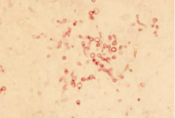
Phaeohyphomycoses are rare opportunistic fungal infections caused by numerous genera of fungal moulds that characteristically produce melanin-pigmented ‘dematiaceous’ (dark-coloured) hyphal elements in tissues and in culture (Figures 1 and 2); yeast-like forms have also been found in some cases.1Hyalohyphomycoses are caused by several genera of fungi that are non-pigmented, being transparent or hyaline in tissues.1
Both are ubiquitous saprophytic agents. The number of reports of infections is increasing
in humans and animals, often associated with immunosuppressive treatment or an immunosuppressive condition. In human medicine, they are currently considered emerging fungal infections.2,3
Infections are acquired from
trau-matic implantation from the environment (soil and decomposed plants). Direct transmission between hosts does not occur.1
The taxonomy of the aetiological agents is complicated, and names have often been changed. More than 100 species classified within 60 genera have been described as agents of phaeohyphomycosis in animals and humans. Pathogens for dogs and cats include species from Alternaria, Bipolaris, Cladophialophora and Curvularia. Genera with species causing disease in cats, but not in dogs, are Exophiala, Fonsecaea, Macrophomina, Microsphaerosis, Moniliella, Phialophora, Phoma, Scolecobasidium and Stemphylium.
Journal of Feline Medicine and Surgery (2013) 15, 628–630
C L I N I C A L
R E V I E W
RARE OPPORTUNISTIC MYCOSES IN CATS:
PHAEOHYPHOMYCOSIS AND
HYALOHYPHOMYCOSIS
ABCD guidelines on prevention
and management
Albert Lloret, Katrin Hartmann, Maria Grazia Pennisi, Lluis Ferrer*, Diane Addie, Sándor Belák, Corine Boucraut-Baralon, Herman Egberink, Tadeusz Frymus, Tim Gruffydd-Jones, Margaret J Hosie, Hans Lutz, Fulvio Marsilio, Karin Möstl, Alan D Radford, Etienne Thiry, Uwe Truyen and Marian C Horzinek
628
JFMSCLINICAL PRACTICEEuropean Advisory Board on Cat Diseases www.abcd-vets.org Corresponding author: Albert Lloret
Email: Albert.LLoret@uab.cat
European Advisory Board on Cat Diseases
The European Advisory Board on Cat Diseases (ABCD) is a body of experts in immunology, vaccinology and clinical feline medicine that issues guidelines on prevention and management of feline infectious diseases in Europe, for the benefit of the health and welfare of cats. The guidelines are based on current scientific knowledge of the diseases and available vaccines concerned.
The latest version of the rare opportunistic mycoses in cats guidelines is available at www.abcd-vets.org
DOI: 10.1177/1098612X13489227 © Published by SAGE on behalf of ISFM and AAFP 2013
Overview: Phaeohyphomycoses and hyalohyphomycoses are rare opportunistic infections acquired from the environment. More cases have been reported in recent years in humans and cats.
Disease signs: Single or multiple nodules or ulcerated plaques (which may be pigmented) in the skin are the typical lesions. In some cases the infection disseminates or involves the central nervous system (CNS).
Diagnosis: Diagnosis is based on fungal detection by cytology and/or histology. Culture provides definitive diagnosis and species identification.
Treatment: Treatment involves surgical excision in cases of localised skin disease followed by systemic antifungal therapy, with itraconazole as the agent of first choice. Relapses after treatment are common. Itraconazole and other systemic antifungal agents have been used to treat systemic or neurological cases, but the response is unpredictable. The prognosis is guarded to poor in cats with multiple lesions and systemic or neurological involvement.
Zoonotic risk: There is no zoonotic risk associated with contact with infected cats.
*The ABCD is grateful to Professor Lluís Ferrer, of the Foster Hospital for Small Animals, Cummings School of Veterinary Medicine, Tufts University, USA, who, though not a member of the Board, contributed to this article.
at Universite de Liege on September 3, 2013
jfm.sagepub.com
JFMSCLINICAL PRACTICE
629
Genera with species causing hyalohyphomycosis in dogs and cats include Fusarium, Acremonium, Paecilomyces, Pseudallescheria, Sagemonella, Phialosimplex and Scedosporium.
Feline phaeohyphomycosis probably has a worldwide dis -tribution as sporadic cases have been reported from North America, Spain,4 Italy,5,6 Australia,7 Canada,8 the UK9,10 and Japan.11
A retrospective study from the
UK evaluating 77 cats with nodular granulo-matous skin lesions caused by fungal infection found that the most frequent cause was hyalohypho mycosis. Phaeohyphomycosis and deep pseudomycetomas were less frequently diagnosed.9
Pathogenesis
Infection occurs mainly through contact or skin puncture, especially through trauma involving wood.1 Respiratory tract colonisa-tion is suspected to occur in systemic cases. In the rare cases of CNS infection, the route of exposure has not been elucidated, but an extension from sinuses, the orbit and middle ear has been suggested.1,12Local infections are rarely associated with systemic diseases or immunosuppression.1The infrequent cases of systemic disseminated infection may or may not be associated with immunosuppression.
Clinical presentation
Nodules or masses in the skin or nasal mucosa are the most common clinical problem. Ulcerated, crusting or fistulating nodules, non-ulcerated subcutaneous nodules and/or plaques, which can be focal or multifocal and locally invasive, are typical lesions.4–11 The lesions may appear pigmented,1but are other-wise not different from chronic bacterial infec-tion or cystic skin lesions. In most cases they
R E V I E W/ABCD guidelines on rare opportunistic mycoses
are found in the facial region, and on the distal part of the extremities or the tail. A typ-ical presentation is a nodule on the bridge of the nose.4A case of a focal pulmonary granu-loma caused by Cladophialophora bantiana has been reported in a cat.13
A few cases in the literature concern fungal infections that were responsible for multifocal neurological signs due to encephalitis or brain abscesses12 or for disseminated disease,14,15 especially in association with immunosup-pression. In these cases the causative organism has been identified as Cladosporium species. Most cases have been diagnosed post mortem.
Diagnosis
Diagnosis is based on visualisation of the fungal organism on cytology and/or histol-ogy, which usually shows a nodular to diffuse pyogranulomatous inflammation pattern. In tissue, the presence of pigmented fungal structures in the centre of the pyogranulomatous reaction is highly suggestive of phaeo -hyphomycosis (Figure 3).1,4–11 Special fungal stains such as Gomori methenamine silver or periodic acid-Schiff can enhance the diagnos-tic sensitivity.
Definitive diagnosis relies on fungal culture and identification of the fungal species based on morphology and pigmentation features by specialised laboratories.1
Molecular techniques have only seldom been used to identify pathogenic fungal species.11
Treatment
No prospective studies exist on the treatment of feline phaeohyphomycosis or hyalohypho -mycosis. Recommendations are based on case reports.
The approach for local lesions is aggressive surgical excision, as these rarely respond to antifungal treatment. After surgery of a single lesion, if multiple lesions exist or in cases of disseminated infection, itraconazole is the treatment of choice [EBM grade IV].
In human
medicine,
these are
currently
considered
emerging
fungal
infections.
Figure 1 Dark-coloured, periodic acid-Schiff-positive, fungal structures in a tissue sample. Courtesy of Alessandra Fondati
Figure 2‘Dematiaceous’ (dark-coloured) colony.
Courtesy of Alessandra Fondati
Figure 3 Pigmented fungal structures in a tissue sample.
Courtesy of Lluís Ferrer
EBM grades The ranking system for grading the level of evidence of various statements within this article is described on page 533 of this Special Issue.
at Universite de Liege on September 3, 2013
jfm.sagepub.com
630
JFMSCLINICAL PRACTICER E V I E W/ABCD guidelines on rare opportunistic mycoses
Disseminated or neurological cases are poorly responsive to treatment. Ketoconazole, amphotericin B and posaconazole have been used in a few cases [EBM grade IV].1,13
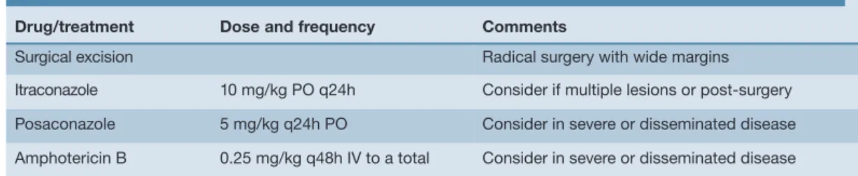
Table 1 lists the treatment options for these infections.
< Sporadic cases of these fungal diseases have been reported in cats.
< Skin nodules and ulcers, especially involving the facial area, distal extremities and tail, are the most frequent lesions.
< Dissemination and CNS signs may occur in rare cases, especially with Cladosporium species infection.
< Histology and culture are the most useful diagnostic tests. Some fungi show a typical pigmentation, which is helpful for diagnosis.
< A combination of surgery and systemic antifungal treatment (itraconazole) may cure cases with localised lesions.
< Cases of disseminated skin disease or systemic disease have a poor prognosis; new azole drugs like
posaconazole should be considered in these cats.
KEY
POINTS
Funding
The authors received no specific grant from any funding agency in the public, commercial or not-for-profit sectors for the preparation of this article. The ABCD is supported by Merial, but is a scientifically independent body.
Conflict of interest
The authors do not have any potential conflicts of interest to declare.
References
1 Grooters AM and Foil CS. Miscellaneous
fun-gal infections. In: Greene CE (ed). Infectious
diseases of the dog and the cat. 3rd ed. St Louis: Saunders Elsevier, 2006, pp 637–650.
2 Revankar SG, Patterson JE, Sutton DA, Pullen R and Rinaldi MG. Disseminated
phaeohy-phomycosis: review of an emerging mycosis. Clin Infect Dis 2002; 34: 467–476.
3 Fothergill AW. Identification of dematiaceous
fungi and their role in human disease. Clin Infect Dis 1996; 22: S179–S184.
4 Fondati A, Gallo MG, Romano E and Fondevila D.
A case of feline phaeohyphomycosis due to
Fonsecaea pedrosoi. Vet Dermatol 2001; 2: 297–301.
5 Beccati M, Vercelli A, Peano A and Gallo MG.
Phaeohyphomycosis by Phialophora
verru-cosa: first European case in a cat. Vet Rec 2005;
157: 93–94.
6 Abramo F, Bastelli F, Nardoni S and Mancianti F. Feline cutaneous phaeohyphomycosis due
to Cladophyalophora bantiana. J Feline Med Surg 2002; 4: 157–163.
7 Kettlewell P, McGinnis MR and Wilkinson GT.
Phaeohyphomycosis caused by Exophiala
spinifera in two cats. J Med Vet Mycol 1989; 27:
257–264.
8 Outerbridge CA, Myers SL and Summerbell RC. Phaeohyphomycosis in a cat. Can Vet J 1995; 36: 629–630.
9 Miller RI. Nodular granulomatous fungal skin
disease of cats in the United Kingdom: a retro-spective review. Vet Dermatol 2010; 21: 130–135.
10 Knights CB, Lee K, Rycroft AN, Patterson-Kane JC and Baines SJ. Phaeohyphomycosis caused
by Ulocladium species in a cat. Vet Rec 2008;
162: 415–416.
11 Maeda H, Shibuya H, Yamaguchi Y, Miyoshi T, Irie M and Sato T. Feline digital phaeohypho
-mycosis due to Exophiala jeanselmei. J Vet Med Sci 2008; 70: 1395–1397.
12 Bouljihad M, Lindeman CJ and Hayden DW.
Pyogranulomatous meningoencephalitis associated with dematiaceous fungal (Cladophialophora bantiana) infection in a domestic cat. J Vet Diagn Invest 2002; 14: 70–72.
13 Evans N, Gunew M, Marshall R, Martin P and Barrs V. Focal pulmonary granuloma caused
by Cladophialophora bantiana in a domestic shorthair cat. Med Mycol 2011; 49: 194–197.
14 Elies L, Balandraud V, Boulouha L, Crespeau F and Guillot J. Fatal systemic
phaeohyphomyco-sis in a cat due to Cladophialophora bantiana. J Vet Med A Physiol Pathol Clin Med 2003; 50: 50–53. 15 Mariani CL, Platt SR, Scase TJ, Howerth EW, Chrisman CL and Clemmons RM. Cerebral
phaeohyphomycosis caused by Cladosporium
spp. in two domestic shorthair cats. J Am Anim
Hosp Assoc 2002; 38: 225–230.
Available online at jfms.com Reprints and permission: sagepub.co.uk/journalsPermissions.nav Zoonotic risk
These fungal agents are acquired from the environment and are not transmitted among cats or to humans.1
Drug/treatment Dose and frequency Comments
Surgical excision Radical surgery with wide margins Itraconazole 10 mg/kg PO q24h Consider if multiple lesions or post-surgery Posaconazole 5 mg/kg q24h PO Consider in severe or disseminated disease Amphotericin B 0.25 mg/kg q48h IV to a total Consider in severe or disseminated disease
Treatment of phaeohyphomycosis and hyalohyphomycosis Table 1
at Universite de Liege on September 3, 2013
jfm.sagepub.com