HAL Id: tel-02443976
https://tel.archives-ouvertes.fr/tel-02443976
Submitted on 17 Jan 2020
HAL is a multi-disciplinary open access
archive for the deposit and dissemination of sci-entific research documents, whether they are pub-lished or not. The documents may come from
L’archive ouverte pluridisciplinaire HAL, est destinée au dépôt et à la diffusion de documents scientifiques de niveau recherche, publiés ou non, émanant des établissements d’enseignement et de
Electrochemical Immunosensors and Electrolyte Gated
Organic Field-Effect Transistors
Thi Thuy Khue Nguyen
To cite this version:
Thi Thuy Khue Nguyen. Detection of Water Pollutants using Label-free Electrochemical Immunosen-sors and Electrolyte Gated Organic Field-Effect Transistors. Other. Université Sorbonne Paris Cité, 2018. English. �NNT : 2018USPCC187�. �tel-02443976�
de l’Université Sorbonne Paris Cité
Préparée à l’Université Paris Diderot
Ecole doctorale: Chimie Physique et Chimie Analytique de Paris Centre
(ED 388)
Laboratoire Interfaces Traitements Organisation et DYnamique des Systèmes – ITODYS
Détection de polluants dans l'eau potable.
Développement d'un immunocapteur sur la base
d'un transistor organique à effet de champ
à grille électrolytique
Par Thi Thuy Khue NGUYEN
Thèse de doctorat de Chimie
Dirigée par Benoît PIRO
Présentée et soutenue publiquement à Paris le 22 Octobre, 2018
Président du jury François MAUREL Professeur, Univ. Paris Diderot
Rapporteur Florence LAGARDE Directrice de Recherches CNRS, Univ. Lyon 1
Rapporteur Hafsa KORRI-YOUSSOUFI Chargée de Recherches CNRS, Univ. Paris Saclay
Examinateur Corinne LAGROST Directrice de Recherches CNRS, Univ. Rennes 1
Examinateur Emmanuel BERGERET Maître de conférences HDR, Univ. Aix-Marseille
de l’Université Sorbonne Paris Cité
Préparée à l’Université Paris Diderot
Ecole doctorale: Chimie Physique et Chimie Analytique de Paris Centre
(ED 388)
Laboratoire Interfaces Traitements Organisation et DYnamique des Systèmes – ITODYS
Detection of Water Pollutants using Label-free
Electrochemical Immunosensors and Electrolyte
Gated Organic Field-Effect Transistors
Par Thi Thuy Khue NGUYEN
Thèse de doctorat de Chimie
Dirigée par Benoît PIRO
Présentée et soutenue publiquement à Paris le 22 Octobre, 2018
Président du jury François MAUREL Professeur, Univ. Paris Diderot
Rapporteur Florence LAGARDE Directrice de Recherches CNRS, Univ. Lyon 1
Rapporteur Hafsa KORRI-YOUSSOUFI Chargée de Recherches CNRS, Univ. Paris Saclay
Examinateur Corinne LAGROST Directrice de Recherches CNRS, Univ. Rennes 1
Examinateur Emmanuel BERGERET Maître de conférences HDR, Univ. Aix-Marseille
ACKNOWLEDGEMENTS
My PhD researches have been accomplished in the laboratory ITODYS (Interfaces, Traitements, Organisation et Dynamique des Systèmes) at University Paris Diderot, Sorbonne Paris Cité.
I would like to express my very great appreciation to my supervisor Prof. Benoît PIRO for giving me the opportunity to carry out my PhD and for his guidance, advices, supports, encouragements and inspiration during 3 years of PhD. He is one of the most patient and enthusiastic professor that I have ever known.
I am particularly grateful for the director of the laboratory, Prof. François MAUREL for his acception so that I could perform my PhD in ITODYS, and also for his help during my thesis process.
I would like to offer my special thanks to Prof. Minh Chau PHAM. She has given me precious advices and care in keeping my progress on schedule.
I wish to thank Dr. Guillaume ANQUETIN who, among other advices, guided my steps in organic compound syntheses, NMR and other characterization techniques.
My thanks are extended to the members of the Bioelectronics and Smart Surfaces group: Dr. Steeve REISBERG, Dr. Vincent NOEL, Dr. Giorgio MATTANA. and Dr. Samia ZRIG for the comfortable working environment.
I am heartily thankful to Dr. Jean-Marc NOEL, Mr. Alexandre CHEVILLOT, Dr. Philippe DECORSE (XPS), Mr. Pierre-François QUENIN, Ms. Brigitte EFTASSIOU, Ms. Brigitte DELAVAQUERIE and Mr. Jean-Claude PERTAYS for technical supports and helps.
I would like to thank Dr. Thanh HA-DUONG for the parties and kindness that helped me to participate in the ‘lab-life’.
My warm thanks go to Ha Anh NGUYEN, Thuan Nguyen PHAM TRUONG and Alexandra TIBALDI for their food and friendship.
I acknowledge the Vietnam International Education Development (VIED)-Ministry of Education and Training of Vietnam for offering me a PhD grant through the University of Science and Technology of Hanoi (USTH) program.
I take this opportunity to express my sincerest love and thanks to my family and my close friends, for the love and understanding.
ABSTRACT
Today, with the increase of the world population, the consumption of drugs and chemicals in agriculture has dramatically increased. It becomes a worrisome issue because a large amount of these molecules, excreted to the environment, are not well eliminated by water-treatment plants (when they exist) and are therefore released without control into the ecosystem. In large quantities, these chemicals are poisonous for living organisms, including humans. Classical analytical methods for the detection of these chemicals already exist such as gas chromatography, high-performance liquid chromatography, possibly coupled with mass spectrometry, etc. However, despite their precision and reliability, these techniques are difficult to apply for on-site monitoring and costly, and they must be manipulated by skilled people. For this reason, my thesis proposes novel analytical approaches, which are easier to use and eventually cheap, to detect such pollutants in water (ground water or drinking water). In the first part of my work, I developed an original immunosensor based on a competitive complexation and on an electrochemical (amperometric) transduction, for detection of diclofenac (DCF), which is a non-steroidal anti-inflammatory drug generally employed to protect patients from inflammation and relieve pain. The working electrode electrografted with two functional diazonium salts, one as molecular probe (a diclofenac derivative coupled with an arylamine) and the other as redox probe (a quinone) also coupled with an arylamine, was able to transduce the hapten-antibody association into a change in electroactivity. The transduction was designed to deliver a current increase upon detection of diclofenac (“signal-on” detection). The detection limit is ca. 20 fM in tap water, which is competitive compared to other label-free eletrochemical immunosensors.
In the following part of my thesis, I kept the same transduction approach (competitive immunoassay) but applied to an Electrolyte-Gated Organic Field-Effect Transistor (EGOFET) based on poly(N-alkyldiketopyrrolo-pyrrole dithienylthieno[3,2-b]thiophene) (DPP-DTT) as organic semiconductor, whose gate electrode was modified by electrografting a functional diazonium salt capable to bind an antibody specific to 2,4-dichlorophenoxyacetic acid (2,4-D), an herbicide well-known to be a soil and water pollutant. Molecular docking computations were performed to rationalize the design of the functional diazonium salt and improve the antibody capture on the gate surface.
In the last part of my work, I proposed an approach which takes profit not only of the capacitive coupling of the EGOFET but also of its sensitivity to electrostatic charges accumulated on the gate surface. To illustrate this in the field of sensors, I used a short peptide (Gly-Gly-His, GGH), known to selectively bind to copper ions Cu2+. The peptide was grafted on the gate electrode of the transistor by direct electrooxidation of the primary amine of the first glycine moiety. I demonstrated that GGH-modified EGOFETs can transduce Cu2+ complexation through significant changes of their output and transfer characteristics, in particular their threshold voltage (VTh).
KEYWORDS
2,4-dichlorophenoxyacetic acid detection, Cu2+ detection, diclofenac detection, displacement immunoassay, electrochemical immunosensor, peptide sensor, electrolyte-gated organic field-effect transistor, gate functionalization.
RESUME
Aujourd'hui, avec l'augmentation de la population mondiale, la consommation de médicaments et de produits phytosanitaires en agriculture a considérablement augmenté. Cela devient inquiétant car une grande partie de ces molécules, rejetée dans l'environnement, n’est pas bien éliminée par les stations d'épuration (lorsqu'elles existent). En trop grande quantité, ces produits deviennent des poisons pour tous les organismes vivants, y compris l’Homme. Des méthodes analytiques classiques pour la mesure de ces produits chimiques existent déjà (méthodes séparatives classiques telles que la chromatographie en phase gazeuse, la chromatographie liquide à haute performance, éventuellement couplées à la spectrométrie de masse, etc.). Cependant, même si elles sont extrêmement précises et fiables, ces techniques sont difficiles à appliquer pour la surveillance sur site et sont généralement coûteuses. Pour cette raison, J’ai orienté ma thèse vers de nouvelles approches analytiques, plus simples d’utilisation pour des opérateurs non qualifiés et potentiellement moins chères, pour détecter de petites molécules en milieu aqueux telles que ces polluants.
Dans une première partie de mon travail, j’ai développé un immunocapteur basé sur une complexation compétitive originale et sur une transduction électrochimique (ampérométrique), pour la détection du diclofénac (DCF), un anti - inflammatoire non stéroïdien généralement utilisé pour soulager la douleur. L'électrode de travail a été fonctionnalisée par deux sels de diazonium, l'un utilisé comme sonde moléculaire (un dérivé du diclofénac couplé à une arylamine) et l'autre comme sonde redox (une quinone) également couplée à une arylamine, capable de transduire l'association haptène-anticorps par une variation de son électroactivité ; en particulier, la transduction a été conçue pour délivrer une augmentation de courant lors de la détection du diclofénac (soit une détection « signal-on »). J’ai montré une limite de détection d’environ 20 fM dans l'eau du robinet, ce qui rend ce type de capteur très compétitif.
Dans la suite de mon travail, j'ai conservé la même approche de transduction originale (immunoreconnaissance compétitive) mais appliquée à un transistor à effet de champ organique à grille électrolytique (EGOFET) dont le semiconducteur est le poly(N-alkyldiketopyrrolo-pyrrole dithiénylthiéno [3,2-b] thiophène) (DPP-DTT) et dont l'électrode de grille a été fonctionnalisée par électrogreffage d'un sel de diazonium fonctionnel capable de lier un anticorps spécifique de l'acide 2,4-dichlorophénoxyacétique (2,4-D), un herbicide
courant. Le design de la sonde moléculaire a été rationalisée par modélisation moléculaire afin d’optimiser la capture de l’anticorps en surface de grille.
Dans la dernière partie de mon travail, je propose une approche qui met à profit à la fois le couplage capacitif de l'EGOFET mais aussi sa sensibilité aux charges électrostatiques accumulées en surface de grille. J'ai immobilisé en surface de grille un peptide court (Gly-Gly-His, GGH) connu pour avoir une forte affinité envers les ions cuivre Cu2+. Le peptide a été immobilisé par électro-oxydation directe de l'amine primaire du premier fragment glycine. J’ai démontré que les dispositifs EGOFET, modifiés par GGH, peuvent transduire la complexation de Cu2+ par des variations significatives de leurs caractéristiques de sortie et de transfert, en particulier par un décalage de la tension de seuil (VTh).
MOT-CLÉS
Détection de l’acide 2,4-dichlorophenoyacétique, détection de l’ion Cu2+, détection du dichlofenac, immunoreconnaissance compétitive, immunocapteur électrochimique, capteur peptidique, transistor à effet de champ organique à grille électrolytique, fonctionnalisation de grilles.
ABBREVATIONS
2,4-D 2,4-dichlorophenoxyacetic acid α6T α-sexithiophene Ab Antibody Ab2,4-D Anti-2,4-dichlorophenoxyacetic antibody AbDCF Anti-diclofenac antibodyACTH Adrenocorticotropin hormone
AFM Atomic force microscopy
AFP α-fetoprotein
Ag Antigen
AHA Aminohexanoic acid
AP Alkaline phosphatase
APTES 3-aminopropyltriethoxysilane
ATZ Atrazine
AuNPs Gold nanoparticles
alk
BPA Alkylbisphenol A
BA 4-formylphenyl boronic acid
BPA Bisphenol A
BQ Benzoquinone
BSA Bovine serum albumin
CA Cysteamine
CA-125 Cancer antigen 125
CEA Carcinoembryonic antigen
Chi-TIC Chitosan-titanium carbide
CRP C-reactive protein
CV Cyclic voltammetry
DBA Diclofenac binding aptamer
DCF Diclofenac
DCF-Probe N-((1-(4-aminophenyl)-1H-1,2,3-triazol-4-yl)methyl)-2-(2-((2,6-dichlorophenyl)amino)phenyl)acetamide
DNA Deoxyribonucleic acids
DRS Diffuse reflectance
DPP-DTT Poly(N-alkyldiketopyrrolopyrrole dithienylthieno[3,2-b]
thiophene
DPV Differential pulse voltammetry
DTSP Dithiobissuccinimidyl propionate
EDC 1-Ethyl-3-(3-dimethylaminopropyl)-carbodiimide
EDL Electrical double layer
EGFR Epidermal growth factor receptor
EGOFET Electrolyte-Gated Organic Field-Effect Transistor
EIS Electrochemical impedance spectroscopy
ELISA Enzyme-linked immunosorbent assay
E-Probe (2-((4-aminophenyl)sulfanyl)-8-hydroxy-1,4-naphthoquinone)
FAAS Flame atomic absorption spectrometry
Fc Ferrocene
FET Field-Effect Transistor
FTO Fluorine-doped Tin Oxide
GCE Glassy carbon electrode
GC Gas chromatography
GGH Gly-Gly-His (glycine-glycine-histidine)
GOPS 3-glycidyloxypropyltrimethoxysilane
GOx Glucose oxidase
GPP Glycosylated Penta peptides
HbA1c Glycosylated hemoglobin
HE4 Human epididymis-specific protein 4
HIV Human immunodeficiency virus
HL Helmholtz layer
HRP Horseradish peroxidase
HOMO Highest occupied molecular orbital
HPLC High performance liquid chromatography
HQ Hydroquinone
IgG Immunoglobulins G
ITO Indium tin oxide
IUPAC International Union of Pure and Applied Chemists
LG-FETs Liquid-Gated Field-Effect Transistors
LoD Limit of detection
LUMO Lowest unoccupied molecular orbital
MB Methylene blue
MS Mass spectrometry
MWCNT Multi-walled carbon nanotube
NHS N-hydroxysuccinimide
NIOSH, USA National Institute for Occupational Safety and Health
NSAID Non-Steroidal Anti-Inflammatory Drug
NTs Nanoparticles
ODN Oligonucleotide
OECT Organic electrochemical transistor
OFET Organic field-effect transistor
OSC Organic semiconductor
OTFT Organic thin film transistor
P3HT Poly(3-hexylthiophene)
PATP p-aminothiophenol
PBA Phenyl butyric acid
PBS Phosphate buffer solution
pBTTT Poly(2,5-bis(3-alkylthiophen-2-yl)thieno[3,2-b]-thiophene)
PEDOT Poly(3,4-ethylenedioxythiophene)
PE-CVD Plasma-enhanced chemical vapor deposition
PI Photocurrent
PL Phospholipid
pOBP Odorant binding protein
PPC Phenyl phosphoryl choline
RCA Rolling circle amplification
SAM Self-assembled monolayer
scFv Single chain variable fragments
SWASV Square wave anodic stripping voltammetry
QCM Quartz crystal microbalance
TBBT 4,4′-thiobisbenzenethiol
TC Tetracycline
TNF-α Tumor necrosis factor α
WHO World Health Organization
CONTENTS
Acknowledgements ii
Abstract iv
Résumé vi
Abbrevations xii
List of Figures xiv
List of Tables xviii
List of Equations xix
General Introduction 1
Chapter I Bibliography 5
1 Biosensors: Generalities ...5
1.1 Generalities on biosensors ...5
1.2 Immunoassays and immunosensors...6
1.3 Antibody-antigen interactions ...7
2 Electrochemical immunosensors ...9
2.1 General reviews describing electrochemical immunosensors ...9
2.2 Competitive assay ...10
2.3 Sandwich assay ...12
2.4 Displacement assay...13
2.5 Enzyme labels and enzyme-based immunosensors ...15
2.6 Enzyme – free immunosensors ...21
2.7 Applification of immunosensors for pesticides detection ...26
3 Electrolyte-gated organic field-effect transistors (EGOFETs) ...27
3.1 General concepts of field-effect transistors ...27
3.2 EGOFETs ...28
3.3 Semiconducting materials ...31
Chapter II Enzyme-less Electrochemical Displacement Heterogeneous Immunosensor for
diclofenac detection 72
1 Introduction ...72
1.1 Diclofenac ...72
1.2 Biosensors for detection of diclofenac ...74
2 References ...82
3 Article (Biosens. Bioelectron. 2017, 97, 246-252) ...84
Chapter III Gate Functionalization of Electrolyte-Gated Organic Field-Effect Transistor Using Diazonium Chemistry: Application to Biodetection of 2,4-Dichlorophenoxyacetic acid (2,4-D) 112 1 Introduction ...112
2 Interest to detect 2.4-D and existing procedures and devices ...113
3 References ...125
4 Article (Biosens. Bioelectron. 2018, 113, 32-38) ...129
Chapter IV Peptide-Modified Electrolyte-Gated Organic Field Effect Transistors. Application to Cu2+ Detection 159 1 Introduction ...159
2 References ...168
3 Article (under review, Biosens. Bioelectron., October 2018) ...170
LIST OF FIGURES
Figure I.1. Schematic representation of a biosensor 6 Figure I.2. Schematic representation of heavy and light chains combined to form the most
common antibody (IgG) 7
Figure I.3. A competitive homogeneous immunoassay, unlabeled analyte displaces bound
labelled analyte, which is then detected or measured. 10
Figure I.4. Illustration of the different steps and protocols involved in an estradiol
competitive immunosensor 11
Figure I.5. Illustration of the sandwich asssay reported by Sing et al., 2013 12
Figure I.6. General illustration of a displacement assay . 13
Figure I.7. Displacement assay coupled to an electrochemical transduction, by Tran et al. 14
Figure I.8. Scheme of an electrochemical immunosensor 14
Figure I.9. Illustration of the different steps and protocols for adrenocorticotropin hormone
(ACTH) detection via electrochemical immunosensor 15
Figure I.10. Different steps construct the amperometric immunosensor for AXL detection 16
Figure I.11. Illustration of antibodies detection of HIV 17
Figure I.12. Illustration of antigen detections 19
Figure I.13. Some examples of enzyme-free immunosensor applications 22
Figure I.14. Illustration of detection of hemagglutinin from avian influenza virus H5N 25
Figure I.15. General scheme of an OFET (a) and an EGOFET (b) 28
Figure I.16. Illustration of the compact and diffuse layers corresponding to an OFET and an
EGOFET, respectively 29
Figure I.17. Scheme of the first water-gated EGOFET. The semiconductor was rubrene and
the gate a platinum wire 30
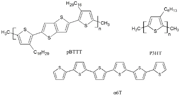
Figure I.19. Lamelar structure of P3HT, normal or parallel to the substrate, respectively 34 Figure I.20. Lamelar structure of P3HT(a), pBTTT-C16 (b), normal or parallel to the
substrate 35
Figure I.21. Chemical structure and product image of DPP-DTT polymers 36
Figure I.22. Flexible DPP-DTT top-gate OFETs fabricated on flexible substrate 37
Figure I.23. Characteristics of an OTFT device with a DPP-DTT thin film annealed at 200 °C
for 15 min 38
Figure I.24. The EGOFETwas described by Buth et al. in 2012 39
Figure I.25. The EGOFET device detected DNA 41
Figure I.26. P3HT organic semiconductor 43
Figure I.27. IDS–VDS curves of EGOFET devices 43
Figure I.28. P3HT:P3HT-COOH:P3HT-biotin terpolymer 44
Figure I.29. Illustration of CRP detection 46
Figure I.30. Illustration of streptavidin detection via biotin-streptavidin binding 47
Figure I.31. pBTTT-based EGOFET for protein (streptavidin) detection 48
Figure I.32. The gate-modified EGOFET for interleukin detection 50
Figure I.33. EGOFET where the gate is modified by a SAM of odorant binding protein
(pOBP-SAM) 51
Figure II.1. Identified metabolites of diclofenac and its percentages of oral dosage 73 Figure II.2. Dose-response curve for diclofenac spiked into ultrapure water samples 75
Figure II.3. The aptamer-base diclofenac sensor (Kheyrabadi et al., 2011) 76
Figure II.4. Label-free aptasensor for DCF detection 77
Figure II.5. DCF concentration calibration curve obtained in pasteurised milk 78
Figure II.6. Electropolymerized polypyrrole films-based sensor for DCF detection 78
Figure II.7. Oxidation mechanism of DCF 79
Figure II.8. Surface functionalization strategies for immobilization of AuNPs on gold and
Figure II.9. ECL detection mechanism for DCF based on GCE/MWCNTs-AuNPs/coating
antigen/BSA/GO-g-C3N4 labeled DCF antibody 81
Figure III.1. The magnetic electrochemical immunosensor for 2,4-D detection 113
Figure III.2. SPR sensorgram observed under a flow of 2,4-D-Ab solutions 114
Figure III.3. Variation of normalized SPR angle shift with the concentration of 2,4-D in the
competitive immunosensing experiments 115
Figure III.4. Gold nanoparticle-catalysed chemiluminescence reaction mechanism 116 Figure III.5. Schematic illustration for (A) fabrication and (B) detection mechanism of the
photoelectrochemical sensor 117
Figure III.6. Photoelectrochemical sensor characteristics 117
Figure III.7. Output curves (left) and transfer curves (right) for a water-gated OFET based on
P3HT-biotin 120
Figure III.8. EGOFET to determine the C-reactive protein 121
Figure III.9. Detection bisphenol via EGOFET device 122
Figure III.10. EGOFET in order to sense dopamine 123
Figure III.11. EGOFET for monitoring tumor necrosis factor alpha (TNFα) 124
Figure IV.1. Cu2+ detection applying ligation DNAzyme complex assembled onto gold
nanoparticles 160
Figure IV.2. Chemical structure of PMTPS, illustration of the formation of AD-TAB and
sensing mechanism for the detection of Cu2+ 161
Figure IV.3. The electrochemical sensor for detection of Cu2+ applying PATP immobilized
on AuNPs decorated with hydrogenated TiS2 nanosheets 162
Figure IV.4. Calibration curve corresponding to the detection of Cu2+ of the electrochemical
sensor investigated by Gan et al. (2016) 162
Figure IV.5. Principle of the sensor applying L-histidine immobilized on gold-labeled
multiwalled CNTs proposed by Zhu et al. in 2017 163
Figure IV.7. Characteristics of hydrogel-modified gate electrode of EGOFET 164 Figure IV.8. Plots of ID of hydrogel-modified gate electrode of EGOFET versus time 164 Figure IV.9. Detecting copper results obtained to copper-binding tripeptide
Gly-Gly-His-based sensor, by Yang et al. (2003) 165
Figure IV.10. Detecting copper results obtained to copper-binding tripeptide
LIST OF TABLES
Table I-1. Figures of merit of enzyme-based and enzyme-free immunosensors using
conventional electrode substrates 25
Table I-2. Characteristics of some electrochemical immunosensors 26
Table I-3. Influence of probe grafting and target hybridization on device performance with PBS as electrolyte, and influence of probe hybridization with water as electrolyte 42
Table II-1. Physical, chemical and pharmacological properties of diclofenac 72
Table II-2. Annual consumed volumes of diclofenac in some Western countries. 73
Table II-3. Aquatic toxicity data of diclofenac in the literature 74
LIST OF EQUATIONS
Equation I-1 8
Equation I-2 8
GENERAL INTRODUCTION
This thesis has been aacomplished in the group “Bioelectronics and Smart Surfaces” since November 2015. It focuses, in the first part, on the design and fabrication of an enzymeless electrochemical displacement heterogeneous immunosensor for diclofenac detection, and also in the second part, on gate functionalization of an Electrolyte-Gated Organic Field-Effect Transistor applied to biodetection of 2,4-dichlorophenoxyacetic acid (2,4-D) and Cu2+ in tap water.
For the development of the enzymeless electrochemical displacement heterogeneous immunosensor (Chapter II), the electrode interface was functionalized by an hapten which is an analogue of 2,4-D. The hapten is a molecule very close to 2,4-D but slightly different in its chemical structure so that the affinity of the antibody for this hapten is slightly lower than for the unmodified 2,4-D. The 2,4-D antibody is immobilized to the hapten; when antigens are present in the solution, a competition for binding the antibody between the antigen and the hapten takes place, so that the antibody is removed off the surface because it preferentialy binds to the free antigen. This reduces the steric hindrance on the electroactive surface and causes the current to increase.
For the development of the Electrolyte-Gated Organic Field-Effect Transistor (Chapters III and IV), we considered that this device is a capacitance-coupled one which can be modeled as two capacitors in series. The first corresponds to the gate/electrolyte interface while the second corresponds to the electrolyte/semiconductor interface. It is known that the drain current of such a transistor depends on the total capacitance. Therefore, if we are able to change this capacitance, we will be able to modulate the drain current. We decided to work on the gate/electrolyte capacitance because it is easier to chemically modify the gate than to the semiconductor (and also because it was much less investigated in the literature). To neglect the electrolyte/semiconductor interface capacitance, that of the other interface must be much smaller (this is because 1/CTotal = 1/COSC + 1/CGate; if CGate << COSC, then CTotal ≈ CGate). For this reason, we used microelectrodes as gate, of 100 to 250 µm in diameter.
drain current; this has been studied in the most recent literature, in particular for application in biosensing where large probes or targets are able to modify the double layer capacitance of the gate. However, the effect of charges carried by these probes or targets have been poorly investigated, or only for application to DNA sensors. In my work on EGOFETs, I investigated both effects: bulky molecules on the interfacial capacitance (Chapter III) and electrostatic effect of ions complexed in peptides (Chapter IV).
My thesis therefore consists in three parts. 1. First part
Diclofenac is one of the most popular compounds used as Non-Steroidal Anti-Inflammatory Drug (NSAID). It is generally employed to protect patients from inflammation and relieve pain, e.g. for arthritis, acute injuries, menstrual pain or even nocturnal enuresis. Along with the increase of human population, the consumption of this drug has become a worrisome issue as diclofenac residues are excreted directly from the body to the environment via urine and faeces, in large quantities compared to the oral dosage. This molecule is not well eliminated by water-treatment plants, so it is released into the ecosystem and becomes a poisoning agent to living organisms. Recently, scientists have developed several methods for the assessment of diclofenac ecotoxicology. However, there are only few studies applying electrochemical sensors, as well as electronic devices, to detect diclofenac. Moreover, the sensitivity is low, with limits of detection of ca. a few tens of nM. It is, therefore, necessary to develop novel techniques to improve the limit of detection. In this first part of my thesis, I will report the development of an original approach for an electrochemical immunosensor to detect diclofenac.
For this, we designed two precursors of a diazonium salt: the first is N-((1-(4-aminophenyl)-1H-1,2,3-triazol-4-yl)methyl)-2-(2-((2,6-dichlorophenyl)amino)phenyl)acetamide used to immobilize a molecular probe on the surface of the working electrode (further referred to as DCF-Probe) onto which the diclofenac antibody (AbDCF) will bind; the second is (2-[(4-aminophenyl)sulfanyl]-8-hydroxy-1,4-naphthoquinone) (further referred to as E-Probe), a redox probe used to transduce the AbDCF/DCF-Probe binding into a change of redox current. The E-Probe contains quinone groups, which are known to be sensitive to ion strength, pH or to local ion transport. In my model, the binding of AbDCF to DCF-Probe prevents the access of cations to the redox probe (i.e. decreases the apparent diffusion coefficient), so a faradic
current decrease is observed (due to the slowing down of the overall charge transfer rate). Conversely, if DCF is added into the surrounding electrolyte, it competes with the immobilized DCF-Probe and induces dissociation of AbDCF from the electrode surface (i.e. displaces the AbDCF/DCF-Probe complexation equilibrium); this phenomenon leads to a decrease of the overall steric hindrance, which in turn leads to an observable current increase.
2. Second part
World population development requires a more intensive agriculture, in particular the use of herbicides and pesticides. Consequently, large amounts of pollutants have been released into the environment all over the world. 2,4-D is a widely-used herbicide employed to control broad leave in agriculture. Unfortunately, this compound presents a serious harm for the health of people working in-situ with high concentrations of 2,4-D for prolonged contact time, as reported by the National Institute for Occupational Safety and Health (NIOSH, USA) in 1996. It is, therefore, necessary to develop assays to detect 2,4-D. In this second part of my thesis, I would like to introduce a novel approach applying the gate functionalization of an Electrolyte-Gated Organic Field-Effect Transistor (EGOFET). In this device, metallic contacts were done using classical photolithography in cleanroom, while the organic semiconductor (poly(N-alkyldiketopyrrolopyrrole dithienylthieno[3,2-b]thiophene) (DPP-DTT) was deposited by spin coating. These EGOFETs use water as electrolyte; they were operated in ambient conditions, without taking particular care for humidity or oxygen levels. A precursor of the 2,4-D diazonium salt was designed and used as immobilized molecular probe (further referred to as 2,4-D-Probe) onto which the 2,4-D antibody (Ab2,4- D) can bind (both are immobilized on the gate). The binding of Ab2,4-D to 2,4-D-Probe was shown to modulate the capacitance of the gate, leading to change in drain current (while no gate current is measured, which demonstrates that the device operates in the field-effect mode). Conversely, 2,4-D target added into the surrounding electrolyte was shown to compete with 2,4-D-Probe and induce dissociation of Ab2,4-D (i.e. displaces the Ab2,4-D from the electrode surface). This phenomenon leads to an increase in gate capacitance, which in turn leads to an increase in drain current, shown to depend on the 2,4-D concentration.
concentrations have been observed in human as the disorders in the gastrointestinal, hepatic, and renal systems. In 1993, the WHO (World Health Organization) reported that the level of copper in drinking water has to be at concentrations below 2 mg L-1. However, this value has been still regarded as doubtful due to the limit of detection of the validated measurement protocols, which dictated the recommended value rather than true health reasons. Hence, I propose in the third part of my thesis to develop a sensitive method, in a point-of-use format, for the on-site determination of Cu2+ ions in aqueous media. Following the specific strategy mentioned in the previous part of my work, the gate of EGOFETs were functionalized by short peptides (Gly-Gly-His) known to efficiently complex Cu2+ in aqueous medium.
We propose here the direct electrografting of the Gly-Gly-His tripeptide (0.27 kDa) through the first primary amine-terminated Gly residue (due to steric effects, only primary amines are grafted, with the exclusion of secondary and tertiary amines).This peptide has been investigated these last years, mainly in electrochemical systems. Here, I show that the binding of Cu2+ to Gly-Gly-His slightly modulates the capacitance of the gate but also and maintly lead to a significant shift of the threshold voltage of the device, attributed to the electrostatic effect of the accumulation of copper cations at the gate/electrolyte interface. I demonstrates that such device can be used to detect Cu2+ even in tap water, that is to say in real conditions.
BIBLIOGRAPHY
Chapter I
1
Biosensors: Generalities
In simple words, biosensors are analytical devices that convert a biological interaction into, at the very end, an electrical signal. Biosensors have been applied in many fields, e.g. food industry, medical field, defense, where they provide better sensitivity and ease of use as compared with traditional methods.
In food processing, biosensors are expected for simple, real-time, selective and inexpensive measures (1), i.e. for the detection of pathogens such as E. coli (2). Enzymatic biosensors are also employed in the dairy industry, for example to quantify pesticides in milk (3). In fermentation industries, process safety and product quality are crucial, thus effective monitoring of the fermentation process is imperative; biosensors have attracted a lot of attention in online monitoring in fermentation process due to their simplicity and quick response (4). Biosensors are also being employed to detect heavy metals (5-7) or pesticides (8-10) in drinking water.
In the medical field, applications of biosensors are growing rapidly. For example, glucose biosensors are expected to be widely used in clinical applications for diagnosis of diabetes mellitus, which requires precise control over blood-glucose levels. Blood-glucose biosensors usage at home accounts for 85% of this expected gigantic world market (11). Biosensors are also being used to diagnose infectious disease or cancer biomarkers human interleukin 10 (IL-10) (12).
1.1 Generalities on biosensors
Biosensors must absolutely be highly specific, which is usually obtained with bioprobes (enzymes, DNA, proteins, antibodies…) which present a high specificity for a given and unique molecule, i.e. their target. The term “biosensor” was introduced in 1977 by Cammann
(13)
, and biosensors are now well-defined, e.g. by IUPAC
(www.old.iupac.org/publications/books/author/mcnaught.html). Figure I.1 gives the very general scheme of a biosensor, for the three typical bioprobes: antibodies (in immunosensors), enzymes (in enzymatic biosensors) and DNA strands (in genosensors).
Figure I.1. Schematic representation of a biosensor (14)
Materials, transducing devices and immobilization methods involved in the fabrication of biosensors require multidisciplinarity in chemistry, biology and engineering. The first enzyme-based sensor was reported by Updike and Hicks in 1967 (15), in which they described an enzymatic glucose sensor. Enzymatic biosensors can be classified by their immobilization methods, i.e. adsorption of enzymes by van der Waals forces, ionic bonding or covalent bonding. The commonly used enzymes for this purpose are oxidoreductases, polyphenol oxidases, peroxidases (16), and aminooxidases (17). Immunosensors were established on the fact that antibodies have high affinity towards their respective antigens, i.e. the antibodies specifically bind to pathogens or toxins, or interact with components of the host's immune system (18). DNA biosensors were devised on the property that a single-stranded nucleic acid is able to recognize and bind to its complementary strand, even in a complex matrix containing a number of other molecules or non-complementary DNA strands. The interaction is due to the formation of stable hydrogen bonds between the two nucleic acid strands (19-24). There are several approaches to transduce the various molecular recognitions involved in these biosensors into a measurable signal. These approaches will not be reviewed here but the reader can refer to the excellent review from Thompson and Krull (25), for example.
1.2 Immunoassays and immunosensors
Immunosensors are biosensors where antibody-antigen interactions (the antibody often being used as capture probe and the antigen being the target analyte) occurring on the sensor
surface are used to get a specific recognition. All concepts necessary to understand how immunosensors work are given below.
From the IUPAC gold book [1], an antigen is “a substance that stimulates the immune system
to produce a set of specific antibodies and that combines with the antibody through a specific binding site or epitope”. Also, from the same IUPAC document, an antibody is “a protein (immunoglobulin) produced by the immune system of an organism in response to exposure to a foreign molecule (antigen) and characterized by its specific binding to a site of that molecule (antigenic determinant or epitope)”. The general structure of an antibody is given
on Figure I.2 below.
Figure I.2. Schematic representation of heavy and light chains combined to form the most common antibody (IgG) (26).
1.3 Antibody-antigen interactions
An antibody molecule includes two identical, of equal binding affinity, antigen-recognization sites consisting of the NH2-terminated paratope. Their role is to bind antigens at this region and form a stable antibody-antigen complex. Antibody-antigen interactions are of different nature; one can consider that hydrogen bonds, hydrophobic interactions, van der Waals and colombic interactions are involved (27-29).
Equation I-1
Where ka is the association rate constant and kd is the dissociation rate constant. The equilibrium constant Ka can therefore be written:
Equation I-2
The dissociation constant (Kd = 1/ Ka) of a large number of antibody-antigen pairs have already been determined (30, 31) and are in all case very low, in the pM range (the dissociation constant of the avidin-biotin pair, one of the strongest known non-covalent bonds, is in the fM range).
2
Electrochemical immunosensors
2.1 General reviews describing electrochemical immunosensors
Electrochemical immunosensors are electrochemical biosensors where antibody-antigen interactions (the antibody being the capture probe and the antigen the target analyte) occurring on an electrode surface are transduced into an electrochemical signal. Compared to other transduction strategies, electrochemical transducing techniques are fast, sensitive, require simple equipment, use small quantity of material and can be portable. The field of electrochemical immunosensors is very rich and dynamic. To give a general but however concise overview of the current state-of-the-art, I focused this bibliographic section on reviews of the last 5 years only.
A relatively brief review was published by Ricci et al. in 2012 (32), presenting a guidance to all researchers who would like to enter the electrochemical immunosensor domain, particularly focusing on practical aspects. Another review, published by Yang et al. also in 2012 (33), focused on new trends in signal amplification in enzyme-based immunosensors (combination of enzymatic reactions, multienzyme labels, use of magnetic bead, etc.). Several reviews focus on precise applications. For example, Wan et al. (28) dealt in 2013 with generalities on point-of-care diagnostics for early detection of diseases. Bahadir et al. (34) did the same in 2015 for early clinical diagnostics of cancer and cardiac diseases. Chikkaveeraiah
et al. (35) reviewed in 2012 the most recent advances at that time in electrochemical immunosensors for detection of cancer protein biomarkers, with strategies to increase densities of capture molecules and sensitivities. On their side, in 2013, Vidal et al. (36) reviewed electrochemical affinity biosensors for detection of mycotoxins in food. Also in 2013, Diaconu et al. (37) reviewed electrochemical immunosensors for precise applications in breast and ovarian cancer. At last, Campuzano et al. (38) reviewed very recently (2017) electrochemical bioaffinity sensors for salivary biomarkers.
Various transduction strategies have been employed up to now, on various antibody-antigens interactions. Here are some examples only. Hayat et al. employed anti-okadaic acid antibody to detect Okadaic acid (39, 40). Antibodies were immobilized onto electrodes via protein A interactions (40) and amine groups (39). Jarocka et al. mentioned interaction of anti- hemagglutinin H5 antibody (MAb 6-9-1) and his-tagged hemagglutinin antigen (41, 42). Lim et
meat. Articles dealing with electrochemical imunosensors have continued to be published, even very recently in 2018, with the works of Sharma et al. (44) (prostate specific antigen detection), Khoshroo et al. (45) (carbohydrate antigen detection); Zeng et al. (46) (cytokeratin 19 fragment antigen); Amani et al. (47) (alpha-fetoprotein detection, a cancer biomarker); Rauf
et al. (48) (Mucin 1 detection, also a cancer biomarker), etc.
Several assay formats were used for transduction, e.g. competitive, sandwich, displacement, using enzymatic or enzymeless amplification, or even without amplification.
2.2 Competitive assay
In a competitive immunoassay, unlabelled analyte in a sample competes with labelled analyte to bind an antibody. The amount of labelled, unbound analyte is then measured. In theory, the more analyte in the sample, the more labelled analyte gets displaced and then measured; hence, the amount of labelled unbound analyte is proportional to the amount of analyte in the sample.
Figure I.3. A competitive homogeneous immunoassay, unlabeled analyte displaces bound labelled analyte, which is then detected or measured.
Competitive assays are performed using conjugated antigens. A good example of this approach is given on Figure I.3. Below are cited a few recent articles dealing with electrochemical competitive immunosensors.
M. Moreno Guzman et al. published an example of competitive electrochemical immunosensor in 2012, for adrenocorticotropin hormone (ACTH) detection. Competition was made between a mixture of the standard ACTH with its biotin-conjugated ACTH counterpart, for complexation on the specific antibody (anti-ACTH antibody) immobilized on the electrode surface (49). Similarly, Liu et al (50), the same year, published a competitive inhibition assay for detection of haemoglobin variant (HbA1c, a diabete marker protein). They immobilized glycosylated pentapeptides (GPP) on the electrode, then incubated it with a mixture of HbA1c and anti-HbA1c antibody. Another example is competition between HRP-estradiol and estradiol to bind to biotinylated anti-estradiol immobilized on the electrode (Figure I.4) (51). Conzuelo et al. also used this type of assay, for tetracycline detection [31].
Figure I.4. Illustration of the different steps and protocols involved in an estradiol competitive immunosensor.
2.3 Sandwich assay
The basic principle relies on, first, the immobilization of a primary antibody on the electrode surface, then its binding with the antigen, and finally, complexation with a second antibody; this antibody is generally labelled with a redox enzyme which produces a detectable signal at the electrode.
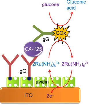
For example, Singh et al. described in 2013 an immunosensor based on a sandwich architecture, to determine the cancer antigen 125 (CA-125, a protein) (52) (Figure I.5). They first immobilized the anti-CA-125 antibody on the electrode surface through biotin-avidin interactions, then incubated it in a medium containing the CA-125, for its first complexation. In the third step, they added the second antibody labelled with glucose oxidase (GOx). For transduction, the authors used Ru(NH3)63+ as mediator; recylcing of the mediator on the electrode was detected and quantified amperometrically.
Figure I.5. Illustration of the sandwich asssay reported by Sing et al., 2013 (52).
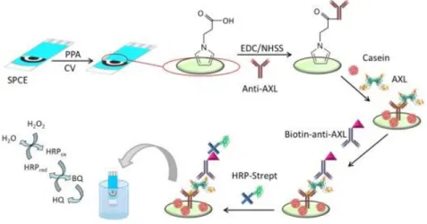
More recently, in 2017, Serafin et al. published a similar approach to detect the receptor tyrosine kinase AXL (53). In this work, the capture antibody was immobilized on an electropolymerized poly(pyrrolepropionic acid) film, and the second antibody was labelled not with GOx but with horseradish peroxidase (HRP), which is the most common enzyme
label. Hydroquinone (HQ) was used as mediator: HRP oxidizes HQ into benzoquinone (BQ), which is reduced amperometrically on the electrode and produces a detectable current.
2.4 Displacement assay
The displayment assay is a quite typical method applied to immunosensing. It is made of two main steps. A complex is made between the specific capture antibody attached to the surface and a labelled hapten (i.e. a molecule which is similar to the target analyte, having a quite good affinity for the antibody) (step 1, Figure I.6). Second step, in the presence of the true analyte, the first complex is dissociated in favour of the new complex between the antibody and the analyte. The labelled hapten leaves the electrode, so that its electrochemical signal decreases (step 2, Figure I.6). To insure the best efficiency, the antibody must have a higher affinity for the analyte than for the labelled hapten.
Figure I.6. General illustration of a displacement assay .
To illustrate this strategy, one can take the example of Tran et al. who published an electrochemical displacement immunoassay in 2013 (54). For detection of atrazine (a common pesticide), he employed a displayment strategy with an immobilized hapten (hydroxyatrazine) on an electroactive conducting polymer (Figure I.7).
Figure I.7. Displacement assay coupled to an electrochemical transduction, by Tran et al. (54).
Khor et al. also proposed a displayment assay protocol for determination of antibiotics (55) (Figure I.8), which could be applied for detection of any type of small molecules.
Figure I.8. Scheme of an electrochemical immunosensor: (A) shows the electrode surface after the antibody/epitope complexation with the corresponding attenuated electrochemical signal; (B) shows the detection of a small analyte via a displacement assay where the antibody is released, and the increase in eletroactivity as a function of time (55).
2.5 Enzyme labels and enzyme-based immunosensors
As explained before, labeling of analytes or secondary antibodies is often necessary for achieving an electrochemical transduction. Redox molecules can be used as labels; however, to achieve amplification, it is preferable to use redox enzymes which are capable to produce a large amount of electroactive molecules from only one antibody or one analyte molecule. For example, Guzman et al. reported that they could determine adrenocorticotropin hormone using alkaline phosphatase (AP)-labeled streptavidin and 1-naphtyl phosphate as the enzyme substrate. 1-Naphtol was generated by the enzyme reaction, and detected on the electrode surface (Figure I.9) (49).
Figure I.9. Illustration of the different steps and protocols for adrenocorticotropin hormone (ACTH) detection via electrochemical immunosensor involved in the development of a disposable screen-printed electrodes modified with phenylboronic acid (49).
In addition to alkaline phosphatase, horseradish peroxidase (HRP) is also a popular enzyme for labelling, due to its robustness and high turnover. In this case, H2O2 is the enzyme substrate and most of the case, hydroquinone (HQ) is used as co-substrate: its catalytic oxidation produces benzoquinone (BQ) which can be detected amperometrically and be re-reduced into HQ. As examples, one can cite Ojeda et al. (51) for detection of estradiol, Conzuelo et al. for determination of antibiotic residues in milk (56) or Serafin et al. for
tyrosine kinase (by the presence of AXL versus anti-AXL) (Figure I.10) or natriuretic peptide detection (53, 57).
Figure I.10. Different steps construct the amperometric immunosensor for AXL detection involving pPPA-modified SPCEs and covalent immobilization of anti-AXL (53).
Glucose oxidase (GOx) is less used as label but, due to its wide availability and good robustness, several articles have reported its use. For example, GOx-conjugated antibody cancer 125 was used by Singh et al. (52). Similarly, β-galactosidase was employed very recently (2018) by Sharma et al. (57).
Enzymes are generally used not for detection itself but for transduction and amplification, taking profit of the enzyme turn-over (for one event of biorecognition, one antibody or antigen captured, several molecules of enzyme product are produced at the vicinity of the electrode surface and electrochemically detected). HRP, horse radish peroxidase, is the most used enzyme label in immunosensors because it is commercially available and catalyzes the oxidation of numerous chromogenic substrates from H2O2 reduction. However, some other enzymes are also encountered such as alkaline phosphatase (AP) and glucose oxidase (GOx).
Detection of antibodies
The term “immunosensor” could mean that the target could be either an antigen (Ag) or an antibody (Ab) as for example in the framework of serological diagnosis, i.e. the research and the determination of specific antibodies linked to a pathogenic infection. Even though the
most popular approach remains targeting antigens using antibodies, some works report targeting antibodies.
In 2013, Bhimji et al. (41) described an interesting route to detect human immunodeficiency virus (HIV) antibodies by immobilization of antigenic peptides derived from a complex transmembrane protein of HIV-1 gp41 or HIV-2 gp36, covalently attached to a SU-8 substrate (an epoxy photoresist) close to microelectrodes. The detection of HIV antibodies was achieved using an alkaline phosphatase (AP)-conjugated secondary antibody antibody (Figure I.11). The linear detection range was reported between 1 ng mL-1 and 1 μg mL–1, with a limit of detection (LoD) of 1 ng mL–1 (6.7 pM).
Figure I.11. Illustration of antibodies detection of HIV. (a) Au microelectrodes (b) Immunorecognition (c) Transduction (d) Resulting DPV (41).
More recently in 2016, Montes et al. (58) used a composite graphite-epoxy substrate into which an HRP-labelled antibody was incorporated, for detection of IgG. Transduction was classical, with a competitive assay using H2O2 and hydroquinone (HQ) in solution. Amperometric measurements (reduction of benzoquinone BQ into HQ at the electrode) led to
other significantly cited work reporting enzyme-based immunosensor for detection of antibodies. In fact, detection of antigens is much more popular.
Detection of antigens
For example, in 2012, Ojeda et al. (51) described an electrochemical immunosensor for estradiol sensing, based on carbon screen-printed electrodes (SPE) sequentially modified with p-aminobenzoic acid, streptavidin and biotinylated anti-estradiol. Transduction was performed by applying a competitive immunoassay between peroxidase-labeled estradiol (HRP-estradiol) and estradiol for the binding sites of the immobilized antibodies. The reaction between estradiol and biotinylated anti-estradiol was amperometrically detected by addition of H2O2 in the presence of HQ. The linear range was between 1 and 250 pg mL−1 and the LoD was 0.77 pg mL−1.
Also in 2012, Qi et al. (59) reported an array of carbon SPE for simultaneous detection of several tumor biomarkers such as carcinoembryonic antigen (CEA) and α-fetoprotein (AFP). Electrodes were modified by grafting p-phenylenediamine via the diazonium route, followed by crosslinking the primary capture antibody using a Schiff base reaction. Transduction was made by using a sandwich assay with HRP-labelled secondary antibodies (Figure I.12 (C)). The detection range was from 0.10 to 50 ng mL−1 and the LoD of ca. 40 pg mL−1. Still in 2012, Moreno-Guzmán et al. (49) described a competitive electrochemical immunosensor for adrenocorticotropin hormone (ACTH) using disposable phenylboronic-modified carbon SPE used to efficiently immobilize ACTH antibodies. Transduction was designed using a competitive equilibrium for the binding sites of the immobilized antibody, between the target ACTH and a biotinylated ACTH (Figure I.12 (A)). The electroanalytical response was generated by using an AP-labelled streptavidin and 1-naphtyl phosphate as enzyme substrate. Differential pulse voltammetry (DPV) was used to monitor the enzyme activity (instead of classical CV to suppress the capacitive component). A very low LoD of 18 fg mL-1 was obtained.
Figure I.12. Illustration of antigen detections: (A) Reactions involved in the ACTH immunosensor using SPEs modified with phenylboronic acid (49). (B) Above, scheme of the immunosensor principle for SA and TC antibiotics. Below, surface chemistry involved for covalent binding of Protein G (56). (C) Detection principle of the screen-printed bi-analyte array, including a BSA
A competitive immunosensing approach was also followed by Conzuelo et al. (56) in 2013, for determination of sulfonamide (SA) and tetracycline (TC), two antibiotics which could be present in milk. The originality was to use Protein G coupled to 4-aminobenzoic acid electrografted on the electrode, as anchoring point for oriented immobilization of anti-SA and anti-TC (Figure I.12 (B)). Using HRP-labelled TC, they obtained a LoD of ca. 1 nM (ca. 200-500 pg mL-1) for both SA and TC. As for the previous ACTH competitive detection, no linear range was given, probably because it is relatively difficult to obtain such linear response with competitive transduction. For detection of cancer antigen 125 (CA-125), Singh et al. (52) described a Ru(NH3)63+-mediated glucose oxidase (GOx) labelling instead of routinely used enzymes such as HRP or AP. The LoD, for an incubation period of 5 min, was slightly lower 0.1 U mL-1 for CA-125, comparable to the other reported electrochemical immunosensors. However, the authors claimed a shorter incubation time compared to HRP or AP amplifications. More generally, for enzyme-based amplification routes, the turnover of the enzyme (more precisely the Kcat/KM ratio, which should be as high as possible) is a crucial parameter for a good amplification. Jiang et al. (60) described an electrochemical immunosensor for detection of tumor necrosis factor (TNF-) with a strategy to avoid non-specific adsorption. For this purpose, they used an original layer of phenyl phosphoryl choline (PPC) and phenyl butyric acid (PBA). The capture antibody was grafted on the working ITO electrode along with this anti-adsorption layer and, in a sandwich configuration, the signaling (labelled) antibody was coupled to HRP. H2O2 was added in solution as well as ferrocene (Fc) to reuse HRP. The immunosensor was shown to detect TNF-α with a LoD of 10 pg mL-1 with a wide linear range between 0.01 ng mL-1 to 500 ng mL-1. More recently, Serafín et al. (53) reported in 2017 a tyrosine kinase immunosensor involving a sandwich architecture with a capture antibody covalently immobilized on poly(pyrrolepropionic acid)-modified electrodes and a HRP-labeled secondary antibody. The LoD was 337 pg mL−1. Due to the relative instability, limited robustness and severe limitation of the operating conditions required for most enzymes, it is of interest to develop enzyme-free catalytically-amplified immunosensors, which could be considered as one of the most promising perspectives for electrochemical immunosensors. Another strategy to get rid of enzymes is to design non-amplified sensors. These two approaches are reviewed below.
2.6 Enzyme-free immunosensors
Ciani et al. (61) developed gold SPE electrodes modified with specific thiolated antibodies for direct detection of infection biomarkers, using electrochemical impedance spectroscopy (EIS), more particularly measuring the charge transfer resistance of Fe(CN)63-/4- on the gold electrode depending on the presence or not of the targeted proteinic antigen (Triggering Receptor-1 Expressed on Myeloid cells,Matrix MetalloPeptidase 9 and N-3-oxo-dodecanoyl-l-HomoSerineLactone). Limits of detection were between the pM and the nM range, depending on the target. Also with an enzyme-free transduction procedure, Tran et al. (54) described a label-free electrochemical competitive immunosensor based on an electroactive conducting polymer coupled with a slightly modified atrazine molecule, a common pesticide (Figure I.13 (A)). This quinone-based polymer presented a current decrease following anti-atrazine antibody complexation, and a current increase after anti-atrazine addition in solution, with a very low detection limit of 1 pM, i.e. 0.2 pg mL−1 estimated by square wave voltammetry (SWV). One originality relies on the fact that the redox probe is not diffusing in solution but immobilized on the electrode surface, and another originality is the competitive equilibrium between an immobilized mimic of the target (so-called hapten) and the diffusing target to detect.
Figure I.13. Some examples of enzyme-free immunosensor applications: (A) Strategy for the electrochemical detection of atrazine based on the change in electroactivity of a polymer film poly(juglone-ATZ); (1) polymer/hapten-modified electrode; (2) after complexation with anti-ATZ; (3) after addition of ATZ in solution (54). (B) Scheme of the competitive inhibition assay for detecting HbA1c (anti-HbA1c: blue Y; HbA1c: red triangle; GPP: pink triangle, surface bound)(50). (C) RCA-based immunosensor for HE4 detection (62).
In the same year (2012), a similar idea was developed by Liu et al. (50) They reported an electrochemical immunosensor for detecting glycosylated hemoglobin (HbA1c) based on glassy carbon electrodes (GCEs) modified with a mixed layer of oligo(phenylethynylene) and oligo(ethyleneglycol), obtained by electrografting of the corresponding aryl diazonium salts. 1,1′-Di(aminomethyl)ferrocene and an epitope, the glycosylated pentapeptide (GPP) VHLTP, were covalently attached to oligo(phenylethynylene). GPP is a peptide mimetic to HbA1c, to which an anti-HbA1c antibody could bind. As for Tran et al., HbA1c was detected by a competitive assay based on the competition for binding to anti-HbA1c between the analyte in solution, HbA1c, and the surface bound GPP peptide. Exposure of the GPP-modified interface to the mixture of anti-HbA1c IgG antibody and HbA1c resulted in the attenuation
of Fc electroactivity due to steric hindrance generated by the antibody bound to the surface (Figure I.13 (B)). The authors found that HbA1c could be detected from 4.5% to 15.1% of total hemoglobin in serum. The same authors, in the same year, adapted this method to AuNPs-modified surfaces (reference cited later in the section). Also, to avoid addition of a diffusing redox probe in solution, Wang et al. (63) reported later a similar approach, based on an electroactive polymer onto which an antibody was coupled, to detect bisphenol A (BPA) by competitive binding assay with a detection limit of 2 pg mL−1 using SWV. A current decrease was obtained upon anti-BPA binding and an opposite current increase upon BPA addition in solution. These authors also described a similar approach for detection of acetaminophen (64) with the detection limit of ca. 10 pM (1.5 pg mL-1). An original approach using DNA was reported by Lu et al. (62). They described detection of human epididymis-specific protein 4 (HE4) with a chitosan-titanium carbide-modified ITO electrode (Chi-TIC/ITO) onto which AuNPs were deposited. The capture antibody was adsorbed onto the Au and TiC NPs. For transduction and amplification, secondary antibodies were labelled with DNA strands, followed by rolling circle amplification (RCA). Using doxorubicin as DNA intercalator and DPV for detection, the redox current responded to HE4 linearly in the concentration range of 3–300 pM, with a LoD of 0.06 pM (3-300 ng L-1 and 0.06 pg mL-1, respectively) (Figure I.13 (C)). This is a good example of enzyme-free amplification; amplification is provided by the numerous sites where doxorubicin can intercalate on each DNA strand. The high surface density of doxorubicin achieved by this strategy provided high currents leading to the high sentivitity of the method.
However, because this kind of amplification implies several successive steps, simple non-amplified procedures stayed popular, and particularly EIS combined with a diffusing redox probe. Hayat et al. (40) described the immobilization of anti-okadaic acid antibody on 4-carboxyphenyl film. The Ab-Ag binding was transduced simply using electrochemical impedance spectroscopy with Fe(CN)63-/4- as diffusing redox probe. The increase in electron transfer resistance was linearly proportional to the okadaic acid concentration in the range 0.195–12.5 μg L-1, with a LoD of 0.3 ng mL-1. In 2013, Vasudev et al. (65) described a similar procedure for epidermal growth factor receptor (EGFR) detection, by immobilizing anti-EGFR antibody on dithiobissuccinimidyl propionate (DTSP) SAM on Au electrodes. EIS measures with Fe(CN)63-/4- as redox probe exhibited a linear range from 1 pg mL-1 to
but replaced conventional Au electrode by microfabricated interdigitated ones. As a proof-of-concept, cortisol antibodies were immobilized using the same SAM as previously described. Cortisol (MW of 362 g mol-1) was detected using CVover a linear range of 10 pM to 100 nM (3.6 pg mL-1-36 ng mL-1).
This example is probably the occasion to recall here that the relative size of the target molecule compared to that of an antibody should determine the choice of the transduction architecture of an electrochemical immunosensor. Indeed, the most popular Ab reported in biosensors are immunoglobulins G (IgG), with a typical molecular weight of 150 kDa, i.e. a volume of between 300 and 700 nm3 or a projected area, on the supporting surface, of ca. 60 nm2. This should be compared to the molecular weight of the target antigen. If this one is for example a small protein of 30 kDa, it corresponds to a projected area of 22 nm2, i.e. ca 30% of the antibody’s. Nevertheless, for a molecule (such as pesticide, industrial or pharmaceutical pollutant) of 200 g mol-1, this ratio falls to 1% of the antibody’s projected surface, which is negligible and cannot play a significant role in changing the steric hindrance at the solution/electrode interface; for such situations, other transduction schemes should be considered, for example, by playing not only on steric hindrance but also on electrostatic repulsions, or reducing the size of the capture antibody by using only Ab fragments or an analog of protein (a peptidomimetic). For example, for detection of a bulky protein (porcine serum albumin), Lim et al. (67) reported a carbon nanofiber-modified SPE electrofunctionalized with a 4-carboxyphenyl diazonium salt onto which antibodies were covalently bound. Taking profit of the strong affinity of serum albumins towards anions, an anionic redox probe was used in solution. An increase in cathodic peak current was measured after immunocomplex formation between antibodies and proteins. The linear range was from 0.5 to 500 pg mL-1 and the LoD of 0.5 pg mL-1. Also, using Fe(CN)63−/4− as an electroactive diffusing probe, Jarocka et al. (68) reported detection of hemagglutinin from avian influenza virus H5N1. Gold electrodes were modified with a SAM of 4,4′-thiobisbenzenethiol (TBBT), itself modified by gold nanoparticles (AuNPs) and single chain variable fragments of antibodies (scFv) against hemagglutinin H5 (Figure I.14 (A)). Interactions between the scFv of antibodies and hemagglutinin were sensed by EIS, giving a LoD of 0.6 pg mL-1 and a linear range from 4.0 to 20.0 pg mL-1. This scFv makes 25 kDa and corresponds to the variable domains; it is the smallest fragment that holds a complete binding site of an antibody and therefore retains its specificity (Figure I.14 (B)).
Figure I.14. Illustration of detection of hemagglutinin from avian influenza virus H5N1: (A) Hemagglutinin H5 immunosensor based on a SAM of 4,4’-thiobisbenzenethiol carrying single chain variable fragments (scFv) of antibodies as probes (68). (B) Schematic representation of a scFv fragment. A scFv makes 25 kDa and corresponds to the VH+VL domains. From Antibody Design Laboratories. (http://www.abdesignlabs.com/technical-resources/scfv-cloning/); access January 29th 2017.
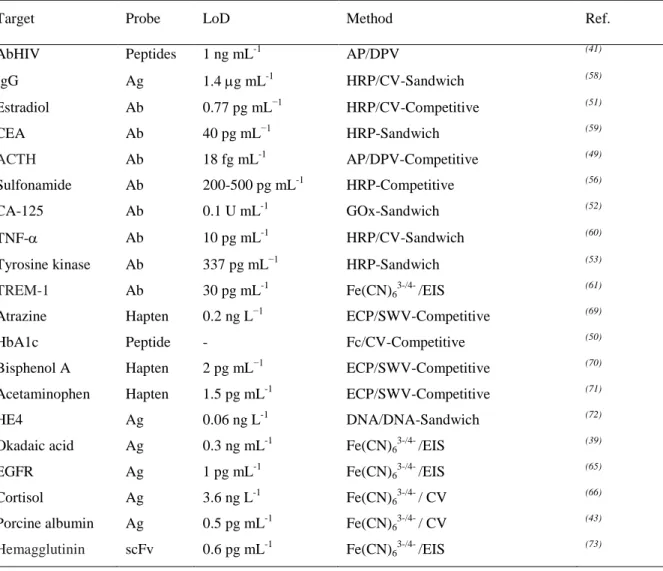
Table I-1. Figures of merit of enzyme-based and enzyme-free immunosensors using conventional electrode substrates.
Target Probe LoD Method Ref.
AbHIV Peptides 1 ng mL-1 AP/DPV (41) IgG Ag 1.4 g mL-1 HRP/CV-Sandwich (58) Estradiol Ab 0.77 pg mL−1 HRP/CV-Competitive (51) CEA Ab 40 pg mL−1 HRP-Sandwich (59) ACTH Ab 18 fg mL-1 AP/DPV-Competitive (49) Sulfonamide Ab 200-500 pg mL-1 HRP-Competitive (56) CA-125 Ab 0.1 U mL-1 GOx-Sandwich (52) TNF- Ab 10 pg mL-1 HRP/CV-Sandwich (60) Tyrosine kinase Ab 337 pg mL−1 HRP-Sandwich (53)
TREM-1 Ab 30 pg mL-1 Fe(CN)63-/4- /EIS (61)
Atrazine Hapten 0.2 ng L−1 ECP/SWV-Competitive (69) HbA1c Peptide - Fc/CV-Competitive (50) Bisphenol A Hapten 2 pg mL−1 ECP/SWV-Competitive (70) Acetaminophen Hapten 1.5 pg mL-1 ECP/SWV-Competitive (71) HE4 Ag 0.06 ng L-1 DNA/DNA-Sandwich (72) Okadaic acid Ag 0.3 ng mL-1 Fe(CN)63-/4- /EIS (39)
EGFR Ag 1 pg mL-1 Fe(CN)63-/4- /EIS (65)